Diffusion MRI: From Principles To Modelling And … · BASICS OF DIFFUSION MRI ... can help us...
18
1 Biomedical Imaging | www.smgebooks.com Copyright Arrigo A.This book chapter is open access distributed under the Creative Commons Attribution 4.0 International License, which allows users to download, copy and build upon published articles even for commercial purposes, as long as the author and publisher are properly credited. Gr up SM Diffusion MRI: From Principles To Modelling And Clinical Applications INTRODUCTION Diffusion weighted imaging (DWI) is the set of methodologies developed to investigate structural properties of brain tissues by analyzing water molecules diffusion process. Indeed, presence of biological barriers influences water displacement both in terms of velocity and directionality. DWI has the unique ability to provide, non-invasively and in-vivo, clues at microscopic level starting from data acquired at macroscopic scales. A number of models have been proposed in literature to highlight different aspects of diffusion. The main focus is put on white matter (WM), since it is even possible to visualize main WM pathways thanks to particular techniques that go under the name of tractography. Nevertheless, DWI has been successfully adopted to probe grey matter (GM), as well as other non-brain tissues, like skeletal muscles, peripheral nervous system, myocardium or kidneys. Alessandro Arrigo 1 *, Alessandro Calamuneri 1 and Enricomaria Mormina 1 1 Department of Biomedical Sciences and Morphological and Functional Images, University of Messina, Italy *Corresponding author: Alessandro Arrigo, University of Messina, Department of Biomedi- cal Sciences and Morphological and Functional Imaging, Via Consolare Valeria, 1 Messina, 98125, Italy, Email: [email protected] Published Date: February 19, 2016
-
Upload
nguyendien -
Category
Documents
-
view
220 -
download
1
Transcript of Diffusion MRI: From Principles To Modelling And … · BASICS OF DIFFUSION MRI ... can help us...
1Biomedical Imaging | www.smgebooks.comCopyright Arrigo A.This book chapter is open access distributed under the Creative Commons Attribution 4.0 International License, which allows users to download, copy and build upon published articles even for commercial purposes, as long as the author and publisher are properly credited.
Gr upSMDiffusion MRI: From Principles To Modelling
And Clinical Applications
INTRODUCTIONDiffusion weighted imaging (DWI) is the set of methodologies developed to investigate
structural properties of brain tissues by analyzing water molecules diffusion process. Indeed, presence of biological barriers influences water displacement both in terms of velocity and directionality. DWI has the unique ability to provide, non-invasively and in-vivo, clues at microscopic level starting from data acquired at macroscopic scales. A number of models have been proposed in literature to highlight different aspects of diffusion. The main focus is put on white matter (WM), since it is even possible to visualize main WM pathways thanks to particular techniques that go under the name of tractography. Nevertheless, DWI has been successfully adopted to probe grey matter (GM), as well as other non-brain tissues, like skeletal muscles, peripheral nervous system, myocardium or kidneys.
Alessandro Arrigo1*, Alessandro Calamuneri1 and Enricomaria Mormina1
1Department of Biomedical Sciences and Morphological and Functional Images, University of Messina, Italy
*Corresponding author: Alessandro Arrigo, University of Messina, Department of Biomedi-cal Sciences and Morphological and Functional Imaging, Via Consolare Valeria, 1 Messina, 98125, Italy, Email: [email protected]
Published Date: February 19, 2016
2Biomedical Imaging | www.smgebooks.comCopyright Arrigo A.This book chapter is open access distributed under the Creative Commons Attribution 4.0 International License, which allows users to download, copy and build upon published articles even for commercial purposes, as long as the author and publisher are properly credited.
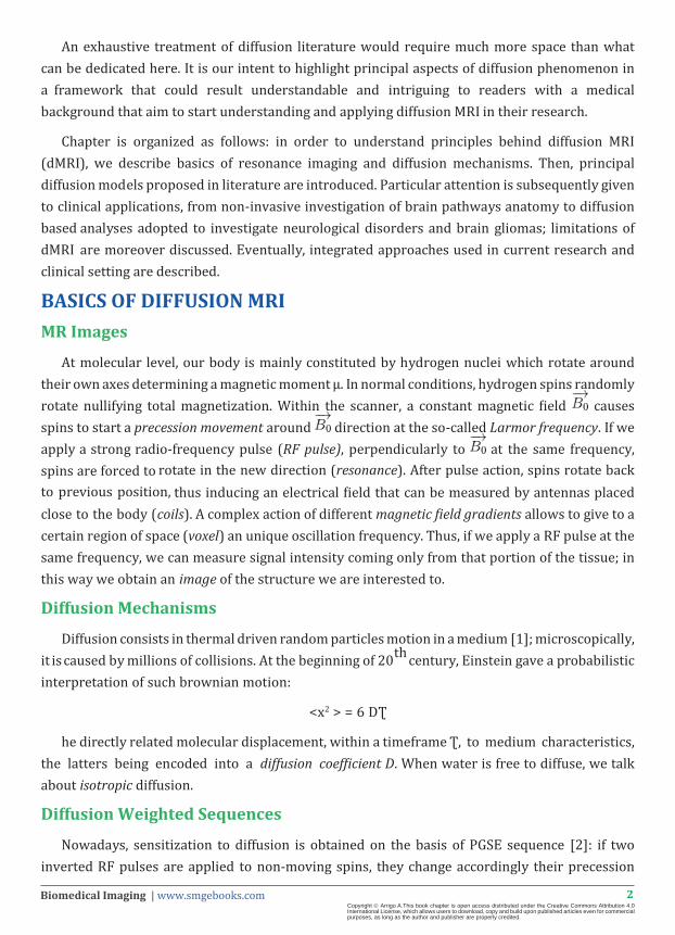
An exhaustive treatment of diffusion literature would require much more space than what can be dedicated here. It is our intent to highlight principal aspects of diffusion phenomenon in a framework that could result understandable and intriguing to readers with a medical background that aim to start understanding and applying diffusion MRI in their research.
Chapter is organized as follows: in order to understand principles behind diffusion MRI (dMRI), we describe basics of resonance imaging and diffusion mechanisms. Then, principal diffusion models proposed in literature are introduced. Particular attention is subsequently given to clinical applications, from non-invasive investigation of brain pathways anatomy to diffusion based analyses adopted to investigate neurological disorders and brain gliomas; limitations of dMRI are moreover discussed. Eventually, integrated approaches used in current research and clinical setting are described.
BASICS OF DIFFUSION MRIMR Images
At molecular level, our body is mainly constituted by hydrogen nuclei which rotate around their own axes determining a magnetic moment µ. In normal conditions, hydrogen spins randomly rotate nullifying total magnetization. Within the scanner, a constant magnetic field causes spins to start a precession movement around direction at the so-called Larmor frequency. If we apply a strong radio-frequency pulse (RF pulse), perpendicularly to at the same frequency, spins are forced to rotate in the new direction (resonance). After pulse action, spins rotate back to previous position, thus inducing an electrical field that can be measured by antennas placed close to the body (coils). A complex action of different magnetic field gradients allows to give to a certain region of space (voxel) an unique oscillation frequency. Thus, if we apply a RF pulse at the same frequency, we can measure signal intensity coming only from that portion of the tissue; in this way we obtain an image of the structure we are interested to.
Diffusion Mechanisms
Diffusion consists in thermal driven random particles motion in a medium [1]; microscopically, it is caused by millions of collisions. At the beginning of 20th century, Einstein gave a probabilistic interpretation of such brownian motion:
<x2 > = 6 DƮ
he directly related molecular displacement, within a timeframe Ʈ, to medium characteristics, the latters being encoded into a diffusion coefficient D. When water is free to diffuse, we talk about isotropic diffusion.
Diffusion Weighted Sequences
Nowadays, sensitization to diffusion is obtained on the basis of PGSE sequence [2]: if two inverted RF pulses are applied to non-moving spins, they change accordingly their precession
3Biomedical Imaging | www.smgebooks.comCopyright Arrigo A.This book chapter is open access distributed under the Creative Commons Attribution 4.0 International License, which allows users to download, copy and build upon published articles even for commercial purposes, as long as the author and publisher are properly credited.
phase, eventually having the same phase. Particles can be spatially encoded along a certain direction with a diffusion gradient: if particles move in between the application of inverted pulses, spins are no longer in phase. Such dephasing causes a signal loss that can be compared with an equivalent configuration in which diffusion gradient is not applied. Signals measured with and without diffusion gradient are related by a mono-exponential decay law driven by two quantities: b-value, which encloses (among others) sequence specifics, and the apparent diffusion coefficient D (or ADC) [3]. “Apparent” term is used since ADC depends both on the medium properties and on the particular timeframe spins are allowed to diffuse before signal acquisition [1]. From the physical point of view, ADC represents average diffusivity of water molecules in a given direction, and it is measured in m2/s:
Sg =S0 exp (-b D)
Modelling and Quantifying DiffusionWhat do we measure with diffusion MRI?
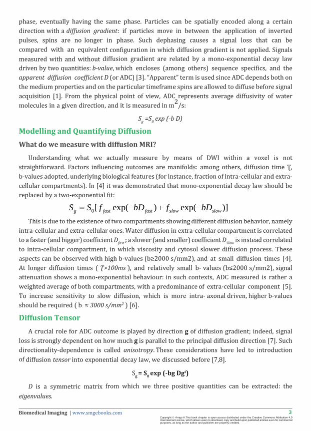
Understanding what we actually measure by means of DWI within a voxel is not straightforward. Factors influencing outcomes are manifolds: among others, diffusion time Ʈ, b-values adopted, underlying biological features (for instance, fraction of intra-cellular and extra-cellular compartments). In [4] it was demonstrated that mono-exponential decay law should be replaced by a two-exponential fit:
This is due to the existence of two compartments showing different diffusion behavior, namely intra-cellular and extra-cellular ones. Water diffusion in extra-cellular compartment is correlated to a faster (and bigger) coefficient Dfast ; a slower (and smaller) coefficient DSlow is instead correlated to intra-cellular compartment, in which viscosity and cytosol slower diffusion process. These aspects can be observed with high b-values (b≥2000 s/mm2), and at small diffusion times [4]. At longer diffusion times ( Ʈ>100ms ), and relatively small b- values (b≤2000 s/mm2), signal attenuation shows a mono-exponential behaviour: in such contexts, ADC measured is rather a weighted average of both compartments, with a predominance of extra-cellular component [5]. To increase sensitivity to slow diffusion, which is more intra- axonal driven, higher b-values should be required ( b ≈ 3000 s/mm2 ) [6].
Diffusion TensorA crucial role for ADC outcome is played by direction g of diffusion gradient; indeed, signal
loss is strongly dependent on how much g is parallel to the principal diffusion direction [7]. Such directionality-dependence is called anisotropy. These considerations have led to introduction of diffusion tensor into exponential decay law, we discussed before [7,8].
Sg = S0 exp (-bg Dgt)
D is a symmetric matrix from which we three positive quantities can be extracted: the eigenvalues.
0[ exp( ) exp( )]g fast fast slow slowS S f bD f bD= − + −

4Biomedical Imaging | www.smgebooks.comCopyright Arrigo A.This book chapter is open access distributed under the Creative Commons Attribution 4.0 International License, which allows users to download, copy and build upon published articles even for commercial purposes, as long as the author and publisher are properly credited.
They correspond to diffusivities along three orthogonal directions in space. To better understand meaning of tensor model we can consider its geometric representation, an ellipsoid (Figure 1a): if we imagine a particle quite in a certain position at time 0, it turns out that ellipsoid surface represents boundaries of a region where particle could have moved in a given time frame , with equal probability, because of diffusion process [7]. Hence, ellipsoid shape can help us understanding how tensor model probes brain tissues: if minimal or no obstacles are encountered, like in cerebrospinal fluid (CSF), ellipsoid is spherically shaped (Figure 1b). Instead, in presence of anisotropic diffusion, ellipsoid is more squashed along principal diffusion direction (Figure 1c). From tensor eigenvalues, a number of useful parameters have been introduced to highlight different aspects of diffusion, like axon integrity and myelination [9-12]. These indices are shown in Table 1; their meaning and biological interpretation are matter of discussion of next sections. For the moment it is worth to notice that, in a given voxel, the more a principal unique diffusion direction hinders diffusion, the higher the anisotropy in that voxel.
Figure 1: Diffusion Tensor and its geometric meaning. a) A tensor (upmost part of the panel) obtained from a simulation in which axons are coherently pointing in a given direction; for visualization purposes, axons are drawn in bottom part of the panel. A principal preferred diffusion direction is thus detected; diffusivity along that direction is quantified by highest eigenvalue λ1. b) In an isotropic scenario, ellipsoid is spherically shaped; a principal diffusion direction is not detected by tensor. c) Similarly to panel a, a tensor obtained from a voxel showing anisotropic diffusion is shown.

5Biomedical Imaging | www.smgebooks.comCopyright Arrigo A.This book chapter is open access distributed under the Creative Commons Attribution 4.0 International License, which allows users to download, copy and build upon published articles even for commercial purposes, as long as the author and publisher are properly credited.
Table 1: List of tensor based parameters most commonly used in literature.
Beyond Tensor: Tissue Heterogeneity and Complex Modeling
At normal imaging scales (2mm), a typical WM voxel contains several biological structures, like axons, astrocytes, and glia. In this perspective, water molecules diffusion is hindered in extra-axonal space and restricted in the intra-axonal space [13]. If in a voxel a coherent structure is detected, namely a single group of axons (fiber bundle), tensor model is well suited to investigate diffusion. On the contrary, if complex fibers geometry occurs, like crossing, branching or kissing bundles, tensor model loses its meaningfulness as it represents a non-well defined average behavior coming from heterogeneous structures [14]. It has been demonstrated [15] that more than 90% of WM voxels actually contain fibers with complex configurations. For these reasons more sophisticated models have been proposed in literature: some of them still explore water spins displacement, like diffusion kurtosis [16], multitensor [17] or ball-and-stick models [18], QBI [19], DSI [20]. Others directly attempt to individuate discrete sets of fiber bundles, like in case of spherical deconvolution [21-23]. Recently, a multi-compartment model, called NODDI [24] has been introduced, with the goal to provide a new framework for analysis of WM and GM. It is important to notice that most of these models require sequences and acquisition times that can rarely be achieved in clinical practice; for this reason diffusion tensor is still today a widely accepted choice.

6Biomedical Imaging | www.smgebooks.comCopyright Arrigo A.This book chapter is open access distributed under the Creative Commons Attribution 4.0 International License, which allows users to download, copy and build upon published articles even for commercial purposes, as long as the author and publisher are properly credited.
To conclude we want to remark that, for a principled diffusion based quantification of brain tissues, one should have clear in mind that an unique solution does not exist; model choice has to be dictated by hardware constraints as well as biological question one aims to answer to.
Tractography
Tractography is the set of techniques able to detect and trace WM fiber bundles by means of dMRI [25]. Regardless to the diffusion model adopted, the main goal is to individuate and follow a discrete set of fibers starting from a certain spatial location (seed). Outcomes are a series of streamlines that can highlight major WM routes. With tensor model, we are most likely able to detect, within a voxel, only one main principal direction [25] (Figure 2). By using more sophisticated approaches, like Q-ball [19], CSD [21,22,26] or DSI [20], we can distinguish fibers arranged in more complex geometries (Figure 2). Usually stopping criteria are adopted, both based on spatial priors and diffusion based, to avoid tracts reaching unwanted regions, like deep GM or CSF. Tractography can be categorized into deterministic or probabilistic: in the former case, only one streamlining process is initialized from a seed voxel [25,27,28]; in the latter, several processes are run and different routes can be followed on the basis of how uncertainty is modeled in diffusion data [29-31]. Tractography has been extensively used in last years to explore brain structural connectivity in healthy brains and pathologies by means of connectivity analyses [32].
Figure 2: Tractographic reconstruction of right and left corticospinal tract (CST) in a case of high-grade glioma (brown ROI).
The neoplasm is shown in all radiological planes by means of FLAIR images (a-b-c). DTI-based reconstructions (d) showed a poor CST reconstruction in the healthy side, if compared to CSD-based one (e). Moreover, DTI resulted inadequate to detect CST in the affected side, in terms of both bundle’s involvement and anatomical relationship with neoplasm.

7Biomedical Imaging | www.smgebooks.comCopyright Arrigo A.This book chapter is open access distributed under the Creative Commons Attribution 4.0 International License, which allows users to download, copy and build upon published articles even for commercial purposes, as long as the author and publisher are properly credited.
CLINICAL APPLICATIONSDiffusion-based techniques are widely used in clinical settings, since they are able to provide
very useful information both in physiological and pathological contexts. A typical diffusion study workflow with possible variables that should be considered by operators is shown in Figure 3. The description of principal application fields of dMRI are given below.
Figure 3: Workflow of a diffusion-based study in clinical settings.
Blue boxes show the main steps usually followed during the study; green boxes show the main variables that can be chosen before as well as during the analyses. First of all, DWI images are acquired (STEP 1) and corrected for distortion artifacts. The choice of MRI scanner and acquisition parameters may influence the study repeatability [33] as well as sensitivity and reliability reached by post-processing analyses [34]. Moreover, operator has to decide whether to work in native or normalized space as well as the method to co-register DWI images to T1w ones (this step is useful to obtain anatomical landmarks).
8Biomedical Imaging | www.smgebooks.comCopyright Arrigo A.This book chapter is open access distributed under the Creative Commons Attribution 4.0 International License, which allows users to download, copy and build upon published articles even for commercial purposes, as long as the author and publisher are properly credited.
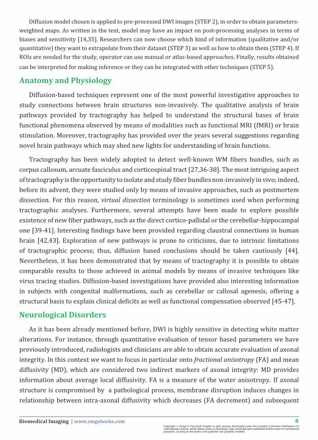
Diffusion model chosen is applied to pre-processed DWI images (STEP 2), in order to obtain parameters- weighted maps. As written in the text, model may have an impact on post-processing analyses in terms of biases and sensitivity [14,35]. Researchers can now choose which kind of information (qualitative and/or quantitative) they want to extrapolate from their dataset (STEP 3) as well as how to obtain them (STEP 4). If ROIs are needed for the study, operator can use manual or atlas-based approaches. Finally, results obtained can be interpreted for making inference or they can be integrated with other techniques (STEP 5).
Anatomy and Physiology
Diffusion-based techniques represent one of the most powerful investigative approaches to study connections between brain structures non-invasively. The qualitative analysis of brain pathways provided by tractography has helped to understand the structural bases of brain functional phenomena observed by means of modalities such as functional MRI (fMRI) or brain stimulation. Moreover, tractography has provided over the years several suggestions regarding novel brain pathways which may shed new lights for understanding of brain functions.
Tractography has been widely adopted to detect well-known WM fibers bundles, such as corpus callosum, arcuate fasciculus and corticospinal tract [27,36-38]. The most intriguing aspect of tractography is the opportunity to isolate and study fiber bundles non-invasively in vivo; indeed, before its advent, they were studied only by means of invasive approaches, such as postmortem dissection. For this reason, virtual dissection terminology is sometimes used when performing tractographic analyses. Furthermore, several attempts have been made to explore possible existence of new fiber pathways, such as the direct cortico-pallidal or the cerebellar-hippocampal one [39-41]. Interesting findings have been provided regarding claustral connections in human brain [42,43]. Exploration of new pathways is prone to criticisms, due to intrinsic limitations of tractographic process; thus, diffusion based conclusions should be taken cautiously [44]. Nevertheless, it has been demonstrated that by means of tractography it is possible to obtain comparable results to those achieved in animal models by means of invasive techniques like virus tracing studies. Diffusion-based investigations have provided also interesting information in subjects with congenital malformations, such as cerebellar or callosal agenesis, offering a structural basis to explain clinical deficits as well as functional compensation observed [45-47].
Neurological Disorders
As it has been already mentioned before, DWI is highly sensitive in detecting white matter alterations. For instance, through quantitative evaluation of tensor based parameters we have previously introduced, radiologists and clinicians are able to obtain accurate evaluation of axonal integrity. In this context we want to focus in particular onto fractional anisotropy (FA) and mean diffusivity (MD), which are considered two indirect markers of axonal integrity: MD provides information about average local diffusivity. FA is a measure of the water anisotropy. If axonal structure is compromised by a pathological process, membrane disruption induces changes in relationship between intra-axonal diffusivity which decreases (FA decrement) and subsequent

9Biomedical Imaging | www.smgebooks.comCopyright Arrigo A.This book chapter is open access distributed under the Creative Commons Attribution 4.0 International License, which allows users to download, copy and build upon published articles even for commercial purposes, as long as the author and publisher are properly credited.
increasing of global diffusivity due to diminished hindrance (MD increment) [48] (Figure 4). Presence of edema, axonal swelling, ischemic injury or other conditions, such as alterations of other glial components, also participate to diffusion parameters alterations [48]. A recent validation study has demonstrated that diffusion parameters measurements can be considered effectively useful biomarker for evaluating white matter damages; combined use of FA and MD with other parameters, such as AD and RD, may thus allow to increase sensitivity in detecting and distinguish different types of brain alterations [49].
Figure 4: Main causes of diffusion parameters changes in pathological contexts.
Thanks to such powerful tool, almost all neurological disorders can be investigated. Since it is impossible to fully describe all results achieved in this field, only illustrative cases of the most common neurological diseases are provided. Parkinson’s disease (PD) is a common neurological disorder characterized by dopaminergic neurons loss.
Diffusion-based evaluations have contributed both to understand structural changes as well as to differentiate different forms of PD. First of all, by analyzing tensor parameter changes in a voxel-based fashion, dMRI has been used to distinguish typical PD from atypical parkinsonian syndromes (like Multiple System Atrophy and Progressive Supranuclear Palsy). In literature, following structures have been investigated: frontal lobe, substantia nigra, corpus callosum, cerebellum, cerebellar peduncles, pons, internal capsule, thalamus, basal ganglia [50-60]. Moreover, these techniques are able to differentiate degenerative parkinsonian syndromes from vascular ones [61] as well as from patients affected by familiar essential tremor [62]. In parkinsonian patients tractographic reconstructions have been found useful for evaluating white matter changes, in this case by means of tract-based approaches, as well as to perform pre-surgical planning in cases of deep brain stimulation surgery [51,63-66].

10Biomedical Imaging | www.smgebooks.comCopyright Arrigo A.This book chapter is open access distributed under the Creative Commons Attribution 4.0 International License, which allows users to download, copy and build upon published articles even for commercial purposes, as long as the author and publisher are properly credited.
Diffusion-based evaluations are widespread used in cognitive disorders as well; for instance, they contributed to demonstrate that Alzheimer’s disease (AD) is only related to GM alterations. GM and WM analyses have helped indeed to better characterize AD etiopathogenesis as well as its progression [67,68]. Tractographic and diffusion parameters evaluations of brain pathways have revealed wide alterations of WM connections, helping to localize which brain circuits are mainly affected, such as frontal, temporal and limbic ones [69-71]. It has been also possible to differentiate AD from other cognitive diseases, such as mild cognitive impairment and vascular dementia [72,73]. Attempts have been made in order to early detect of WM alterations at pre-clinical stages [74] as well as to predict of atrophic progression [75-77].
dMRI has proven its ability to better understand and characterize stroke. First of all, a breakthrough has been achieved for accurate diagnosis and staging of ischemic stroke; indeed, evaluation of the triphasic progression of apparent ADC allows in differentiating between acute, sub-acute and chronic stages [78]. Moreover, the combined use of diffusion-based acquisition and perfusion-based one allows the evaluation of the so called ischemic penumbra, i.e. a hypo-perfused area surrounding the ischemic core which can be recovered by means of an immediate therapeutical intervention [79]. Similar useful information can be obtained in hemorrhagic stroke [80]. Moreover, tractographic and quantitative evaluations of eloquent WM pathways can help to predict functional recovery after acute event as well as patients’ outcome [81-83]. Summary of the most common clinical applications based on tensor parameters changes are shown in Table 2.
Table 2: Clinical applications of diffusion-based qualitative and quantitative analyses in common brain diseases. Main diffusion parameters changes are reported as well.
Brain Disease Clinical Application Main Diffusion Parameters Changes
Brain Neoplasms and Metastases
Detection and Evaluation of Eloquent
Pathways in Pre-Surgical Planning
FA decrement and MD increment due to loss of structural integrity, mass effect on white matter bundles and peritumoral edema.
Neurodegenerativ Disorders
White and Grey Matters Changes Detection through connectivity, voxel-based, tract- based and quantitative analyses
FA decrement and MD increment due to loss of axonal structural integrity, precipitation of pathological components, neuroinflammation.
Ischemic Cerebrovascular Disorders
White and Grey Matters Changes Detection through connectivity, voxel-based, tract- based and quantitative analyses
ADC decrement (acute phase); ADC normalization (subacute phase); ADC increment (chronic phase). FA decrement due to structural damage.
Hemorrhagic Cerebrovascular Disorders
White and Grey Matters Changes Detection through connectivity, voxel-based, tract- based and quantitative analyses
FA decrement and MD increment.
Pre-Surgical Planning
DWI has been adopted to perform pre-surgical investigation of brain pathways, i.e. pre-surgical planning. This is required both in neoplastic patients as well as in all other cases requiring neurosurgical intervention, such as epilepsy surgery or the abovementioned deep brain stimulation. The goal is to optimize surgery preserving brain functions. This is particularly important for eloquent pathways, such as corticospinal tract, arcuate fasciculus and optic

11Biomedical Imaging | www.smgebooks.comCopyright Arrigo A.This book chapter is open access distributed under the Creative Commons Attribution 4.0 International License, which allows users to download, copy and build upon published articles even for commercial purposes, as long as the author and publisher are properly credited.
radiations, whose damages have a bad impact on patients’ quality of life. In combination with other techniques such as intra-operative stimulation and neuronavigation systems, tractographic pre-surgical planning of brain neoplasm has been demonstrated to improve patients’ prognosis by optimizing maximal safe resection margins and also significantly reducing post-surgical deficits [84,85]. Thanks to dMRI, surgical improvement as well as brain functions preservation in patients with drug-resistant epilepsy [86,87] or brainstem cavernous malformations [88] has been shown.
The use of more advanced diffusion models (such as CSD) has been demonstrated to be able to further improve information obtained for pre-surgical planning by reducing limitations of DTI- based approaches [89,90]. In particular, moving beyond DTI, more advanced approaches have been found to be more sensitive in distinguishing different neoplastic effects on WM bundles, such as dislocation, infiltration, disruption and edematous involvement.
Validation and Limitations of Diffusion MRI Tractography
The proper validation of diffusion MRI tractography has represented an issue from the beginning of its discovery. Since an adequate gold standard is lacking, different studies have tried to validate the proper recognition of complex axonal configurations both in human and in animal models. U s i n g a h i g h l y g y r a t e d p o r c i n e b r a i n , D y r b y a n d c o l l e a g u e s [ 9 1 ] have assessed the anatomical validity and reproducibility of in vitro multi-fiber probabilistic tractography against invasive tracers. They have demonstrated that probabilistic MRI tractography is able to reliably detect specific pathways. In a similar recent study performed on macaque’s brain again [92], a good fidelity of dMRI tractography in estimating the presence or absence of WM connections has been suggested. Moreover, a strong agreement between DTI fitted principal diffusion direction and myelin orientations has been demonstrated at high spatial resolution and ultra-high field (9.4Tesla) in post mortem human brain tissue [93].
As we have already mentioned in the text, dMRI tractography suffers from a number of limitations; in some cases those may be knock out with different MR sequences expedients, choice of the tractographic reconstruction algorithms and proper MRI scanner settings. On the other hand, numerous inherent limitations – that cannot be overcome with current technologies – affect tractography. Most of them are related to the intrinsic inability to demonstrate the existence of synapses and gap junctions, as well as the signal directionality of transmission inside a tract [94]. Hence, one has to bear in mind that a reconstructed streamline should never be considered a real WM tract, but rather the mathematically driven most likely path that may suggest existence of an underlying given anatomical pathway. All these limitations are needed to be particularly taken into account in the evaluation of pathological conditions, such as stroke, demyelinating diseases or brain neoplasms.

12Biomedical Imaging | www.smgebooks.comCopyright Arrigo A.This book chapter is open access distributed under the Creative Commons Attribution 4.0 International License, which allows users to download, copy and build upon published articles even for commercial purposes, as long as the author and publisher are properly credited.
Integrated Approaches
In this section dMRI integration with other investigative approaches is briefly discussed. Their combined use has allowed to investigate different brain characteristics both in physiology and in brain diseases, providing an improvement in knowledge of brain processes and neurological pathologies. As a fundamental premise, although this section has been developed considering these techniques coupled in order to simplify the discussion, it should be underlined that often integrated methods include more than two different approaches.
Diffusion with fMRI
Functional MRI (fMRI) is a technique able to analyze blood oxygenation level dependent (BOLD) signal within each voxel in order to localize brain structures recruited during a given task. Combining this functional information with structural one provided by diffusion, it is possible to achieve different goals. First of all, one of the most challenging questions regarding tractography regards ROIs placement for tracking fibers bundles. Manual and automatic approaches are widely used, both suffering from different limitations (i.e. operator dependent bias for manual selection and imperfect ROIs matching for automatic ones). In this context, the use of fMRI-derived ROIs for reconstructing a specific circuit may offer a more subject-specific investigative approach [95], although it requires an increased scan time. Moreover, it is possible to combine functional connectivity with structural one in order to obtain a global view of specific brain processes investigating also the relationship between structure and function of a given circuit [96]. Furthermore, diffusion-fMRI combined approach has been found very useful for study of brain diseases, allowing correlations between fMRI signals changes and structural integrity alterations as well as detection of aberrant circuits [97-99].
Diffusion with brain stimulation
The integration of diffusion-based techniques with both non-invasive and invasive brain stimulation ones is largely adopted in research, clinical as well as surgical contexts. Diffusion combined with transcranial magnetic stimulation (TMS) can provide useful information regarding physiological aspects of a given circuit as well as connectivity changes induced by neuroplasticity processes; this multimodal non-invasive approach has been found also a useful tool to assess functional recovery after acute events as well as the effects of rehabilitation protocols [100-102].
On the other side, invasive deep brain stimulation used together with dMRI has allowed a better detection of eloquent bundles before neurosurgery as well as a better surgical management of neoplastic patients both in terms of surgical effectiveness and in reduction of postoperative deficits [66,103]. From this point of view, it is worth to notice that the integration of diffusion MRI data into neuronavigation systems has significantly improved surgical outcomes in terms of neoplasm’s resection, functional deficits and patients’ survival [84].

13Biomedical Imaging | www.smgebooks.comCopyright Arrigo A.This book chapter is open access distributed under the Creative Commons Attribution 4.0 International License, which allows users to download, copy and build upon published articles even for commercial purposes, as long as the author and publisher are properly credited.
Diffusion with EEG
Electroencephalography (EEG) used in combination with DWI offers several interesting hints both for researchers and clinicians. Indeed, information obtained by means of this multi-methodological approach have been incorporated in order to investigate networks and brain connectivity; for instance, it has been possible to draw inferences regarding the plausibility of EEG electrical activations with respect to a given pathway [104,105]. In pathological contexts, such as AD, mild traumatic brain injury and epilepsy, dMRI-EEG combined investigation have permitted to introduce new features for diagnosis, altered pathways localization, management and progression assessment [106-110].
Diffusion with neuropsychological tests
A number of neuropsychological tests are commonly used to evaluate cognitive functions. By attributing scores for each skill investigated, it is possible to have a view of cognitive deficits in brain diseases. In this context, diffusion-based tractographic and quantitative evaluations are employed to verify whether the clinical assessment is supported by brain structural alterations; diffusion tensor parameters evaluation have been demonstrated to provide correlations between loss of brain structural integrity and cognitive impairment [111-115]. Moreover, this combined analysis is also able to verify progression of cognitive impairment as well as to understand whether cognitive rehabilitations protocols provide clinical recovery due to neuroplasticity [116]. Finally, moving beyond pathological contexts, this kind of evaluation has been demonstrated to provide information regarding physiological progression of aging effects on brain connectivity and functions [117].
CONCLUSIONIn this chapter a didactic and complete description of diffusion MRI approaches was reported.
We showed diffusion theoretic principles in an understandable way for clinicians. Moreover, we described the main clinical applications of such approaches, alone or in combination with other investigative techniques; we reported also differences and possible analysis improvement provided by the choice of advanced diffusion modeling algorithms. Finally, we described how diffusion techniques have been histologically validated as well as the principal limitations of diffusion-based approaches.
References1. Beaulieu C. The basis of anisotropic water diffusion in the nervous system - a technical review. NMR Biomed. 2002; 15: 435-455.
2. Stejskal EO, Tanner JE. Spin Diffusion Measurements: Spin Echoes in the Presence of a Time‐Dependent Field Gradient. J Chem Phys. 1965; 42: 288-292.
3. Le Bihan D, Breton E, Lallemand D, Grenier P, Cabanis E, Laval-Jeantet M. MR imaging of intravoxel incoherent motions: application to diffusion and perfusion in neurologic disorders. Radiology. 1980; 161: 401-407.
4. Niendorf T, Dijkhuizen RM, Norris DG, van Lookeren Campagne M, Nicolay K. Biexponential diffusion attenuation in various states of brain tissue: Implications for diffusion-weighted imaging. Magn Reson Med. 1996; 36: 847-857.
14Biomedical Imaging | www.smgebooks.comCopyright Arrigo A.This book chapter is open access distributed under the Creative Commons Attribution 4.0 International License, which allows users to download, copy and build upon published articles even for commercial purposes, as long as the author and publisher are properly credited.
5. Clark CA, Le Bihan D. Water diffusion compartmentation and anisotropy at high b values in the human brain. Magn Reson Med. 2000; 44: 852-859.
6. Raffelt D, Tournier JD, Rose S, Ridgway GR, Henderson R, Crozier S, et al. Apparent Fibre Density: A novel measure for the analysis of diffusion-weighted magnetic resonance images. NeuroImage. 2012; 59: 3976-3994.
7. Basser PJ, Mattiello J, LeBihan D. Estimation of the effective self-diffusion tensor from the NMR spin echo. J Magn Reson B. 1994; 103: 247-254.
8. Basser PJ, Mattiello J, LeBihan D. MR diffusion tensor spectroscopy and imaging. Biophys J. 1994; 66: 259-267.
9. Pierpaoli C, Basser PJ. Toward a quantitative assessment of diffusion anisotropy. Magn Reson Med. 1996; 36: 893-906.
10. Westin CF, Peled S, Gudbjartsson H, Kikinis R, Jolesz FA. Geometrical diffusion measures for MRI from tensor basis analysis. Proc ISMRM. 1997; 97: 1742.
11. Basser PJ, Jones DK. Diffusion-tensor MRI: theory, experimental design and data analysis - a technical review. NMR Biomed. 2002; 15: 456-467.
12. Song SK, Sun SW, Ramsbottom MJ, Chang C, Russell J, Cross AH, et al. Dysmyelination Revealed through MRI as Increased Radial (but Unchanged Axial) Diffusion of Water. NeuroImage. 2002; 17: 1429-1436.
13. Assaf Y, Freidlin RZ, Rohde GK, Basser PJ. New modeling and experimental framework to characterize hindered and restricted water diffusion in brain white matter. Magn Reson Med. 2004; 52: 965-978.
14. Farquharson S, Tournier JD, Calamante F, Fabinyi G, Schneider-Kolsky M. White matter fiber tractography: why we need to move beyond DTI. J Neurosurg. 2013; 118: 1367-1377.
15. Jeurissen B, Leemans A, Tournier JD, Jones DK, Sijbers J. Investigating the prevalence of complex fiber configurations in white matter tissue with diffusion magnetic resonance imaging. Hum Brain Mapp. 2013; 34: 2747-2766.
16. Jensen JH, Helpern JA, Ramani A, Lu H, Kaczynski K. Diffusional kurtosis imaging: The quantification of non-gaussian water diffusion by means of magnetic resonance imaging. Magn Reson Med. 2005; 53: 1432-1440.
17. Tuch DS, Reese TG, Wiegell MR, Makris N, Belliveau JW, Wedeen VJ, et al. High angular resolution diffusion imaging reveals intravoxel white matter fiber heterogeneity. Magn Reson Med. 2002; 48: 577-582.
18. Behrens TE, Woolrich MW, Jenkinson M, Johansen-Berg H, Nunes RG. Characterization and propagation of uncertainty in diffusion-weighted MR imaging. Magn Reson Med. 2003; 50: 1077-1088.
19. Tuch DS1. Q-ball imaging. Magn Reson Med. 2004; 52: 1358-1372.
20. Wedeen VJ, Wang RP, Schmahmann JD, Benner T, Tseng WY. Diffusion spectrum magnetic resonance imaging (DSI) tractography of crossing fibers. Neuroimage. 2008; 41: 1267-1277.
21. Tournier JD, Calamante F, Gadian DG, Connelly A. Direct estimation of the fiber orientation density function from diffusion-weighted MRI data using spherical deconvolution. NeuroImage. 2004; 23: 1176-1185.
22. Tournier JD, Yeh CH, Calamante F, Cho KH, Connelly A, Lin CP, et al. Resolving crossing fibres using constrained spherical deconvolution: Validation using diffusion-weighted imaging phantom data. NeuroImage. 2008; 42: 617-625.
23. Dell’Acqua F, Scifo P, Rizzo G, Catani M, Simmons A, Scotti G, et al. A modified damped Richardson–Lucy algorithm to reduce isotropic background effects in spherical deconvolution. NeuroImage. 2010; 49: 1446-1458.
24. Zhang H, Schneider T, Wheeler-Kingshott CA, Alexander DC. NODDI: Practical in vivo neurite orientation dispersion and density imaging of the human brain. NeuroImage. 2012; 61: 1000-1016.
25. Basser PJ, Pajevic S, Pierpaoli C, Duda J, Aldroubi A. In vivo fiber tractography using DT-MRI data. Magn Reson Med. 2000; 44: 625-632.
26. Tournier JD, Calamante F, Connelly A. Robust determination of the fibre orientation distribution in diffusion MRI: Non-negativity constrained super-resolved spherical deconvolution. NeuroImage. 2007; 35: 1459-1472.
27. Mori S, Crain BJ, Chacko VP, Van Zijl PCM. Three-dimensional tracking of axonal projections in the brain by magnetic resonance imaging. Ann Neurol. 1999; 45: 265-269.
28. Lazar M, Weinstein DM, Tsuruda JS, Hasan KM, Arfanakis K. White matter tractography using diffusion tensor deflection. Hum Brain Mapp. 2003; 18: 306-321.
29. Lazar M, Alexander AL. Bootstrap white matter tractography (BOOT-TRAC). Neuroimage. 2005; 24: 524-532.
30. Jones DK. Tractography gone wild: probabilistic fibre tracking using the wild bootstrap with diffusion tensor MRI. IEEE Trans Med Imaging. 2008; 27: 1268-1274.
15Biomedical Imaging | www.smgebooks.comCopyright Arrigo A.This book chapter is open access distributed under the Creative Commons Attribution 4.0 International License, which allows users to download, copy and build upon published articles even for commercial purposes, as long as the author and publisher are properly credited.
31. Jeurissen B, Leemans A, Jones DK, Tournier JD, Sijbers J. Probabilistic fiber tracking using the residual bootstrap with constrained spherical deconvolution. Hum Brain Mapp. 2011; 32: 461-479.
32. Bullmore E, Sporns O. Complex brain networks: graph theoretical analysis of structural and functional systems. Nat Rev Neurosci. 2009; 10: 186-198.
33. Vollmar C, O’Muircheartaigh J, Barker GJ, Symms MR, Thompson P. Identical, but not the same: intra-site and inter-site reproducibility of fractional anisotropy measures on two 3.0T scanners. Neuroimage. 2010; 51: 1384-1394.
34. Chung AW, Thomas DL, Ordidge RJ, Clark CA. Diffusion tensor parameters and principal eigenvector coherence: relation to b-value intervals and field strength. Magn Reson Imaging. 2013; 31: 742-747.
35. Jones DK, Cercignani M. Twenty-five pitfalls in the analysis of diffusion MRI data. NMR Biomed. 2010; 23: 803-820.
36. Catani M, Howard RJ, Pajevic S, Jones DK. Virtual in vivo interactive dissection of white matter fasciculi in the human brain. Neuroimage. 2002; 17: 77-94.
37. Hasan KM, Kamali A, Kramer LA. Mapping the human brain white matter tracts relative to cortical and deep gray matter using diffusion tensor imaging at high spatial resolution. Magn Reson Imaging. 2009; 27: 631-636.
38. Thiebaut de Schotten M, Ffytche DH, Bizzi A, Dell’Acqua F, Allin M, Walshe M, et al. Atlasing location, asymmetry and inter-subject variability of white matter tracts in the human brain with MR diffusion tractography. Neuroimage. 2011; 54: 49-59.
39. Milardi D, Gaeta M, Marino S, Arrigo A, Vaccarino G, et al. Basal ganglia network by constrained spherical deconvolution: a possible cortico-pallidal pathway? Mov Disord. 2015; 30: 342-349.
40. Arrigo A, Mormina E, Anastasi GP, Gaeta M, Calamuneri A, Quartarone A, et al. Constrained spherical deconvolution analysis of the limbic network in human, with emphasis on a direct cerebello-limbic pathway. Front Hum Neurosci. 2014; 8: 987.
41. Dell’Acqua F, Catani M. Structural human brain networks: hot topics in diffusion tractography. Curr Opin Neurol. 2012; 25: 375-383.
42. Milardi D, Bramanti P, Milazzo C, Finocchio G, Arrigo A, Santoro G, et al. Cortical and subcortical connections of the human claustrum revealed in vivo by constrained spherical deconvolution tractography. Cereb Cortex. 2015; 25: 406-14.
43. Torgerson CM, Irimia A, Goh SY, Van Horn JD. The DTI connectivity of the human claustrum. Hum Brain Mapp. 2015; 36: 827-838.
44. Tournier JD, Mori S, Leemans A. Diffusion tensor imaging and beyond. Magn Reson Med. 2011; 65: 1532-1556.
45. Mormina E, Briguglio M, Morabito R, Arrigo A, Marino S. A rare case of cerebellar agenesis: a probabilistic Constrained Spherical Deconvolution tractographic study. Brain Imaging Behav. 2015;.
46. Andrade CS, Figueiredo KG, Valeriano C, Mendoza M, Valente KD. DTI-based tractography of the arcuate fasciculus in patients with polymicrogyria and language disorders. Eur J Radiol. 2015; 84: 2280-2286.
47. Meoded A, Katipally R, Bosemani T, Huisman TA, Poretti A. Structural connectivity analysis reveals abnormal brain connections in agenesis of the corpus callosum in children. Eur Radiol. 2015; 25: 1471-1478.
48. Alexander AL, Hurley SA, Samsonov AA, Adluru N, Hosseinbor AP, Mossahebi P, et al. Characterization of cerebral white matter properties using quantitative magnetic resonance imaging stains. Brain Connect. 2011; 1: 423-46.
49. Lin M, He H, Schifitto G, Zhong J. Simulation of changes in diffusion related to different pathologies at cellular level after traumatic brain injury. Magn Reson Med. 2015;.
50. Cochrane CJ, Ebmeier KP. Diffusion tensor imaging in parkinsonian syndromes: a systematic review and meta-analysis. Neurology. 2013; 80: 857-864.
51. Mormina E, Arrigo A, Calamuneri A, Granata F, Quartarone A, Ghilardi MF, et al. Diffusion tensor imaging parameters’ changes of cerebellar hemispheres in Parkinson’s disease. Neuroradiology. 2015; 57: 327-334.
52. Blain CR, Barker GJ, Jarosz JM, Coyle NA, Landau S. Measuring brain stem and cerebellar damage in parkinsonian syndromes using diffusion tensor MRI. Neurology. 2006; 67: 2199-2205.
53. Paviour DC, Thornton JS, Lees AJ, Jäger HR. Diffusion-weighted magnetic resonance imaging differentiates Parkinsonian variant of multiple-system atrophy from progressive supranuclear palsy. Mov Disord. 2007; 22: 68-74.
54. Nicoletti G, Lodi R, Condino F, Tonon C, Fera F. Apparent diffusion coefficient measurements of the middle cerebellar peduncle differentiate the Parkinson variant of MSA from Parkinson’s disease and progressive supranuclear palsy. Brain. 2006; 129: 2679-2687.
55. Lenfeldt N, Hansson W, Larsson A, Nyberg L, Birgander R. Diffusion tensor imaging and correlations to Parkinson rating scales. J Neurol. 2013; 260: 2823-2830.
16Biomedical Imaging | www.smgebooks.comCopyright Arrigo A.This book chapter is open access distributed under the Creative Commons Attribution 4.0 International License, which allows users to download, copy and build upon published articles even for commercial purposes, as long as the author and publisher are properly credited.
56. Vaillancourt DE, Spraker MB, Prodoehl J, Abraham I, Corcos DM. High-resolution diffusion tensor imaging in the substantia nigra of de novo Parkinson disease. Neurology. 2009; 72: 1378-1384.
57. Piccini P, Brooks DJ. New developments of brain imaging for Parkinson’s disease and related disorders. Mov Disord. 2006; 21: 2035-2041.
58. Ito M, Watanabe H, Kawai Y, Atsuta N, Tanaka F, et al. Usefulness of combined fractional anisotropy and apparent diffusion coefficient values for detection of involvement in multiple system atrophy. J Neurol Neurosurg Psychiatry. 2007; 78: 722-728.
59. Karagulle Kendi AT, Lehericy S, Luciana M, Ugurbil K, Tuite P. Altered diffusion in the frontal lobe in Parkinson disease. AJNR Am J Neuroradiol. 2008; 29: 501-505.
60. Gattellaro G, Minati L, Grisoli M, Mariani C, Carella F, Osio M, et al. White matter involvement in idiopathic Parkinson disease: a diffusion tensor imaging study. AJNR Am J Neuroradiol. 2009; 30: 1222-1226.
61. Deverdun J, Menjot de Champfleur S, Cabello-Aguilar S, Maury F, Molino F, Charif M, et al. Diffusion tensor imaging differentiates vascular parkinsonism from parkinsonian syndromes of degenerative origin in elderly subjects. Eur J Radiol. 2014; 83: 2074-2079.
62. Nicoletti G, Manners D, Novellino F, Condino F, Malucelli E. Diffusion tensor MRI changes in cerebellar structures of patients with familial essential tremor. Neurology. 2010; 74: 988-994.
63. Wiltshire K, Concha L, Gee M, Bouchard T, Beaulieu C. Corpus callosum and cingulum tractography in Parkinson’s disease. Can J Neurol Sci. 2010; 37: 595-600.
64. Sweet JA, Walter BL, Gunalan K, Chaturvedi A, McIntyre CC, Miller JP, et al. Fiber tractography of the axonal pathways linking the basal ganglia and cerebellum in Parkinson disease: implications for targeting in deep brain stimulation. J Neurosurg. 2014; 120: 988-996.
65. Said N, Elias WJ, Raghavan P, Cupino A, Tustison N, Frysinger R, et al. Correlation of diffusion tensor tractography and intraoperative macrostimulation during deep brain stimulation for Parkinson disease. J Neurosurg. 2014; 121: 929-935.
66. Torres CV, Manzanares R, Sola RG. Integrating diffusion tensor imaging-based tractography into deep brain stimulation surgery: a review of the literature. Stereotact Funct Neurosurg. 2014; 92: 282-290.
67. Zhang B, Xu Y, Zhu B, Kantarci K. The role of diffusion tensor imaging in detecting microstructural changes in prodromal Alzheimer’s disease. CNS Neurosci Ther. 2014; 20: 3-9.
68. Genc S, Steward CE. Short-term white matter alterations in Alzheimer’s disease characterized by diffusion tensor imaging. J Magn Reson Imaging. 2016; 43: 627-634.
69. Acosta-Cabronero J, Nestor PJ. Diffusion tensor imaging in Alzheimer’s disease: insights into the limbic-diencephalic network and methodological considerations. Front Aging Neurosci. 2014; 6: 266.
70. Prescott JW, Guidon A, Doraiswamy PM, Roy Choudhury K, Liu C. The Alzheimer structural connectome: changes in cortical network topology with increased amyloid plaque burden. Radiology. 2014; 273: 175-184.
71. Nishioka C, Poh C, Sun SW. Diffusion tensor imaging reveals visual pathway damage in patients with mild cognitive impairment and Alzheimer’s disease. J Alzheimers Dis. 2015; 45: 97-107.
72. Amlien IK, Fjell AM. Diffusion tensor imaging of white matter degeneration in Alzheimer’s disease and mild cognitive impairment. Neuroscience. 2014; 276: 206-215.
73. Román G, Pascual B. Contribution of neuroimaging to the diagnosis of Alzheimer’s disease and vascular dementia. Arch Med Res. 2012; 43: 671-676.
74. Fischer FU, Wolf D, Scheurich A, Fellgiebel A. Alzheimer’s Disease Neuroimaging Initiative. Altered whole-brain white matter networks in preclinical Alzheimer’s disease. Neuroimage Clin. 2015; 8: 660-666.
75. Nir TM, Jahanshad N, Toga AW, Bernstein MA, Jack CR Jr, Weiner MW, et al. Connectivity network measures predict volumetric atrophy in mild cognitive impairment. Neurobiol Aging. 2015; 36 Suppl 1: S113-S120.
76. Hiyoshi-Taniguchi K, Oishi N, Namiki C, Miyata J, Murai T. The Uncinate Fasciculus as a Predictor of Conversion from Amnestic Mild Cognitive Impairment to Alzheimer Disease. J Neuroimaging. 2015; 25: 748-753.
77. Jung WB, Lee YM, Kim YH, Mun CW. Automated Classification to Predict the Progression of Alzheimer’s Disease Using Whole-Brain Volumetry and DTI. Psychiatry Investig. 2015; 12: 92-102.
78. Sotak CH. The role of diffusion tensor imaging in the evaluation of ischemic brain injury - a review. NMR Biomed. 2002; 15: 561-569.
79. Ramos-Cabrer P, Campos F, Sobrino T, Castillo J. Targeting the ischemic penumbra. Stroke. 2011; 42: S7-11.

17Biomedical Imaging | www.smgebooks.comCopyright Arrigo A.This book chapter is open access distributed under the Creative Commons Attribution 4.0 International License, which allows users to download, copy and build upon published articles even for commercial purposes, as long as the author and publisher are properly credited.
80. Chaudhary N, Pandey AS, Gemmete JJ, Hua Y, Huang Y. Diffusion tensor imaging in hemorrhagic stroke. Exp Neurol. 2015; 272: 88-96.
81. Raffin E, Dyrby TB. Diagnostic approach to functional recovery: diffusion-weighted imaging and tractography. Front Neurol Neurosci. 2013; 32: 26-35.
82. Smits M, Jiskoot LC, Papma JM. White matter tracts of speech and language. Semin Ultrasound CT MR. 2014; 35: 504-516.
83. Jang SH1. Diffusion tensor imaging studies on arcuate fasciculus in stroke patients: a review. Front Hum Neurosci. 2013; 7: 749.
84. Wu JS, Zhou LF, Tang WJ, Mao Y, Hu J, Song YY, et al. Clinical evaluation and follow-up outcome of diffusion tensor imaging-based functional neuronavigation: a prospective, controlled study in patients with gliomas involving pyramidal tracts. Neurosurgery. 2007; 61: 935-948; discussion 948-9.
85. González-Darder JM, González-López P, Talamantes F, Quilis V, Cortés V, García-March G, et al. Multimodal navigation in the functional microsurgical resection of intrinsic brain tumors located in eloquent motor areas: role of tractography. Neurosurg Focus. 2010; 28: E5.
86. James JS, Radhakrishnan A, Thomas B, Madhusoodanan M, Kesavadas C, Abraham M, et al. Diffusion tensor imaging tractography of Meyer’s loop in planning resective surgery for drug-resistant temporal lobe epilepsy. Epilepsy Res. 2015;110: 95-104.
87. Jeong JW, Asano E, Juhász C, Chugani HT. Localization of specific language pathways using diffusion-weighted imaging tractography for presurgical planning of children with intractable epilepsy. Epilepsia. 2015; 56: 49-57.
88. Flores BC, Whittemore AR, Samson DS, Barnett SL. The utility of preoperative diffusion tensor imaging in the surgical management of brainstem cavernous malformations. J Neurosurg. 2015; 122: 653-662.
89. Mormina E, Longo M, Arrigo A, Alafaci C, Tomasello F, Calamuneri A, et al. MRI Tractography of Corticospinal Tract and Arcuate Fasciculus in High-Grade Gliomas Performed by Constrained Spherical Deconvolution: Qualitative and Quantitative Analysis. AJNR Am J Neuroradiol. 2015; 36: 1853-1858.
90. Arrigo A, Mormina E, Calamuneri A. The pre-surgical planning of brain neoplasms: from diffusion tensor imaging to more advanced approaches. Radiol Open Journal. 2015; 1: e1- e5.
91. Dyrby TB, Søgaard LV, Parker GJ, Alexander DC, Lind NM. Validation of in vitro probabilistic tractography. Neuroimage. 2007; 37: 1267-1277.
92. Azadbakht H, Parkes LM, Haroon HA, Augath M, Logothetis NK. Validation of High-Resolution Tractography Against In Vivo Tracing in the Macaque Visual Cortex. Cereb Cortex. 2015; 25: 4299-4309.
93. Seehaus A, Roebroeck A, Bastiani M, Fonseca L, Bratzke H. Histological validation of high-resolution DTI in human post mortem tissue. Front Neuroanat. 2015; 9: 98.
94. Chung HW, Chou MC, Chen CY. Principles and limitations of computational algorithms in clinical diffusion tensor MR tractography. AJNR Am J Neuroradiol. 2011; 32: 3-13.
95. Javad F, Warren JD, Micallef C, Thornton JS, Golay X. Auditory tracts identified with combined fMRI and diffusion tractography. Neuroimage. 2014; 84: 562-574.
96. Khalsa S, Mayhew SD, Chechlacz M, Bagary M, Bagshaw AP. The structural and functional connectivity of the posterior cingulate cortex: comparison between deterministic and probabilistic tractography for the investigation of structure-function relationships. Neuroimage. 2014; 102 Pt 1: 118-27.
97. Kuhnt D, Bauer MH, Ganslandt O, Nimsky C. Functional imaging: where do we go from here? J Neurosurg Sci. 2013; 57: 1-11.
98. Chaudhary UJ, Duncan JS. Applications of blood-oxygen-level-dependent functional magnetic resonance imaging and diffusion tensor imaging in epilepsy. Neuroimaging Clin N Am. 2014; 24: 671-94.
99. Pyatigorskaya N, Gallea C, Garcia-Lorenzo D, Vidailhet M, Lehericy S. A review of the use of magnetic resonance imaging in Parkinson’s disease. Ther Adv Neurol Disord. 2014; 7: 206-220.
100. Eliassen JC, Boespflug EL, Lamy M, Allendorfer J, Chu WJ. Brain-mapping techniques for evaluating poststroke recovery and rehabilitation: a review. Top Stroke Rehabil. 2008; 15: 427-450.
101. Andoh J, Matsushita R, Zatorre RJ. Asymmetric Interhemispheric Transfer in the Auditory Network: Evidence from TMS, Resting-State fMRI, and Diffusion Imaging. J Neurosci. 2015; 35: 14602-14611.
102. Bleyenheuft Y, Dricot L, Gilis N, Kuo HC, Grandin C, Bleyenheuft C, et al. Capturing neuroplastic changes after bimanual intensive rehabilitation in children with unilateral spastic cerebral palsy: A combined DTI, TMS and fMRI pilot study. Res Dev Disabil. 2015; 43-44: 136-149.
18Biomedical Imaging | www.smgebooks.comCopyright Arrigo A.This book chapter is open access distributed under the Creative Commons Attribution 4.0 International License, which allows users to download, copy and build upon published articles even for commercial purposes, as long as the author and publisher are properly credited.
103. Garrett MC, Pouratian N, Liau LM. Use of language mapping to aid in resection of gliomas in eloquent brain regions. Neurosurg Clin N Am. 2012; 23: 497-506.
104. Lei X, Wu T, Valdes-Sosa PA. Incorporating priors for EEG source imaging and connectivity analysis. Front Neurosci. 2015; 9: 284.
105. Garcés P, Pereda E, Hernández-Tamames JA, Del-Pozo F. Multimodal description of whole brain connectivity: A comparison of resting state MEG, fMRI, and DWI. Hum Brain Mapp. 2016; 37: 20-34.
106. Irimia A, Van Horn JD. Functional neuroimaging of traumatic brain injury: advances and clinical utility. Neuropsychiatr Dis Treat. 2015; 11: 2355-2365.
107. Wurtman R. Biomarkers in the diagnosis and management of Alzheimer’s disease. Metabolism. 2015; 64: S47-50.
108. Labate A, Cherubini A, Tripepi G, Mumoli L. White matter abnormalities differentiate severe from benign temporal lobe epilepsy. Epilepsia. 2015; 56: 1109-1116.
109. Ramli N, Rahmat K, Lim KS, Tan CT. Neuroimaging in refractory epilepsy. Current practice and evolving trends. Eur J Radiol. 2015; 84: 1791-1800.
110. Bozluolcay M, Elmali AD, Menku SF, Zeydan B, Benbir G, Delil S, et al. Magnetic resonance imaging findings in probable Creutzfeld-Jacob disease: comparison with electroencephalography and cerebrospinal fluid characteristics. Acta Radiol Short Rep. 2014; 3: 2047981614552218.
111. Nestor PG, Kubicki M, Gurrera RJ, Niznikiewicz M, Frumin M. Neuropsychological correlates of diffusion tensor imaging in schizophrenia. Neuropsychology. 2004; 18: 629-637.
112. Nestor PG, Kubicki M, Nakamura M, Niznikiewicz M, McCarley RW. Comparing prefrontal gray and white matter contributions to intelligence and decision making in schizophrenia and healthy controls. Neuropsychology. 2010; 24: 121-129.
113. Lui YW, Nusbaum AO, Barr WB, Johnson G, Babb JS. Correlation of apparent diffusion coefficient with neuropsychological testing in temporal lobe epilepsy. AJNR Am J Neuroradiol. 2005; 26: 1832-1839.
114. Kumar M, Rathore RK, Srivastava A, Yadav SK, Behari S. Correlation of diffusion tensor imaging metrics with neurocognitive function in Chiari I malformation. World Neurosurg. 2011; 76: 189-194.
115. Flügel D, Cercignani M, Symms MR, O’Toole A, Thompson PJ. Diffusion tensor imaging findings and their correlation with neuropsychological deficits in patients with temporal lobe epilepsy and interictal psychosis. Epilepsia. 2006; 47: 941-944.
116. Prosperini L, Piattella MC, Giannì C, Pantano P. Functional and Structural Brain Plasticity Enhanced by Motor and Cognitive Rehabilitation in Multiple Sclerosis. Neural Plast. 2015; 2015: 481574.
117. Fama R, Sullivan EV. Thalamic structures and associated cognitive functions: Relations with age and aging. Neurosci Biobehav Rev. 2015; 54: 29-37.