Differential contribution of subunit interfaces to 9 10...
61
MOL#107482 1 Title Page Differential contribution of subunit interfaces to 9 10 nicotinic acetylcholine receptor function Juan Carlos Boffi, Irina Marcovich, JasKiran K. Gill-Thind, Jeremías Corradi, Toby Collins, María Marcela Lipovsek, Marcelo Moglie, Paola V. Plazas, Patricio O. Craig, Neil S. Millar, Cecilia Bouzat and Ana Belén Elgoyhen. Instituto de Investigaciones en Ingeniería, Genética y Biología Molecular, Dr Héctor N Torres, Consejo Nacional de Investigaciones Científicas y Técnicas, Buenos Aires, Argentina (J.C.B., I.M., M.M. L., M.M., P.V.P. and A.B.E.), Department of Neuroscience, Physiology & Pharmacology, University College London, United Kingdom (J.K.G-T., T.C. and N.S.M.), Departamento de Química Biológica Facultad de Ciencias Exactas y Naturales, Universidad de Buenos Aires, Argentina (P.O.C.), Instituto de Química Biológica, Consejo Nacional de Investigaciones Científicas y Técnicas, Buenos Aires, Argentina (P.O.C.), Instituto de Farmacología, Facultad de Medicina, Universidad de Buenos Aires, Argentina (P.V.P. and A.B.E.) and Instituto de Investigaciones Bioquímicas de Bahía Blanca, Consejo Nacional de Investigaciones Científicas y Técnicas and Departamento de Biología, Bioquímica y Farmacia, Universidad Nacional del Sur, Bahía Blanca, Argentina (J.C. and C.B). This article has not been copyedited and formatted. The final version may differ from this version. Molecular Pharmacology Fast Forward. Published on January 9, 2017 as DOI: 10.1124/mol.116.107482 at ASPET Journals on January 24, 2019 molpharm.aspetjournals.org Downloaded from
Transcript of Differential contribution of subunit interfaces to 9 10...
MOL#107482
1
Title Page
Differential contribution of subunit interfaces to α9α10
nicotinic acetylcholine receptor function
Juan Carlos Boffi, Irina Marcovich, JasKiran K. Gill-Thind, Jeremías Corradi, Toby
Collins, María Marcela Lipovsek, Marcelo Moglie, Paola V. Plazas, Patricio O. Craig,
Neil S. Millar, Cecilia Bouzat and Ana Belén Elgoyhen.
Instituto de Investigaciones en Ingeniería, Genética y Biología Molecular, Dr Héctor N
Torres, Consejo Nacional de Investigaciones Científicas y Técnicas, Buenos Aires,
Argentina (J.C.B., I.M., M.M. L., M.M., P.V.P. and A.B.E.), Department of
Neuroscience, Physiology & Pharmacology, University College London, United
Kingdom (J.K.G-T., T.C. and N.S.M.), Departamento de Química Biológica Facultad de
Ciencias Exactas y Naturales, Universidad de Buenos Aires, Argentina (P.O.C.),
Instituto de Química Biológica, Consejo Nacional de Investigaciones Científicas y
Técnicas, Buenos Aires, Argentina (P.O.C.), Instituto de Farmacología, Facultad de
Medicina, Universidad de Buenos Aires, Argentina (P.V.P. and A.B.E.) and Instituto de
Investigaciones Bioquímicas de Bahía Blanca, Consejo Nacional de Investigaciones
Científicas y Técnicas and Departamento de Biología, Bioquímica y Farmacia,
Universidad Nacional del Sur, Bahía Blanca, Argentina (J.C. and C.B).
This article has not been copyedited and formatted. The final version may differ from this version.Molecular Pharmacology Fast Forward. Published on January 9, 2017 as DOI: 10.1124/mol.116.107482
at ASPE
T Journals on January 24, 2019
molpharm
.aspetjournals.orgD
ownloaded from

MOL#107482
2
Running Title Page
Running Title:
α9α10 nAChR subunit interface
Corresponding author:
Ana Belén Elgoyhen
Vuelta de Obligado 2490
1428 Buenos Aires
Argentina
[email protected], [email protected]
Phone: 5411 47832871
Fax: 5411 47868578
Number of text pages: 50
Number of Tables: 2
Number of Figures: 9
Number of References: 71
Number of words:
Abstract: 250
Introduction: 750
Discussion: 1492
Non standard abbreviations:
α-BTX, α-bungarotoxin; ACh, acetylcholine; BAPTA AM (1,2-Bis(2-
aminophenoxy)ethane-N,N,N′,N′-tetraacetic acid (acetoxymethyl ester)); BBE, best
binding energy; 5-HT3A, serotonin type 3A; nAChR, nicotinic cholinergic receptor.
This article has not been copyedited and formatted. The final version may differ from this version.Molecular Pharmacology Fast Forward. Published on January 9, 2017 as DOI: 10.1124/mol.116.107482
at ASPE
T Journals on January 24, 2019
molpharm
.aspetjournals.orgD
ownloaded from

MOL#107482
3
Abstract
Nicotinic acetylcholine receptors can be assembled from either homomeric or
heteromeric pentameric subunit combinations. At the interface of the extracellular
domains of adjacent subunits lies the acetylcholine binding site, composed of a principal
component provided by one subunit and a complementary component of the adjacent
subunit. Compared to neuronal nAChRs assembled from α and β subunits, the α9α10
receptor is an atypical member of the family. It is a heteromeric receptor composed only
of α subunits. Whereas mammalian α9 subunits can form functional homomeric α9
receptors, α10 subunits do not generate functional channels when expressed
heterologously. Hence, it has been proposed that α10 might serve as a “structural”
subunit, much like a β subunit of heteromeric nAChRs, providing only complementary
components to the agonist binding site. Here we have made use of site-directed
mutagenesis to examine the contribution of subunit interface domains to α9α10
receptors by a combination of electrophysiological and radioligand binding studies.
Characterization of receptors containing Y190T mutations revealed unexpectedly that
both α9 and α10 subunits equally contribute to principal components of the α9α10
nAChR. In addition we have shown that the introduction of a W55T mutation impairs
receptor binding and function in the rat α9 subunit but not in the α10 subunit, indicating
that the contribution of α9 and α10 subunits to complementary components of the
ligand-binding site is non-equivalent. We conclude that this asymmetry, which is
This article has not been copyedited and formatted. The final version may differ from this version.Molecular Pharmacology Fast Forward. Published on January 9, 2017 as DOI: 10.1124/mol.116.107482
at ASPE
T Journals on January 24, 2019
molpharm
.aspetjournals.orgD
ownloaded from
MOL#107482
4
supported by molecular docking studies, results from adaptive amino acid changes
acquired only during the evolution of mammalian α10 subunits.
This article has not been copyedited and formatted. The final version may differ from this version.Molecular Pharmacology Fast Forward. Published on January 9, 2017 as DOI: 10.1124/mol.116.107482
at ASPE
T Journals on January 24, 2019
molpharm
.aspetjournals.orgD
ownloaded from
MOL#107482
5
Introduction
Nicotinic acetylcholine receptors (nAChRs) are members of the pentameric
ligand-gated ion channels family (Nemecz et al., 2016). Seventeen nAChR subunits
(α1- α10, β1-β4, δ, γ and ε) have been identified in vertebrates (Nemecz et al., 2016),
each of which has a large extracellular N-terminal region, four transmembrane helices
(M1-M4) and an intracellular domain (Thompson et al., 2010). At the interface of the
extracellular domains of adjacent subunits lies the acetylcholine (ACh) binding site,
formed by six non-contiguous regions (loops A-F). Each binding site is composed of a
principal component or (+) face provided by one subunit, which contributes three loops
of highly conserved residues (loops A to C), and a complementary component (-) of the
adjacent subunit, which contributes three loops (D to F) that have lower levels of
sequence conservation between subunits (Brejc et al., 2001; Dellisanti et al., 2007;
Unwin, 2005). Consequently, the components of the extracellular inter-subunit binding
sites are non-equivalent and their loops contribute differently to receptor function
(Karlin, 2002).
nAChRs can be assembled from either homomeric or heteromeric subunit
combinations (Millar and Gotti, 2009). Homomeric receptors, such as α7, have five
equivalent ACh binding sites, each formed by the same principal and complementary
components. ACh occupancy of one site is enough for activation of the homomeric
human α7 nAChR and also of a chimeric receptor containing the extracellular domain of
α7 and the transmembrane domain of the serotonin type 3A (5-HT3A) receptor subunit
(Andersen et al., 2013; Rayes et al., 2009). However, whereas occupancy of three non-
This article has not been copyedited and formatted. The final version may differ from this version.Molecular Pharmacology Fast Forward. Published on January 9, 2017 as DOI: 10.1124/mol.116.107482
at ASPE
T Journals on January 24, 2019
molpharm
.aspetjournals.orgD
ownloaded from
MOL#107482
6
consecutive binding sites is required for maximal open channel lifetime of the chimeric
receptor, only one functional agonist binding site is required for maximal open-channel
lifetime in α7.
In contrast to homomeric nAChRs, heteromeric receptors can have no-
nequivalent ACh binding sites provided by different subunit interfaces. For example, the
Torpedo nAChR has two structurally different binding sites provided by α(+)-δ(-) and
the α(+)-γ(-) subunit interfaces (Martinez et al., 2000), which bind agonists with
different affinities (Blount and Merlie, 1989; Prince and Sine, 1999). According to the
known stoichiometries of some neuronal nAChRs (Millar and Gotti, 2009) and, by
analogy to the muscle type receptor, it was originally thought that heteromeric nAChRs
have two agonist binding sites (Sine, 2002). In the case of neuronal nAChRs such as
α4β2, α subunits were thought to only provide the (+) site to the binding interface,
whereas β subunits were thought to provide the (-) site (Arias, 1997; Luetje and Patrick,
1991). However, it was subsequently shown that the composition of binding site
interfaces is more complex. For example, the α4β2 receptor has two alternative
stoichiometries (α4)2(β2)3 and (α4)3(β2)2 leading to different binding site configurations
and resulting in different functional and pharmacological properties (Carbone et al.,
2009; Harpsoe et al., 2011; Mazzaferro et al., 2011). Whereas (α4)2(β2)3 has two agonist
binding sites provided by α(+)-β(-) interfaces, (α4)3(β2)2 has a third α(+)-α(-) binding
interface (Hsiao et al., 2008; Mazzaferro et al., 2011).
The α9α10 receptor is an atypical member of the nAChR family. It is a
heteromeric receptor composed only of α subunits (Elgoyhen et al., 1994; Elgoyhen et
This article has not been copyedited and formatted. The final version may differ from this version.Molecular Pharmacology Fast Forward. Published on January 9, 2017 as DOI: 10.1124/mol.116.107482
at ASPE
T Journals on January 24, 2019
molpharm
.aspetjournals.orgD
ownloaded from
MOL#107482
7
al., 2001; Sgard et al., 2002). Mammalian α9 subunits can form functional homomeric
α9 receptors with an EC50 for ACh similar to that of the heteromeric α9α10 receptor
(Elgoyhen et al., 1994; Elgoyhen et al., 2001). Hence, α9 subunits are capable of
providing principal and complementary components to functional agonist binding sites.
In contrast, rat and human α10 subunits do not lead to functional channels when
expressed heterologously (Elgoyhen et al., 2001; Sgard et al., 2002). Consequently, it
has been proposed that α10 might serve as a “structural” subunit, much like a β subunit
of heteromeric receptors, providing only complementary components to the agonist
binding site (Elgoyhen and Katz, 2012). A (α9)2(α10)3 stoichiometry has been
determined for the rat recombinant receptor (Plazas et al., 2005), although expression of
a 10-fold excess of α9 compared to α10 in Xenopus oocytes can lead to an additional
receptor isoform with the stoichiometry (α9)3(α10)2 (Indurthi et al., 2014). However, the
relative contribution of each subunit to the binding pockets of the heteromeric α9α10
receptor is unknown. By a combination of approaches (site-directed mutagenesis,
expression studies and molecular docking) we show that, contrary to previous
assumptions, α10 subunits do contribute to the principal component of the binding site.
Moreover, the contribution of the α9 and α10 to the complementary component is non-
equivalent. Our results demonstrate the versatility of nAChR subunits to generate
diverse binding site interfaces with potentially different functional and/or
pharmacological properties.
This article has not been copyedited and formatted. The final version may differ from this version.Molecular Pharmacology Fast Forward. Published on January 9, 2017 as DOI: 10.1124/mol.116.107482
at ASPE
T Journals on January 24, 2019
molpharm
.aspetjournals.orgD
ownloaded from
MOL#107482
8
Materials and Methods
Expression of Recombinant Receptors in Xenopus laevis oocytes
cDNAs encoding Gallus gallus (chick) or Rattus norvegicus (rat) α9 and α10
nAChR subunits were subcloned into a modified pGEMHE vector for expression
studies in Xenopus laevis oocytes. Capped cRNAs were in vitro transcribed from
linearized plasmid DNA templates using RiboMAXTM Large Scale RNA Production
System (Promega, Madison, WI, USA). Mutant subunits were produced using Quick
change XL II kit (Stratagene, La Jolla, CA, USA). Amino acid sequences of rat and
chicken α9, α10 and Torpedo α1 subunits were aligned using ClustalW. Residues were
numbered according to the corresponding Torpedo α1 subunit mature protein (Karlin,
2002).
The maintenance of Xenopus laevis and the preparation and cRNA injection of
stage V and VI oocytes have been described in detail elsewhere (Verbitsky et al., 2000).
Typically, oocytes were injected with 50 nl of RNase-free water containing 0.01 to 1.0
ng of cRNA (at a 1:1 molar ratio when pair-wise combined) and maintained in Barth’s
solution at 18°C. Electrophysiological recordings were performed 2 to 6 days after
cRNA injection under two-electrode voltage clamp with an Oocyte Clamp OC-725B or
C amplifier (Warner Instruments Corp., Hamden, CT, USA). Recordings were filtered at
a corner frequency of 10 Hz using a 900BT Tunable Active Filter (Frequency Devices
Inc., Ottawa, IL, USA). Data acquisition was performed using a Patch Panel PP-50
LAB/1 interface (Warner Instruments Corp., Hamden, CT, USA) at a rate of 10 points
per second. Both voltage and current electrodes were filled with 3 M KCl and had
This article has not been copyedited and formatted. The final version may differ from this version.Molecular Pharmacology Fast Forward. Published on January 9, 2017 as DOI: 10.1124/mol.116.107482
at ASPE
T Journals on January 24, 2019
molpharm
.aspetjournals.orgD
ownloaded from
MOL#107482
9
resistances of ~1 MΩ. Data were analyzed using Clampfit from the pClamp 6.1
software. During electrophysiological recordings, oocytes were continuously superfused
(~15 ml/min) with normal frog saline composed of: 115 mM NaCl, 2.5 mM KCl, 1.8
mM CaCl2, and 10 mM HEPES buffer, pH 7.2. ACh was added to the perfusion solution
for application. Unless otherwise indicated, the membrane potential was clamped to -70
mV. To minimize activation of the endogenous Ca2+ sensitive chloride current
(Elgoyhen et al., 2001), all experiments were performed in oocytes incubated with the
Ca2+ chelator BAPTA-AM (100 µM) for 3h before electrophysiological recordings.
Concentration-response curves were normalized to the maximal agonist
response in each oocyte. The mean and S.E.M. of responses are represented. Agonist
concentration-response curves were iteratively fitted, using Prism 5 software (GraphPad
Software Inc., La Jolla, CA, USA), with the equation: I/Imax = AnH/(AnH + EC50nH),
where I is the peak inward current evoked by agonist at concentration A; Imax is the
current evoked by the concentration of agonist eliciting a maximal response; EC50 is the
concentration of agonist inducing half-maximal current response, and nH is the Hill
coefficient. Data were analyzed using Clampfit from the pClamp 6.1 software.
The effects of extracellular Ca2+ on the ionic currents through mutant α9α10
receptors were studied by measuring the amplitudes of the responses to an EC50
concentration of ACh upon varying the concentration of this cation from nominally 0 to
3 mM (Weisstaub et al., 2002). Amplitude values obtained at each Ca2+ concentration
were normalized to that obtained in the same oocyte at a 1.8 mM. Values from different
oocytes were averaged and expressed as the mean ± S.E.M.
This article has not been copyedited and formatted. The final version may differ from this version.Molecular Pharmacology Fast Forward. Published on January 9, 2017 as DOI: 10.1124/mol.116.107482
at ASPE
T Journals on January 24, 2019
molpharm
.aspetjournals.orgD
ownloaded from
MOL#107482
10
Radioligand Binding
Chimeric subunit cDNAs containing the extracellular N-terminal domain of the α9 or
α10 subunit fused to the transmembrane and intracellular domain of the mouse 5-HT3A
subunit have been described previously (Baker et al., 2004). The mammalian cell line
tsA201, derived from the human embryonic kidney 293 cell line, was obtained from Dr.
William Green (University of Chicago, Chicago, IL). Cells were cultured in Dulbecco’s
modified Eagle’s medium (Invitrogen, Paisley, UK) containing 2 mM L-GlutaMAX
(Invitrogen, Paisley, UK) plus 10% heat-inactivated fetal calf serum (Sigma, Poole, UK)
with penicillin (100 U/ml) and streptomycin (100 µg/ml) and were maintained in a
humidified incubator containing 5% CO2 at 37°C. Cells were transiently transfected
using Effectene transfection reagent (QIAGEN, Crawley, UK) according to the
manufacturer’s instructions. In all cases, cells were transfected overnight and assayed
for expression approximately 40 to 48h after transfection. To ensure that the levels of
radioligand binding were not influenced by differences in the amount of subunit cDNA
expressed, when subunits were expressed singly and in combination, the amount of each
subunit plasmid DNA and also the total amount of plasmid DNA was kept constant.
This was achieved by the inclusion of empty plasmid expression vector when single
subunits were transfected.
Binding studies with [3H]-α-bungarotoxin (α-BTX) in cell membrane
preparations were performed essentially as described previously (Harkness and Millar,
2002; Lansdell and Millar, 2000). Membranes (typically 10–100 µg of protein) were
This article has not been copyedited and formatted. The final version may differ from this version.Molecular Pharmacology Fast Forward. Published on January 9, 2017 as DOI: 10.1124/mol.116.107482
at ASPE
T Journals on January 24, 2019
molpharm
.aspetjournals.orgD
ownloaded from
MOL#107482
11
incubated with radioligand (final concentration 20 nM) for 150 min at 4°C in a total
volume of 300 µl in the presence of protease inhibitors leupeptin (2 µg/ml) and
pepstatin (1 µg/ml). Our standard protocol for determining nonspecific binding was the
addition of 1 mM carbachol, 1 mM nicotine, and 10 µM methyllycaconitine to triplicate
samples. Additional experiments were also performed in which non-specific binding of
[3H]-α-BTX was determined by displacement of the radioligand by ACh (1 mM). In all
cases levels of specific binding were determined by subtracting the level of non-specific
binding from the total binding (both of which were determined in triplicate). The data
were determined as means of three independent experiments, each performed in
triplicate. Radioligand binding was assayed by filtration onto 0.5% polyethylenimine-
presoaked Whatman GF/B filters followed by rapid washing (typically 5 washes, each
of 4 ml) with ice-cold 10 mM phosphate buffer using a Brandel cell harvester and
radioactivity determined by scintillation counting. Care was taken to ensure that the
number of receptor binding sites used for binding studies was low enough to avoid
significant (>10%) ligand depletion at low concentrations of radioligand. Preliminary
experiments were conducted to ensure that incubation times were long enough to enable
radioligand binding to reach equilibrium. Protein concentrations were determined using
bovine serum albumin standards (Bio-Rad, Hercules, CA).
Molecular modelling and docking
Homology models of the extracellular domain of the chick and rat α9α10
nAChRs were created with Swiss Model (Arnold et al., 2006; Bordoli et al., 2009;
This article has not been copyedited and formatted. The final version may differ from this version.Molecular Pharmacology Fast Forward. Published on January 9, 2017 as DOI: 10.1124/mol.116.107482
at ASPE
T Journals on January 24, 2019
molpharm
.aspetjournals.orgD
ownloaded from

MOL#107482
12
Schwede et al., 2003) using the monomeric structure of human α9 subunit as template
(PDB ID 4UY2, (Zouridakis et al., 2014)). The monomeric models of these proteins
were then structurally aligned to the pentameric structure of Lymnaea stagnalis AChBP
bound to ACh (PDB ID 3WIP, (Olsen et al., 2014)) using the program STAMP (Russell
and Barton, 1992) from VMD (Humphrey et al., 1996) to obtain pentameric models
with a (α9)2(α10)3 stoichiometry bound to ACh. Four different types of possible binding
site interfaces were included: α9α9, α9α10, α10α9 and α10α10. In each interface, the
first subunit forms the principal face and the second, the complementary face. The
models were energy minimized to relax steric clashes using spdbviewer (Guex and
Peitsch, 1997), and used for docking studies after deletion of ACh from the models.
ACh was docked using AutoDock version 4.3 (Morris et al., 2009) into each of the four
types of interfaces for rat and chick subunits. Two hundred genetic algorithm runs were
performed for each condition. Residues R57, R111 and R117 were set as flexible to
avoid steric and/or electrostatic effects that may impair ACh docking into the binding
site.
Clustering of the results was done with AutoDock based on a root-mean-square
deviation cutoff of 2.0 Å. Docking results were corroborated in three different
procedures. The most representative docking result was plotted with the Discovery
Studio visualizer 3.5 (Accelrys Software, San Diego, CA).
Double mutant cycle analysis
This article has not been copyedited and formatted. The final version may differ from this version.Molecular Pharmacology Fast Forward. Published on January 9, 2017 as DOI: 10.1124/mol.116.107482
at ASPE
T Journals on January 24, 2019
molpharm
.aspetjournals.orgD
ownloaded from
MOL#107482
13
The EC50 values were used to determine the coupling coefficient Ω based on the
equation:
Ω= EC50WRxEC50
TM/ EC50TR xEC50
WM,
where WR corresponds to wild-type, TM to the double mutant (W55TR117M), WM to
the single mutant R117M, and TR to the single mutant W55T. The coupling energy
between residues was calculated by the equation (Schreiber and Fersht, 1995):
ΔΔG= -RTln(Ω).
Statistical analysis
Statistical significance was determined using ANOVA followed by Bonferroni.
Some of our datasets did not fit to a standard Gaussian distribution, tested using
Kolmogorov-Smirnov, D’Agostino-Pearson or Shapiro-Wilk tests. In those cases,
statistical significance was evaluated using non parametric Mann-Whitney tests or
Kruskal-Wallis followed by Dunn’s tests. A p < 0.05 was considered significant.
All drugs were obtained from Sigma-Aldrich (St. Louis, MO, USA), except
otherwise indicated. ACh chloride was dissolved in distilled water as 100 mM stocks
and stored aliquoted at -20°C. BAPTA-AM was stored at -20°C as aliquots of a 100
mM solution in dimethyl sulfoxide (DMSO), thawed and diluted 1000-fold into Barth’s
This article has not been copyedited and formatted. The final version may differ from this version.Molecular Pharmacology Fast Forward. Published on January 9, 2017 as DOI: 10.1124/mol.116.107482
at ASPE
T Journals on January 24, 2019
molpharm
.aspetjournals.orgD
ownloaded from

MOL#107482
14
solution shortly before incubation of the oocytes. ACh solutions in Ringer’s saline were
freshly prepared immediately before application.
Experiments were carried out in accordance with the Guide for the Care and Use
of Laboratory Animals as adopted and promulgated by the U.S. National Institutes of
Health, and were approved by the Institution’s Animal Care and use Committee.
This article has not been copyedited and formatted. The final version may differ from this version.Molecular Pharmacology Fast Forward. Published on January 9, 2017 as DOI: 10.1124/mol.116.107482
at ASPE
T Journals on January 24, 2019
molpharm
.aspetjournals.orgD
ownloaded from

MOL#107482
15
Results
The principal components of α9 and α10 subunits contribute equally to function of
rat α9α10 nAChRs
To determine the contribution of the principal components of the α9 or α10
subunits to ligand binding and α9α10 nAChR function, we generated Y190T mutant
subunits (Torpedo marmorata α1 numbering). Amino acid Y190 is a highly conserved
key residue in loop C of α nAChR subunits (Karlin, 2002). It has been shown to interact
with ACh in a crystal structure of a nAChR homologue from Lymnaea stagnalis (Olsen
et al., 2014) and with α-BTX when crystallized with either the α1 (Dellisanti et al.,
2007) and the α9 receptor subunits (Zouridakis et al., 2014) or a α7/AChBP chimera
(Huang et al., 2013). The substitution of Y190 by threonine profoundly reduces binding
and gating of the muscle AChR (Chen et al., 1995) and prevents agonist-evoked
responses in human α7 and α7/5-HT3A receptors (Andersen et al., 2013; Rayes et al.,
2009). Additionally, as a consequence of loop C movement during ACh binding
stabilization (Gao et al., 2006), Y190 has been reported to disrupt a salt bridge
associated to the closed state of the receptor (Mukhtasimova et al., 2005).
We first evaluated specific total binding of [3H]-α-BTX in nAChRs carrying the
Y190T mutation. As previously described (Baker et al., 2004), due to undetectable
expression levels in cell lines when expressing wild-type α9 or α10 subunits (Baker et
al., 2004), binding studies were performed with chimeric subunits containing the
extracellular domain of rat α9 or α10 subunits fused to the C-terminal domain of the 5-
HT3A subunit (referred to as α9χ and α10χ, respectively). Specific binding of [3H]-α-
This article has not been copyedited and formatted. The final version may differ from this version.Molecular Pharmacology Fast Forward. Published on January 9, 2017 as DOI: 10.1124/mol.116.107482
at ASPE
T Journals on January 24, 2019
molpharm
.aspetjournals.orgD
ownloaded from

MOL#107482
16
BTX was observed in cells transiently transfected with either α9χ or α10χ, indicating
membrane targeting of homomeric receptors (Fig. 1). The co-expression of α9χ and
α10χ resulted in significantly higher levels of [3H]-α-BTX specific binding, which is
likely to be a consequence of more efficient assembly of the chimeric subunits into
heteromeric complexes as previously described (Baker et al., 2004). Specific binding of
[3H]-α-BTX to α9χα10χ was 6-fold higher than observed with α9χ expressed alone (n =
3, p < 0.0001, Kruskal-Wallis followed by Dunn's).
The introduction of the Y190T substitution into either α9χ or α10χ (α9χY190T
and α10χY190T) resulted in a complete loss of specific binding of [3H]-α-BTX, when
expressed as either homomeric or heteromeric (double-mutant) receptors (Fig. 1).
However, when either α9χY190T or α10χY190T were co-expressed with their non-
mutated counterpart subunit (α9χ or α10χ), specific [3H]-α-BTX binding was observed,
indicating that both α9 and α10 subunits can contribute to the principal component of
the extracellular ligand binding site. Specific binding was 6-fold (n = 3) and 4-fold (n =
3) lower for α9χY190Tα10χ and α9χα10χY190T, respectively, compared to wild-type
α9χα10χ (p < 0.0001, Kruskal-Wallis followed by Dunn's). However, specific binding of
α9χY190Tα10χ was 4-fold higher than that observed for homomeric α10χ receptors,
suggesting that mutant (Y190T) subunits efficiently assemble into heteromeric receptors
(p = 0.0472, Mann-Whitney).
In order to examine whether Y190T mutants are capable of forming functional
channels, receptors were heterogously expressed in Xenopus laevis oocytes. Figure 2A
shows representative responses to increasing concentrations of ACh for wild-type and
This article has not been copyedited and formatted. The final version may differ from this version.Molecular Pharmacology Fast Forward. Published on January 9, 2017 as DOI: 10.1124/mol.116.107482
at ASPE
T Journals on January 24, 2019
molpharm
.aspetjournals.orgD
ownloaded from

MOL#107482
17
Y190T mutant receptors. Both α9Y190Tα10 and α9α10Y190T complexes formed
functional channels. Maximal ACh-evoked currents were similar for wild-type α9α10
and α9α10Y190T mutants (Table 1) and were an order of magnitude larger than those
previously reported for α9 homomeric receptors (Elgoyhen et al., 2001), indicating that
the resultant responses are not due to the expression of α9 homomeric wild-type
receptors. Moreover, responses of α9Y190Tα10 receptors derive from the incorporation
of α9Y190T mutant subunits to the heteromeric receptor, since α9Y190T homomeric
receptors lack functional ligand binding sites (Fig. 1) and rat and human α10 homomers
are non-functional (Elgoyhen et al., 2001; Sgard et al., 2002). Double mutant
α9Y190Tα10Y190T receptors failed to respond to either 1 mM or 30 mM ACh (n = 8),
a result consistent with the lack of binding sites (Figure 1). As displayed in Figure 2B,
the Y190T substitution either in α9 or α10 produced a shift of the ACh concentration-
response curve to the right and an increase in the ACh EC50 of two orders of magnitude
(Table 1). The increase in the EC50 of α9α10Y190T mutant receptors compared to wild-
type, once again indicates that Y190T mutants are assembly competent, and that
responses do not derive from homomeric α9 wild-type receptors. Taken together these
results suggest that both α9 and α10 can contribute with their principal components to
the binding site and that the integrity of both of them is necessary for wild-type receptor
function.
To further analyze the participation of the principal components of both α9 and
α10 to receptor function, we mutated to serine the double cysteines of loop C,
C192S/C193S, (CC/SS), a hallmark of nAChR α subunits (Karlin, 2002). Figure 3A
This article has not been copyedited and formatted. The final version may differ from this version.Molecular Pharmacology Fast Forward. Published on January 9, 2017 as DOI: 10.1124/mol.116.107482
at ASPE
T Journals on January 24, 2019
molpharm
.aspetjournals.orgD
ownloaded from
MOL#107482
18
shows representative responses to increasing concentrations of ACh evoked in Xenopus
laevis oocytes expressing mutant receptors bearing the CC/SS substitution in either α9
or α10 subunits, or both. Surprisingly, CC/SS double-mutant receptors were functional.
The CC/SS substitution in either α9 or α10 produced a similar shift of the ACh
concentration-response curve to the right and an increase in the ACh EC50 of one order
of magnitude (EC50: wild-type = 18±3 µM; α9CC/SSα10 = 148±9 µM, n = 8, p <
0.0001; α9α10CC/SS = 147±17 µM, n = 17, p < 0.0001, one way ANOVA followed by
Bonferroni) (Figure 3B, Table 1). Further shift of the concentration-response curve and
an increase of the ACh EC50 were observed in double mutant CC/SS receptors (405±13
µM, n = 6, p < 0.0001 compared to wild-type, one way ANOVA followed by
Bonferroni).
Non-equivalent contribution of α9 and α10 complementary components to rat
α9α10 nAChR receptor function
To determine the contribution of the complementary faces of either α9 or α10 to
rat α9α10 nAChR function, we generated W55T mutant subunits. Amino acid W55 is
highly conserved within loop D of nAChR subunits that contributes to the
complementary face of the ligand binding site (Karlin, 2002). The crystal structure of
the ACh binding protein from Lymnaea stagnalis bound to ACh shows a cation-π
interaction of W55 with this agonist (Olsen et al., 2014). Moreover, the substitution of
W55 by threonine in an α7/5-HT3A chimera renders a receptor that binds α-BTX but
impairs competition of [3H]-α-BTX by ACh, leading to non-functional receptors (Rayes
This article has not been copyedited and formatted. The final version may differ from this version.Molecular Pharmacology Fast Forward. Published on January 9, 2017 as DOI: 10.1124/mol.116.107482
at ASPE
T Journals on January 24, 2019
molpharm
.aspetjournals.orgD
ownloaded from

MOL#107482
19
et al., 2009). In addition, mutagenesis analysis in the Torpedo electric organ nAChR has
demonstrated that W55 is part of the ACh binding pocket of nAChRs (Xie and Cohen,
2001).
Figure 4 shows binding experiments performed with [3H]-α-BTX in wild-type
and W55T mutant α9α10 receptors. In contrast to previous findings reported for an
α7/5-HT3A subunit chimera (Rayes et al., 2009), no detectable specific binding was
observed with homomeric α9χW55T receptors. In contrast, homomeric α10χW55T
receptors showed significant levels of specific binding, similar to levels observed with
homomeric α10χ (2.5±0.6 fmol/mg and 1.4±0.5 fmol/mg respectively, p = 0.229, Mann
Whitney). Consistent with these results, heteromeric receptors containing a mutant
α9χW55T subunit (α9χW55Tα10χ and α9χW55Tα10χW55T) showed binding levels
similar to those observed with either α10χ or α10χW55T when expressed alone (p = 0.1-
0.7, Mann Whitney). Moreover, receptors composed of wild-type α9χ subunits and
mutated 10χ (α9χα10χW55T) displayed specific binding levels similar to those observed
with wild-type heteromeric α9χα10χ receptors (p = 0.114, Mann Whitney). Taken
together, these results indicate that the conserved amino acid W55 in loop D is involved
in the binding site of the α9α10 receptor only when provided by the α9 subunit. This
appears to suggest that the α9 subunit contributes to the complementary component of
the binding site of α9α10 nAChRs and that the (-) face of α9 and α10 are non-
equivalent.
An important question is whether ACh binds to α9χα10χW55T receptors. In
order to discriminate between total and specific binding of [3H]-α-BTX we used a
This article has not been copyedited and formatted. The final version may differ from this version.Molecular Pharmacology Fast Forward. Published on January 9, 2017 as DOI: 10.1124/mol.116.107482
at ASPE
T Journals on January 24, 2019
molpharm
.aspetjournals.orgD
ownloaded from
MOL#107482
20
standard protocol in which a mixture of cold ligands were used to determine non-
specific binding. To confirm whether ACh itself is able to displace binding of [3H]-α-
BTX we repeated these binding experiments and used only ACh to displace bound [3H]-
α-BTX. For both wild-type (α9χα10χ) and mutated (α9χα10χW55T) nAChRs, bound
[3H]-α-BTX was displaced as efficiently with ACh alone as with our standard mixture
of non-radioactive competing ligands, confirming that the ACh binding site is retained
in α9χα10χW55T. This indicates that the W55 mutation has a different effect in α10 to
that observed with the α9 subunit and its previously reported effect in α7 (Rayes et al.,
2009), and suggests that W55 contributes differently to the ACh binding site of the
α9α10 receptor when provided by the α9 or α10 subunit. Since W55 is a highly
conserved key residue present in loop D of nicotinic subunits that contributes to
complementary components of binding sites (Karlin, 2002), the present results are
consistent with the conclusion that α10 either does not contribute to the (-) face of the
binding site of the α9α10 receptor or that W55 of α10 is not readily accessible within
the binding pocket. If the latter were the case, then the contribution of the (-) faces of α9
and α10 to the binding interface are non-equivalent. To further examine among these
possibilities the functional responses of W55T mutated receptors were studied in
Xenopus laevis oocytes.
Figure 5A shows representative responses to increasing concentrations of ACh
in Xenopus laevis oocytes expressing wild-type rat α9α10 receptors or W55T mutant
receptors. Double mutant α9α10 receptors failed to evoke currents at 1 mM or 30 mM
ACh (n = 15). The W55T substitution in α9 produced a displacement of the
This article has not been copyedited and formatted. The final version may differ from this version.Molecular Pharmacology Fast Forward. Published on January 9, 2017 as DOI: 10.1124/mol.116.107482
at ASPE
T Journals on January 24, 2019
molpharm
.aspetjournals.orgD
ownloaded from
MOL#107482
21
concentration-response curve to ACh to the right with a 60-fold increase in the EC50
(EC50: wild-type = 18±3 µM, α9W55Tα10 = 1022±35 µM, p < 0.0001, one way
ANOVA followed by Bonferroni, n = 5-8, Table 1). On the other hand, the W55T
substitution in α10 produced only a slight although non-significant increase in the
receptor EC50 (EC50: WT = 18±3 µM, α9α10W55T = 36±1 µM, p = 0.0665 one way
ANOVA followed by Bonferroni, n = 6, Table 1). Maximal evoked currents of
α9α10W55T receptors were not significantly different from those of wild-type α9α10
receptors (Imax: wild-type = 298±48 nA, α9α10W55T = 177±81 nA, p = 0.1826, Mann-
Whitney, n = 6, Table 1) and one order of magnitude larger than those reported for α9
homomeric receptors (Katz et al., 2000; Rothlin et al., 1999), indicating that α10W55T
is incorporated into a α9α10W55T heteromeric receptor.
To further rule out the possibility that the modest effect observed in responses to
ACh of α9α10W55T receptors is due to the lack of incorporation of the α10W55T
subunit into a heteromeric assembly, we analyzed the Ca2+ sensitivity of the resultant
receptors. Homomeric α9 receptors are only blocked by extracellular Ca2+, whereas
heteromeric α9α10 receptors are potentiated in the sub-mM range and blocked at higher
concentrations of this divalent cation (Katz et al., 2000; Weisstaub et al., 2002). Figure
5C shows the modulation profile obtained at a concentration of ACh close to the EC50
(30 µM) and the application of increasing concentrations of extracellular Ca2+. Peak
current amplitudes at each Ca2+ concentration in each oocyte were normalized to those
obtained at 1.8 mM. Similar to that reported for wild-type receptors (Elgoyhen et al.,
2001; Weisstaub et al., 2002) a biphasic Ca2+ modulation profile was observed with
This article has not been copyedited and formatted. The final version may differ from this version.Molecular Pharmacology Fast Forward. Published on January 9, 2017 as DOI: 10.1124/mol.116.107482
at ASPE
T Journals on January 24, 2019
molpharm
.aspetjournals.orgD
ownloaded from

MOL#107482
22
maximal responses at 0.5 mM. A one way ANOVA followed by multiple comparisons
indicated that the difference in normalized mean current amplitude between nominal 0
mM and 0.5 mM Ca2+ is significant (p = 0.019, Kruskal-Wallis followed by Dunn’s).
This result demonstrates the occurrence of Ca2+ potentiation and thus confirms the
incorporation of α10W55T subunits into pentameric receptors.
The functional results indicate that both α9 and α10 contribute to the (-) face of
the inter-subunit interface, but that their contribution is non-equivalent. Thus, if α10 did
not contribute at all to the (-) face, the shift in the ACh concentration-response curve of
double mutated W55T receptors should resemble that of α9W55T receptors instead of
rendering non-functional receptors (Figure 5B).
α9 and α10 subunits contribute equally to the complementary component of the
ACh binding site in the chicken α9α10 nAChR
The asymmetric contribution of α9 and α10 subunits to the (-) face of the ACh binding
site might result from the adaptive evolution that occurred only in mammalian
CHRNA10 genes. This resulted in important non-synonymous amino acid substitutions
in the coding region of the α10 nAChR subunits, including that of loop D (Elgoyhen
and Franchini, 2011; Franchini and Elgoyhen, 2006; Lipovsek et al., 2012). If this were
the case, then both α9 and α10 should equally contribute to the (-) face of the
intersubunit interface in a non-mammalian vertebrate species. Figure 6A shows
representative responses to increasing concentrations of ACh evoked in Xenopus laevis
oocytes expressing chicken α9α10 wild-type and W55T mutant receptors. Double
This article has not been copyedited and formatted. The final version may differ from this version.Molecular Pharmacology Fast Forward. Published on January 9, 2017 as DOI: 10.1124/mol.116.107482
at ASPE
T Journals on January 24, 2019
molpharm
.aspetjournals.orgD
ownloaded from

MOL#107482
23
mutant receptors failed to evoke currents at 1 mM or 30 mM ACh (n = 10). The W55T
substitution in either α9 or α10 produced similar shifts of the ACh concentration-
response curves to the right (Fig. 6) and a one order of magnitude increase in the
receptor EC50 (EC50: wild-type = 16±2 µM, α9W55Tα10 = 357±75 µM, α9α10W55T =
334±13 µM, p < 0.0001, one way ANOVA followed by Bonferroni, n = 6, Table 1). This
result suggests that, in contrast to the situation with rat α9α10 receptors, in chicken the
(-) face of both α9 and α10 subunits equally contribute to receptor function.
Molecular docking of ACh in α9α10 receptors
To gain further insight into the contribution of the subunit components to ACh
binding, we modeled different subunit arrangements to account for the four possible
subunit interfaces, α9(+)α9(-), α9(+)α10(-), α10(+)α10(-), and α10(+)α9(-), in rat and
chicken receptors, and performed molecular docking studies. To evaluate the capability
of each interface to bind ACh, we compared the best binding energy (BBE, Fig. 7A) and
the frequency of conformations that bind the agonist in the correct orientation in the
binding pocket (Fig. 7B). For all interfaces, the conformations considered as favorable
were those showing the previously described cation-π interactions between the amino
group of ACh and aromatic residues of the binding pocket (W55, Y93, W149, Y190)
(Dougherty, 2007; Hernando et al., 2012) (Fig. 7C). In these conformations, and for all
interfaces, ACh shows the capability to form hydrogen bonds with D119 and Y197,
which are equivalent to conserved H bonds of different nAChRs (Hernando et al., 2012;
Lester et al., 2004; Tomaselli et al., 1991) (Fig. 7C).
This article has not been copyedited and formatted. The final version may differ from this version.Molecular Pharmacology Fast Forward. Published on January 9, 2017 as DOI: 10.1124/mol.116.107482
at ASPE
T Journals on January 24, 2019
molpharm
.aspetjournals.orgD
ownloaded from

MOL#107482
24
The BBE did not show important differences among the different models, except
for the homomeric rat α10α10 interface. At this interface, the BBE was about -3.5
kcal/mol compared to -5 to -6 kcal/mol for all the rest (Fig. 7A).
The main difference of the docking results among interfaces was detected in the
frequency of favorable conformations (Fig. 7B). In rat, the most frequent conformations
with ACh in the correct orientation at the binding site was observed at the interface in
which α10 contributes to the principal and α9 to the complementary face (α10(+)α9(-)
interface), with a BBE of -4.8 kcal/mol (Fig. 7). Models with rat α10 subunit placed in
the complementary face -α9(+)α10(-) or α10(+)α10(-)- showed a significant reduction
of the frequency of conformations with ACh docked in the correct orientation (Fig. 7B).
In the case of α10(+)α10(-), in most of the docking conformations ACh only showed a
favorable orientation at the binding site in less than 2% of the conformations (Fig. 7B).
In chicken heteromeric interfaces, no significant differences were observed in
the frequency of favorable conformations between α9(+)α10(-) and α10(+)α9(-)
interfaces, thus suggesting that, in contrast to the rat nAChR, α10 contributes similarly
to both the principal and complementary faces of the chicken receptor (Fig.7). When
comparing homomeric interfaces, rat α10(+)α10(-) appears to be very unfavorable for
ACh binding (i.e., the lowest frequency of conformations with ACh in the correct
orientation and the highest BBE). In chicken, both homomeric interfaces appear to be
similarly favorable for ACh binding, but less favorable than the heteromeric ones (Fig.
7).
This article has not been copyedited and formatted. The final version may differ from this version.Molecular Pharmacology Fast Forward. Published on January 9, 2017 as DOI: 10.1124/mol.116.107482
at ASPE
T Journals on January 24, 2019
molpharm
.aspetjournals.orgD
ownloaded from
MOL#107482
25
Taken together, the in silico studies support the experimental data indicating that
in rat, the contribution of α9 and α10 to complementary components is non-equivalent.
In contrast, α9 can form relatively appropriate interfaces for ACh binding when placed
at either the principal or complementary faces. Moreover, the modeling supports the
functional data for chicken receptors, where α10 equally contributes to principal and
complementary components.
α10 residue 117 in loop E of the (-) face is a major determinant of functional
differences
Given that the main key interactions at the binding site with aromatic residues
are conserved in all models in conformations where ACh is bound in the correct
orientation (Fig. 7), we analyzed in more detail other residues that might account for the
fact that W55 is not a major determinant of rat α10 subunit complementary components,
compared to rat α9 and chicken α9 and α10. Analysis of the model of ACh bound to the
4 different types of interfaces: α9(+)α9(-), α9(+)α10(-), α10(+)α10(-) and α10(+)α9(-)
show that the residues on a radial distribution of 5 Å are the same for the principal
component (Y93, S148, W149, Y190, C192, and Y197), and for most of the
complementary component (W55, R57, R79, N107, V109, T/M/R117, D119). They
only differ at position 117, where the rat α10 positively charged arginine (R117) which
is highly conserved in mammalian α10 subunits, is substituted by a non-charged
methionine in chicken α10 and a threonine or methionine in non-mammalian α10
subunits (Fig. 7A and 8A; for an extended number of species see (Lipovsek et al., 2014;
This article has not been copyedited and formatted. The final version may differ from this version.Molecular Pharmacology Fast Forward. Published on January 9, 2017 as DOI: 10.1124/mol.116.107482
at ASPE
T Journals on January 24, 2019
molpharm
.aspetjournals.orgD
ownloaded from

MOL#107482
26
Lipovsek et al., 2012)). Interestingly, all α9 subunits carry a threonine at this position.
Moreover, the appearance of the R117 non-synonymous amino acid substitution in
mammalian species has been under positive selection pressure (Franchini and Elgoyhen,
2006). In many docking conformations R117 was placed toward the cavity (Fig. 7C).
Moreover, R117 had to be set as flexible to avoid steric and/or electrostatic effects that
impair ACh docking into the correct binding site (see Materials and Methods). In
addition, rat α10 subunits have a negatively charged glutamic acid residue E59 in loop
D which is highly conserved and has been also positively selected in mammalian
species (Franchini and Elgoyhen, 2006), compared to non-charged residues in non-
mammalian α10 and α9 subunits (Figure 8A).
Because R117 and E59 are charged residues, due to the long-range nature of
electrostatic interactions, we analyzed the distance distribution of protein charged
groups from the positively charged N atom of ACh (Figure 8B). In all interfaces, the
conserved residues observed on a radial distribution of 10 Å from this N atom were
D119(-), R57(-), R79(-), D169(-), and D199(+) in order of increasing distance. Here the
+ and – signs correspond to the presence of residues in either the principal (+) or
complementary (-) faces, respectively and not to the charge of each residue. The most
significant difference was the positively charged R117 at a distance of ~8-9 Å from the
ACh amino group, only present in the complementary site of rat α10. This relative
excess in positively charged residues in rat α10 could result in an unfavorable
interaction with the ligand through electrostatic repulsion and thus may perturb the
binding site. Interestingly, the negatively charged E59 is close to R117. Although this
This article has not been copyedited and formatted. The final version may differ from this version.Molecular Pharmacology Fast Forward. Published on January 9, 2017 as DOI: 10.1124/mol.116.107482
at ASPE
T Journals on January 24, 2019
molpharm
.aspetjournals.orgD
ownloaded from
MOL#107482
27
residue could partially compensate for the positive charge of R117, it is located more
than 10 Å from ACh, and thus its effect on the ligand is lower than that of R117.
Moreover, the analysis of positively and negatively charged residues in the entire N
terminal domain of rat and chick subunits indicates that the global balance is neutral in
rat α10, whereas it is strongly negative in rat α9 and chicken α9 and α10 subunits. The
difference is due to an excess of basic residues (R and K) in rat α10 compared to the
other subunits (Table 2). Overall, these observations further confirm that the
complementary faces of rat α9 and α10 subunits are non-equivalent and that R117 in the
complementary component of α10 might account for functional differences.
We introduced the R117M substitution in the rat α10 subunit and expressed it in
Xenopus oocytes with rat α9 (Fig. 9A). α9α10R117M receptors were functional and
their ACh EC50, although slightly higher, did not significantly differ from that of wild-
type receptors (Table 1). However, when W55 of α10R117M subunits was mutated to
threonine, a 43-fold shift in the ACh concentration-response curve to the right was
observed (EC50: wild-type = 18±3 µM, α9α10 W55T/R117M = 768±135 µM, p =
0.0011, one way ANOVA followed by Bonferroni, n = 5-11, Table 1, Fig. 9). Thus, it
appears that when the R117 is removed, W55 contributes to the (-) face of rat α10
subunits.
The typical way to analyze a system in which two mutations are evaluated,
individually and in tandem, is by mutant cycle analysis (Corradi et al., 2007; Schreiber
and Fersht, 1995). Such analysis reveals whether the contributions from a pair of
residues are additive or if the effects of mutations are coupled. We calculated the
This article has not been copyedited and formatted. The final version may differ from this version.Molecular Pharmacology Fast Forward. Published on January 9, 2017 as DOI: 10.1124/mol.116.107482
at ASPE
T Journals on January 24, 2019
molpharm
.aspetjournals.orgD
ownloaded from

MOL#107482
28
changes due to R117M and W55T mutations in the free energy of the responses using
EC50 values (Fig. 9B). Single mutants α10W55T and α10R117M decreased the free
energy, -0.40 kcal/mol and -0.32 kcal/mol, respectively. The change in the free energy
of the double mutant (-2.19 kcal/mol) was significantly different from the sum of the
changes occurring in the two single mutants. To quantify energetic coupling between
α10W55 and α10R117 we analyzed the changes in the free energy of coupling by
double-mutant thermodynamic cycles. When the EC50 values are cast as a mutant cycle,
the coupling coefficient is 12.4, which corresponds to a free energy of coupling of -1.47
kcal/mol. Taken together, these results indicate that the effects of the mutations are not
independent and that the residues are coupled in their contribution to function (Corradi
et al., 2007; Schreiber and Fersht, 1995).
This article has not been copyedited and formatted. The final version may differ from this version.Molecular Pharmacology Fast Forward. Published on January 9, 2017 as DOI: 10.1124/mol.116.107482
at ASPE
T Journals on January 24, 2019
molpharm
.aspetjournals.orgD
ownloaded from
MOL#107482
29
Discussion
The present work shows that, contrary to previous assumptions, the α10 subunit
contributes to the principal face of the ligand binding site in the heteromeric α9α10
nAChR. Moreover, we show that the contribution of rat α9 and α10 subunits to the
complementary face is non-equivalent. It is worth noting that conotoxin RgIA, which
potently blocks α9α10 nAChRs (Ellison et al., 2006), was initially reported to bind to
the α9(+)α10(-) interface, based on molecular modeling, docking and molecular
dynamics simulations (Perez et al., 2009). However, mutagenesis experiments have
shown that conotoxins RgIA (Azam and McIntosh, 2012; Azam et al., 2015) and Vc1.1
(Yu et al., 2013) bind to the α10(+)α9(-) interface, further indicating that α10
contributes to the principal component of the binding site for antagonist as well as
agonist binding.
The lack of [3H]-α-BTX binding to homomeric (α9χY190T and α10χY190T)
and heteromeric (α9χY190Tα10χY190T) nAChRs is in agreement with the observation
that Y190 in loop C of the principal component interacts with α-BTX when crystallized
with either the α1 (Dellisanti et al., 2007), α9 (Zouridakis et al., 2014), or an α7/AChBP
chimera (Huang et al., 2013). Moreover, Y190 has been shown to interact with ACh in a
crystal structure of a nAChR homologue from Lymnaea stagnalis (Olsen et al., 2014).
Therefore, the lack of binding of [3H]-α-BTX to Y190T mutant receptors most likely
also indicates disrupted ACh binding sites. These binding experiments with Y190T
mutated receptors, together with the expression studies, indicate that both α9 and α10
can contribute to the principal component of the agonist binding site.
This article has not been copyedited and formatted. The final version may differ from this version.Molecular Pharmacology Fast Forward. Published on January 9, 2017 as DOI: 10.1124/mol.116.107482
at ASPE
T Journals on January 24, 2019
molpharm
.aspetjournals.orgD
ownloaded from

MOL#107482
30
The fact that the mutation of the double cysteines to serine (CC/SS), a hallmark
of nAChR α subunits, in either α9 or α10, produced similar rightward shifts in the
concentration-response curves to ACh, further indicates that both subunits can equally
contribute to principal components of the binding site. The observation that
α9CC/SSα10CC/SS double mutant receptors were functional, albeit with a further
increase in the ACh EC50, indicates that the ACh binding pocket is not completely
disrupted in the absence of the continuous double cysteines of the principal component.
This is in line with the observation that in the crystal structure of the Lymnaea stagnalis
nAChR bound to ACh, this agonist is wedged in between the disulfide bridge of the
double cysteine, but interactions occur with aromatic residues (Olsen et al., 2014).
Likewise, mutation of the CC in the Aplysia californica AChBP produce a 10-fold
decrease in affinity but does not abolish ACh binding (Hansen and Taylor, 2007). Thus,
it has been shown that loop C contributes to the molecular recognition of the agonist by
moving into a capped position and locking the agonist in place (Celie et al., 2004; Gao
et al., 2005; Gao et al., 2006; Olsen et al., 2014). Movement of loop C is also involved
in the initial steps that lead from binding to gating of the receptor (Sine and Engel,
2006).
The observation that the W55T mutation in loop D of the complementary
component of the α9 (but not α10) receptor subunit impaired [3H]-α-BTX binding most
likely suggests a disrupted agonist binding site and therefore that α9 contributes to the
complementary component of the ligand binding site. In a crystal structure of α-BTX
bound to a pentameric α7/AChBP chimera, although Y190 in loop C is the main
This article has not been copyedited and formatted. The final version may differ from this version.Molecular Pharmacology Fast Forward. Published on January 9, 2017 as DOI: 10.1124/mol.116.107482
at ASPE
T Journals on January 24, 2019
molpharm
.aspetjournals.orgD
ownloaded from
MOL#107482
31
contributor to high affinity toxin interaction through π-cation and hydrogen bond
interactions (Huang et al., 2013; Sine et al., 2013), W55 contacts F32 of the toxin and
its mutation produces mild but a significant reduction of α-BTX binding affinity (Sine et
al., 2013). The notion that α9 contributes to the complementary face of the binding site
is further supported by the docking analysis where, in rat receptors, the most frequent
conformations with ACh in the correct orientation at the binding site was observed at
the interface in which α10 contributes to the principal (+) and α9 to the complementary
face (-) (α10(+)α9(-) interface). Expression studies of mutant W55T receptors also
indicate that α9 complementary components contribute to receptor function. The
increase in ACh apparent affinity of α9W55Tα10 might also result from reduced gating
kinetics. In this regard, mutations in this residue in the muscle receptor affect channel
gating due to a reduction in the channel opening rate constant (Akk, 2002).
The fact that the α9χα10χW55T mutation bound [3H]α-BTX, and this was
displaced by ACh, together with the finding that the α9α10W55T mutant receptors had
similar ACh apparent affinity and macroscopic currents to wild-type receptors, indicates
either that α10 does not contribute to the complementary face of the binding pocket or
that α10 might inefficiently provide the (-) face since W55 in loop D cannot make the
proper cation-π interactions with ACh. The latter is rather unexpected, since W55 is a
key contributor of the (-) face to ACh binding in all nAChRs (Karlin, 2002; Olsen et al.,
2014). However, it can explain the observation that α10 contributes to the
complementary face in the presence of disrupted α9 (-) faces, as observed in functional
studies with α9W55Tα10 receptors. Therefore, one could conclude that in rat
This article has not been copyedited and formatted. The final version may differ from this version.Molecular Pharmacology Fast Forward. Published on January 9, 2017 as DOI: 10.1124/mol.116.107482
at ASPE
T Journals on January 24, 2019
molpharm
.aspetjournals.orgD
ownloaded from

MOL#107482
32
heteromeric α9α10 receptors, the contribution of α10 to the complementary component
is non-equivalent to that of α9 since it does not involve equally W55, a key residue for
ACh binding and gating. This resembles what has been described for the Torpedo and
muscle embryonic nAChRs, where the contribution of the γ and δ subunits to the (-)
face is non-equivalent (Martin et al., 1996; Sine and Claudio, 1991; Xie and Cohen,
2001). Overall the functional results are in line with the in silico modeling which
showed a significant reduction in the frequency of conformations with ACh docked in
the correct orientation with rat α10 subunit placed in the complementary face,
α9(+)α10(-) or α10(+)α10(-).
The observation that in chicken receptors the introduction of the W55T mutation
in either α9 or α10 produced similar shifts in the ACh apparent affinity of resultant
heteromeric receptors, indicates that both α9 and α10 can equally contribute to the (-)
face of the binding pocket. This is supported by the observation that, contrary to that
observed for rat receptors, in chicken, molecular docking studies indicate that the
frequency of ACh bound in the correct orientation is similar for either α9(+)α10(-) or
α10(+)α9(-) interfaces. This might explain that, in contrast to that observed for rat
subunits (Elgoyhen et al., 2001; Sgard et al., 2002), chicken homomeric α10 receptors
are functional when expressed in Xenopus laevis oocytes (Lipovsek et al., 2014).
The asymmetry between rat and chicken receptors most likely derives from the
acquisition of non-synonymous substitutions in the complementary face of mammalian
α10 subunits (Franchini and Elgoyhen, 2006). R117 present in mammalian α10
subunits, but replaced by a non-charged methionine or threonine in non-mammalian α10
This article has not been copyedited and formatted. The final version may differ from this version.Molecular Pharmacology Fast Forward. Published on January 9, 2017 as DOI: 10.1124/mol.116.107482
at ASPE
T Journals on January 24, 2019
molpharm
.aspetjournals.orgD
ownloaded from
MOL#107482
33
subunits and threonine in vertebrate α9 subunits (Fig. 8), might account for the fact that
W55 does not equivalently contribute to receptor function when comparing rat α10 to
rat α9, chicken α9 and chicken α10 subunits. Its presence might result in a positively
charged environment that would perturb the access of the quaternary ammonium of ACh
to the binding pocket. This resembles what has been recently described in the crystal
structure of the α4β2 nAChR, where three hydrophobic groups on the (-) side of the β2
subunit are replaced by polar side chains on the (-) side of the α4 subunit. It has been
suggested that this difference in chemical environment may affect agonist binding to
α4–α4 interfaces in the (α4)3(β2)2 stoichiometry, being a polar environment less
favorable for agonist binding (Morales-Perez et al., 2016). Understanding the
underlying mechanisms accounting for the perturbation produced by R117 in the (-)
face of the rat α10 subunit would require further experiments, including determination
of the crystal structure of the α9α10 receptor bound to ACh. However by double mutant
cycle analysis we have been able to show that W55 and R117 are coupled with each
other in their contribution to nAChR function. Thus, the mutation at one site has
structural or energetic impact at a second site. Typically, an Ω that deviates significantly
from 1 is interpreted as a direct interaction between residues, such as that provided by a
hydrogen bond or a salt bridge. However, the molecular structure of the α9α10 nAChR
(Fig. 7) shows that W55 and R117 are not in close apposition and appear separated by
about 10 Å, thus suggesting that the coupling does not arise from a direct interaction.
The occurrence of long-range functional coupling between residues in which a direct
This article has not been copyedited and formatted. The final version may differ from this version.Molecular Pharmacology Fast Forward. Published on January 9, 2017 as DOI: 10.1124/mol.116.107482
at ASPE
T Journals on January 24, 2019
molpharm
.aspetjournals.orgD
ownloaded from

MOL#107482
34
interaction is precluded has been described in the mouse muscle nAChR (Gleitsman et
al., 2009).
In conclusion, we demonstrate that whereas both α9 and α10 contribute to the
principal component of α9α10 nAChRs their contribution to the complementary face of
the binding pocket in rat α9α10 nAChRs is non-equivalent. This results from the
adaptive evolutionary amino acid changes acquired by mammalian α10, which rendered
a divergent branch within the clade of vertebrate α10 subunits (Lipovsek et al., 2012).
This article has not been copyedited and formatted. The final version may differ from this version.Molecular Pharmacology Fast Forward. Published on January 9, 2017 as DOI: 10.1124/mol.116.107482
at ASPE
T Journals on January 24, 2019
molpharm
.aspetjournals.orgD
ownloaded from
MOL#107482
35
Authorship Contributions
Participated in research design: Boffi, Gill-Thind, Corradi, Collins, Lipovsek, Moglie,
Plazas, Craig, Millar, Bouzat, Elgoyhen.
Conducted experiments: Boffi, Gill-Thind, Corradi, Craig, Marcovich, Collins.
Performed data analysis: Boffi, Gill-Thind, Corradi, Craig, Moglie, Plazas, Millar,
Bouzat, Elgoyhen.
Wrote or contributed to the writing of the manuscript: Boffi, Bouzat, Millar, Elgoyhen.
This article has not been copyedited and formatted. The final version may differ from this version.Molecular Pharmacology Fast Forward. Published on January 9, 2017 as DOI: 10.1124/mol.116.107482
at ASPE
T Journals on January 24, 2019
molpharm
.aspetjournals.orgD
ownloaded from

MOL#107482
36
References
Akk G (2002) Contributions of the non-alpha subunit residues (loop D) to
agonist binding and channel gating in the muscle nicotinic acetylcholine receptor. J
Physiol 544: 695-705.
Andersen N, Corradi J, Sine SM and Bouzat C (2013) Stoichiometry for
activation of neuronal alpha7 nicotinic receptors. Proc Natl Acad Sci U S A 110: 20819-
20824.
Arias HR (1997) Topology of ligand binding sites on the nicotinic acetylcholine
receptor. Brain Res Brain Res Rev 25: 133-191.
Arnold K, Bordoli L, Kopp J and Schwede T (2006) The SWISS-MODEL
workspace: a web-based environment for protein structure homology modelling.
Bioinformatics 22: 195-201.
Azam L and McIntosh JM (2012) Molecular basis for the differential sensitivity
of rat and human alpha9alpha10 nAChRs to alpha-conotoxin RgIA. J Neurochem 122:
1137-1144.
Azam L, Papakyriakou A, Zouridakis M, Giastas P, Tzartos SJ and McIntosh JM
(2015) Molecular interaction of alpha-conotoxin RgIA with the rat alpha9alpha10
nicotinic acetylcholine receptor. Mol Pharmacol 87: 855-864.
Baker ER, Zwart R, Sher E and Millar NS (2004) Pharmacological properties of
alpha 9 alpha 10 nicotinic acetylcholine receptors revealed by heterologous expression
of subunit chimeras. Mol Pharmacol 65: 453-460.
This article has not been copyedited and formatted. The final version may differ from this version.Molecular Pharmacology Fast Forward. Published on January 9, 2017 as DOI: 10.1124/mol.116.107482
at ASPE
T Journals on January 24, 2019
molpharm
.aspetjournals.orgD
ownloaded from
MOL#107482
37
Blount P and Merlie JP (1989) Molecular basis of the two nonequivalent ligand
binding sites of the muscle nicotinic acetylcholine receptor. Neuron 3: 349-357.
Bordoli L, Kiefer F, Arnold K, Benkert P, Battey J and Schwede T (2009)
Protein structure homology modeling using SWISS-MODEL workspace. Nat Protoc 4:
1-13.
Brejc K, van Dijk WJ, Klaassen RV, Schuurmans M, van Der Oost J, Smit AB
and Sixma TK (2001) Crystal structure of an ACh-binding protein reveals the ligand-
binding domain of nicotinic receptors. Nature 411: 269-276.
Carbone AL, Moroni M, Groot-Kormelink PJ and Bermudez I (2009)
Pentameric concatenated (alpha4)(2)(beta2)(3) and (alpha4)(3)(beta2)(2) nicotinic
acetylcholine receptors: subunit arrangement determines functional expression. Br J
Pharmacol 156: 970-981.
Celie PH, van Rossum-Fikkert SE, van Dijk WJ, Brejc K, Smit AB and Sixma
TK (2004) Nicotine and carbamylcholine binding to nicotinic acetylcholine receptors as
studied in AChBP crystal structures. Neuron 41: 907-914.
Chen J, Zhang Y, Akk G, Sine S and Auerbach A (1995) Activation kinetics of
recombinant mouse nicotinic acetylcholine receptors: mutations of alpha-subunit
tyrosine 190 affect both binding and gating. Biophys J 69: 849-859.
Corradi J, Spitzmaul G, De Rosa MJ, Costabel M and Bouzat C (2007) Role of
pairwise interactions between M1 and M2 domains of the nicotinic receptor in channel
gating. Biophys J 92: 76-86.
This article has not been copyedited and formatted. The final version may differ from this version.Molecular Pharmacology Fast Forward. Published on January 9, 2017 as DOI: 10.1124/mol.116.107482
at ASPE
T Journals on January 24, 2019
molpharm
.aspetjournals.orgD
ownloaded from

MOL#107482
38
Dellisanti CD, Yao Y, Stroud JC, Wang ZZ and Chen L (2007) Crystal structure
of the extracellular domain of nAChR alpha1 bound to alpha-bungarotoxin at 1.94 A
resolution. Nat Neurosci 10: 953-962.
Dougherty DA (2007) Cation-pi interactions involving aromatic amino acids. J
Nutr 137: 1504S-1508S; discussion 1516S-1517S.
Elgoyhen AB and Franchini LF (2011) Prestin and the cholinergic receptor of
hair cells: positively-selected proteins in mammals. Hear Res 273: 100-108.
Elgoyhen AB, Johnson DS, Boulter J, Vetter DE and Heinemann S (1994) Alpha
9: an acetylcholine receptor with novel pharmacological properties expressed in rat
cochlear hair cells. Cell 79: 705-715.
Elgoyhen AB and Katz E (2012) The efferent medial olivocochlear-hair cell
synapse. J Physiol Paris.
Elgoyhen AB, Vetter DE, Katz E, Rothlin CV, Heinemann SF and Boulter J
(2001) alpha10: a determinant of nicotinic cholinergic receptor function in mammalian
vestibular and cochlear mechanosensory hair cells. Proc Natl Acad Sci U S A 98: 3501-
3506.
Ellison M, Haberlandt C, Gomez-Casati ME, Watkins M, Elgoyhen AB,
McIntosh JM and Olivera BM (2006) Alpha-RgIA: a novel conotoxin that specifically
and potently blocks the alpha9alpha10 nAChR. Biochemistry 45: 1511-1517.
Franchini LF and Elgoyhen AB (2006) Adaptive evolution in mammalian
proteins involved in cochlear outer hair cell electromotility. Mol Phylogenet Evol 41:
622-635.
This article has not been copyedited and formatted. The final version may differ from this version.Molecular Pharmacology Fast Forward. Published on January 9, 2017 as DOI: 10.1124/mol.116.107482
at ASPE
T Journals on January 24, 2019
molpharm
.aspetjournals.orgD
ownloaded from
MOL#107482
39
Gao F, Bren N, Burghardt TP, Hansen S, Henchman RH, Taylor P, McCammon
JA and Sine SM (2005) Agonist-mediated conformational changes in acetylcholine-
binding protein revealed by simulation and intrinsic tryptophan fluorescence. J Biol
Chem 280: 8443-8451.
Gao F, Mer G, Tonelli M, Hansen SB, Burghardt TP, Taylor P and Sine SM
(2006) Solution NMR of acetylcholine binding protein reveals agonist-mediated
conformational change of the C-loop. Mol Pharmacol 70: 1230-1235.
Gleitsman KR, Shanata JA, Frazier SJ, Lester HA and Dougherty DA (2009)
Long-range coupling in an allosteric receptor revealed by mutant cycle analysis.
Biophys J 96: 3168-3178.
Guex N and Peitsch MC (1997) SWISS-MODEL and the Swiss-PdbViewer: an
environment for comparative protein modeling. Electrophoresis 18: 2714-2723.
Hansen SB and Taylor P (2007) Galanthamine and non-competitive inhibitor
binding to ACh-binding protein: evidence for a binding site on non-alpha-subunit
interfaces of heteromeric neuronal nicotinic receptors. J Mol Biol 369: 895-901.
Harkness PC and Millar NS (2002) Changes in conformation and subcellular
distribution of alpha4beta2 nicotinic acetylcholine receptors revealed by chronic
nicotine treatment and expression of subunit chimeras. J Neurosci 22: 10172-10181.
Harpsoe K, Ahring PK, Christensen JK, Jensen ML, Peters D and Balle T (2011)
Unraveling the high- and low-sensitivity agonist responses of nicotinic acetylcholine
receptors. J Neurosci 31: 10759-10766.
This article has not been copyedited and formatted. The final version may differ from this version.Molecular Pharmacology Fast Forward. Published on January 9, 2017 as DOI: 10.1124/mol.116.107482
at ASPE
T Journals on January 24, 2019
molpharm
.aspetjournals.orgD
ownloaded from

MOL#107482
40
Hernando G, Berge I, Rayes D and Bouzat C (2012) Contribution of subunits to
Caenorhabditis elegans levamisole-sensitive nicotinic receptor function. Mol Pharmacol
82: 550-560.
Hsiao B, Mihalak KB, Magleby KL and Luetje CW (2008) Zinc potentiates
neuronal nicotinic receptors by increasing burst duration. J Neurophysiol 99: 999-1007.
Huang S, Li SX, Bren N, Cheng K, Gomoto R, Chen L and Sine SM (2013)
Complex between alpha-bungarotoxin and an alpha7 nicotinic receptor ligand-binding
domain chimera. Biochem J 454: 303-310.
Humphrey W, Dalke A and Schulten K (1996) VMD: visual molecular
dynamics. J Mol Graph 14: 33-38, 27-38.
Indurthi DC, Pera E, Kim HL, Chu C, McLeod MD, McIntosh JM, Absalom NL
and Chebib M (2014) Presence of multiple binding sites on alpha9alpha10 nAChR
receptors alludes to stoichiometric-dependent action of the alpha-conotoxin, Vc1.1.
Biochem Pharmacol 89: 131-140.
Karlin A (2002) Emerging structure of the nicotinic acetylcholine receptors. Nat
Rev Neurosci 3: 102-114.
Katz E, Verbitsky M, Rothlin CV, Vetter DE, Heinemann SF and Elgoyhen AB
(2000) High calcium permeability and calcium block of the alpha9 nicotinic
acetylcholine receptor. Hear Res 141: 117-128.
Lansdell SJ and Millar NS (2000) The influence of nicotinic receptor subunit
composition upon agonist, alpha-bungarotoxin and insecticide (imidacloprid) binding
affinity. Neuropharmacology 39: 671-679.
This article has not been copyedited and formatted. The final version may differ from this version.Molecular Pharmacology Fast Forward. Published on January 9, 2017 as DOI: 10.1124/mol.116.107482
at ASPE
T Journals on January 24, 2019
molpharm
.aspetjournals.orgD
ownloaded from

MOL#107482
41
Lester HA, Dibas MI, Dahan DS, Leite JF and Dougherty DA (2004) Cys-loop
receptors: new twists and turns. Trends Neurosci 27: 329-336.
Lipovsek M, Fierro A, Perez EG, Boffi JC, Millar NS, Fuchs PA, Katz E and
Elgoyhen AB (2014) Tracking the molecular evolution of calcium permeability in a
nicotinic acetylcholine receptor. Mol Biol Evol 31: 3250-3265.
Lipovsek M, Im GJ, Franchini LF, Pisciottano F, Katz E, Fuchs PA and Elgoyhen
AB (2012) Phylogenetic differences in calcium permeability of the auditory hair cell
cholinergic nicotinic receptor. Proc Natl Acad Sci U S A 109: 4308-4313.
Luetje CW and Patrick J (1991) Both alpha- and beta-subunits contribute to the
agonist sensitivity of neuronal nicotinic acetylcholine receptors. J Neurosci 11: 837-845.
Martin M, Czajkowski C and Karlin A (1996) The contributions of aspartyl
residues in the acetylcholine receptor gamma and delta subunits to the binding of
agonists and competitive antagonists. J Biol Chem 271: 13497-13503.
Martinez KL, Corringer PJ, Edelstein SJ, Changeux JP and Merola F (2000)
Structural differences in the two agonist binding sites of the Torpedo nicotinic
acetylcholine receptor revealed by time-resolved fluorescence spectroscopy.
Biochemistry 39: 6979-6990.
Mazzaferro S, Benallegue N, Carbone A, Gasparri F, Vijayan R, Biggin PC,
Moroni M and Bermudez I (2011) Additional acetylcholine (ACh) binding site at
alpha4/alpha4 interface of (alpha4beta2)2alpha4 nicotinic receptor influences agonist
sensitivity. J Biol Chem 286: 31043-31054.
This article has not been copyedited and formatted. The final version may differ from this version.Molecular Pharmacology Fast Forward. Published on January 9, 2017 as DOI: 10.1124/mol.116.107482
at ASPE
T Journals on January 24, 2019
molpharm
.aspetjournals.orgD
ownloaded from

MOL#107482
42
Millar NS and Gotti C (2009) Diversity of vertebrate nicotinic acetylcholine
receptors. Neuropharmacology 56: 237-246.
Morales-Perez CL, Noviello CM and Hibbs RE (2016) X-ray structure of the
human alpha4beta2 nicotinic receptor. Nature 538: 411-415.
Morris GM, Huey R, Lindstrom W, Sanner MF, Belew RK, Goodsell DS and
Olson AJ (2009) AutoDock4 and AutoDockTools4: Automated docking with selective
receptor flexibility. J Comput Chem 30: 2785-2791.
Mukhtasimova N, Free C and Sine SM (2005) Initial coupling of binding to
gating mediated by conserved residues in the muscle nicotinic receptor. J Gen Physiol
126: 23-39.
Nemecz A, Prevost MS, Menny A and Corringer PJ (2016) Emerging Molecular
Mechanisms of Signal Transduction in Pentameric Ligand-Gated Ion Channels. Neuron
90: 452-470.
Olsen JA, Balle T, Gajhede M, Ahring PK and Kastrup JS (2014) Molecular
recognition of the neurotransmitter acetylcholine by an acetylcholine binding protein
reveals determinants of binding to nicotinic acetylcholine receptors. PLoS One 9:
e91232.
Perez EG, Cassels BK and Zapata-Torres G (2009) Molecular modeling of the
alpha9alpha10 nicotinic acetylcholine receptor subtype. Bioorg Med Chem Lett 19:
251-254.
This article has not been copyedited and formatted. The final version may differ from this version.Molecular Pharmacology Fast Forward. Published on January 9, 2017 as DOI: 10.1124/mol.116.107482
at ASPE
T Journals on January 24, 2019
molpharm
.aspetjournals.orgD
ownloaded from
MOL#107482
43
Plazas PV, Katz E, Gomez-Casati ME, Bouzat C and Elgoyhen AB (2005)
Stoichiometry of the alpha9alpha10 nicotinic cholinergic receptor. J Neurosci 25:
10905-10912.
Prince RJ and Sine SM (1999) Acetylcholine and epibatidine binding to muscle
acetylcholine receptors distinguish between concerted and uncoupled models. J Biol
Chem 274: 19623-19629.
Rayes D, De Rosa MJ, Sine SM and Bouzat C (2009) Number and locations of
agonist binding sites required to activate homomeric Cys-loop receptors. J Neurosci 29:
6022-6032.
Rothlin CV, Katz E, Verbitsky M and Elgoyhen AB (1999) The alpha9 nicotinic
acetylcholine receptor shares pharmacological properties with type A gamma-
aminobutyric acid, glycine, and type 3 serotonin receptors. Mol Pharmacol 55: 248-254.
Russell RB and Barton GJ (1992) Multiple protein sequence alignment from
tertiary structure comparison: assignment of global and residue confidence levels.
Proteins 14: 309-323.
Schreiber G and Fersht AR (1995) Energetics of protein-protein interactions:
analysis of the barnase-barstar interface by single mutations and double mutant cycles. J
Mol Biol 248: 478-486.
Schwede T, Kopp J, Guex N and Peitsch MC (2003) SWISS-MODEL: An
automated protein homology-modeling server. Nucleic Acids Res 31: 3381-3385.
This article has not been copyedited and formatted. The final version may differ from this version.Molecular Pharmacology Fast Forward. Published on January 9, 2017 as DOI: 10.1124/mol.116.107482
at ASPE
T Journals on January 24, 2019
molpharm
.aspetjournals.orgD
ownloaded from
MOL#107482
44
Sgard F, Charpantier E, Bertrand S, Walker N, Caput D, Graham D, Bertrand D
and Besnard F (2002) A novel human nicotinic receptor subunit, alpha10, that confers
functionality to the alpha9-subunit. Mol Pharmacol 61: 150-159.
Sine SM (2002) The nicotinic receptor ligand binding domain. J Neurobiol 53:
431-446.
Sine SM and Claudio T (1991) Gamma- and delta-subunits regulate the affinity
and the cooperativity of ligand binding to the acetylcholine receptor. J Biol Chem 266:
19369-19377.
Sine SM and Engel AG (2006) Recent advances in Cys-loop receptor structure
and function. Nature 440: 448-455.
Sine SM, Huang S, Li SX, daCosta CJ and Chen L (2013) Inter-residue coupling
contributes to high-affinity subtype-selective binding of alpha-bungarotoxin to nicotinic
receptors. Biochem J 454: 311-321.
Thompson AJ, Lester HA and Lummis SC (2010) The structural basis of
function in Cys-loop receptors. Q Rev Biophys 43: 449-499.
Tomaselli GF, McLaughlin JT, Jurman ME, Hawrot E and Yellen G (1991)
Mutations affecting agonist sensitivity of the nicotinic acetylcholine receptor. Biophys J
60: 721-727.
Unwin N (2005) Refined structure of the nicotinic acetylcholine receptor at 4A
resolution. J Mol Biol 346: 967-989.
This article has not been copyedited and formatted. The final version may differ from this version.Molecular Pharmacology Fast Forward. Published on January 9, 2017 as DOI: 10.1124/mol.116.107482
at ASPE
T Journals on January 24, 2019
molpharm
.aspetjournals.orgD
ownloaded from

MOL#107482
45
Verbitsky M, Rothlin CV, Katz E and Elgoyhen AB (2000) Mixed nicotinic-
muscarinic properties of the alpha9 nicotinic cholinergic receptor. Neuropharmacology
39: 2515-2524.
Weisstaub N, Vetter DE, Elgoyhen AB and Katz E (2002) The alpha9alpha10
nicotinic acetylcholine receptor is permeable to and is modulated by divalent cations.
Hear Res 167: 122-135.
Xie Y and Cohen JB (2001) Contributions of Torpedo nicotinic acetylcholine
receptor gamma Trp-55 and delta Trp-57 to agonist and competitive antagonist function.
J Biol Chem 276: 2417-2426.
Yu R, Kompella SN, Adams DJ, Craik DJ and Kaas Q (2013) Determination of
the alpha-conotoxin Vc1.1 binding site on the alpha9alpha10 nicotinic acetylcholine
receptor. J Med Chem 56: 3557-3567.
Zouridakis M, Giastas P, Zarkadas E, Chroni-Tzartou D, Bregestovski P and
Tzartos SJ (2014) Crystal structures of free and antagonist-bound states of human
alpha9 nicotinic receptor extracellular domain. Nat Struct Mol Biol 21: 976-980.
This article has not been copyedited and formatted. The final version may differ from this version.Molecular Pharmacology Fast Forward. Published on January 9, 2017 as DOI: 10.1124/mol.116.107482
at ASPE
T Journals on January 24, 2019
molpharm
.aspetjournals.orgD
ownloaded from
MOL#107482
46
Footnotes
This work was supported by Agencia Nacional de Promoción Científica y
Tecnológica, Argentina, Consejo Nacional de Investigaciones Científicas y Técnicas,
Argentina Universidad Nacional del Sur, Argentina and National Institute on Deafness
and Other Communication Disorders [RO1DC001508].
T.C. was supported by a Biotechnology and Biological Sciences Research
Council (BBSRC) doctoral training account PhD studentship [BB/D526961/1]. J.K.G.-
T. was supported by a BBSRC Collaborative Award in Science and Engineering (CASE)
PhD studentship [BB/F017146/1] with additional financial support from Eli Lilly & Co.
Ltd.
Reprint Requests to: Ana Belén Elgoyhen, Vuelta de Obligado 2490, 1428
Buenos Aires, Argentina, [email protected], [email protected]
JCB present address: Department of Functional Neuroanatomy, Institute for
Anatomy and Cell Biology, University of Heidelberg, Germany.
MML present address: Centre for Developmental Neurobiology, King's College,
London.
This article has not been copyedited and formatted. The final version may differ from this version.Molecular Pharmacology Fast Forward. Published on January 9, 2017 as DOI: 10.1124/mol.116.107482
at ASPE
T Journals on January 24, 2019
molpharm
.aspetjournals.orgD
ownloaded from
MOL#107482
47
Legends to Figures
Figure 1: Effect of the Y190T mutation on [3H]-α-BTX binding. Specific binding levels
of [3H]-α-BTX (final concentration 20 nM) to wildtype and mutated (Y190T) subunit
combinations expressed in mammalian tsA201 cells. Data are means and S.E.M. of
three independent experiments, each of which was performed in triplicate.
Figure 2: Effect of the Y190T mutation on the response to ACh of rat α9α10 receptors.
A) Representative traces of responses evoked by increasing ACh concentrations in
oocytes expressing wild-type (upper panel), α9Y190Tα10 (middle panel) and
α9α10Y190T (lower panel) receptors. B) Concentration-response curves to ACh
performed in oocytes expressing wild-type (), α9Y190Tα10 () and α9α10Y190T (◊)
receptors. Peak current values were normalized and referred to the maximal peak
response to ACh in each case. The mean and S.E.M. of 5 to 8 experiments per group are
shown.
Figure 3: Effect of the CC192/193SS (CC/SS) mutations on the response to ACh of rat
α9α10 receptors. A) Representative traces of responses evoked by increasing ACh
concentrations in oocytes expressing α9CC/SSα10 (upper panel), α9α10CC/SS (middle
panel) and α9CC/SSα10CC/SS (lower panel) receptors. B) Concentration-response
curves to ACh performed in oocytes expressing wild-type (), α9CC/SSα10 (),
α9α10CC/SS (◊) and α9CC/SSα10CC/SS () receptors. Peak current values were
This article has not been copyedited and formatted. The final version may differ from this version.Molecular Pharmacology Fast Forward. Published on January 9, 2017 as DOI: 10.1124/mol.116.107482
at ASPE
T Journals on January 24, 2019
molpharm
.aspetjournals.orgD
ownloaded from
MOL#107482
48
normalized and referred to the maximal peak response to ACh in each case. The mean
and S.E.M. of 6 to 17 experiments per group are shown.
Figure 4: Effect of the W55T mutation on [3H]-α-BTX binding. Specific binding of
[3H]-α-BTX (final concentration 20 nM) to wildtype and mutated (W55T) subunit
combinations expressed in mammalian tsA201 cells. Data are means and S.E.M. of
three independent experiments, each of which was performed in triplicate.
Figure 5: Effect of the W55T mutation on the response to ACh of rat α9α10 receptors.
A) Representative traces of responses evoked by increasing ACh concentrations in
oocytes expressing α9W55Tα10 (upper panel) and α9α10W55T (lower panel) receptors.
B) Concentration-response curves to ACh performed in oocytes expressing wild-type
(), α9W55Tα10 () and α9α10W55T (◊) receptors. Peak current values were
normalized and referred to the maximal peak response to ACh. The mean and S.E.M. of
5 to 8 experiments per group are shown. C) Bar diagram illustrating the modulation of
the α9α10W55T receptor by extracellular Ca2+ exerts. Current amplitudes obtained at
the different Ca2+ concentrations in each oocyte were normalized with respect to that
obtained at 1.8 mM in the same oocyte. The mean and S.E.M. of 3 experiments per
group are shown.
Figure 6: Effect of the W55T mutation on the response to ACh of chicken α9α10
receptors. A) Representative traces of responses evoked by increasing ACh
This article has not been copyedited and formatted. The final version may differ from this version.Molecular Pharmacology Fast Forward. Published on January 9, 2017 as DOI: 10.1124/mol.116.107482
at ASPE
T Journals on January 24, 2019
molpharm
.aspetjournals.orgD
ownloaded from
MOL#107482
49
concentrations in oocytes expressing wild-type (upper panel), α9W55Tα10 (middle
panel) and α9α10W55T (lower panel) chick receptors. B) Concentration-response
curves to ACh performed in oocytes expressing wild-type (), α9W55Tα10 () and
α9α10W55T (◊) chick receptors. Peak current values were normalized and referred to
the maximal peak response to ACh. The mean and S.E.M. of 6 experiments per group
are shown.
Figure 7: Docking of ACh into homology-modeled α9/α10 binding-site interfaces. ACh
was docked in the correct orientation into the two possible models for heteromeric
interfaces of rat and chicken receptors. The best binding energy (BBE) (A) and the
percentage of favorable conformations (B) for bound ACh were averaged from three
different runs for each interface. C). Representative models of ACh docked into the
different interfaces. The main π-cation interactions are shown with straight lines and the
H-bonds with dashed lines.
Figure 8: α9 and α10 subunit sequence alignments and distribution of charged residues.
A) Sequence alignments of part of the (-) face of α9 and α10 from different vertebrate
species. Conserved W55 and mammalian positively selected E59 and R117 are shaded.
B) Distance (Å) of protein charged groups from the nitrogen atom of ACh in chicken
and rat receptors. The analysis was made using the theoretical models constructed by
homology modeling described in Methods. The results are shown for the four types of
interfaces: α9(+)α9(-), α9(+)α10(-), α10(+)α10(-), and α10(+)α9(-). Positively charged
This article has not been copyedited and formatted. The final version may differ from this version.Molecular Pharmacology Fast Forward. Published on January 9, 2017 as DOI: 10.1124/mol.116.107482
at ASPE
T Journals on January 24, 2019
molpharm
.aspetjournals.orgD
ownloaded from
MOL#107482
50
groups are represented in black circles, whereas the negatively charged groups are
represented in white circles. The identity of each residue is shown.
Figure 9: Effect of the R117M mutation on rat α9α10 receptors. A) Concentration-
response curves to ACh performed in oocytes expressing wild-type (), α9α10R117M
() and α9α10W55T/R117M (◊) double-mutant rat receptors. Peak current values were
normalized and referred to the maximal peak response to ACh. The mean and S.E.M. of
5 to 11 experiments per group are shown. B) Scheme for double mutant cycle analysis.
ΔΔG values corresponding to each mutant are shown. These values were calculated as -
RTln(EC50 mutant/ EC50 wild-type). The coupling parameter, Ω, was calculated as
indicated in Materials and Methods.
This article has not been copyedited and formatted. The final version may differ from this version.Molecular Pharmacology Fast Forward. Published on January 9, 2017 as DOI: 10.1124/mol.116.107482
at ASPE
T Journals on January 24, 2019
molpharm
.aspetjournals.orgD
ownloaded from
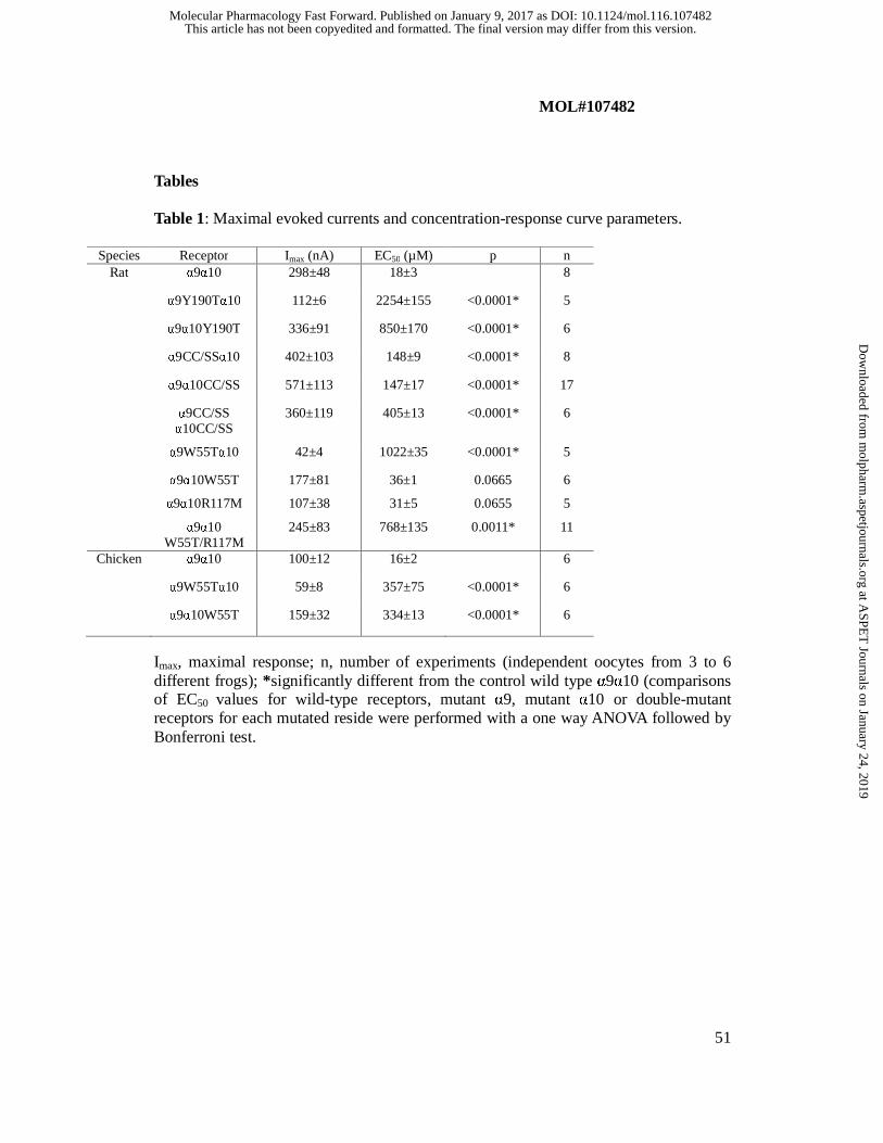
MOL#107482
51
Tables Table 1: Maximal evoked currents and concentration-response curve parameters.
Species Receptor Imax (nA) EC50 (µM) p n Rat α9α10 298±48 18±3 8
α9Y190Tα10 112±6 2254±155 <0.0001* 5
α9α10Y190T 336±91 850±170 <0.0001* 6
α9CC/SSα10 402±103 148±9 <0.0001* 8
α9α10CC/SS 571±113 147±17 <0.0001* 17
α9CC/SS α10CC/SS
360±119 405±13 <0.0001* 6
α9W55Tα10 42±4 1022±35 <0.0001* 5
α9α10W55T
α9α10R117M
α9α10 W55T/R117M
177±81
107±38
245±83
36±1
31±5
768±135
0.0665
0.0655
0.0011*
6
5
11
Chicken α9α10 100±12 16±2 6
α9W55Tα10 59±8 357±75 <0.0001* 6
α9α10W55T 159±32 334±13 <0.0001* 6
Imax, maximal response; n, number of experiments (independent oocytes from 3 to 6 different frogs); *significantly different from the control wild type α9α10 (comparisons of EC50 values for wild-type receptors, mutant α9, mutant α10 or double-mutant receptors for each mutated reside were performed with a one way ANOVA followed by Bonferroni test.
This article has not been copyedited and formatted. The final version may differ from this version.Molecular Pharmacology Fast Forward. Published on January 9, 2017 as DOI: 10.1124/mol.116.107482
at ASPE
T Journals on January 24, 2019
molpharm
.aspetjournals.orgD
ownloaded from
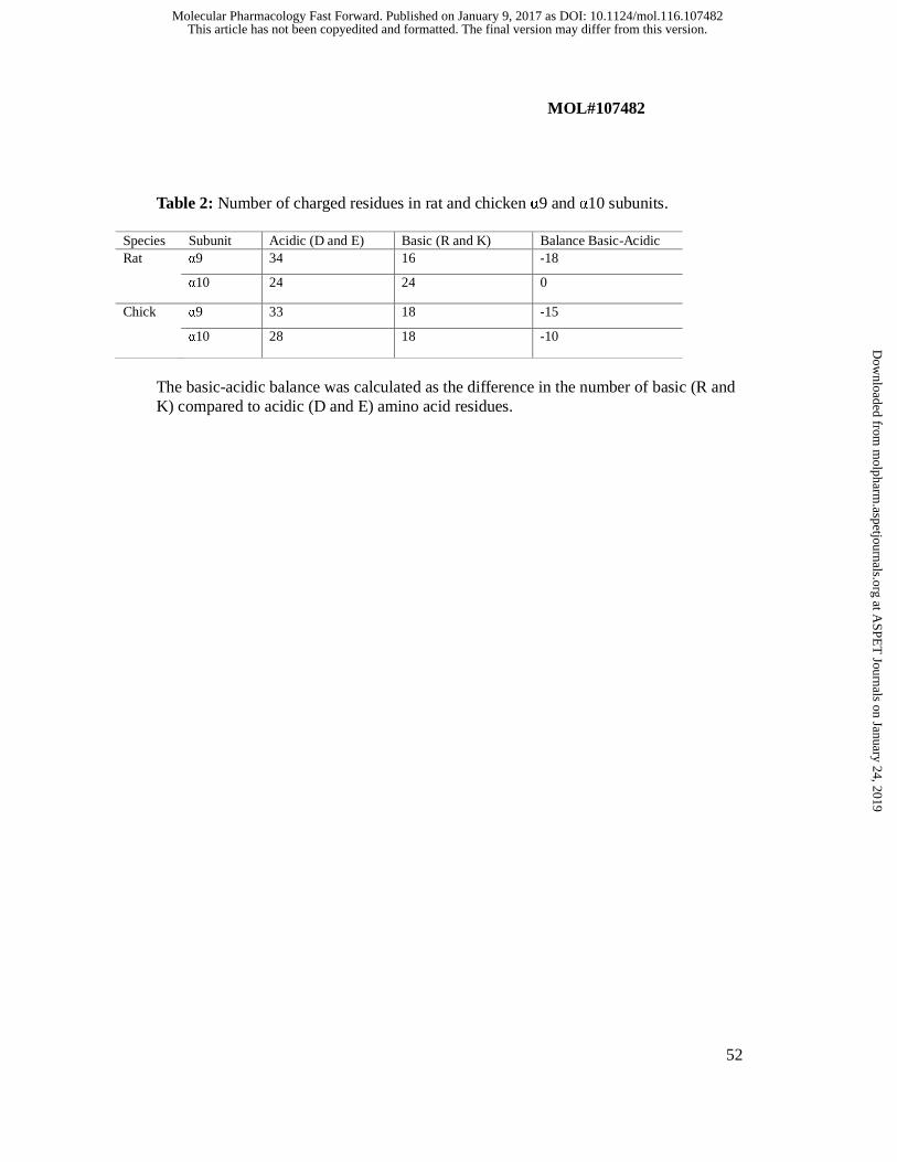
MOL#107482
52
Table 2: Number of charged residues in rat and chicken α9 and α10 subunits.
Species Subunit Acidic (D and E) Basic (R and K) Balance Basic-Acidic Rat α9 34 16 -18
α10 24 24 0
Chick α9 33 18 -15
α10 28 18 -10
The basic-acidic balance was calculated as the difference in the number of basic (R and K) compared to acidic (D and E) amino acid residues.
This article has not been copyedited and formatted. The final version may differ from this version.Molecular Pharmacology Fast Forward. Published on January 9, 2017 as DOI: 10.1124/mol.116.107482
at ASPE
T Journals on January 24, 2019
molpharm
.aspetjournals.orgD
ownloaded from

This article has not been copyedited and formatted. The final version may differ from this version.Molecular Pharmacology Fast Forward. Published on January 9, 2017 as DOI: 10.1124/mol.116.107482
at ASPE
T Journals on January 24, 2019
molpharm
.aspetjournals.orgD
ownloaded from
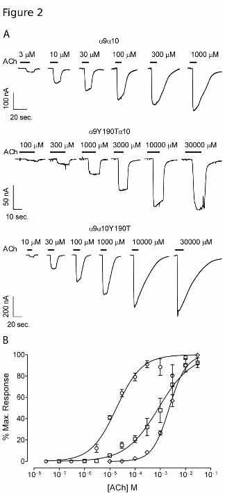
This article has not been copyedited and formatted. The final version may differ from this version.Molecular Pharmacology Fast Forward. Published on January 9, 2017 as DOI: 10.1124/mol.116.107482
at ASPE
T Journals on January 24, 2019
molpharm
.aspetjournals.orgD
ownloaded from

This article has not been copyedited and formatted. The final version may differ from this version.Molecular Pharmacology Fast Forward. Published on January 9, 2017 as DOI: 10.1124/mol.116.107482
at ASPE
T Journals on January 24, 2019
molpharm
.aspetjournals.orgD
ownloaded from
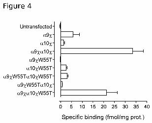
This article has not been copyedited and formatted. The final version may differ from this version.Molecular Pharmacology Fast Forward. Published on January 9, 2017 as DOI: 10.1124/mol.116.107482
at ASPE
T Journals on January 24, 2019
molpharm
.aspetjournals.orgD
ownloaded from

This article has not been copyedited and formatted. The final version may differ from this version.Molecular Pharmacology Fast Forward. Published on January 9, 2017 as DOI: 10.1124/mol.116.107482
at ASPE
T Journals on January 24, 2019
molpharm
.aspetjournals.orgD
ownloaded from
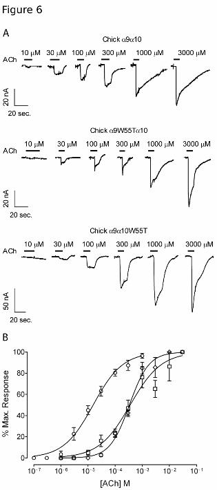
This article has not been copyedited and formatted. The final version may differ from this version.Molecular Pharmacology Fast Forward. Published on January 9, 2017 as DOI: 10.1124/mol.116.107482
at ASPE
T Journals on January 24, 2019
molpharm
.aspetjournals.orgD
ownloaded from
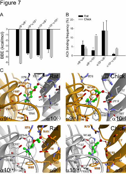
This article has not been copyedited and formatted. The final version may differ from this version.Molecular Pharmacology Fast Forward. Published on January 9, 2017 as DOI: 10.1124/mol.116.107482
at ASPE
T Journals on January 24, 2019
molpharm
.aspetjournals.orgD
ownloaded from

This article has not been copyedited and formatted. The final version may differ from this version.Molecular Pharmacology Fast Forward. Published on January 9, 2017 as DOI: 10.1124/mol.116.107482
at ASPE
T Journals on January 24, 2019
molpharm
.aspetjournals.orgD
ownloaded from

This article has not been copyedited and formatted. The final version may differ from this version.Molecular Pharmacology Fast Forward. Published on January 9, 2017 as DOI: 10.1124/mol.116.107482
at ASPE
T Journals on January 24, 2019
molpharm
.aspetjournals.orgD
ownloaded from