Dietary behaviour of man-eating lions as revealed by ... eating lions.pdf · lions as revealed by...
7
1 SCIENTIFIC REPORTS | 7: 904 | DOI:10.1038/s41598-017-00948-5 www.nature.com/scientificreports Dietary behaviour of man-eating lions as revealed by dental microwear textures Larisa R. G. DeSantis 1 & Bruce D. Patterson 2 Lions (Panthera leo) feed on diverse prey species, a range that is broadened by their cooperative hunting. Although humans are not typical prey, habitual man-eating by lions is well documented. Fathoming the motivations of the Tsavo and Mfuwe man-eaters (killed in 1898 in Kenya and 1991 in Zambia, respectively) may be elusive, but we can clarify aspects of their behaviour using dental microwear texture analysis. Specifically, we analysed the surface textures of lion teeth to assess whether these notorious man-eating lions scavenged carcasses during their depredations. Compared to wild-caught lions elsewhere in Africa and other large feliforms, including cheetahs and hyenas, dental microwear textures of the man-eaters do not suggest extreme durophagy (e.g. bone processing) shortly before death. Dental injuries to two of the three man-eaters examined may have induced shifts in feeding onto softer foods. Further, prompt carcass reclamation by humans likely limited the man-eaters’ access to bones. Man-eating was likely a viable alternative to hunting and/or scavenging ungulates due to dental disease and/or limited prey availability. Lions (Panthera leo) once inhabited much of Africa, southeastern Europe, and southwestern Asia 1 . Currently, lions (Panthera leo) occupy savannas and deserts in sub-Saharan Africa (excluding rainforests and the Sahara), with an isolated population located in the Gir Forest of India. ey are highly social, and males and females each live in persistent bonded groups 2 . Lion behaviours, diets, and social groupings all vary enormously in response to spatial or temporal shiſts in prey availability and habitat structure 3 . Smaller groups and females prey mainly on zebra and wildebeest whereas larger groups and males feed differentially on buffalo 4, 5 . Lions are known to con- sume a diverse suite of prey with preferences for gemsbok, buffalo, wildebeest, giraffe, zebra, omson’s gazelle, warthog, kongoni, and topi 4, 6 . Further, habitat and droughts can affect lion preferences and prey vulnerability to predation, with lions increasing the proportion of elephant calves consumed during droughts 7 . eir current range collapse, to 20% of historic values, is driven not by limited adaptability but rather by habitat loss and frag- mentation, prey depletion, and direct persecution 8 . Man-eating, or consumption of humans as women and children are oſten victims, has occasionally been a die- tary strategy of lions and other pantherines 9, 10 . Two notorious lions (popularized in the 1996 film e Ghost and the Darkness) terrorized people near Tsavo by repeatedly killing and consuming railway workers in 1898, and one from Mfuwe, Zambia consumed six people as recently as 1991 11 . Colonel J. H. Patterson, who eventually killed the Tsavo man-eaters in December 1898, estimated that they had killed and eaten 135 people 12 . However, stable iso- tope analysis of their hair and bone collagen suggests that they had consumed ~35 people, representing roughly 30% of the first man-eater’s diet (FMNH 23970) and ~13% of the second man-eater’s diet (FMNH 23969) 13 . e reasons for the lions’ differential reliance on humans, and for man-eating by lions in general, remain unclear. Many hypotheses have been proposed regarding the motivations of the man-eating lions, including an extended drought, a 1898 rinderpest outbreak that ravaged prey populations, various cultural causes, and/or dental dis- ease 11, 14, 15 . Evidence of dental disease is quite clear in two of the three man-eating lions. One lion (the first Tsavo man-eater), with a broken canine, developed a periapical abscess and lost three lower right incisors 15 (see Fig. 1). e pronounced toothwear and extensive cranial remodeling suggests that the lion had broken his canine sev- eral years earlier 15 . e second Tsavo man-eater had minor injuries including a fractured upper leſt carnassial and subsequent pulp exposure, although these types of injuries are fairly common and were unaccompanied by 1 Department of Earth and Environmental Sciences, Vanderbilt University, Nashville, TN, 37235-1805, USA. 2 Integrative Research Center, Field Museum of Natural History, Chicago, IL, 60605-2496, USA. Correspondence and requests for materials should be addressed to L.R.G.D. (email: [email protected]) Received: 29 November 2016 Accepted: 20 March 2017 Published: xx xx xxxx OPEN
Transcript of Dietary behaviour of man-eating lions as revealed by ... eating lions.pdf · lions as revealed by...
1Scientific RepoRts | 7: 904 | DOI:10.1038/s41598-017-00948-5
www.nature.com/scientificreports
Dietary behaviour of man-eating lions as revealed by dental microwear texturesLarisa R. G. DeSantis1 & Bruce D. Patterson2
Lions (Panthera leo) feed on diverse prey species, a range that is broadened by their cooperative hunting. Although humans are not typical prey, habitual man-eating by lions is well documented. Fathoming the motivations of the Tsavo and Mfuwe man-eaters (killed in 1898 in Kenya and 1991 in Zambia, respectively) may be elusive, but we can clarify aspects of their behaviour using dental microwear texture analysis. Specifically, we analysed the surface textures of lion teeth to assess whether these notorious man-eating lions scavenged carcasses during their depredations. Compared to wild-caught lions elsewhere in Africa and other large feliforms, including cheetahs and hyenas, dental microwear textures of the man-eaters do not suggest extreme durophagy (e.g. bone processing) shortly before death. Dental injuries to two of the three man-eaters examined may have induced shifts in feeding onto softer foods. Further, prompt carcass reclamation by humans likely limited the man-eaters’ access to bones. Man-eating was likely a viable alternative to hunting and/or scavenging ungulates due to dental disease and/or limited prey availability.
Lions (Panthera leo) once inhabited much of Africa, southeastern Europe, and southwestern Asia1. Currently, lions (Panthera leo) occupy savannas and deserts in sub-Saharan Africa (excluding rainforests and the Sahara), with an isolated population located in the Gir Forest of India. They are highly social, and males and females each live in persistent bonded groups2. Lion behaviours, diets, and social groupings all vary enormously in response to spatial or temporal shifts in prey availability and habitat structure3. Smaller groups and females prey mainly on zebra and wildebeest whereas larger groups and males feed differentially on buffalo4, 5. Lions are known to con-sume a diverse suite of prey with preferences for gemsbok, buffalo, wildebeest, giraffe, zebra, Thomson’s gazelle, warthog, kongoni, and topi4, 6. Further, habitat and droughts can affect lion preferences and prey vulnerability to predation, with lions increasing the proportion of elephant calves consumed during droughts7. Their current range collapse, to 20% of historic values, is driven not by limited adaptability but rather by habitat loss and frag-mentation, prey depletion, and direct persecution8.
Man-eating, or consumption of humans as women and children are often victims, has occasionally been a die-tary strategy of lions and other pantherines9, 10. Two notorious lions (popularized in the 1996 film The Ghost and the Darkness) terrorized people near Tsavo by repeatedly killing and consuming railway workers in 1898, and one from Mfuwe, Zambia consumed six people as recently as 199111. Colonel J. H. Patterson, who eventually killed the Tsavo man-eaters in December 1898, estimated that they had killed and eaten 135 people12. However, stable iso-tope analysis of their hair and bone collagen suggests that they had consumed ~35 people, representing roughly 30% of the first man-eater’s diet (FMNH 23970) and ~13% of the second man-eater’s diet (FMNH 23969)13. The reasons for the lions’ differential reliance on humans, and for man-eating by lions in general, remain unclear. Many hypotheses have been proposed regarding the motivations of the man-eating lions, including an extended drought, a 1898 rinderpest outbreak that ravaged prey populations, various cultural causes, and/or dental dis-ease11, 14, 15.
Evidence of dental disease is quite clear in two of the three man-eating lions. One lion (the first Tsavo man-eater), with a broken canine, developed a periapical abscess and lost three lower right incisors15 (see Fig. 1). The pronounced toothwear and extensive cranial remodeling suggests that the lion had broken his canine sev-eral years earlier15. The second Tsavo man-eater had minor injuries including a fractured upper left carnassial and subsequent pulp exposure, although these types of injuries are fairly common and were unaccompanied by
1Department of Earth and Environmental Sciences, Vanderbilt University, Nashville, TN, 37235-1805, USA. 2Integrative Research Center, Field Museum of Natural History, Chicago, IL, 60605-2496, USA. Correspondence and requests for materials should be addressed to L.R.G.D. (email: [email protected])
Received: 29 November 2016
Accepted: 20 March 2017
Published: xx xx xxxx
OPEN

www.nature.com/scientificreports/
2Scientific RepoRts | 7: 904 | DOI:10.1038/s41598-017-00948-5
disease16. Similarly, the man-eater from Mfuwe had fractured its right mandibular ramus. These injuries may have been decisive factors influencing their consumption of humans. While it is difficult to assess the motiva-tions of the man-eating lions, we can clarify aspects of their behaviour prior to their death. Most notably, we can assess if their circumstances caused them to rely more heavily on scavenging carcasses shortly before they were killed, as suggested by contemporary reports of the sounds of bone-crunching on the edge of camp12. Dental microwear texture analysis (DMTA) can clarify the textural properties of consumed food, including durophagy in carnivorans, and clearly distinguishes feliforms that eat primarily flesh (the cheetah, Acinonyx jubatus), from generalists (P. leo), and various hyenas which are known to fully consume carcasses, including bone17, 18.
In contrast to two-dimensional dental microwear, which relies on human identification and counting of microscopic wear features such as pits and scratches, 3D DMTA quantifies surfaces using scale-sensitive frac-tal analysis19–22. Complexity (Asfc) distinguishes taxa that consume brittle foods from taxa that consume softer ones19–23. Anisotropy (epLsar), the degree to which features share similar orientations, instead indicates tough food consumption when values are high19–23. Textural fill volume (Tfv) is a measure of the difference in vol-ume filled by large (10 µm) and small (2 µm) diameter square cuboids; high values indicate many deep features
Figure 1. Images of injuries to Tsavo’s 1st man-eater (a), FMNH 23970 and the Mfuwe man-eater (b), FMNH 163109. Image (a), Field Museum of Natural History image Z-94320_11c by John Weinstein documents a broken lower right canine (which had a periapical abscess) and loss of the lower three right incisors - presumably from the kick of a struggling prey - and subsequent over-eruption of the upper right incisors and rotation of the upper right canine both labially and mesially in the absence of the interlocking lower canine. In (b), multiple oval-shaped intraosseous lesions are visible on the right mandible, superficial to an occluded mandibular canal and associated with a chronically draining fistula15. Again, these injuries are consistent with blunt trauma from a powerful ungulate kick.

www.nature.com/scientificreports/
3Scientific RepoRts | 7: 904 | DOI:10.1038/s41598-017-00948-5
between these sizes21, 23. For extant carnivorous taxa, increased complexity and increased textural fill volume are associated with increased durophagy17, 18, 23–26.
Here, we compare dental microwear attributes of man-eating lions from Tsavo (from 1898) and from Mfuwe (from 1991) to wild-caught lions from throughout their range to assess if the dental microwear textures of man-eaters suggest extreme durophagy shortly before death. A secondary aim is to improve our understanding of how age, sex, and body size may influence access to carcasses in wild-caught lions. We test the hypothesis that the diets of man-eating lions consisted primarily of hard objects (e.g. bone), and that these lions mainly engaged in scavenging (perhaps out of desperation) prior to their death.
ResultsResults are illustrated in Figs 2 and 3 and summarized in Table 1 (all primary data are included in electronic sup-plementary materials, Supplemental Tables 1 and 2). As previously documented, complexity values of A. jubatus (median = 2.180) are significantly lower than for P. leo (p < 0.001) and all hyenas (p-values are all < 0.0001)18. P. leo (excluding the man-eaters) have complexity values that range from 0.258 to 11.096 (median = 3.669; Table 1) and are significantly lower than all hyenas (median values range from 5.316 in Parahyaena brunnea, 6.328 in Hyaena hyaena, to 7.354 in Crocuta crocuta; p-values are all < 0.05). Anisotropy values are indistinguishable between all feliform species here examined (as noted in prior work18). Textural fill volume of wild-caught feli-forms is lowest in A. jubatus, followed by P. leo, with A. jubatus and P. leo having significantly lower Tfv values than all hyenas (p < 0.05). The captive lions have the lowest Tfv values (median = 3.486).
The man-eating lions have DMTA values (Asfc, epLsar, and Tfv) indistinguishable from other wild-caught P. leo (all p-values > 0.69; Tables 1 and S1; Fig. 2), and captive and man-eating lions have nearly identical mean Asfc values (3.266 and 3.097, respectively). All man-eater Asfc values (ranging from 2.581 to 3.403) fall below the mean and median values for all hyenas18. Man-eaters are indistinguishable from all extant taxa in all DMTA attributes (likely due to their limited sample size, n = 3); however, statistical comparisons of Asfc values of man-eating
Figure 2. Digital elevation models of microwear surfaces of (a,b) wild-caught lions (FMNH 20762; FMNH 33479), (c) a captive lion (FMNH 54639), and (d–f) man-eating lions (Tsavo 1st man-eater, FMNH 23970; Tsavo 2nd man-eater, FMNH 23969; and Mfuwe man-eater, FMNH 163109). All models noted here represent 204 × 274 μm in area with relevant z-scale bars noted for each image (μm).
www.nature.com/scientificreports/
4Scientific RepoRts | 7: 904 | DOI:10.1038/s41598-017-00948-5
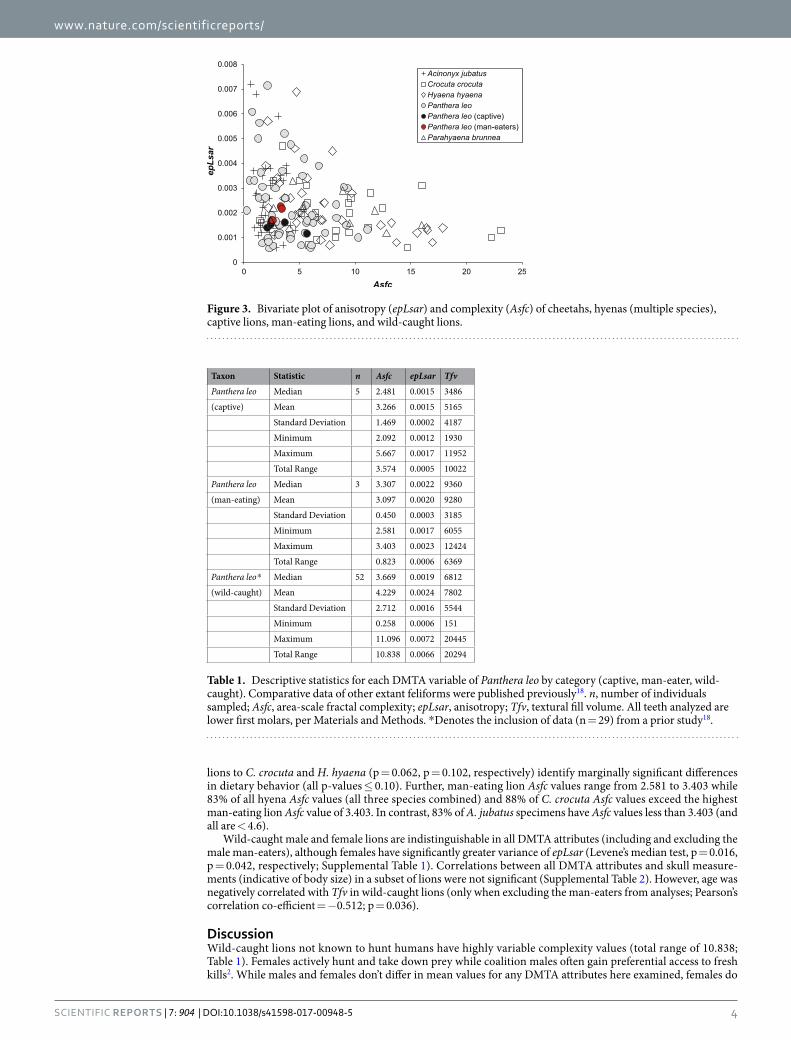
lions to C. crocuta and H. hyaena (p = 0.062, p = 0.102, respectively) identify marginally significant differences in dietary behavior (all p-values ≤ 0.10). Further, man-eating lion Asfc values range from 2.581 to 3.403 while 83% of all hyena Asfc values (all three species combined) and 88% of C. crocuta Asfc values exceed the highest man-eating lion Asfc value of 3.403. In contrast, 83% of A. jubatus specimens have Asfc values less than 3.403 (and all are < 4.6).
Wild-caught male and female lions are indistinguishable in all DMTA attributes (including and excluding the male man-eaters), although females have significantly greater variance of epLsar (Levene’s median test, p = 0.016, p = 0.042, respectively; Supplemental Table 1). Correlations between all DMTA attributes and skull measure-ments (indicative of body size) in a subset of lions were not significant (Supplemental Table 2). However, age was negatively correlated with Tfv in wild-caught lions (only when excluding the man-eaters from analyses; Pearson’s correlation co-efficient = −0.512; p = 0.036).
DiscussionWild-caught lions not known to hunt humans have highly variable complexity values (total range of 10.838; Table 1). Females actively hunt and take down prey while coalition males often gain preferential access to fresh kills2. While males and females don’t differ in mean values for any DMTA attributes here examined, females do
Figure 3. Bivariate plot of anisotropy (epLsar) and complexity (Asfc) of cheetahs, hyenas (multiple species), captive lions, man-eating lions, and wild-caught lions.
Taxon Statistic n Asfc epLsar Tfv
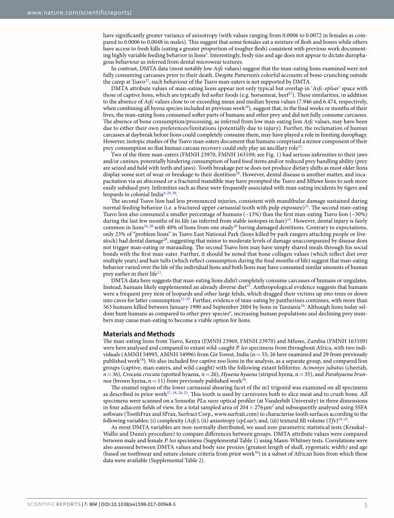
Panthera leo Median 5 2.481 0.0015 3486
(captive) Mean 3.266 0.0015 5165
Standard Deviation 1.469 0.0002 4187
Minimum 2.092 0.0012 1930
Maximum 5.667 0.0017 11952
Total Range 3.574 0.0005 10022
Panthera leo Median 3 3.307 0.0022 9360
(man-eating) Mean 3.097 0.0020 9280
Standard Deviation 0.450 0.0003 3185
Minimum 2.581 0.0017 6055
Maximum 3.403 0.0023 12424
Total Range 0.823 0.0006 6369
Panthera leo* Median 52 3.669 0.0019 6812
(wild-caught) Mean 4.229 0.0024 7802
Standard Deviation 2.712 0.0016 5544
Minimum 0.258 0.0006 151
Maximum 11.096 0.0072 20445
Total Range 10.838 0.0066 20294
Table 1. Descriptive statistics for each DMTA variable of Panthera leo by category (captive, man-eater, wild-caught). Comparative data of other extant feliforms were published previously18. n, number of individuals sampled; Asfc, area-scale fractal complexity; epLsar, anisotropy; Tfv, textural fill volume. All teeth analyzed are lower first molars, per Materials and Methods. *Denotes the inclusion of data (n = 29) from a prior study18.
www.nature.com/scientificreports/
5Scientific RepoRts | 7: 904 | DOI:10.1038/s41598-017-00948-5
have significantly greater variance of anisotropy (with values ranging from 0.0006 to 0.0072 in females as com-pared to 0.0006 to 0.0048 in males). This suggest that some females eat a mixture of flesh and bones while others have access to fresh kills (eating a greater proportion of tougher flesh) consistent with previous work document-ing highly variable feeding behavior in lions3. Interestingly, body size and age does not appear to dictate duropha-gous behaviour as inferred from dental microwear textures.
In contrast, DMTA data (most notably low Asfc values) suggest that the man-eating lions examined were not fully consuming carcasses prior to their death. Despite Patterson’s colorful accounts of bone-crunching outside the camp at Tsavo12, such behaviour of the Tsavo man-eaters is not supported by DMTA.
DMTA attribute values of man-eating lions appear not only typical but overlap in ‘Asfc-eplsar’ space with those of captive lions, which are typically fed softer foods (e.g. horsemeat, beef27). These similarities, in addition to the absence of Asfc values close to or exceeding mean and median hyena values (7.946 and 6.474, respectively, when combining all hyena species included in previous work18), suggest that, in the final weeks or months of their lives, the man-eating lions consumed softer parts of humans and other prey and did not fully consume carcasses. The absence of bone consumption/processing, as inferred from low man-eating lion Asfc values, may have been due to either their own preferences/limitations (potentially due to injury). Further, the reclamation of human carcasses at daybreak before lions could completely consume them, may have played a role in limiting durophagy. However, isotopic studies of the Tsavo man-eaters document that humans comprised a minor component of their prey consumption so that human carcass recovery could only play an ancillary role13.
Two of the three man-eaters (FMNH 23970, FMNH 163109; see Fig. 1) had serious infirmities to their jaws and/or canines, potentially hindering consumption of hard food items and/or reduced prey handling ability (prey are seized and held with teeth and jaws). Tooth breakage per se does not produce dietary shifts as most older lions display some sort of wear or breakage to their dentition28. However, dental disease is another matter, and inca-pacitation via an abscessed or a fractured mandible may have prompted the Tsavo and Mfuwe lions to seek more easily subdued prey. Infirmities such as these were frequently associated with man-eating incidents by tigers and leopards in colonial India9, 29, 30.
The second Tsavo lion had less pronounced injuries, consistent with mandibular damage sustained during normal feeding behavior (i.e. a fractured upper carnassial tooth with pulp exposure)15. The second man-eating Tsavo lion also consumed a smaller percentage of humans (~13%) than the first man-eating Tsavo lion (~30%) during the last few months of its life (as inferred from stable isotopes in hair)13. However, dental injury is fairly common in lions16, 28 with 40% of lions from one study28 having damaged dentitions. Contrary to expectations, only 23% of “problem lions” in Tsavo East National Park (lions killed by park rangers attacking people or live-stock) had dental damage28, suggesting that minor to moderate levels of damage unaccompanied by disease does not trigger man-eating or marauding. The second Tsavo lion may have simply shared meals through his social bonds with the first man-eater. Further, it should be noted that bone collagen values (which reflect diet over multiple years) and hair tufts (which reflect consumption during the final months of life) suggest that man-eating behavior varied over the life of the individual lions and both lions may have consumed similar amounts of human prey earlier in their life13.
DMTA data here suggests that man-eating lions didn’t completely consume carcasses of humans or ungulates. Instead, humans likely supplemented an already diverse diet31. Anthropological evidence suggests that humans were a frequent prey item of leopards and other large felids, which dragged their victims up into trees or down into caves for latter consumption32–35. Further, evidence of man-eating by pantherines continues, with more than 563 humans killed between January 1990 and September 2004 by lions in Tanzania10. Although lions today sel-dom hunt humans as compared to other prey species6, increasing human populations and declining prey num-bers may cause man-eating to become a viable option for lions.
Materials and MethodsThe man-eating lions from Tsavo, Kenya (FMNH 23969, FMNH 23970) and Mfuwe, Zambia (FMNH 163109) were here analysed and compared to extant wild-caught P. leo specimens from throughout Africa, with two indi-viduals (AMNH 54995, AMNH 54996) from Gir Forest, India (n = 55; 26 here examined and 29 from previously published work18). We also included five captive zoo lions in the analysis, as a separate group, and compared lion groups (captive, man-eaters, and wild-caught) with the following extant feliforms: Acinonyx jubatus (cheetah, n = 36), Crocuta crocuta (spotted hyaena, n = 26), Hyaena hyaena (striped hyena, n = 35), and Parahyaena brun-nea (brown hyena, n = 11) from previously published work18.
The enamel region of the lower carnassial shearing facet of the m1 trigonid was examined on all specimens as described in prior work17, 18, 24, 25. This tooth is used by carnivores both to slice meat and to crush bone. All specimens were scanned on a Sensofar PLu neox optical profiler (at Vanderbilt University) in three dimensions in four adjacent fields of view, for a total sampled area of 204 × 276 µm2 and subsequently analysed using SSFA software (ToothFrax and SFrax, Surfract Corp., www.surfrait.com) to characterise tooth surfaces according to the following variables: (i) complexity (Asfc); (ii) anisotropy (epLsar); and, (iii) textural fill volume (Tfv)19–23.
As most DMTA variables are non-normally distributed, we used non-parametric statistical tests (Kruskal–Wallis and Dunn’s procedure) to compare differences between groups. DMTA attribute values were compared between male and female P. leo specimens (Supplemental Table 1) using Mann-Whitney tests. Correlations were also assessed between DMTA values and body size proxies (greatest length of skull, zygomatic width) and age (based on toothwear and suture closure criteria from prior work36) in a subset of African lions from which these data were available (Supplemental Table 2).
www.nature.com/scientificreports/
6Scientific RepoRts | 7: 904 | DOI:10.1038/s41598-017-00948-5
References 1. Barnett, R. et al. Phylogeography of lions (Panthera leo ssp.) reveals three distinct taxa and a late Pleistocene reduction in genetic
diversity. Mol. Ecol 18, 1668–1677, doi:10.1111/mec.2009.18.issue-8 (2009). 2. Schaller, G. B. The Serengeti Lion: A Study of Predator-Prey Relations. (University of Chicago Press, 1972). 3. Patterson, B. D. On the nature and significance of variability in lions (Panthera leo). Evol. Biol 34, 61–71, doi:10.1007/s11692-007-
9003-6 (2007). 4. Scheel, D. & Packer, C. Variation in predation by lions: tracking a movable feast in Serengeti II: Dynamics, Management, and
Conservation of an Ecosystem (eds. Sinclair, A. R. E. & Arcese, P.) 299–314 (University of Chicago Press, 1995). 5. Funston, P. J., Mills, M. G. L. & Biggs, H. C. Factors affecting the hunting success of male and female lions in the Kruger National
Park. J. Zool 253, 419–431, doi:10.1017/S0952836901000395 (2001). 6. Hayward, M. W. & Kerley, G. I. Prey preferences of the lion (Panthera leo). J. Zool. 267, 309–322, doi:10.1017/S0952836905007508
(2005). 7. Loveridge, A. J., Hunt, J. E., Murindagomo, F. & Macdonald, D. W. Influence of drought on predation of elephant (Loxodonta
africana) calves by lions (Panthera leo) in an African wooded savannah. J. Zool. 270, 523–530, doi:10.1371/journal.pone.0055182 (2006).
8. Riggio, J. et al. The size of savannah Africa: a lion’s (Panthera leo) view. Biodivers. Conserv. 22, 17–35, doi:10.1007/s10531-012-0381-4 (2013).
9. Corbett, J. E. Man-eaters of Kumaon. (Oxford University Press, 1946). 10. Packer, C., Ikanda, D., Kissui, B. & Kushnir, H. Lion attacks on humans in Tanzania. Nature 436, 927–928, doi:10.1038/436927a
(2005). 11. Patterson, B. D. The lions of Tsavo. (McGraw-Hill, 2004). 12. Patterson, J. H. The man-eating lions of Tsavo. (Field Museum of Natural History, Zoology, 1925). 13. Yeakel, J. D. et al. Cooperation and individuality among man-eating lions. Proc. Natl. Acad. Sci. USA 106, 19040–19043, doi:10.1073/
pnas.0905309106 (2009). 14. Kerbis Peterhans, J. C. & Gnoske, T. P. The science of ‘man-eating’ among lions Panthera leo with a reconstruction of the natural
history of the ‘man-eaters of Tsavo’. J. East Afr. Natl. Hist 90, 1–40, doi:10.2982/0012-8317(2001)90[1:TSOMAL]2.0.CO;2 (2001). 15. Neiburger, E. J. & Patterson, B. D. The man-eaters with bad teeth. NY State Dent. J 66, 26–29 (2000). 16. Van Valkenburgh, B. Costs of carnivory: tooth fracture in Pleistocene and Recent carnivorans. Biol. J. Linn. Soc 96, 68–81,
doi:10.1111/j.1095-8312.2008.01108.x (2009). 17. Schubert, B. W., Ungar, P. S. & DeSantis, L. R. G. Carnassial microwear and dietary behaviour in large carnivorans. J. Zool 280,
257–263, doi:10.1111/j.1469-7998.2009.00656.x (2010). 18. DeSantis, L. R. G. et al. Assessing niche conservatism using a multi-proxy approach: dietary ecology of extinct and extant spotted
hyenas. Paleobio, (https://doi.org/10.1017/pab.2016.45) (2017). 19. Ungar, P. S., Brown, C. A., Bergstrom, T. S. & Walker, A. Quantification of Dental Microwear by Tandem Scanning Confocal
Microscopy and Scale‐Sensitive Fractal Analyses. Scanning 25, 185–193, doi:10.1002/sca.4950250405 (2003). 20. Scott, R. S. et al. Dental microwear texture analysis shows within-species diet variability in fossil hominins. Nature 436, 693–695,
doi:10.1038/nature03822 (2005). 21. Scott, R. S. et al. Dental microwear texture analysis: technical considerations. J. Hum. Evol. 51, 339–349, doi:10.1016/j.
jhevol.2006.04.006 (2006). 22. DeSantis, L. R. G. et al. Direct comparisons of 2D and 3D dental microwear proxies in extant herbivorous and carnivorous
mammals. PLoS One 8(8), e71428, doi:10.1371/journal.pone.0071428 (2013). 23. DeSantis, L. R. G. Dental microwear textures: reconstructing diets of fossil mammals. Surf. Topo.: Metr. Prop 4, 023002 (2016). 24. DeSantis, L. R. G., Schubert, B. W., Scott, J. R. & Ungar, P. S. Implications of diet for the extinction of saber-toothed cats and
American lions. PLoS One 7(12), e52453, doi:10.1371/journal.pone.0052453 (2012). 25. DeSantis, L. R. G. & Haupt, R. J. Cougars’ key to survival through the Late Pleistocene extinction: insights from dental microwear
texture analysis. Biology. Lett 10(4), 20140203–20140203, doi:10.1098/rsbl.2014.0203 (2014). 26. DeSantis, L. R. G. et al. Dental Microwear Textures of Carnivorans from the La Brea Tar Pits, California, and Potential Extinction
Implications in La Brea and beyond: the paleontology of asphalt-preserved biotas (ed. Harris, J. M.) 37–52 (Los Angeles: Natural History Museum of Los Angeles County, 2015)
27. Patterson, B. D., Kays, R. W., Kasiki, S. M. & Sebestyen, V. M. Developmental effects of climate on the lion’s mane (Panthera leo). J. Mamm 87, 193–200, doi:10.1644/05-MAMM-A-226R2.1 (2006).
28. Patterson, B. D., Neiburger, E. J. & Kasiki, S. M. Tooth breakage and dental disease as causes of carnivore-human conflicts. J. Mamm 84, 190–196, doi:10.1644/1545-1542(2003)084<0190:TBADDA>2.0.CO;2 (2003).
29. Corbett, J. & Sheppard, R. The man-eating leopard of Rudraprayag. (Oxford University Press, 1948). 30. Corbett, J. The Temple Tiger and more Man-eaters of Kumaon. (Oxford University Press, 1954). 31. Packer, C., Swanson, A., Ikanda, D. & Kushnir, H. Fear of darkness, the full moon and the nocturnal ecology of African lions. PLoS
One 6(7), e22285, doi:10.1371/journal.pone.0022285 (2011). 32. Brain, C. K. New finds at the Swartkrans australopithecine site. Nature 225, 1112–1119, doi:10.1038/2251112a0 (1970). 33. Vrba, E. S. Some evidence of chronology and palaeoecology of Sterkfontein, Swartkrans and Kromdraai from the fossil Bovidae.
Nature 254, 301–304, doi:10.1038/254301a0 (1975). 34. de Ruiter, D. J. & Berger, L. R. Leopards as taphonomic agents in dolomitic caves—implications for bone accumulations in the
hominid-bearing deposits of South Africa. J. Archaeol. Sci. 27, 665–684, doi:10.1006/jasc.1999.0470 (2000). 35. Pickering, T. R., Domı́nguez-Rodrigo, M., Egeland, C. P. & Brain, C. K. Beyond leopards: tooth marks and the contribution of
multiple carnivore taxa to the accumulation of the Swartkrans Member 3 fossil assemblage. J. Hum. Evol. 46, 595–604, doi:10.1016/j.jhevol.2004.03.002 (2004).
36. Smuts, G. L., Anderson, J. L. & Austin, J. C. Age determination of the African lion (Panthera leo). J. Zool 185, 115–146, doi:10.1111/j.1469-7998.1978.tb03317.x (1978).
AcknowledgementsWe are grateful to many curators and collections staff, including those from the Field Museum of Natural History, for access and assistance with specimens. This study was supported by the National Science Foundation (EAR1053839 to LRGD), Vanderbilt University, and the Brown Fund of the Field Museum of Natural History.
Author ContributionsBoth authors (L.R.G.D. and B.D.P.) contributed to the design of the paper. L.R.G.D. performed analyses and wrote the paper with input from B.D.P.

www.nature.com/scientificreports/
7Scientific RepoRts | 7: 904 | DOI:10.1038/s41598-017-00948-5
Additional InformationSupplementary information accompanies this paper at doi:10.1038/s41598-017-00948-5Competing Interests: The authors declare that they have no competing interests.Publisher's note: Springer Nature remains neutral with regard to jurisdictional claims in published maps and institutional affiliations.
Open Access This article is licensed under a Creative Commons Attribution 4.0 International License, which permits use, sharing, adaptation, distribution and reproduction in any medium or
format, as long as you give appropriate credit to the original author(s) and the source, provide a link to the Cre-ative Commons license, and indicate if changes were made. The images or other third party material in this article are included in the article’s Creative Commons license, unless indicated otherwise in a credit line to the material. If material is not included in the article’s Creative Commons license and your intended use is not per-mitted by statutory regulation or exceeds the permitted use, you will need to obtain permission directly from the copyright holder. To view a copy of this license, visit http://creativecommons.org/licenses/by/4.0/. © The Author(s) 2017