Diet and Non-alcoholic Steatohepatitis (NASH): A … and Non-alcoholic Steatohepatitis (NASH): A...
72
Diet and Non-alcoholic Steatohepatitis (NASH): A Risk Factor for Liver Cancer Donald B. Jump, Ph.D. Nutrition Program Oregon State University Corvallis, OR October 2014
Transcript of Diet and Non-alcoholic Steatohepatitis (NASH): A … and Non-alcoholic Steatohepatitis (NASH): A...

Diet and Non-alcoholic Steatohepatitis (NASH):
A Risk Factor for Liver Cancer
Donald B. Jump, Ph.D. Nutrition Program
Oregon State University Corvallis, OR
October 2014

Outline • Background on Primary Liver Cancer • Background on Non-Alcoholic Fatty
Liver Disease [NAFLD] and NASH – Link between NASH and Liver Cancer
• Prevention of NASH – Inflammation – Fibrosis – Metabolomics
• Is NASH Reversible? • Wrap Up

Primary Liver Cancer • Hepatocellular carcinoma
– Most common primary liver cancer; involves hepatic parenchymal cells – 5th most common human cancer – 3rd most frequent cause of cancer death world wide – Best Pract Res Clin Gastroenterol 28(5):753-770 [2014] – http://www.cancer.gov/cancertopics/pdq/treatment/adult-primary-liver/Patient/page1#Keypoint2
• Cholangiocarcinoma – 2nd most common liver cancer; involves biliary epithelial cells – Accounts for ~10% of primary liver cancers – J Hepatobiliary Pancreat Sci In press [2014]
• Chronic liver disease sets the stage for dis-regulated regeneration of
hepatic and biliary epithelia.
• Current treatment options are limited to surgery and drugs (sorafenib)

Risk Factors for Hepatocellular Carcinoma
Gastroenterology 142: 1411-1413 [2012]
USA & Europe
Sub-Saharan Africa

Risk Factors for Hepatocellular Carcinoma
Gastroenterology 142: 1411-1413 [2012]
Major risk factor of HCC

Risk Factors for Hepatocellular Carcinoma
Gastroenterology 142: 1411-1413 [2012]
Cholesterol
Non-alcoholic Fatty Liver Disease (NAFLD)
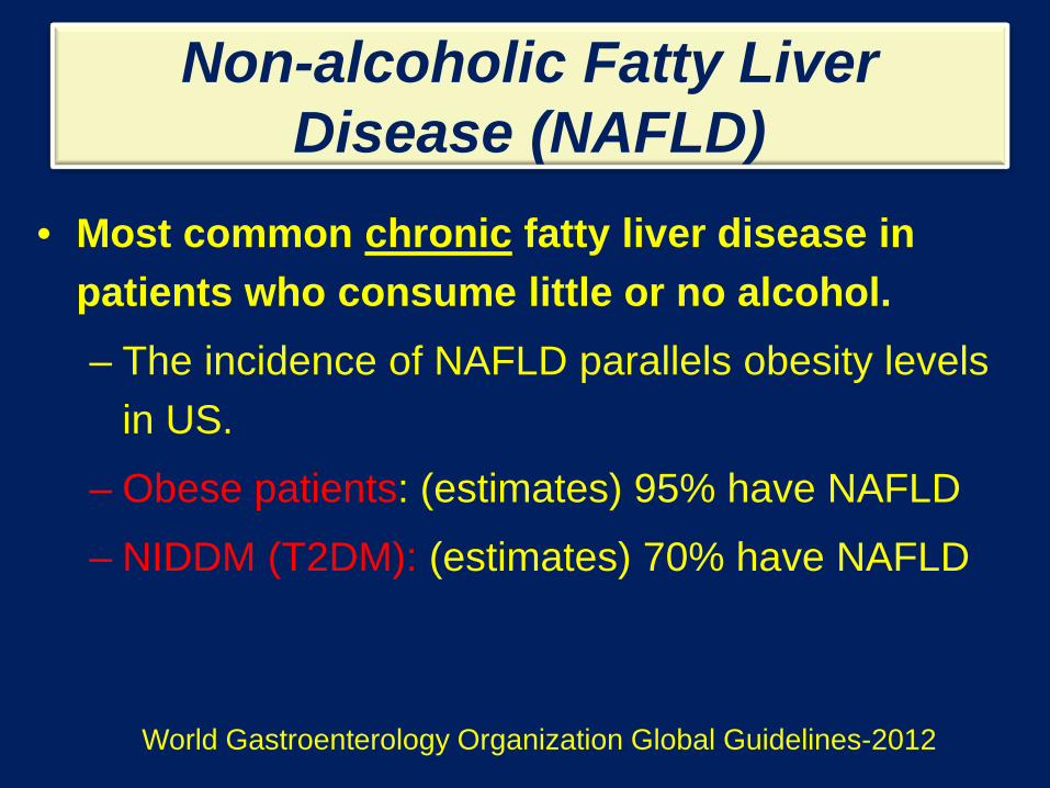
• Most common chronic fatty liver disease in patients who consume little or no alcohol. – The incidence of NAFLD parallels obesity levels
in US.
– Obese patients: (estimates) 95% have NAFLD
– NIDDM (T2DM): (estimates) 70% have NAFLD
World Gastroenterology Organization Global Guidelines-2012

NAFLD- NASH-Cirrhosis • A spectrum of diseases from benign fatty liver
(steatosis) to non-alcoholic steatohepatitis (NASH), i.e., hepatic inflammation. – Chronic inflammation promotes liver damage and fibrosis (scar). – 20-30% of patients diagnosed with NAFLD develop NASH
– NASH can progress to cirrhosis, a major risk factor for primary hepatocellular cancer.
– Advanced cirrhosis, resulting from NASH, is predicted to be a major cause for liver transplants by 2020.
World Gastroenterology Organization Global Guidelines-2012
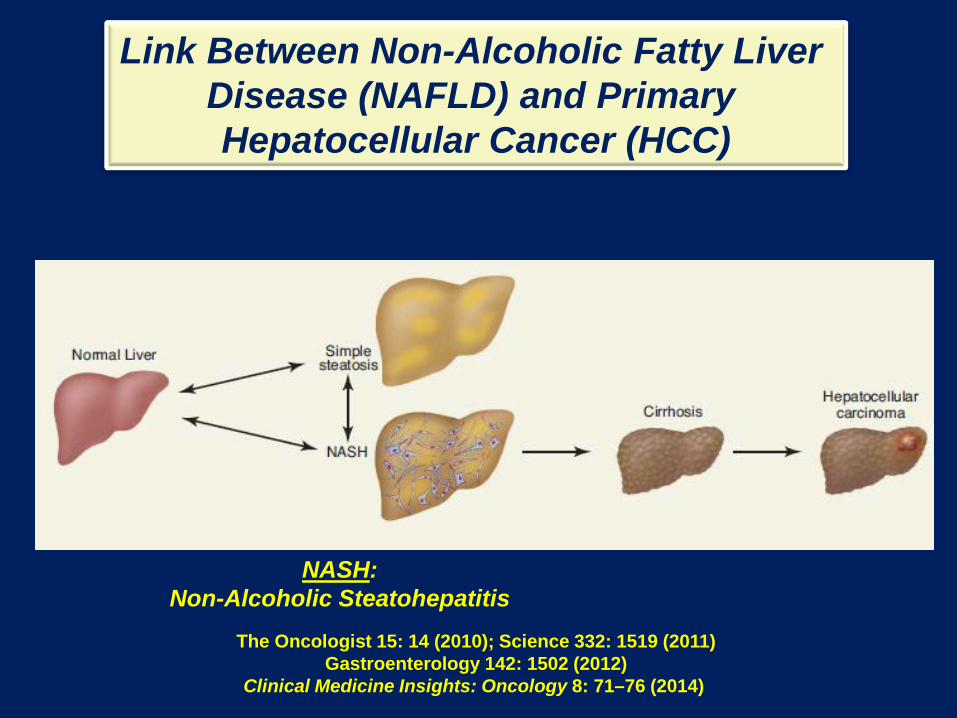
Link Between Non-Alcoholic Fatty Liver Disease (NAFLD) and Primary Hepatocellular Cancer (HCC)
NASH: Non-Alcoholic Steatohepatitis
The Oncologist 15: 14 (2010); Science 332: 1519 (2011) Gastroenterology 142: 1502 (2012)
Clinical Medicine Insights: Oncology 8: 71–76 (2014)

Link Between Non-Alcoholic Fatty Liver Disease (NAFLD) and Primary Hepatocellular Cancer (HCC)
6-30% of the General Population Develop NAFLD. Incidence Parallels the Incidence of Obesity & T2DM.
The Oncologist 15: 14 (2010); Science 332: 1519 (2011) Gastroenterology 142: 1502 (2012)
Clinical Medicine Insights: Oncology 8: 71–76 (2014)
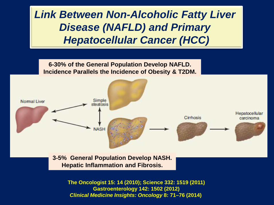
3-5% General Population Develop NASH. Hepatic Inflammation and Fibrosis.
10-30% of NASH Patients Develop Hepatic Cirrhosis
Link Between Non-Alcoholic Fatty Liver Disease (NAFLD) and Primary Hepatocellular Cancer (HCC)
6-30% of the General Population Develop NAFLD. Incidence Parallels the Incidence of Obesity & T2DM.
The Oncologist 15: 14 (2010); Science 332: 1519 (2011) Gastroenterology 142: 1502 (2012)
Clinical Medicine Insights: Oncology 8: 71–76 (2014)

6-30% of the General Population Develop NAFLD. Incidence Parallels the Incidence of Obesity & T2DM.
The Oncologist 15: 14 (2010); Science 332: 1519 (2011) Gastroenterology 142: 1502 (2012)
Clinical Medicine Insights: Oncology 8: 71–76 (2014)
3-5% General Population Develop NASH. Hepatic Inflammation and Fibrosis.
2-4% of NASH Patients Develop HCC
10-30% of NASH Patients Develop Hepatic Cirrhosis
Link Between Non-Alcoholic Fatty Liver Disease (NAFLD) and Primary Hepatocellular Cancer (HCC)

Hepatic Fibrosis is a Precursor to HCC
Cohen, Hobbs & Horton, Science 332: 1519 (2011)

Modified From Annual Review of Pathology: Mechanisms of Disease (2010) 5: 145-171
Benign Steatosis NASH
Factors Driving NAFLD Progression to NASH: Diet (High fat, sucrose, cholesterol)
Body Fat & Its Distribution Insulin Resistance
Inflammatory factors, like Gut-derived microbial components, e.g., Endotoxin
Genetics

1
Visceral Obesity
SFA/MUFA>>>PUFA
Modified From Annual Review of Pathology: Mechanisms of Disease (2010) 5: 145-171
Benign Steatosis NASH
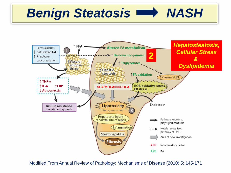
2 Hepatosteatosis, Cellular Stress
& Dyslipidemia
SFA/MUFA>>>PUFA
Modified From Annual Review of Pathology: Mechanisms of Disease (2010) 5: 145-171
Benign Steatosis NASH
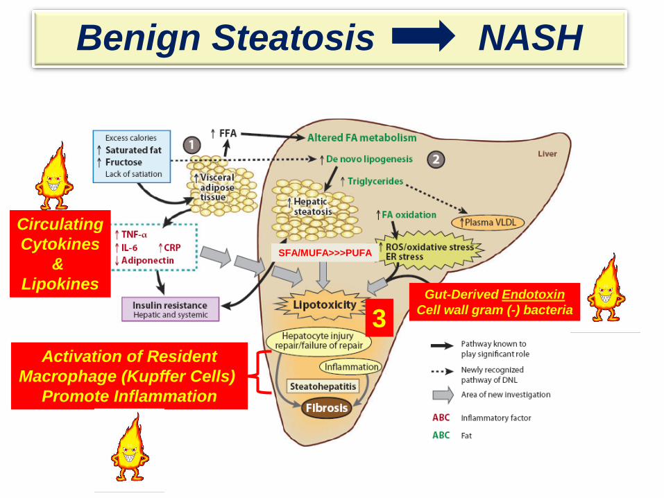
3 Activation of Resident
Macrophage (Kupffer Cells) Promote Inflammation
Circulating Cytokines
& Lipokines
SFA/MUFA>>>PUFA
Benign Steatosis NASH
Gut-Derived Endotoxin Cell wall gram (-) bacteria

Hepatic Damage Cell Injury
& Repair (Fibrosis)
Modified From Annual Review of Pathology: Mechanisms of Disease (2010) 5: 145-171
SFA/MUFA>>>PUFA
Benign Steatosis NASH
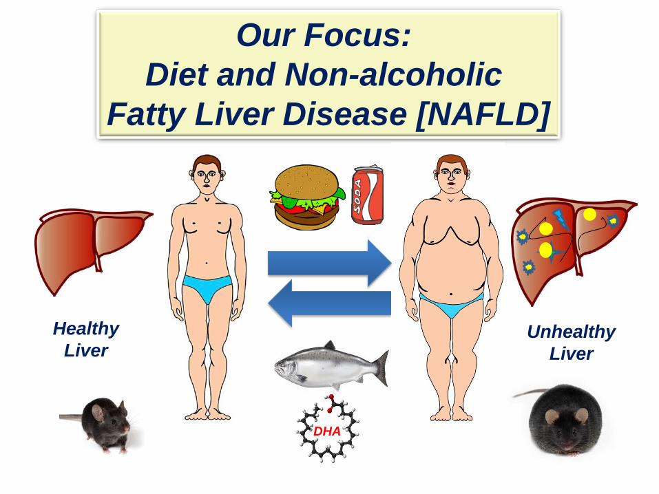
Our Focus: Diet and Non-alcoholic
Fatty Liver Disease [NAFLD]
Healthy Liver
Unhealthy Liver
DHA

Prevention of Diet-Induced NAFLD & NASH Using ω3 Fatty Acids
• Can dietary intervention prevent western diet-induced NAFLD/NASH? – Western diet (WD) moderately high fat,
sucrose & cholesterol – ω3 Polyunsaturated fatty acids; ω3 PUFA
CH3
COOH
CH3
HOOC
Docosahexaenoic Acid 22:6,ω3; DHA
20:5,ω3 EPA
Eicosapentaenoic Acid 20:5,ω3; EPA
Chris Depner, Ph.D.
22:6,ω3 DHA
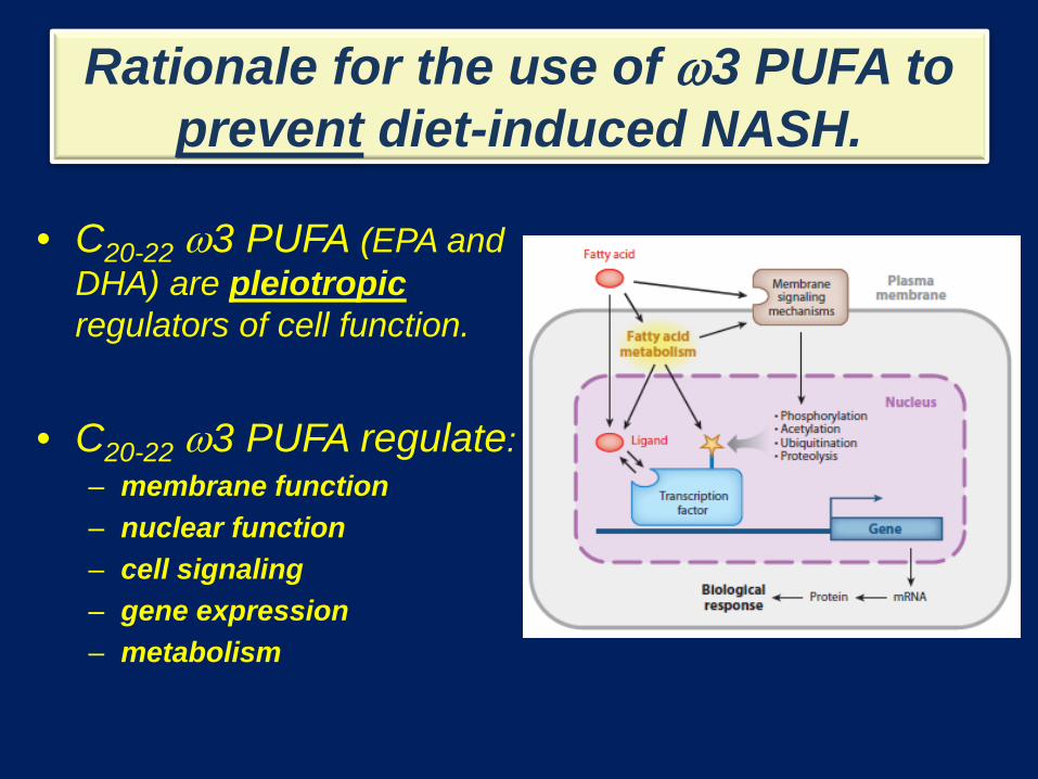
Rationale for the use of ω3 PUFA to prevent diet-induced NASH.
• C20-22 ω3 PUFA (EPA and DHA) are pleiotropic regulators of cell function.
• C20-22 ω3 PUFA regulate: – membrane function – nuclear function – cell signaling – gene expression – metabolism

Rationale for the use of ω3 PUFA to prevent diet-induced NASH.
• NASH is a chronic inflammatory disease
• C20-22 ω3 PUFA are: – Anti-inflammatory
– Inhibit fatty acid synthesis – Induce triglyceride catabolism – Induce fatty acid oxidation
– Lowers blood triglycerides
• (Lovaza®, GSK, [EPA + DHA]) • Human dose: 1-2% total calories/d
GRAS & Established Hypolipemic Nutrient/Drug
Potential to Lower
Hepatic Lipid

Approach We developed a mouse model of NASH that recapitulates many of the clinical features of human NAFLD/NASH.
– LDLR-/- mouse + the “Western Diet”
– Clinical features included: • hepatosteatosis • ballooning hepatocytes • Inflammation • oxidative stress • fibrosis

Distribution of Calories
Western Diet
43% Carbohydrate Sugar>complex CHO
15% Protein
42% Fat SFA>>>PUFA
Red Meat Processed Meat
Fried Food (French Fries) High-Fat Dairy Products
Refined Grains Sweets & Desserts Sugary Beverages
Cholesterol: >300 mg/day

American Heart Assoc. & American Diabetes Assoc. Dietary Recommendations
Distribution of Calories
50% Carbohydrate
Complex CHO>>Sugar
15% Protein
<35% Fat SFA=MUFA=PUFA
Western Diet
43% Carbohydrate Sugar>complex CHO
15% Protein
42% Fat SFA>>>PUFA
Lean Meats (Poultry) Oily Fish (ω3 PUFA)
Vegetables Whole Grains
Low Fat Dairy Products Fruit
Red Meat Processed Meat
Fried Food (French Fries) High-Fat Dairy Products
Refined Grains Sweets & Desserts Sugary Beverages
Cholesterol: <300 mg/day Cholesterol: >300 mg/day

Obese Mice LDL-R-/-
Overnight Fast
Blood, Urine, Liver Obese
Lean
Lean LDL-R-/- Male Mice
42 + 2 g
28 + 2 g
4 WD Groups 1. Olive 2. EPA 3. DHA 4. EPA + DHA N=8/group 16 weeks
N=8; Chow group 16 weeks
The NASH Model
Diets: Chow (Purina 5053) & Western Diet (WD) WD is supplemented with olive oil, EPA and/or DHA;
supplemental fats are 2% energy; all WD are isocaloric.
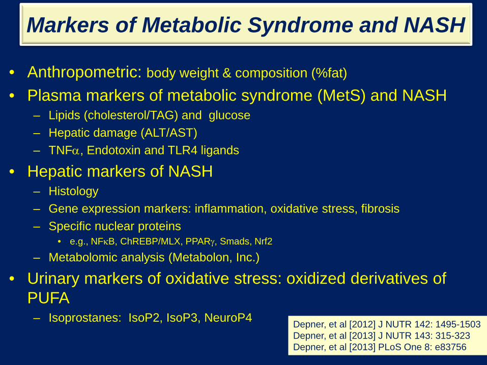
Markers of Metabolic Syndrome and NASH
• Anthropometric: body weight & composition (%fat)
• Plasma markers of metabolic syndrome (MetS) and NASH – Lipids (cholesterol/TAG) and glucose – Hepatic damage (ALT/AST) – TNFα, Endotoxin and TLR4 ligands
• Hepatic markers of NASH – Histology – Gene expression markers: inflammation, oxidative stress, fibrosis – Specific nuclear proteins
• e.g., NFκB, ChREBP/MLX, PPARγ, Smads, Nrf2
– Metabolomic analysis (Metabolon, Inc.)
• Urinary markers of oxidative stress: oxidized derivatives of PUFA – Isoprostanes: IsoP2, IsoP3, NeuroP4 Depner, et al [2012] J NUTR 142: 1495-1503
Depner, et al [2013] J NUTR 143: 315-323 Depner, et al [2013] PLoS One 8: e83756

Leukocyte Infiltration
Inflammation Induced by the Western Diet Fatty & Inflammed Liver Normal Liver
H/E Stain
PV
PV
CV
Hepatocytes
Lipid Droplet
Leukocytes

Fibrosis Induced by the Western Diet Normal Liver
Trichrome Stain
CV
CV
PV Fibrosis
PV
Branching Fibrosis
PV
Fatty & Fibrotic Liver
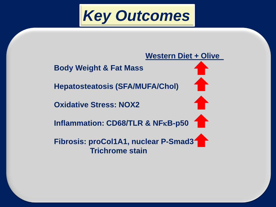
Key Outcomes
Western Diet + Olive Body Weight & Fat Mass Hepatosteatosis (SFA/MUFA/Chol) Oxidative Stress: NOX2 Inflammation: CD68/TLR & NFκB-p50 Fibrosis: proCol1A1, nuclear P-Smad3 Trichrome stain

Western Diet + Olive Western Diet + DHA Body Weight & Fat Mass Hepatosteatosis (SFA/MUFA/Chol) Oxidative Stress: NOX2 Inflammation: CD68/TLR & NFκB-p50 Fibrosis: proCol1A1, nuclear P-Smad3 Trichrome staining
No Change from WD + O
Key Outcomes
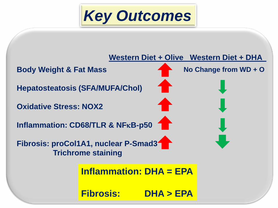
Western Diet + Olive Western Diet + DHA Body Weight & Fat Mass Hepatosteatosis (SFA/MUFA/Chol) Oxidative Stress: NOX2 Inflammation: CD68/TLR & NFκB-p50 Fibrosis: proCol1A1, nuclear P-Smad3 Trichrome staining
No Change from WD + O
Key Outcomes
Inflammation: DHA = EPA Fibrosis: DHA > EPA
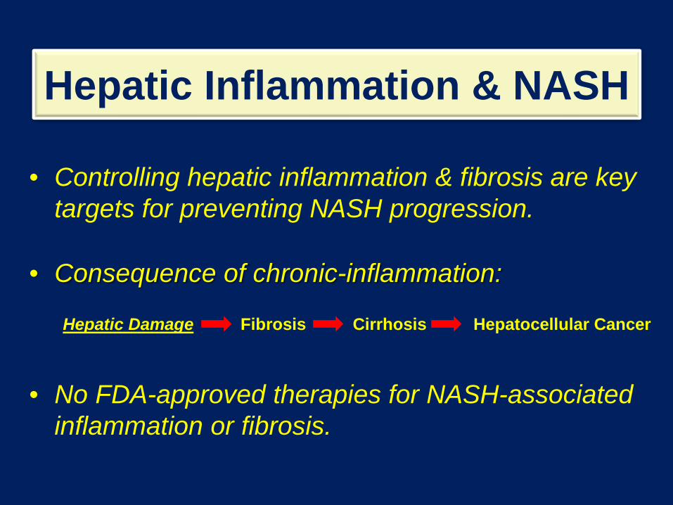
Hepatic Inflammation & NASH
• Controlling hepatic inflammation & fibrosis are key targets for preventing NASH progression.
• Consequence of chronic-inflammation: Hepatic Damage Fibrosis Cirrhosis Hepatocellular Cancer
• No FDA-approved therapies for NASH-associated
inflammation or fibrosis.

Hepatic Inflammation & NASH • Sources of inflammatory factors:
– Adipose tissue • Inflammatory cytokines: TNFα, IL6, Leptin • Insulin sensitizer: Adiponectin
– Innate immune cells, e.g., Kupffer & monocytes/macrophage – Metabolism: advanced glycation end products derived from glucose – Damaged/dead cells; cellular debris – Gut: bacterial components,
• e.g., endotoxin (cell wall component of gram (-) bacteria)
• Mediators of inflammation – Toll-like receptors (TLR2, TLR4, TLR9, etc) – Inflammatory cytokine and chemokine receptors; cell signaling – Inflammatory lipids (oxidized fatty acids and cholesterol)
• Receptor mediated mechanisms • Non-receptor mediated mechanisms
NOX
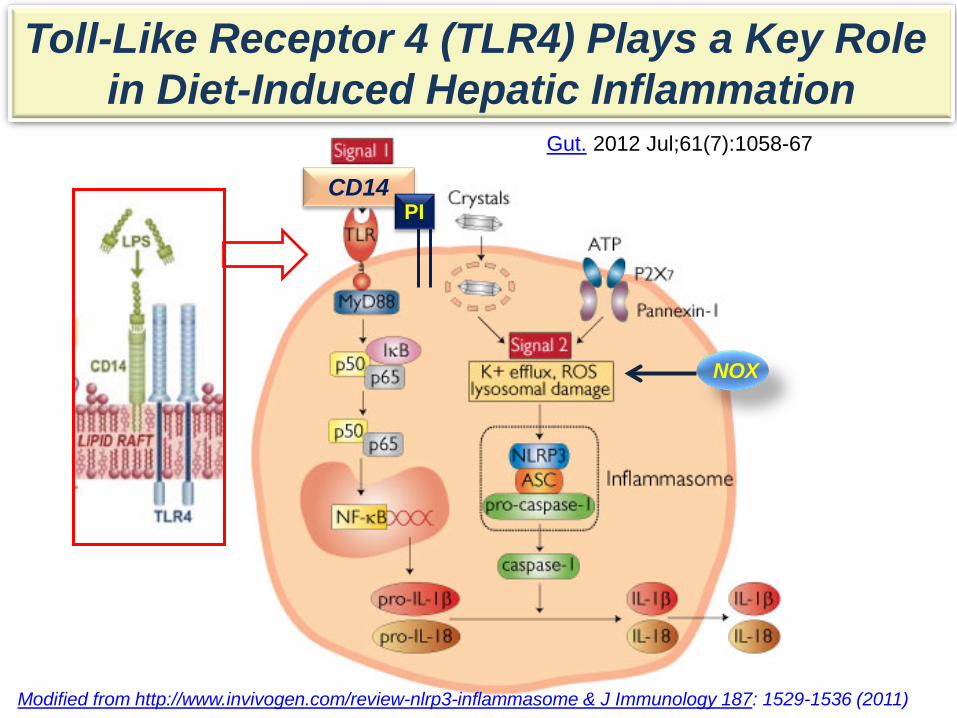
Toll-Like Receptor 4 (TLR4) Plays a Key Role in Diet-Induced Hepatic Inflammation
Modified from http://www.invivogen.com/review-nlrp3-inflammasome & J Immunology 187: 1529-1536 (2011)
CD14 PI
Gut. 2012 Jul;61(7):1058-67

*
*
*
#
#
#
# #
*, P<0.05 versus Chow # P< 0.05 versus WD + O
mR
NA
Abun
danc
e Fo
ld C
hang
e
TLR4
The Western Diet Induces Hepatic TLR Components
0
2
4
6
8
10
TLR-2 TLR-4 TLR9 CD-14 MD-2 MYD88
NPWD + OWD + EWD + DWD + ED
Required for Endotoxin Activation of TLR4

Nuclear Abundance of NFκB Subunits J Nutr 143: 315-323 (2013)
0
1
2
3NPWD + OOWD + EPAWD + DHAWD + EPA + DHA
* *
NFκ
B S
ubun
its
Fold
of N
P-Fe
d M
ice
NFκB-p50 NFκB-p65 NFκB-p105
NFκB p105 mRNA Abundance (Precursor of p50)
Nuclear Abundance of NFκB Subunits

0
10
20
30
MCP1 Clec4F CD 68 F4/80 Clec10a TNFa IL-6 IL1B IL-10 NLRP1 PAI1 SAA1
NP WD + O WD + E WD + D WD + ED
#
#
#
*
* *
* *
* * * *
# # # # #
#
*
#
Hepatic Inflammation Markers
*
mR
NA
Abun
danc
e Fo
ld C
hang
e
Key Cellular & Plasma Factors Involved in Hepatic Inflammation
NP = Chow (Purina 5053)
Kupffer Macrophage

NOX
The Western Diet Induces Hepatic Inflammation
CD14 PI
What are the signals?

NOX
Does Western Diet Increase Blood Endotoxin?
LPS/Endotoxin CD14
PI
WESTERN DIET
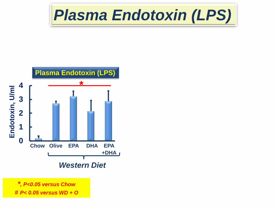
* Plasma Endotoxin (LPS)
*, P<0.05 versus Chow # P< 0.05 versus WD + O
0
1
2
3
4
Western Diet
Chow Olive EPA DHA EPA +DHA
Endo
toxi
n, U
/ml
Plasma Endotoxin (LPS)

TLRs Function within Membrane Lipid Rafts
CD14 PI
DHA is assimilated into membrane phospholipids.
DHA disrupts lipid raft
structure & TLR4 signaling
LPS/Endotoxin
CH3
HOOC
DHA

NOX
Western Diet Effects on Blood Endotoxin and Hepatic Inflammation
CD14 PI
WESTERN DIET LPS/Endotoxin
NOX
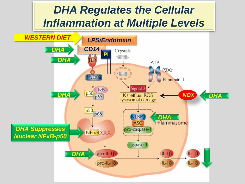
DHA Regulates the Cellular Inflammation at Multiple Levels
CD14 PI
WESTERN DIET
DHA
DHA DHA
DHA Suppresses Nuclear NFκB-p50
DHA
DHA
DHA
LPS/Endotoxin

NOX
DHA Attenuates the Cellular Response to Inflammatory Stimuli!
CD14 PI
WESTERN DIET
DHA
DHA DHA
DHA Suppresses Nuclear NFκB-p50
DHA
DHA
DHA
DHA does not attenuate plasma Endotoxin (LPS) or TLR4 Agonist
LPS/Endotoxin
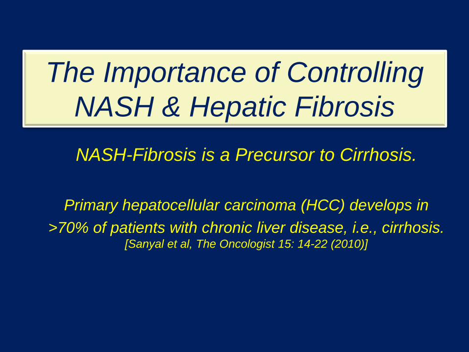
The Importance of Controlling NASH & Hepatic Fibrosis NASH-Fibrosis is a Precursor to Cirrhosis.
Primary hepatocellular carcinoma (HCC) develops in
>70% of patients with chronic liver disease, i.e., cirrhosis. [Sanyal et al, The Oncologist 15: 14-22 (2010)]

WD + DHA WD + Olive Oil
Fibrosis Induced by the Western Diet

0
1
2
3
0
10
20
30
40
50
Col1A1 Col1A2 Col4A1 Col7A1 TIMP-1
Chow WD + O WD + E WD + D WD + ED
mR
NA
Abu
ndan
ce-F
old
Cha
nge
TGFβ1
*
*
* *
*
#
#
#
#
DHA Suppresses WD-Induced Fibrosis
Collagen Subtypes
Metabolomic Analysis [Metabolon, Inc.]
Goal:
To identify metabolites linked to WD-induced hepatic damage.
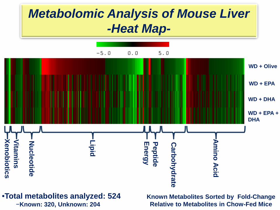
•Total metabolites analyzed: 524 −Known: 320, Unknown: 204
WD + EPA
WD + DHA
WD + EPA + DHA
Known Metabolites Sorted by Fold-Change Relative to Metabolites in Chow-Fed Mice
Metabolomic Analysis of Mouse Liver -Heat Map-
Amino Acid
Peptide
Carbohydrate
Energy
Lipid
Nucleotide
Vitamins
Xenobiotics
WD + Olive
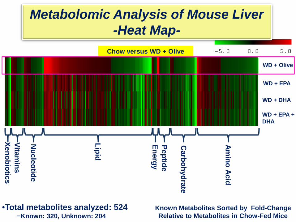
•Total metabolites analyzed: 524 −Known: 320, Unknown: 204
Chow versus WD + Olive
WD + EPA
WD + DHA
WD + EPA + DHA
Known Metabolites Sorted by Fold-Change Relative to Metabolites in Chow-Fed Mice
WD + Olive
Metabolomic Analysis of Mouse Liver -Heat Map-
Peptide
Carbohydrate
Lipid
Nucleotide
Vitamins
Xenobiotics
Amino Acid
Energy

LIPIDS 136 Biochemicals
VITAMINS & COFACTORS 16 Biochemicals
Increase
Decrease
No Change
40 27 5
11 4
Metabolites Affected by WD and Reversal by WD + DHA
Increase
Decrease
No Change
4
9 3
3 2
Decrease
No Change
15 19 3
AMINO ACIDS 78 Biochemicals
CARBOHYDRATES 34 Biochemicals
Increase
Decrease
No Change
62 31 16
43 31

WD + Olive
WD + EPA
WD + DHA
WD + EPA + DHA
Heat Map of Lipid Metabolites
C18-20 SFA, MUFA
& Sphingolipids
C20-22 ω3 PUFA
Oleic Acid
Palmitoyl Sphingomyelin
C20-22 ω6 PUFA

WD + Olive
WD + EPA
WD + DHA
WD + EPA + DHA
Heat Map of Lipid Metabolites
C18-20 SFA, MUFA
& Sphingolipids
C20-22 ω6 PUFA
palmitoyl sphingomyelin
Chow WDOlive WDEPA WDDHAWDEPA + DH0
0.5
1
1.5
2
2.5
Western Diet
Ch Ol EPA DHA EPA +DHA
Fatty
Aci
d C
lass
µm
oles
/g p
rote
in
0
2500
5000
7500
10000 Saturated MUFAN-3 PUFA N-6 PUFA Pal-Sphingomyelin
Ch Ol EPA DHA EPA
Western Diet +DHA

0
10
20
30
40
50
60
0 2 4 6
MC
P1 m
RN
A
ChowWD + OWD + EWD + DWD + E + D
0
10
20
30
40
50
0 2 4 6
ProC
ol1a
1 m
RN
A
Sphingomyelin, fold change
ChowWD + OWD + EWD + DWD + E + D
0
2
4
6
8
10
0 2 4 6
NO
X2 m
RN
A
Sphingomyelin, fold change
ChowWD + OWD + EWD + DWD + E + D
r2= 0.7 P<0.0001
r2= 0.58 P<0.0001
r2= 0.57 P<0.0001
r2= 0.47 P<0.0001
Hepatic Sphingomyelin is Associated with Inflammation, Oxidative Stress & Fibrosis
0
2
4
6
8
0 2 4 6
TRL4
mR
NA
ChowWD + OWD + EWD + DWD + E + D

Linkage Between Hepatic Sphingomyelin & TRL4 Function
• Sphingomyelin (SM) is a membrane sphingolipid
• SM is also a component of membrane microdomains (lipid rafts); Lipid rafts consists of SM, cholesterol, and phospholipids with saturated fatty acyl chains: – TLR-4 functions within lipid rafts
• DHA disrupts optimal lipid raft-dependent clustering of
proteins involved in cell signaling.
J Biol. Chem. 277: 8755-8758 (2002); J Biol. Chem. 277: 25843-25846 (2002) Prostaglandin, Leukotrienes & Essential Fatty Acid 82: 159-164 (2010); J Immunol 187: 1529-1535 (2011)

DHA>EPA May be Useful in the Prevention of NASH

Is NASH Reversible?
Kelli A. Lytle, R.D. Ph.D. Candidate
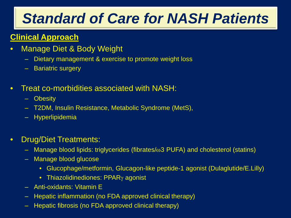
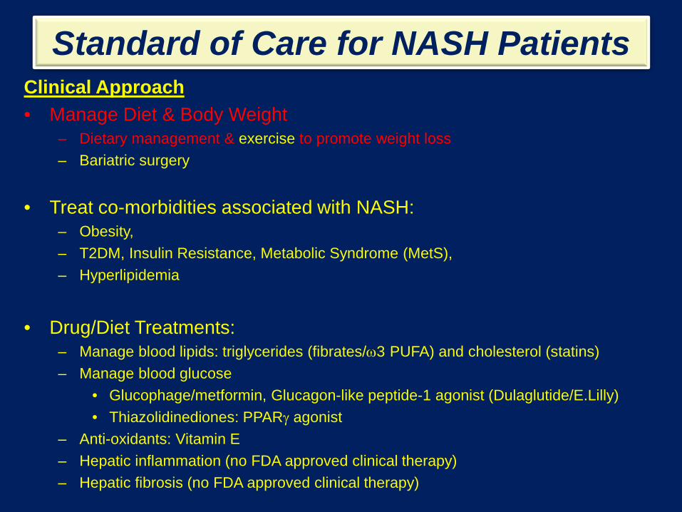
Standard of Care for NASH Patients Clinical Approach • Manage Diet & Body Weight
– Dietary management & exercise to promote weight loss – Bariatric surgery
• Treat co-morbidities associated with NASH: – Obesity – T2DM, Insulin Resistance, Metabolic Syndrome (MetS), – Hyperlipidemia
• Drug/Diet Treatments:
– Manage blood lipids: triglycerides (fibrates/ω3 PUFA) and cholesterol (statins) – Manage blood glucose
• Glucophage/metformin, Glucagon-like peptide-1 agonist (Dulaglutide/E.Lilly) • Thiazolidinediones: PPARγ agonist
– Anti-oxidants: Vitamin E – Hepatic inflammation (no FDA approved clinical therapy) – Hepatic fibrosis (no FDA approved clinical therapy)
Clinical Approach • Manage Diet & Body Weight
– Dietary management & exercise to promote weight loss – Bariatric surgery
• Treat co-morbidities associated with NASH: – Obesity, – T2DM, Insulin Resistance, Metabolic Syndrome (MetS), – Hyperlipidemia
• Drug/Diet Treatments:
– Manage blood lipids: triglycerides (fibrates/ω3 PUFA) and cholesterol (statins) – Manage blood glucose
• Glucophage/metformin, Glucagon-like peptide-1 agonist (Dulaglutide/E.Lilly) • Thiazolidinediones: PPARγ agonist
– Anti-oxidants: Vitamin E – Hepatic inflammation (no FDA approved clinical therapy) – Hepatic fibrosis (no FDA approved clinical therapy)
Standard of Care for NASH Patients
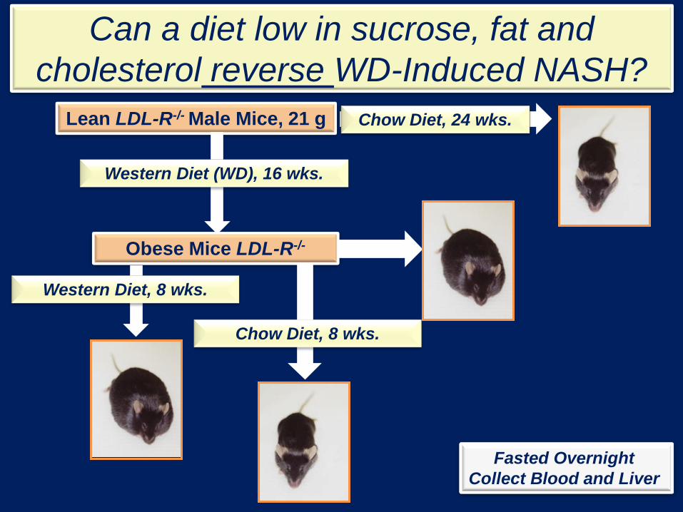
Can a diet low in sucrose, fat and cholesterol reverse WD-Induced NASH?
Obese Mice LDL-R-/-
Fasted Overnight Collect Blood and Liver
Lean LDL-R-/- Male Mice, 21 g
Chow Diet, 8 wks.
Western Diet (WD), 16 wks.
Chow Diet, 24 wks.
Western Diet, 8 wks.

Key Outcomes Western Diet Western Diet + DHA
Body Weight/Fat Mass
Plasma Markers of Hepatic Damage (ALT/AST)
Plasma Endotoxin & TLR4 Ligands
Hepatosteatosis
Oxidative Stress/NOX2
Inflammation/TLR, CD68
Fibrosis/ProCol1A
No Effect
No Effect

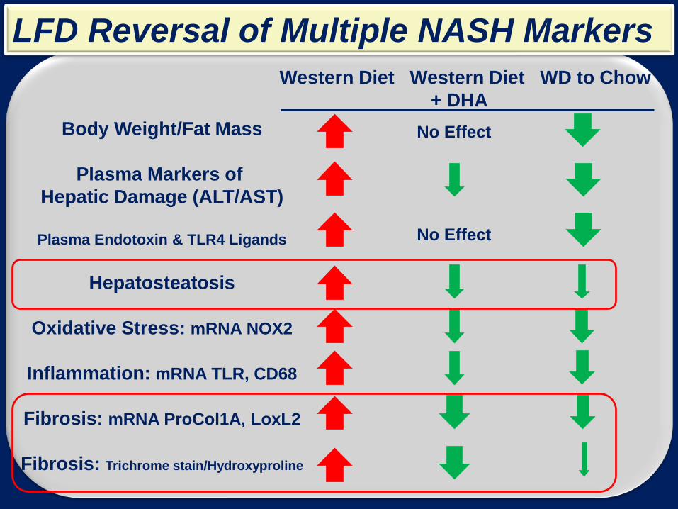
Diet Can Reverse Multiple NASH Markers Western Diet Western Diet WD to Chow + DHA
Body Weight/Fat Mass
Plasma Markers of Hepatic Damage (ALT/AST)
Plasma Endotoxin & TLR4 Ligands
Hepatosteatosis
Oxidative Stress: mRNA NOX2
Inflammation: mRNA TLR, CD68
Fibrosis: mRNA ProCol1A, LoxL2
Fibrosis: Trichrome stain/Hydroxyproline
No Effect
No Effect
Western Diet Western Diet WD to Chow + DHA
Body Weight/Fat Mass
Plasma Markers of Hepatic Damage (ALT/AST)
Plasma Endotoxin & TLR4 Ligands
Hepatosteatosis
Oxidative Stress: mRNA NOX2
Inflammation: mRNA TLR, CD68
Fibrosis: mRNA ProCol1A, LoxL2
Fibrosis: Trichrome stain/Hydroxyproline
No Effect
No Effect
LFD Reversal of Multiple NASH Markers

What Have We Learned? • The Western Diet (WD) induces a robust NASH
phenotype in LDLR-/- mice
• DHA>EPA attenuates WD-induced NASH: – EPA is anti-inflammatory, but not anti-fibrotic – DHA is both anti-inflammatory and anti-fibrotic – Neither EPA nor DHA regulate body weight or blood levels of glucose or
endotoxin
• A diet low in saturated fat, sucrose and cholesterol
reverses many (but not all) WD-induced NASH markers. – Partial reversal of hepatosteatosis and fibrosis. – Plasma parameters are not an accurate measure of hepatic lipid
content or fibrosis. – Better plasma markers of liver status are required.

A Recommendation • Obese adults and children are at risk for
NASH – A diet low in fat, sucrose and cholesterol – Diet enriched in DHA
– Compliance remains a problem with dietary management.

Dietary Management Can Prevent and Reverse NAFLD-NASH.
Prevention
Intervention
Add: DHA @ 2% total Calories
Reduce Dietary: Sucrose
Saturate/Trans Fat Cholesterol

Whether dietary management, alone, can reverse cirrhosis or HCC
remains an unanswered question.
Dietary Intervention???
Prevention
Intervention
Personnel
OSU Personnel Christopher M. Depner
Kelli A. Lytle Sasmita Tripathy
Moises Torres-Gonzalez Kenneth Philbrick
Carmen Wong
Off-Campus Personnel Kurt M. Bohren Metabolon, Inc. Elizabeth Kensicki Metabolon, Inc. Ginger L. Milne Vanderbilt U Chris B. Newgard Duke U James R. Bain Duke U Robert D. Stevens Duke U Andrew S Greenberg Tufts U Li-Shin Huang Columbia U
Research Support
DK 43220; DK 094600
2009-65200-05846

DHA
Healthy
Questions
NASH