Sustaining Activism by Jeffrey W. Rubin and Emma Sokoloff-Rubin
Upload
henry-rodgersCategory
view
217download
0
DICOM Ontology (DO) Project
Daniel L. Rubin M.D., M.S.Clinical Asst. Professor of Radiology
Research Scientist, Center for Biomedical Informatics Research
Stanford University
David S. Channin, MDAssociate Professor of Radiology
Northwestern University
Curtis P. Langlotz, MD, PhDAssociate Professor of Radiology
University of Pennsylvania
Charles E. Kahn, Jr., MD, MSProfessor of Radiology
Medical College of Wisconsin
David Clunie, MDChief Technology Officer
RadPharm Inc.

DICOM
• DICOM is global standard for medical imaging• Comprises combined knowledge from
engineers and radiologists from academia and industry
• Subsets of DICOM used extensively in all caBIG Imaging Workspace projects

Many projects refer to parts of DICOM
The pixel at the tip of the arrow [coordinates (x,y)] inthis image[DICOM: 1.2.814.234543.23243]represents the Ascending Thoracic Aorta[SNOMED:A3310657]

AIM Schema includes information from DICOM
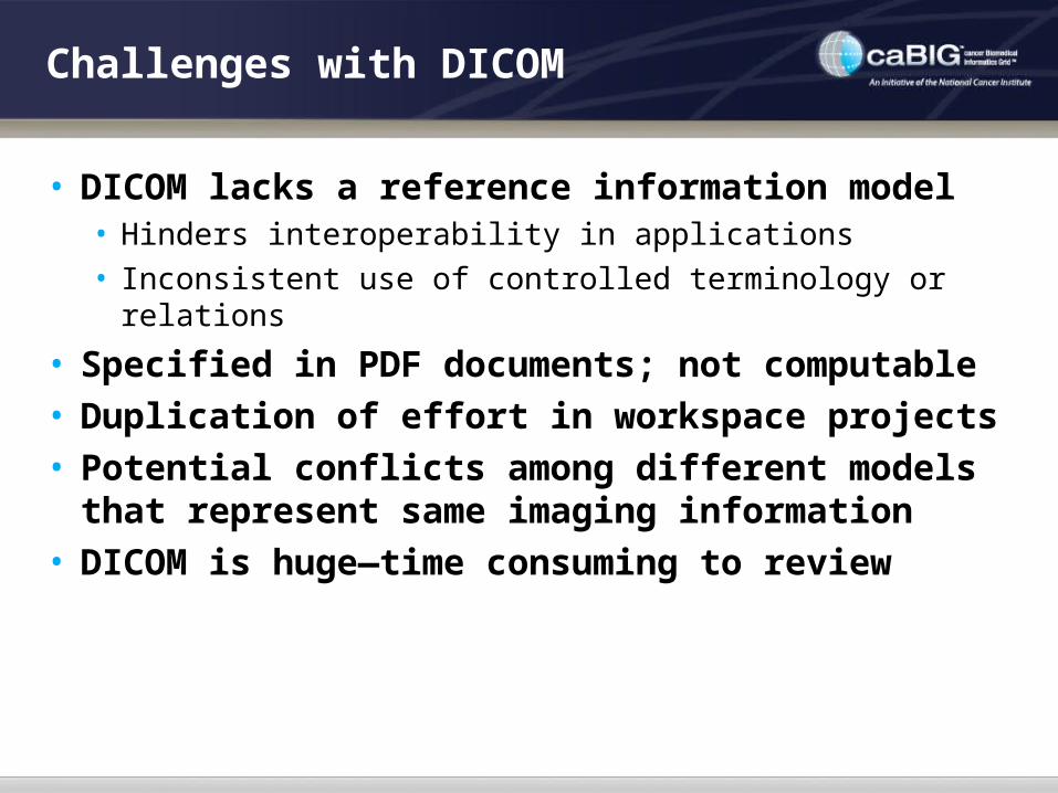
Challenges with DICOM
• DICOM lacks a reference information model• Hinders interoperability in applications• Inconsistent use of controlled terminology or relations
• Specified in PDF documents; not computable• Duplication of effort in workspace projects• Potential conflicts among different models
that represent same imaging information• DICOM is huge—time consuming to review
The DICOM Standard is caBIG

DO Project Goals
• Create ontology based on DICOM• Make explicit the entities and relations in DICOM• Harmonize with other ontologies (RadLex, SNOMED)• Harmonize with other imaging projects (AIM)
• Evaluate impact by studying use cases related to existing projects in the workspace

DO ProjectPhase I Goals
• Requirements gathering• What subset of DICOM is relevant to caBIG?• What content should be included in the DICOM ontology?• Who should build this?
• Define scope of Phase II• The parts of DICOM that will be reviewed• The actual building of the ontology• Controlled terminology identification/reconciliation tasks• Evaluation via use cases
• Proof-of-concept presentation at RSNA 2007

Phase I Activities to define scope of Phase II

DICOM Ontology Project Activities
• Determine the requirements for the DICOM Ontology
• Identify the expertise of participants required to create the DICOM Ontology (DO)
• Determine the extent to which DICOM uses controlled terminology
• Assess the gap between the DO and caBIG™ terminology resources (EVS and caDSR)
• Specify operations on the ontology (e.g., DO XML)

DO Progress #1
• SCOPE DEFINITION of the DICOM standard to define the work to be done in Phase II• We reviewed DICOM Information Object Definitions
(IODs)—both image- and non-image objects relevant to clinical trials
• We excluded • Non-radiology image as well as RT objects (could be added
in a future phase, especially the RT objects)
• We included• All image types in clinical trials.
• Relevant parts of DICOM used for DO will be part 3 and some of part 16

Some included IODs
• A.2 COMPUTED RADIOGRAPHY IMAGE INFORMATION OBJECT DEFINITION• A.3 COMPUTED TOMOGRAPHY IMAGE INFORMATION OBJECT DEFINITION• A.4 MAGNETIC RESONANCE IMAGE INFORMATION OBJECT DEFINITION• A.5 NUCLEAR MEDICINE IMAGE INFORMATION OBJECT DEFINITION• A.6 ULTRASOUND IMAGE INFORMATION OBJECT DEFINITION• A.7 ULTRASOUND MULTI-FRAME IMAGE INFORMATION OBJECT DEFINITION• A.14 X-RAY ANGIOGRAPHIC IMAGE INFORMATION OBJECT DEFINITION• A.15 X-RAY ANGIOGRAPHIC BI-PLANE IMAGE INFORMATION OBJECT DEFINITION • A.16 X-RAY RF IMAGE INFORMATION OBJECT DEFINITION• A.21 POSITRON EMISSION TOMOGRAPHY IMAGE INFORMATION OBJECT
DEFINITION• A.26 DIGITAL X-RAY IMAGE INFORMATION OBJECT DEFINITION• A.27 DIGITAL MAMMOGRAPHY X-RAY IMAGE INFORMATION OBJECT DEFINITION• A.36 ENHANCED MR INFORMATION OBJECT DEFINITIONS• A.38 ENHANCED COMPUTED TOMOGRAPHY IMAGE INFORMATION OBJECT
DEFINITION• A.47 ENHANCED X-RAY ANGIOGRAPHIC IMAGE INFORMATION OBJECT DEFINITION• A.48 ENHANCED X-RAY RF IMAGE INFORMATION OBJECT DEFINITION

Some excluded IODs
• A.17 RT IMAGE INFORMATION OBJECT DEFINITION• A.18 RT DOSE INFORMATION OBJECT DEFINITION• A.19 RT STRUCTURE SET INFORMATION OBJECT DEFINITION• A.20 RT PLAN INFORMATION OBJECT DEFINITION• A.29 RT BEAMS TREATMENT RECORD INFORMATION OBJECT DEFINITION• A.30 RT BRACHY TREATMENT RECORD INFORMATION OBJECT DEFINITION• A.31 RT TREATMENT SUMMARY RECORD INFORMATION OBJECT DEFINITION• A.49 RT ION PLAN INFORMATION OBJECT DEFINITION• A.50 RT ION BEAMS TREATMENT RECORD INFORMATION OBJECT DEFINITION• A.9 STANDALONE OVERLAY INFORMATION OBJECT DEFINITION• A.10 STANDALONE CURVE INFORMATION OBJECT DEFINITION• A.12 STANDALONE MODALITY LUT INFORMATION OBJECT DEFINITION• A.13 STANDALONE VOI LUT INFORMATION OBJECT DEFINITION• A.22 STANDALONE PET CURVE INFORMATION OBJECT DEFINITION• A.23 STORED PRINT INFORMATION OBJECT DEFINITION• A.24 HARDCOPY GRAYSCALE IMAGE INFORMATION OBJECT DEFINITION• A.25 HARDCOPY COLOR IMAGE INFORMATION OBJECT DEFINITION

DO Progress #2
• We defined required expertise for building DO• Intimate familiarity with DICOM standard and its
documentation, and how to turn that documentation into software applications
• Expertise in imaging informatics and in using DICOM for developing software
• Ontology building expertise• Terminology expertise (radiology-related terminologies)• Expertise in caBIG methodologies (specifically caDSR,
EVS)

DO Progress #3
• Determining the extent to which DICOM uses controlled terminology• The Phase II protocol will require harmonization of
DICOM with RadLex, SNOMED and LOINC, ISO standards (e.g., country codes).
• Assess the gap between the DO and caBIG™ terminology resources (EVS and caDSR)• An important, but time-consuming task, needing to be
done in a future phase• Will be simpler after first harmonizing with RadLex

Requirements for Phase II
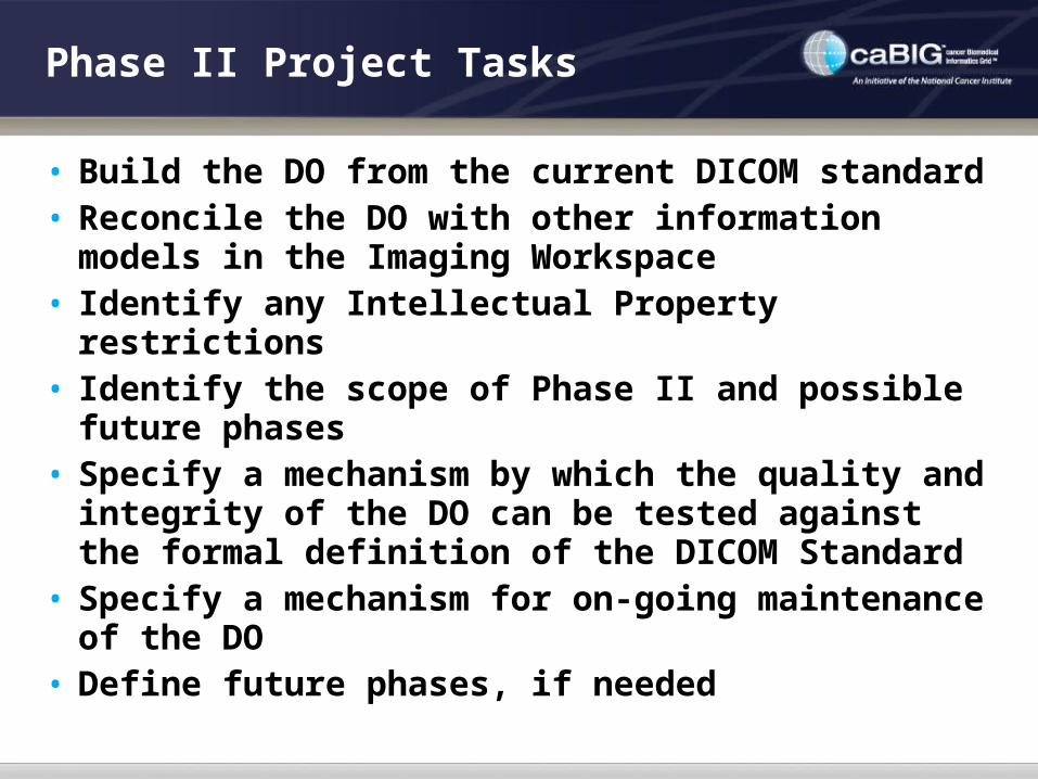
Phase II Project Tasks
• Build the DO from the current DICOM standard• Reconcile the DO with other information
models in the Imaging Workspace• Identify any Intellectual Property restrictions• Identify the scope of Phase II and possible
future phases• Specify a mechanism by which the quality and
integrity of the DO can be tested against the formal definition of the DICOM Standard
• Specify a mechanism for on-going maintenance of the DO
• Define future phases, if needed

Steps for Building the DO
• Translate DICOM standard to structured format
• Critical review of the DICOM standard once DICOM is in ontological format
• Assemble the ontological components and build the DO

Translating DICOM to Structured Format
• Ontology building could be daunting—the DICOM standard is huge (17 parts, approx 2500 pages) and distributed as text documents.
• Information pertaining to the DO is spread across several relevant parts • Part 3: information models, information object definitions (IODs)
and modules and macros • Part 6: data dictionary, including the type and multiplicity of each
data element used as an attribute within objects defined in Part 3• Part 16: value sets (context groups) referenced by the information
objects in Part 3 and templates for structured reports• Approach: translate the DICOM standard into a
structured format that can be imported into an ontology authoring tool such as Protégé
• Leverage existing XML transformations, modifying them to translate DICOM to RDF so that it can be directly be imported into Protégé, and then manipulated to create DO

Critical Review of DICOM while Building Ontology
• Encode DICOM E-R Diagrams• These currently exist in non-computable format in DICOM
standard• Will produce computable representations of E-R diagrams
and harmonize with other semantics components of the DO
• Reconcile DO for modeling inconsistencies in DICOM• Overloaded data elements in the same concept at different
levels of DICOM Pixel Spacing (0028,0030) are defined• Same concept but different value sets of same level of info
model Rescale Type (0028,1054)• Same concept, but different descriptions Imager Pixel
Spacing (0018,1164) is defined • Same concept, but different conditionality Lossy Image
Compression Ratio (0028,2112)

Proposed Strategy for Evaluation
• Use cases: AIM, NCIA, IQ, XIP, AVT, gACRIN projects. Does DO provide all the imaging knowledge needed? • AIM—references to image context information• NCIA—info about image technique, acquisition,
demographics• IQ—same as NCIA• XIP—any structured information relating to • AVT—attributes related to comparing images at
different time points• gACRIN—more accurate modeling of ACRIN
warehouse

Acknowledgements and Support
• caBIG Imaging Workspace, Subcontract from Booz-Allen & Hamilton, Inc.


![DICOM Conformance Statement9d48995e-cb8b-4ac4-ae9b... · 2020. 2. 20. · DICOM protocol. 1.5 References [DICOM PS 3 2006] The Digital Imaging and Communications in Medicine (DICOM)](https://static.fdocuments.in/doc/165x107/60e78a442d236e0f92518d06/dicom-conformance-statement-9d48995e-cb8b-4ac4-ae9b-2020-2-20-dicom-protocol.jpg)