Diagnostic Reference Levels
43
Page 1 of 43 Diagnostic Reference Levels Guidance on the establishment, use and review of diagnostic reference levels for medical exposure to ionising radiation Updated July 2021
Transcript of Diagnostic Reference Levels

Page 1 of 43
Diagnostic Reference Levels
Guidance on the establishment, use and review of
diagnostic reference levels for medical exposure to
ionising radiation
Updated July 2021

Diagnostic Reference Levels
Health Information and Quality Authority
2 of 43
About the Health Information and Quality Authority (HIQA)
The Health Information and Quality Authority (HIQA) is an independent statutory
authority established to promote safety and quality in the provision of health and
social care services for the benefit of the health and welfare of the public.
HIQA’s mandate to date extends across a wide range of public, private and voluntary
sector services. Reporting to the Minister for Health and engaging with the Minister
for Children, Equality, Disability, Integration and Youth, HIQA has responsibility for
the following:
Setting standards for health and social care services — Developing
person-centred standards and guidance, based on evidence and international
best practice, for health and social care services in Ireland.
Regulating social care services — The Office of the Chief Inspector within
HIQA is responsible for registering and inspecting residential services for older
people and people with a disability, and children’s special care units.
Regulating health services — Regulating medical exposure to ionising
radiation.
Monitoring services — Monitoring the safety and quality of health services
and children’s social services, and investigating as necessary serious concerns
about the health and welfare of people who use these services.
Health technology assessment — Evaluating the clinical and cost-
effectiveness of health programmes, policies, medicines, medical equipment,
diagnostic and surgical techniques, health promotion and protection activities,
and providing advice to enable the best use of resources and the best
outcomes for people who use our health service.
Health information — Advising on the efficient and secure collection and
sharing of health information, setting standards, evaluating information
resources and publishing information on the delivery and performance of
Ireland’s health and social care services.
National Care Experience Programme — Carrying out national service-
user experience surveys across a range of health services, in conjunction with
the Department of Health and the HSE.

Diagnostic Reference Levels
Health Information and Quality Authority
Page 3 of 43
Table of Contents
About the Health Information and Quality Authority (HIQA) ...................... 2
1. Introduction .............................................................................................. 4
2. Purpose of this document ......................................................................... 4
3. Introduction to DRLs ................................................................................ 6
4. National DRLs ........................................................................................... 8
4.1 Background ................................................................................................ 8
4.2 Establishment and review of National DRLs by HIQA ..................................... 8
5. Local facility DRLs ................................................................................... 10
5.1 Establishing local facility DRLs ................................................................... 10
5.2 Using local facility DRLs ............................................................................. 14
5.3 Reviewing local facility DRLs ...................................................................... 15
5.4 Corrective actions ..................................................................................... 18
6. Conclusion............................................................................................... 20
References .................................................................................................. 21
Appendix 1: Glossary of terms ................................................................... 24
Appendix 2: National DRLs ......................................................................... 27
Table 3. National DRL category, quantity, symbol and units .............................. 27
Table 4. National adult dental DRLs ................................................................. 29
Table 5. National adult general radiography DRLs ............................................. 30
Table 6. National mammography DRLs............................................................. 31
Table 7. National adult fluoroscopy DRLs ......................................................... 32
Table 8. National interventional radiology DRLs ................................................ 33
Table 9. National interventional cardiology DRLs ............................................... 34
Table 10. National adult computed tomography (CT) DRLs ................................ 35
Table 11. National adult nuclear medicine DRLs ................................................ 37
Table 12. National adult DXA DRLs .................................................................. 38
Table 13. National paediatric CT DRLs.............................................................. 39
Table 14. National paediatric radiography and fluoroscopy DRLs ........................ 40
Revision History .......................................................................................... 42

Diagnostic Reference Levels
Health Information and Quality Authority
4 of 43
1. Introduction
Medical exposure to ionising radiation is the largest contributor of artificial ionising
radiation exposure to Irish people.1 To ensure that individuals are protected when
undergoing medical exposures, the core components of radiation protection
principles — justification* and optimisation† — must be used.2 An essential part of
ensuring medical exposures are optimised is the establishment, use and regular
review of diagnostic reference levels (DRLs) for medical radiological procedures.2,3,4
The concept of using DRLs for medical radiological procedures was adopted by the
European Directive 97/43/Euratom in 1997.5 DRLs are dose levels that are not
expected to be exceeded for standard diagnostic and interventional radiological
procedures when good practice regarding diagnostic and technical performance is
applied.2,3 Where a DRL is exceeded on a consistent basis, a review must be carried
out to determine the cause and corrective actions must be taken without undue
delay.6
2. Purpose of this document
In 2018, European legislation on the regulation of medical exposures to ionising
radiation was transposed into Irish law. The European Union (Basic Safety Standards
for Protection Against Dangers Arising from Medical Exposure to Ionising Radiation)
Regulations 2018 and 2019 (referred to in this document as the “regulations”)
designate the Health Information and Quality Authority (HIQA) as the competent
authority for the establishment and review of national DRLs.6,7,8
* Justification is the process of weighing up the potential benefit of a medical exposure against the detriment for that individual. † Optimisation is the process by which doses that are as low as reasonably achievable (ALARA).
What are DRLs?
DRLs are dose levels set to aid optimisation of diagnostic and interventional
medical exposures. They provide a standard for comparison to help ensure the
radiation protection of patients undergoing these types of medical radiological
procedures.
Why should DRLS be used for medical radiological procedures?
DRLs help to ensure that the radiation dose received by patients for a specific
type of medical radiological procedure is optimised.

Diagnostic Reference Levels
Health Information and Quality Authority
Page 5 of 43
The purpose of this guidance is to fulfil HIQA’s obligation under the regulations to
publish national DRLs, including details of the review of these national DRLs and
guidance on the establishment, review and use of DRLs. Similarly, undertakings
must ensure that local facility DRLs are established, regularly reviewed and used,
having regard to national DRLs. The regulations also require the undertaking to
make this guidance document available to practitioners, medical physics experts and
persons delegated the practical aspects of medical radiological procedures.6
What does this mean for HIQA?
HIQA, as the competent authority, must establish national DRLs. Under the
regulations HIQA must publish:
national DRLs
the details of any regular reviews carried out of these national DRLs
guidance in relation to the establishment, review and use of diagnostic
reference levels for use by undertakings, practitioners, medical physics
experts, and persons delegated the practical aspects of medical
radiological procedures.6
What does this mean for undertakings?
Undertakings must ensure that this guidance document is made available to
practitioners, medical physics experts and persons delegated the practical
aspects of medical radiological procedures. Undertakings must also establish
local facility DRLs, ensure that these are regularly reviewed and used by
persons conducting medical radiological procedures.

Diagnostic Reference Levels
Health Information and Quality Authority
6 of 43
3. Introduction to DRLs
The Basic Safety Standards define DRLs as:
dose levels in medical radiodiagnostic or interventional radiology practices,
or, in the case of radio-pharmaceuticals, levels of activity, for typical
examinations for groups of standard-sized patients or standard phantoms for
broadly defined types of equipment.2
This means that a DRL is a dose level that is set for typical diagnostic and
interventional radiological procedures. For setting DRLs, a standard sized patient is
considered to be 55 to 85 kg.9
It is important to note that DRLs are not dose limits or dose constraints. They should
only be used as an indicator of a dose level that, if exceeded, should lead to a
review of the medical radiological procedure to ensure that optimisation was
adequate.2,3,4,5,6 The use of DRLs is an important way of ensuring that medical
exposures are optimised and helps to ensure that the ‘as low as reasonably
achievable’ (ALARA) principle‡ is used to deliver maximum diagnostic yield at the
lowest radiation dose necessary.3
The assessment and evaluation of patient doses for medical radiological procedures
is a requirement under the regulations and undertakings can partly achieve this
through the establishment of local facility DRLs.6 Once established, local facility DRLs
should be compared with national DRL values. If local facility DRLs consistently
exceed or are substantially lower than national DRL values, an investigation must be
conducted to ensure optimal practices and intended outcomes are delivered.3, 4 An
assessment of clinical image quality should be included as part of any local review of
dose levels to ensure the intended benefit from the medical exposure is still
achieved. Objective measures for the assessment of image quality must be used
where available. Where issues are identified, an investigation or risk assessment
must be undertaken to identify possible causes. Corrective actions to mitigate the
risks to patients should be implemented.3, 4, 10, 11
‡ The ALARA principle is a key principle of optimisation, where all doses due to medical exposure should be kept as low as reasonably achievable, consistent with obtaining the intended outcomes of the exposure.
Clinical audits and DRLs
The process for reviewing patient doses, comparing to national DRLs,
identifying issues, taking corrective actions as necessary and repeating this
cycle regularly is consistent with established clinical audit models.10,12, 14

Diagnostic Reference Levels
Health Information and Quality Authority
Page 7 of 43
The process for reviewing patient doses and DRLs is consistent with established
clinical audit models.10, 12, 14 The establishment and ongoing review of DRLs should
be embedded into an undertaking’s clinical audit schedule. Any clinical audit model
for the review of DRLs should be multidisciplinary and adhere to local and national
clinical audit procedures, where available.

Diagnostic Reference Levels
Health Information and Quality Authority
8 of 43
4. National DRLs
4.1 Background
National DRLs established by the competent authority represent common national
diagnostic and interventional radiological procedures and or the clinical task for
which medical exposures are performed. When developing national DRLs, priority is
given to medical exposures with the highest frequency and or that result in the
highest patient radiation dose and for which the assessment of DRL quantities is
practicable.3
In 2013, the Health Service Executive (HSE) Medical Exposure Radiation Unit
(MERU) published national DRLs for a range of standard medical radiological
procedures.13 These national DRLs were updated in 2017 following the publication of
HSE MERU’s Patient Radiation Protection Manual 2017 14 and National Survey on
Population Dose from Computed Tomography 2017.15
The National Survey on Population Dose from Computed Tomography also
introduced the concept of defining medical radiological procedures by clinical task or
based on clinical indications (Clinical DRLs).15 Traditional DRLs built for specific
anatomical locations may have different clinical indications, use different protocols
and subsequently deliver different exposure levels. The use of clinical DRLs has
been acknowledged as more appropriate in CT and interventional radiology 17
4.2 Establishment and review of National DRLs by HIQA
In February 2020, HIQA adopted national DRLs following consultation with its expert
advisory group (EAG), which includes representation from relevant professional
bodies. These national DRLs were developed based on the categories, procedures
and or clinical tasks included in:
the existing national DRLs established by HSE MERU14, 15
Public Health England’s DRL guidance16
EUCLID ongoing research and DRL review17
European guidelines on paediatric DRLs4
the Administration of Radioactive Substances Advisory Committee (ARSAC)
guidance18
other published international literature.19, 20
In July 2021, these national DRLs were updated following HIQA’s national review of
DRLs in general radiography, mammography and dual-energy X-ray absorptiometry
(DXA) scanning. 21 As the organisation responsible for the establishment, review and

Diagnostic Reference Levels
Health Information and Quality Authority
Page 9 of 43
publication of national DRLs, HIQA carry out a cyclical thematic request for dose
information to update national DRL values. Undertakings will also be required to
contribute to these national reviews by submitting further radiation dose data when
requested by HIQA under Regulation 18(2).
The information that may be required by HIQA to complete national DRL reviews
includes:
median dose quantity values, where available, for diagnostic and
interventional medical radiological procedures or clinical tasks published by
HIQA (National DRLs)
median dose quantity values for diagnostic and interventional medical
radiological procedures or clinical tasks which deliver high radiation doses
(not included in national DRLs)
frequency per annum of these medical radiological procedures
equipment details (manufacturer, model, detector types and software
available).
Undertakings should note that this list is not exhaustive and may be subject to
change.
The collected data will be collated and used to establish a national 75th percentile
DRL value. For mammography screening, the 95th percentile may also be considered
as a standard metric due to more defined and extensive quality assurance
programmes.
HIQA’s ongoing DRL review process will create an opportunity to identify and
address any gaps in current DRL data available, particularly in differentiation of
medical radiological equipment types, software available and new or evolving
radiological procedures.
What is a percentile?
A percentile is the value below which a percentage of data falls. For example, a
value is the 75th percentile where 75% of the data is below that value.
What is a national DRL value?
A national DRL value is a dose level which is set at the 75th percentile of the
distribution of the medians obtained from national surveys or other means.

Diagnostic Reference Levels
Health Information and Quality Authority
10 of 43
The national DRLs adopted and established by HIQA, including further information
on DRL categories, quantities, symbols and units, can be found in Appendix 2.
5. Local facility DRLs
5.1 Establishing local facility DRLs
All undertakings have a requirement under the regulations to establish local facility
DRLs, irrespective of size and complexity. Local facility DRLs should be established
for procedures or clinical tasks quoted by national DRLs (when these procedures are
undertaken locally). However, some procedures or clinical tasks may not be included
in the national DRLs as they may be unique to a facility, hospital or medical
speciality and expertise. Where this is the case, particularly when these procedures
involve high patient doses, local facility DRLs for such procedures or clinical tasks
should be established by the undertaking.
The process for establishing local facility DRLs is detailed in Table 1. Establishing
local facility DRLs is the first step in the cyclical DRL process or audit cycle. A typical
value should be used for setting an individual undertaking’s local DRLs. A typical
value is defined as the median value of the distribution of DRL quantities for medical
radiological procedures.3
Does the use of local facility DRLs apply to all healthcare providers?
What does this mean for undertakings?
Undertakings must ensure that local facility DRLs are established, regularly
reviewed and used, taking corrective action where necessary

Diagnostic Reference Levels
Health Information and Quality Authority
Page 11 of 43
Dose management systems, where available, can be used to contribute to the
establishment and review of local facility DRLs. However, its application may not be
practicable in all installations, for example, dental surgeries, due to cost. It should be
noted that there is no regulatory requirement for undertakings to acquire or use
such software however the availability and use of such systems demonstrates an
example of proactive dose monitoring and prospective optimisation.
Retrospective analysis of radiology information systems (RIS) and hospital
information systems (HIS) may allow access to dose data on large numbers of
patients to facilitate the establishment of local facility DRLs.
Manual recording of patient doses may also be used by undertakings to contribute to
the establishment of local facility DRLs. Undertakings must consider the resources
available and establish appropriate dose data collection methods to enable the
establishment and review of local facility DRLs.
For the establishment of nuclear medicine local facility DRLs, patient reviews are not
necessary. Injected activity or injected activity per patient weight for each standard
examination should be recorded.
When establishing intra-oral dental DRLs, dosimetric measurements made by a
medical physicist are favoured over patient studies. To establish intra-oral dental
DRLs, measurements of incident air kerma (Ka, i) can be made at standard settings
with a suitable calibrated detector placed at the end of the spacer cone of the X-ray
set.
What value should be used when establishing facility DRLs?
A median value should be used when setting an individual undertaking’s
facility DRLs and should generally be used for all undertakings.
(To find the median, the data should be arranged in order from least to
greatest. If there is an even number of items in the distribution of the dose
quantities collected, then the median is found by taking the mean (average)
of the two middlemost quantities collected. If there are an odd number of
items the median is the middlemost quantity)

Diagnostic Reference Levels
Health Information and Quality Authority
12 of 43
Recommended dose quantities for the establishment of local DRLs can be found in
Appendix 2.
What is a DRL quantity?
A DRL quantity is a commonly used and easily measured or determined metric
(for example, dose length product (DLP) in computed tomography (CT)) which
indicates the amount of ionising radiation used to perform the medical
radiological procedure.3

Diagnostic Reference Levels
Health Information and Quality Authority
Page 13 of 43
Table 1. How to establish local facility DRLs
How to establish local DRLs
Step 1
Create a list of medical radiological procedures to be included
The list of medical radiological procedures should consist of procedures or
clinical tasks:
that national DRL values exist for that are commonly carried out or associated with high patient
doses.
This may result in some procedures or clinical tasks included in local
facility DRLs that are not included in the national DRLs.
Step 2
Establish what needs to be recorded
This should include the dose quantities used to measure that amount of
dose used to carry out the procedure and or clinical task for which a DRL
is being established. Calibration of all dosimeters used for patient
dosimetry should be performed regularly and verified periodically by a
medical physicist3 to ensure accuracy, consistency and reproducibility of
results. Further information on what dose quantities to use is included in
Appendix 2.
Diagnostic Reference Levels
Health Information and Quality Authority
14 of 43
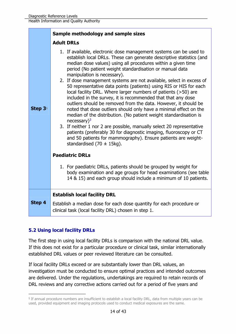
Step 3§
Sample methodology and sample sizes
Adult DRLs
1. If available, electronic dose management systems can be used to establish local DRLs. These can generate descriptive statistics (and median dose values) using all procedures within a given time period (No patient weight standardisation or manual data manipulation is necessary).
2. If dose management systems are not available, select in excess of 50 representative data points (patients) using RIS or HIS for each local facility DRL. Where larger numbers of patients (>50) are included in the survey, it is recommended that that any dose outliers should be removed from the data. However, it should be noted that dose outliers should only have a minimal effect on the median of the distribution. (No patient weight standardisation is necessary)3
3. If neither 1 nor 2 are possible, manually select 20 representative patients (preferably 30 for diagnostic imaging, fluoroscopy or CT and 50 patients for mammography). Ensure patients are weight-standardised (70 ± 15kg).
Paediatric DRLs
1. For paediatric DRLs, patients should be grouped by weight for
body examination and age groups for head examinations (see table 14 & 15) and each group should include a minimum of 10 patients.
Step 4
Establish local facility DRL
Establish a median dose for each dose quantity for each procedure or
clinical task (local facility DRL) chosen in step 1.
5.2 Using local facility DRLs
The first step in using local facility DRLs is comparison with the national DRL value.
If this does not exist for a particular procedure or clinical task, similar internationally
established DRL values or peer reviewed literature can be consulted.
If local facility DRLs exceed or are substantially lower than DRL values, an
investigation must be conducted to ensure optimal practices and intended outcomes
are delivered. Under the regulations, undertakings are required to retain records of
DRL reviews and any corrective actions carried out for a period of five years and
§ If annual procedure numbers are insufficient to establish a local facility DRL, data from multiple years can be used, provided equipment and imaging protocols used to conduct medical exposures are the same.

Diagnostic Reference Levels
Health Information and Quality Authority
Page 15 of 43
make these records available to HIQA on request. Failure to do so is an offence
under the regulations.
All individuals who carry out medical radiological procedures should be familiar with
the important role of DRLs in optimisation of medical radiological procedures for the
protection and safety of patients. The concept and proper use of DRLs should be
included in the education and training programmes of practitioners (including
dentists), medical physics experts, individuals that conduct medical exposures and
others as appropriate, who are involved in medical exposure to ionising radiation.6
Local facility DRLs should be made available to appropriate staff but should not be
used for individual patients or as trigger (alert or alarm) level for individual patients
as some radiation doses may be justified given their size or the complexity of the
procedure.3 It is important to note that DRLs should be used as a supplement to
professional judgement to aid in the optimisation of medical exposures to ionising
radiation.3,4,9,10 The priority for any diagnostic and or interventional medical
radiological procedure is to obtain the required medical information while using the
lowest dose achievable.2,5,11
Local facility DRLs must be established and used, and their adherence regularly
reviewed, for different pieces of medical radiological equipment, procedures and
clinical tasks.3 This will allow more in-depth analysis of patient dose (for the same
procedure/clinical task) in order to evaluate:
specific equipment (for example, comparing the dose routinely given by two
different dental X-ray sets)
devices utilised to capture the image (for example, computed radiography
compared to direct digital radiography or flat panel detector fluoroscopy
compared to image intensifier fluoroscopy)
use of available software (for example, iterative reconstruction compared to
filtered back projection).
5.3 Reviewing local facility DRLs
The development of DRLs is a cyclical process and local facility DRLs must be
regularly reviewed by the undertaking.6 Generally, local facility medical DRLs should
DRLs are not dose limits
DRLs are not individual dose limits for patients or procedures. They should be
used as a supplement to professional judgement to aid in the optimisation of
medical exposures to ionising radiation.
Diagnostic Reference Levels
Health Information and Quality Authority
16 of 43
be reviewed annually and local facility dental DRLs should be reviewed every two
years. DRLs should also be reviewed when new technologies are introduced or a
medical radiological procedure is changed to ensure that there is adequate
optimisation of medical radiological procedures to protect patients. 3, 4
DRL values are not static. The DRL process does not stop after one review but
should be subject to regular review and incorporated into an undertaking’s quality
assurance programme.
Under the regulations, undertakings are required to retain records of such DRL
reviews and any corrective actions carried out for a period of five years and make
these records available to HIQA on request.6
How often should facility DRLs be reviewed?
Facility DRLs should be annually reviewed or after the introduction of new
equipment, software or techniques.
What records should be kept?
Under the regulations, undertakings are required to retain records of DRL
reviews and any corrective actions carried out for a period of five years and
make these records available to HIQA on request.

Diagnostic Reference Levels
Health Information and Quality Authority
Page 17 of 43
Figure 1. DRL process
1. Select procedures and or clinical tasks for inclusion
2. Establish dose quantities to record
3. Identify sample size for inclusion
4. Collect dose quantities
5. Analyse and establish facility DRL
6. Compare to national DRLs
7. Investigate variances to identify cause
8. Take corrective actions, where necessary
9. Make DRL data available to appropriate staff
10. Review regularly

Diagnostic Reference Levels
Health Information and Quality Authority
18 of 43
5.4 Corrective actions
When a DRL value is identified as being consistently exceeded, an investigation of
equipment and practices must be conducted immediately to ensure optimisation of
safety and protection of patients. When the investigation determines the reason that
the DRL is consistently exceeded, corrective actions must be taken without undue
delay.
The undertaking must ensure that this review is appropriate and investigates the
reasons the DRL was consistently exceeded. An appropriate review should involve:
a multidisciplinary team with appropriate expertise and experience
a designated individual of suitable seniority identified as having responsibility
on the undertakings behalf for implementing the corrective actions identified.
Furthermore, review should identify:
the precise cause
the corrective actions to be taken, which should be outlined as SMART
objectives (specific, measurable, achievable, realistic and timely).
This investigation into the cause should, at a minimum, examine:
the measurement methodology used to assess the DRL quantities
the characteristics and performance of medical radiological equipment
the case-mix included in the sample size
the technical parameters used in the medical radiological procedure
the technique(s) used in the medical radiological procedure
medical radiological procedure protocols and adherence to the use of these
protocols.
When is a DRL value considered to be ‘consistently exceeded’?
A DRL value is considered to be ‘consistently exceeded’ when the median dose
quantity is greater than the established DRL value.

Diagnostic Reference Levels
Health Information and Quality Authority
Page 19 of 43
Table 2. Examples of possible causes of why a DRL is consistently
exceeded (assuming appropriate measurement methodology)
Possible causes for consistently exceeding a DRL
A protocol is not adapted to account for paediatric patient characteristics
Unsatisfactory equipment performance
Incorrect technical parameters selected
For example, when the cause is related to unsatisfactory equipment performance, a
review should be conducted by a medical physics expert in conjunction with the
equipment manufacturer. The equipment manufacturer should consider their
regulatory requirements to report such issues to the Health Products Regulatory
Authority (HPRA). If it is determined that the equipment is old or substandard, the
corrective action may require the replacement or upgrading of the equipment. If the
cause is not related to age or substandard equipment, then the quality assurance
programmes for the equipment should be reviewed. Where performance testing
indicates that the equipment is functioning adequately, the criteria used to
determine what adequate performance is may need to be reviewed and updated.
Where this change to the performance criteria for the equipment indicates an issue,
steps must be taken without undue delay to improve equipment which is inadequate
or defective.
Furthermore, where the cause is related to the practical aspects of carrying out the
medical radiological procedure, a review of the protocols for the medical radiological
procedure and adherence to these protocols should be conducted and staff
competence and training should be considered. The corrective actions taken in such
cases may include:
the revision and adjustment of the protocol
additional training of staff and reassessment of competencies
increasing awareness around the use and adherence to these protocols.

Diagnostic Reference Levels
Health Information and Quality Authority
20 of 43
6. Conclusion
National DRL values are set for common procedures and clinical tasks undertaken in
Ireland. These allow undertakings to compare local facility DRLs, representative of
patient dose, to a national standard. Where local facility DRLs are deemed too high
or low, an immediate investigation into the cause is required. Corrective actions
identified must be recorded. An undertaking must ensure that practitioners
(including dentists), medical physics experts and individuals that conduct medical
exposures are informed of national DRLs and local facility DRLs must be established
to facilitate patient dose optimisation.

Diagnostic Reference Levels
Health Information and Quality Authority
Page 21 of 43
References**
1. Radiological Protection Institute of Ireland. Radiation Doses Received by the Irish Population. Dublin: Radiological Protection Institute of Ireland; 2014 June. Available online from: http://www.epa.ie/pubs/reports/radiation/RPII_Radiation_Doses_Irish_Population_2014.pdf. Accessed on: 27 November 2018.
2. Council Directive 2013/59/EURATOM of 5 December 2013 laying down basic safety standards for protection against the dangers arising from exposure to ionising radiation, and repealing Directives 89/618/Euratom, 90/641/Euratom, 96/29/Euratom, 97/43/Euratom and 2003/122/Euratom. L 13/1. Luxemburg: Offical Journal of the European Union; 2013.
3. ICRP. Diagnostic Reference Levels in Medical Imaging. ICRP Publication 135. Ann ICRP. 2017; Oct;46(1): 1-144.
4. European Commission. Radiation Protection No. 185 European Guidelines on Diagnostic Reference Levels for Paediatric Imaging. Luxembourg Publications Office of the European Union; 2018.
5. Council Directive 97/43/ EURATOM of 30 June 1997 on health protection of individuals against the dangers of ionizing radiation inrelation to medical exposure, and repealing Directive 84/466/Euratom. L 180/22. Luxemburg: Offical Journal of the European Union; 1997.
6. European Union (Basic Safety Standards for Protection Against Dangers Arising from Medical Exposure to Ionising Radiation) Regulations 2018, SI No. 256 of 2019. Dublin: The Stationary Office; 2019. Available online from: http://www.irishstatutebook.ie/2018/en/si/0256.html. Accessed on: 03 September 2019.
7. European Union (Basic Safety Standards for Protection Against Dangers Arising from Medical Exposure to Ionising Radiation)(Amendment)Regulations 2019, SI No. 332 of 2019. Dublin: The Stationary Office; 2019. Available online from: http://www.irishstatutebook.ie/eli/2019/si/332/made/en/pdf. Accessed on: 03 September 2019.
8. European Union (Basic Safety Standards for Protection Against Dangers Arising from Medical Exposure to Ionising Radiation)(Amendment)(No.2) Regulations 2019, SI No. 413 of 2019. Dublin: The Stationary Office; 2019. Available online from: http://www.irishstatutebook.ie/eli/2019/si/413/made/en/pdf Accessed on: 03 September 2019.
** If not already cited, all online references were accessed at the time of preparing this handbook. Please note that online addresses may change over time.

Diagnostic Reference Levels
Health Information and Quality Authority
22 of 43
9. European Commission.Radiation Protection No. 180 Diagnostic Reference Levels in Thirty-six European Countries, Part 2/2. Luxembourg Publications Office of the European Union; 2014.
10. Institute of Phyisics and Engineering in Medicine. Report 88 Guidance on the Establishment and Use of Diagnostic Reference Levels for Medical X-ray Examinations. York: Institute of Phyiscs and Engineering in Medicine; 2004.
11. Roch P, Celier D, Dessaud C, Etard C. Using diagnostic reference levels to evaluate the improvement of patient dose optimisation and the influence of recent technologies in radiography and computed tomography. Eur J Radiol. 2018; Jan;98: 68-74. Available online from: http://www.ncbi.nlm.nih.gov/pubmed/29279172. Accessed on: 27 November 2018.
12. European Commission.Radiation Protection No. 159 European Commission Guidelines on Clincial Audit for Medical Radiological Practices (Diagnostic Radiology, Nuclear Medicine and Radiotherapy). Luxembourg Publications Office of the European Union; 2009. Available online from: https://ec.europa.eu/energy/sites/ener/files/documents/159.pdf. Accessed on 03 Septemeber 2019.
13. Medical Exposure Radiation Unit. Template for developing a Patient Radiation Protection Manual For facilities using medical ionising radiation. Dublin: Health Service Executive; 2013. Available online from: https://www.hse.ie/eng/about/who/qualityandpatientsafety/safepatientcare/medexpradiatonunit/rp%20manual%202013.pdf. Accessed on: 03 September 2019.
14. Medical Exposure Radiation Unit. Patient Radiation Protection Manual 2017. Dublin: Health Service Executive; 2017. Available online from: https://www.hse.ie/eng/about/qavd/meru/radiation-protection-manual-2017.pdf Accessed on: 03 September 2019.
15. Medical Exposure Radiation Unit. National Survey on Population Dose from Computed Tomography 2017. Dublin: Health Service Executive; 2017.
16. Public Health England. National Diagnostic Reference Levels (NDRLs) from 19 August 2019 [Online]. Available from: https://www.gov.uk/government/publications/diagnostic-radiology-national-diagnostic-reference-levels-ndrls/ndrl. Assessed: 13 November 2019.
17. EUCLID. European Study on Clinical Diagnostic Reference Levels for X-ray Medical Imaging. Report and review on existing clinical DRLs 2018 [Online]. Available from: http://www.eurosafeimaging.org/wp/wp-content/uploads/2017/09/D2.1_Report-and-review-on-existing-clinical-DRLs_final_published-on-website.pdf. Assessed: 25 July 2019.

Diagnostic Reference Levels
Health Information and Quality Authority
Page 23 of 43
18. ARSAC. Notes for guidance on the clinical administration of radiopharmaceuticals and use of sealed radioactive sources 2019 [Online]. Available from: https://assets.publishing.service.gov.uk/government/uploads/system/uploads/attachment_data/file/778215/ARSAC_NfG_2019.pdf. Assessed: 3 September 2019.
19. Siiskonen T, Ciraj-Bjelac , Dabin J, Diklic A, Domienik-Andrzejewska J, Farah J et al. Establishing the European diagnostic reference levels for interventional cardiology. Phys Med. 2018 Oct;54:42-48.
20. Maccia C, Malchair F, Gobert I, Louvard Y, Lefevre T. Assessment of Local Dose Reference Values for Recanalization of Chronic Total Occlusions and Other Occlusions in a High-Volume Catheterization Center. Am J Cardiol. 2015 Oct;116:1179-1184.
21. HIQA. National diagnostic reference levels (DRLs) for general radiograpy, mammography and DXA scanning. Dublin: HIQA; 2021.
22. Ruiz-Cruces R, Vano E, Carrera-Magariño F, Moreno-Rodriguez F, Soler-Cantos MM, Canis-Lopez M et al. Diagnostic reference levels and complexity indices in interventional radiology: a national programme. Eur Radiol. 2016 Dec;26:4268-4276.

Diagnostic Reference Levels
Health Information and Quality Authority
24 of 43
Appendix 1: Glossary of terms
Air kerma — the kinetic energy released per unit mass of air; measured in Gray
(Gy).
Clinical audit — a systematic examination or review of medical radiological
procedures which seeks to improve the quality and outcome of patient care through
structured review, whereby medical radiological practices, procedures and results
are examined against agreed standards for good medical radiological procedures,
with modification of practices, where appropriate, and the application of new
standards if necessary.
Computed radiography (CR) — a digital image acquisition and processing system
for radiography that uses computers and laser technology.
Diagnostic reference levels — dose levels in medical radiodiagnostic or
interventional radiology practices, or, in the case of radio-pharmaceuticals, levels of
activity, for typical examinations for groups of standard-sized patients or standard
phantoms for broadly defined types of equipment.
Direct digital radiography — a digital image acquisition and processing system
for radiography that captures data and immediately transfers it to a computer
system.
Dose constraint — a constraint set as a prospective upper bound of individual
doses, used to define the range of options considered in the process of optimisation
for a given radiation source in a planned exposure situation.
Dose limit — the value of the effective dose (where applicable) or the equivalent
dose in a specified period which shall not be exceeded for an individual.
Dual-energy X-ray absorptiometry (DXA or DEXA) — a type of medical
exposure typically used to assess bone density in service users where low bone
density or osteoporosis is suspected.
Filtered back projection — means an analytic reconstruction algorithm which
applies a filter to remove blurring.
Flat panel detector — a class of solid-state X-ray digital radiography used in both
radiography and fluoroscopy (as an alternative to X-ray image intensifiers in
fluoroscopy equipment).
Gray (Gy) — a unit of measurement for absorbed dose. It is equivalent to one joule
of energy absorbed per kilogram of material.
Image intensifier — a device to facilitate visual real-time imaging.
Diagnostic Reference Levels
Health Information and Quality Authority
Page 25 of 43
Interventional radiology — the use of X-ray imaging techniques to facilitate the
use of devices in the body for diagnostic or treatment purposes.
Ionising radiation — energy transferred in the form of particles or electromagnetic
waves of a wavelength of 100 nanometres or less (a frequency of 3 × 10¹⁵ hertz or
more) capable of producing ions directly or indirectly.
Iterative reconstruction — the use of iterative algorithms to reconstruct 2D and
3D images in certain imaging techniques.
Mammography — the specialised area of radiology involved in the imaging of
breast tissue.
Mean glandular dose —the absorbed dose of radiation to the breast in
mammography.
Median — is the middle number in a sorted list of numbers.
Medical exposure — exposure incurred by patients or asymptomatic individuals as part of their own medical or dental diagnosis or treatment, and intended to benefit their health, as well as exposure incurred by carers and
comforters and by volunteers in medical or biomedical research.
Medical radiological — pertaining to radiodiagnostic and radiotherapeutic procedures, and interventional radiology or other medical uses of ionising radiation
for planning, guiding and verification purposes.
Medical radiological procedure — any procedure giving rise to medical exposure.
MERU — the HSE Medical Exposure Radiation Unit audited radiation practice in
medical radiological installations in Ireland on behalf of the Department of Health
prior to the commencement of European Union (Basic Safety Standards for
Protection Against Dangers Arising from Medical Exposure to Ionising Radiation)
Regulations 2018 and 2019.
Patient entrance reference point — the position at which the cumulative air
kerma for interventional X-ray equipment is measured in order to reasonably
represent the air kerma incident on the patient's skin surface.
Quality assurance — all those planned and systematic actions necessary to provide adequate assurance that a structure, system, component or procedure will perform satisfactorily in compliance with agreed standards. Quality control is a part of quality assurance.
Quality control — the set of operations (programming, coordinating,
implementing) intended to maintain or to improve quality. It includes monitoring,

Diagnostic Reference Levels
Health Information and Quality Authority
26 of 43
evaluation and maintenance at required levels of all characteristics of performance
of equipment that can be defined, measured, and controlled.
Undertaking — a person or body who has a legal responsibility for carrying out, or
engaging others to carry out, a medical radiological procedure, or the practical
aspects of a medical radiological procedure, as defined by the regulations.

Diagnostic Reference Levels
Health Information and Quality Authority
Page 27 of 43
Appendix 2: National DRLs
Table 3. National DRL category, quantity, symbol and units
DRL
Category
DRL Quantity DRL
Quantity
Symbol
Other
Common
Symbols
DRL
Quantity
Unit
Dental
radiography
Incident air kerma Ka,i
IAK
mGy
Air kerma-area product
(Panoramic)
PKA KAP, DAP Gy.cm²
Radiography Air kerma-area product PKA KAP, DAP Gy.cm²
Mammography Entrance-surface air
kerma,
Ka,e, ESAK, ESD mGy
Incident air kerma OR Ka,i, IAK mGy
Mean glandular dose DG MGD, AGD mGy
Diagnostic
fluoroscopy
Air kerma-area product PKA KAP, DAP Gy.cm²
Incident air kerma at the
patient entrance reference
point
± Ka,r CAK mGy
Fluoroscopy time FT s
Interventional
radiology
Air kerma-area product PKA KAP, DAP Gy.cm²
Incident air kerma at the
patient entrance reference
point
Ka,r CAK mGy
Number of images (in cine
or digital subtraction
angiography runs)
n
Computed
tomography
Computed tomography
dose index (volume)
CTDI vol mGy

Diagnostic Reference Levels
Health Information and Quality Authority
28 of 43
DRL
Category
DRL Quantity DRL
Quantity
Symbol
Other
Common
Symbols
DRL
Quantity
Unit
Dose–length product DLP mGy.cm
Nuclear
medicine
Administered
activity/Activity per body
weight
MBq or
MBq/kg

Diagnostic Reference Levels
Health Information and Quality Authority
Page 29 of 43
Table 4. National adult dental DRLs 16
Procedure/Clinical task DRL
Quantity
DRL
Intra oral Ka,i 1.2 mGy
Panoramic PKA 81 mGy.cm²
Lateral cephalometric radiograph PKA 35 mGy.cm²
Dental CBCT (prior to placement of a maxillary
molar implant)
PKA 265 mGy.cm²

Diagnostic Reference Levels
Health Information and Quality Authority
30 of 43
Table 5. National adult general radiography DRLs 21
Procedure/Clinical task DRL
Quantity
DRL
Chest PA PKA 0.12 Gy.cm²
Chest AP PKA 0.13 Gy.cm²
Portable Chest AP PKA 0.16 Gy.cm²
Abdomen AP PKA 1.7 Gy.cm²
Pelvis AP PKA 1.91 Gy.cm²
Cervical spine AP PKA 0.16 Gy.cm²
Cervical spine LAT PKA 0.19 Gy.cm²
Thoracic spine AP PKA 0.76 Gy.cm²
Thoracic spine LAT PKA 1.8 Gy.cm²
Lumbar spine AP PKA 1.6 Gy.cm²
Lumbar spine LAT PKA 2.24 Gy.cm²
Extremities (Foot/ankle/wrist/hand) PKA 0.06 Gy.cm²

Diagnostic Reference Levels
Health Information and Quality Authority
Page 31 of 43
Table 6. National mammography DRLs 21
Procedure/Clinical task DRL
Quantity
DRL
Single mediolateral oblique (MLO) view DG 2.2 mGy
Single craniocaudal (CC) view DG 2.2mGy
Breast Tomosynthesis – MLO view DG 2.8 mGy
Breast Tomosynthesis – CC view DG 2.8 mGy

Diagnostic Reference Levels
Health Information and Quality Authority
32 of 43
Table 7. National adult fluoroscopy DRLs 14,16
Procedure/Clinical task DRL
Quantity
DRL
Barium (or water soluble) swallow PKA 7.5 Gy.cm²
Barium (or water soluble) meal PKA 12 Gy.cm²
Barium (or water soluble) meal and swallow PKA 10 Gy.cm²
Barium (or water soluble) follow through PKA 8.4 Gy.cm²
Barium (or water soluble) enema PKA 21 Gy.cm²
Barium swallow (Video) PKA 3.4 Gy.cm²
All IV lines PKA 3 Gy.cm²
Femoral angiography PKA 56 Gy.cm²
Nephrostography PKA 9 Gy.cm²
Nephrostomy PKA 13 Gy.cm²
Bilary tract PKA 16 Gy.cm²
Bilary intervention PKA 43 Gy.cm²
Facet joint injection PKA 6 Gy.cm²

Diagnostic Reference Levels
Health Information and Quality Authority
Page 33 of 43
Table 8. National interventional radiology DRLs 14,17
Procedure/Clinical task DRL
Quantity
DRL
Iliac artery stenting PKA 170* Gy.cm²
Transjugular intrahepatic portosystemic shunt
(TIPS)/Portal hypertension
PKA 186 Gy.cm²
Transcatheter arterial chemoembolization (TACE)
Liver/localisation and treatment of hepato cellular
carcinoma
PKA 300 Gy.cm²
Cerebral trans arterial embolization (TAE) PKA 62 Gy.cm²
Aortic endovascular aneurysm repair (EVAR) PKA 159 Gy.cm²
*Classified as a medium level of complexity22

Diagnostic Reference Levels
Health Information and Quality Authority
34 of 43
Table 9. National interventional cardiology DRLs 14,19,20
Procedure/Clinical task DRL
Quantity
DRL
Angiography coronary arteries PKA 55 Gy.cm²
Percutaneous coronary intervention (PCI) PKA 75 Gy.cm²
PCI – chronic total occlusion (CTO) PKA 350 Gy.cm²
Transcatheter aortic valve implantation (TAVI) PKA 130 Gy.cm²
Pacemaker insertion PKA 12 Gy.cm²
Cardiac electrophysiological (EP) study PKA 12 Gy.cm²

Diagnostic Reference Levels
Health Information and Quality Authority
Page 35 of 43
Table 10. National adult computed tomography (CT) DRLs 15,16
Procedure/Clinical task DRL
Quantity
DRL
Brain (routine imaging/head trauma/
haemorrhagic assessment)
DLP 908 mGy.cm
Brain acute stroke (all phases/series) DLP 970 mGy.cm
Sinuses/chronic sinusitis DLP 184 mGy.cm
C spine/trauma — detection or exclusion of a
fracture
DLP 473 mGy.cm
Thorax DLP 310 mGy.cm
Thorax Hi resolution/interstitial lung disease DLP 337 mGy.cm
Thorax CTPA/detection or exclusion of pulmonary
embolus
DLP 346 mGy.cm
Thorax/pulmonary metastases DLP 258 mGy.cm
Abdomen and pelvis DLP 556 mGy.cm
Abdomen liver /abdominal metastases in
colorectal cancer
DLP 554 mGy.cm
Abdomen/appendicitis-detection or exclusion DLP 486 mGy.cm
KUB-urogram (All phases/series) DLP 1150
mGy.cm
KUB/exclusion or detection of a urinary calculus DLP 263 mGy.cm
Thorax, abdomen and pelvis (TAP) DLP 770 mGy.cm
Thorax, abdomen and pelvis (TAP)/oncological
follow up
DLP 635 mGy.cm
Virtual colonoscopy (All phases/series)/polyps,
tumour
DLP 950 mGy.cm
Diagnostic Reference Levels
Health Information and Quality Authority
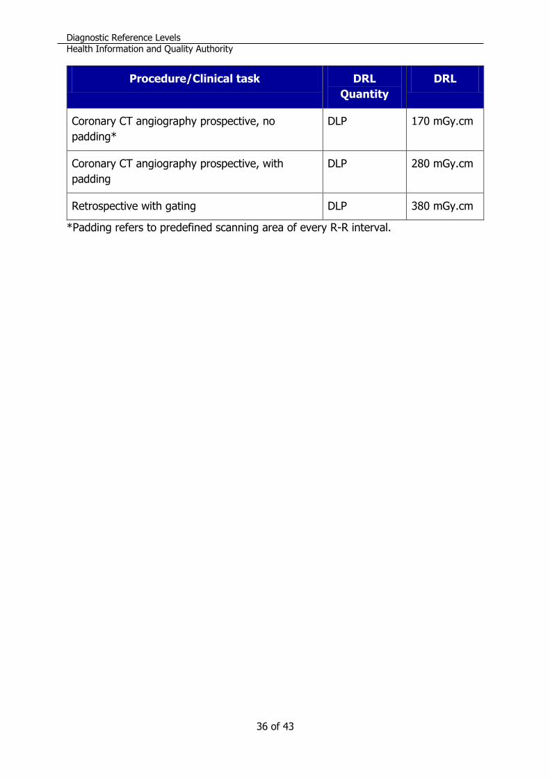
36 of 43
Procedure/Clinical task DRL
Quantity
DRL
Coronary CT angiography prospective, no
padding*
DLP 170 mGy.cm
Coronary CT angiography prospective, with
padding
DLP 280 mGy.cm
Retrospective with gating DLP 380 mGy.cm
*Padding refers to predefined scanning area of every R-R interval.
Diagnostic Reference Levels
Health Information and Quality Authority
Page 37 of 43
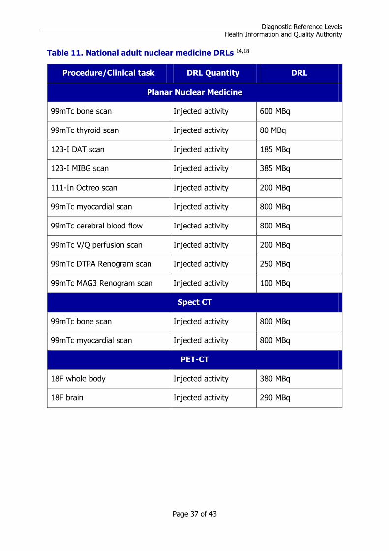
Table 11. National adult nuclear medicine DRLs 14,18
Procedure/Clinical task DRL Quantity DRL
Planar Nuclear Medicine
99mTc bone scan Injected activity 600 MBq
99mTc thyroid scan Injected activity 80 MBq
123-I DAT scan Injected activity 185 MBq
123-I MIBG scan Injected activity 385 MBq
111-In Octreo scan Injected activity 200 MBq
99mTc myocardial scan Injected activity 800 MBq
99mTc cerebral blood flow Injected activity 800 MBq
99mTc V/Q perfusion scan Injected activity 200 MBq
99mTc DTPA Renogram scan Injected activity 250 MBq
99mTc MAG3 Renogram scan Injected activity 100 MBq
Spect CT
99mTc bone scan Injected activity 800 MBq
99mTc myocardial scan Injected activity 800 MBq
PET-CT
18F whole body Injected activity 380 MBq
18F brain Injected activity 290 MBq

Diagnostic Reference Levels
Health Information and Quality Authority
38 of 43
Table 12. National adult DXA DRLs 21
Procedure/Clinical task DRL
Quantity
DRL
Lumbar Spine/Bone Density Analysis (BDA) PKA 20 mGy.cm²
Single Hip/ BDA PKA 15 mGy.cm²
Distal forearm/ BDA PKA 7 mGy.cm²
Lumbar spine/Vertebral Fracture analysis (VFA) PKA 71 mGy.cm²
Diagnostic Reference Levels
Health Information and Quality Authority
Page 39 of 43
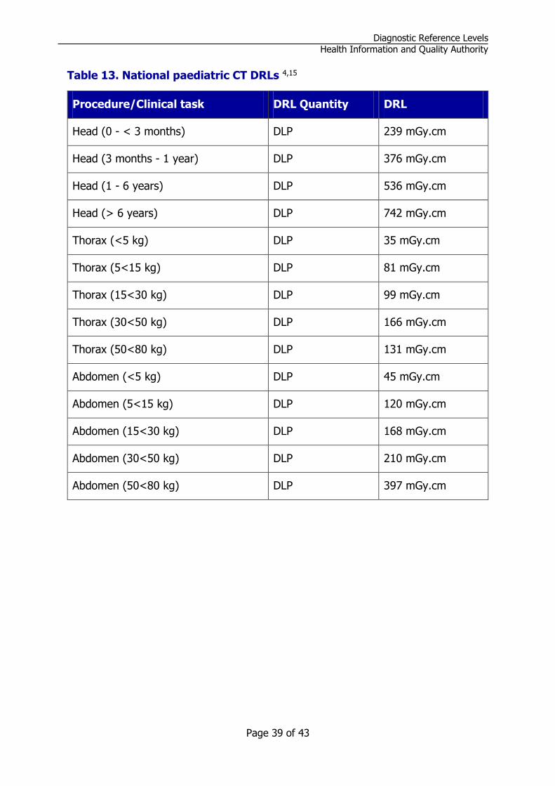
Table 13. National paediatric CT DRLs 4,15
Procedure/Clinical task DRL Quantity DRL
Head (0 - < 3 months) DLP 239 mGy.cm
Head (3 months - 1 year) DLP 376 mGy.cm
Head (1 - 6 years) DLP 536 mGy.cm
Head (> 6 years) DLP 742 mGy.cm
Thorax (<5 kg) DLP 35 mGy.cm
Thorax (5<15 kg) DLP 81 mGy.cm
Thorax (15<30 kg) DLP 99 mGy.cm
Thorax (30<50 kg) DLP 166 mGy.cm
Thorax (50<80 kg) DLP 131 mGy.cm
Abdomen (<5 kg) DLP 45 mGy.cm
Abdomen (5<15 kg) DLP 120 mGy.cm
Abdomen (15<30 kg) DLP 168 mGy.cm
Abdomen (30<50 kg) DLP 210 mGy.cm
Abdomen (50<80 kg) DLP 397 mGy.cm

Diagnostic Reference Levels
Health Information and Quality Authority
40 of 43
Table 14. National paediatric radiography and fluoroscopy DRLs 4, 21
Procedure/Clinical task DRL Quantity DRL
Thorax AP/PA (<5 kg) PKA 9 mGy.cm²
Thorax AP/PA (5-<15 kg) PKA 17 mGy.cm²
Thorax AP/PA (15-<30 kg) PKA 22 mGy.cm²
Thorax AP/PA (30-<50 kg) PKA 50 mGy.cm²
Thorax AP/PA (50-80 kg) PKA 70 mGy.cm²
Abdomen AP (<5 kg) PKA 13 mGy.cm²
Abdomen AP (5-<15 kg) PKA 63 mGy.cm²
Abdomen AP (15-<30 kg) PKA 100 mGy.cm²
Abdomen AP (30-<50 kg) PKA 286 mGy.cm²
Abdomen AP (50-80 kg) PKA 457 mGy.cm²
Pelvis AP (<5 kg) PKA 27 mGy.cm²
Pelvis AP (5-<15 kg) PKA 39 mGy.cm²
Pelvis AP (15-<30 kg) PKA 111 mGy.cm²
Pelvis AP (30-<50 kg) PKA 412 mGy.cm²
Pelvis AP (50-80 kg) PKA 800 mGy.cm²
Cervical Spine AP (15-<30 kg) PKA 28 mGy.cm²
Cervical Spine AP (30-<50 kg) PKA 59 mGy.cm²
Cervical Spine AP (50-80 kg) PKA 96 mGy.cm²
Cervical Spine LAT (15-<30 kg) PKA 41 mGy.cm²
Cervical Spine LAT (30-<50 kg) PKA 54 mGy.cm²
Cervical Spine LAT (50-80 kg) PKA 68 mGy.cm²
Thoracic Spine AP (30-<50 kg) PKA 244 mGy.cm²
Diagnostic Reference Levels
Health Information and Quality Authority
Page 41 of 43
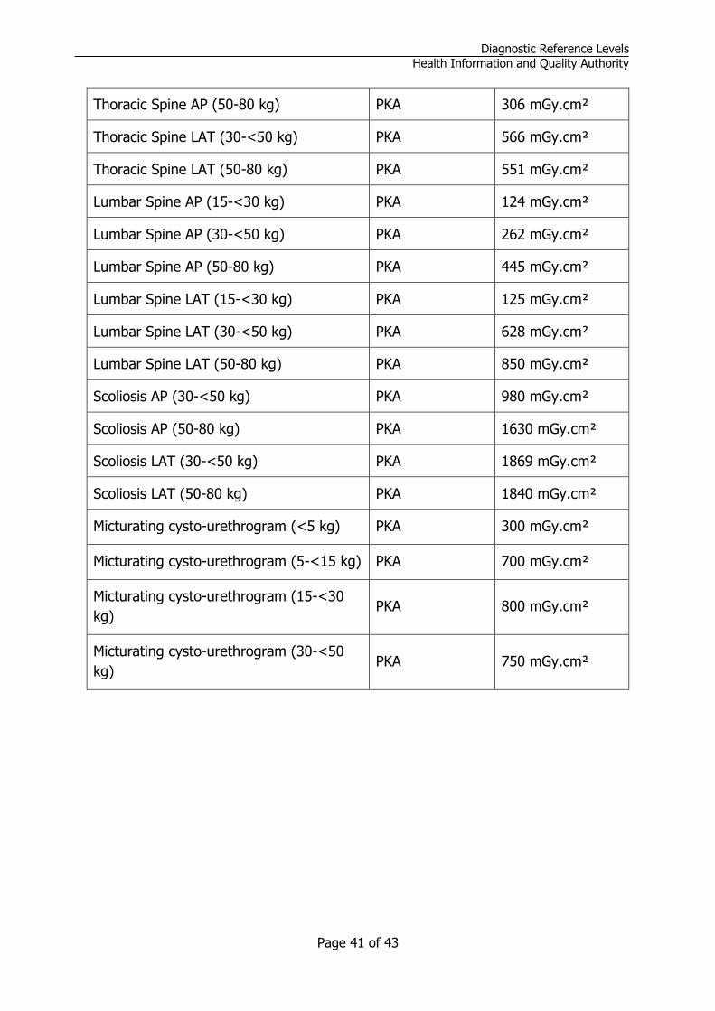
Thoracic Spine AP (50-80 kg) PKA 306 mGy.cm²
Thoracic Spine LAT (30-<50 kg) PKA 566 mGy.cm²
Thoracic Spine LAT (50-80 kg) PKA 551 mGy.cm²
Lumbar Spine AP (15-<30 kg) PKA 124 mGy.cm²
Lumbar Spine AP (30-<50 kg) PKA 262 mGy.cm²
Lumbar Spine AP (50-80 kg) PKA 445 mGy.cm²
Lumbar Spine LAT (15-<30 kg) PKA 125 mGy.cm²
Lumbar Spine LAT (30-<50 kg) PKA 628 mGy.cm²
Lumbar Spine LAT (50-80 kg) PKA 850 mGy.cm²
Scoliosis AP (30-<50 kg) PKA 980 mGy.cm²
Scoliosis AP (50-80 kg) PKA 1630 mGy.cm²
Scoliosis LAT (30-<50 kg) PKA 1869 mGy.cm²
Scoliosis LAT (50-80 kg) PKA 1840 mGy.cm²
Micturating cysto-urethrogram (<5 kg) PKA 300 mGy.cm²
Micturating cysto-urethrogram (5-<15 kg) PKA 700 mGy.cm²
Micturating cysto-urethrogram (15-<30
kg) PKA 800 mGy.cm²
Micturating cysto-urethrogram (30-<50
kg) PKA 750 mGy.cm²

Diagnostic Reference Levels
Health Information and Quality Authority
42 of 43
Revision History
Publication date Title /version Summary of changes
February 2020 Guidance on the
establishment, use and review
of diagnostic reference levels
for medical exposure to
ionising radiation, version 1
First publication
March 2021 Guidance on the
establishment, use and review
of diagnostic reference levels
for medical exposure to
ionising radiation, version 1.1
Section 4.2, Mammography 95th percentile dose added as a complementary metric
Section 5.1, Footnote added to address low annual procedure numbers in the establishment of local facility DRLs
Section 5.4, Addition of regulatory requirements of equipment manufacturers to report issues to the Health Products Regulatory Authority (HPRA)
July 2021 Guidance on the establishment, use and review of diagnostic reference levels for medical exposure to ionising radiation, version 1.2
Section 4.2, Update to include general radiography, mammography and DXA national DRL survey
Appendix 2, Update tables 5, 6 and 13. Addition of table 12

Page 43 of 43