Diagnostic Performance of Routine Brain MRI Sequences for ... · with routine MRIs performed within...
7
ORIGINAL RESEARCH ADULT BRAIN Diagnostic Performance of Routine Brain MRI Sequences for Dural Venous Sinus Thrombosis X D. Patel, X M. Machnowska, X S. Symons, X R. Yeung, X A.J. Fox, X R.I. Aviv, and X P. Jabehdar Maralani ABSTRACT BACKGROUND AND PURPOSE: Signs suggestive of unexpected dural venous sinus thrombosis are detectable on routine MR imaging studies without MRV. We assessed performance characteristics and interrater reliability of routine MR imaging for the diagnosis of dural venous sinus thrombosis, focusing on the superior sagittal, transverse, and sigmoid sinuses. MATERIALS AND METHODS: This case series included 350 patients with MRIs performed with contrast-enhanced MRV and 79 patients with routine MRIs performed within 48 hours of a CTV from 2008 to 2014 (total, n 429). Routine MR images were separated from the contrast-enhanced MRVs and CTVs. Three neuroradiologists, blinded to clinical data, independently reviewed the MRIs for signs of dural venous sinus thrombosis, including high signal on sagittal T1, loss of flow void on axial T2, high signal on FLAIR, high signal on DWI, increased susceptibility effects on T2*-weighted gradient recalled-echo imaging, and filling defects on axial contrast-enhanced spin-echo T1WI and/or volumetric gradient-echo T1WI. Two neuroradiologists independently reviewed contrast-enhanced MRVs and CTVs to determine the consensus gold standard. Interrater reliability was calculated by using the coefficient. RESULTS: Contrast-enhanced MRV and CTV confirmed that dural venous sinus thrombosis was present in 72 of 429 cases (16.8%). The combination of routine MR sequences had an overall sensitivity of 79.2%, specificity of 89.9%, and moderate interrater reliability ( 0.50). The 3 readers did not have similar performance characteristics. 69.4% of positive cases had clinical suspicion of dural venous sinus thrombosis indicated on imaging requisition. CONCLUSIONS: Routine MR images can suggest dural venous sinus thrombosis with high specificity in high-risk patients, even in cases without clinical suspicion. ABBREVIATIONS: CE-3D-T1WI volumetric contrast-enhanced GRE T1-weighted imaging; CEMRV contrast-enhanced MRV; CE-SE-T1WI contrast-enhanced spin-echo T1-weighted imaging; DVST dural venous sinus thrombosis; GRE T2*-weighted gradient recalled-echo imaging D ural venous sinus thrombosis (DVST) is a condition that ranges from being undiagnosed to leading to serious mor- bidity and mortality, including venous infarction and intracranial hemorrhage. 1 DVST has a highly variable clinical presentation, from asymptomatic to acute or subacute headaches, signs or symptoms of increased intracranial pressure, focal neurologic deficits, or seizures. 2,3 The diagnosis is important to consider even in younger patients, with risk factors such as genetic and acquired prothrombotic states, trauma, and infections or inflammatory conditions. 1,2 Although early and accurate diagnosis is important for initia- tion of treatment and prevention of serious complications, the median diagnostic delay from symptom onset is 7 days. 1-4 When DVST is suspected, the current criterion standard for diagnosis is either contrast-enhanced MRV (CEMRV) or CTV. 5 However, the imaging signs of unexpected DVST are seen during interpretation of routine MR imaging studies performed without dedicated CEMRV, concurrent CTV, or relevant clinical history. Typical findings might include visualization of intraluminal thrombus or a filling defect and lack of flow, or flow voids. 2 Signs suggestive of DVST on standard brain MR imaging are reported in the litera- ture with highly variable diagnostic accuracy and can be difficult to interpret in daily practice. 2,6-8 Understanding the diagnostic performance of routine MR imaging for the evaluation of DVST is especially important when DVST is not clinically suspected or CT or MR venography has not been ordered. A false-positive inter- Received February 1, 2016; accepted after revision April 25. From the Department of Medical Imaging, University of Toronto, Sunnybrook Health Sciences Centre, Toronto, Ontario, Canada. Paper previously presented at: Annual Meeting of the American Society of Neuro- radiology and the Foundation of the ASNR Symposium, April 25–30, 2016; Chicago, Illinois. Please address correspondence to Pejman Jabehdar Maralani, MD, FRCPC, De- partment of Medical Imaging, University of Toronto, Sunnybrook Health Sci- ences Centre, AG 270C, 2075 Bayview Ave, Toronto, ON, M4N 3M5; e-mail: [email protected] http://dx.doi.org/10.3174/ajnr.A4843 2026 Patel Nov 2016 www.ajnr.org
Transcript of Diagnostic Performance of Routine Brain MRI Sequences for ... · with routine MRIs performed within...

ORIGINAL RESEARCHADULT BRAIN
Diagnostic Performance of Routine Brain MRI Sequences forDural Venous Sinus Thrombosis
X D. Patel, X M. Machnowska, X S. Symons, X R. Yeung, X A.J. Fox, X R.I. Aviv, and X P. Jabehdar Maralani
ABSTRACT
BACKGROUND AND PURPOSE: Signs suggestive of unexpected dural venous sinus thrombosis are detectable on routine MR imagingstudies without MRV. We assessed performance characteristics and interrater reliability of routine MR imaging for the diagnosis of duralvenous sinus thrombosis, focusing on the superior sagittal, transverse, and sigmoid sinuses.
MATERIALS AND METHODS: This case series included 350 patients with MRIs performed with contrast-enhanced MRV and 79 patientswith routine MRIs performed within 48 hours of a CTV from 2008 to 2014 (total, n � 429). Routine MR images were separated from thecontrast-enhanced MRVs and CTVs. Three neuroradiologists, blinded to clinical data, independently reviewed the MRIs for signs of duralvenous sinus thrombosis, including high signal on sagittal T1, loss of flow void on axial T2, high signal on FLAIR, high signal on DWI, increasedsusceptibility effects on T2*-weighted gradient recalled-echo imaging, and filling defects on axial contrast-enhanced spin-echo T1WIand/or volumetric gradient-echo T1WI. Two neuroradiologists independently reviewed contrast-enhanced MRVs and CTVs to determinethe consensus gold standard. Interrater reliability was calculated by using the � coefficient.
RESULTS: Contrast-enhanced MRV and CTV confirmed that dural venous sinus thrombosis was present in 72 of 429 cases (16.8%). Thecombination of routine MR sequences had an overall sensitivity of 79.2%, specificity of 89.9%, and moderate interrater reliability (� � 0.50).The 3 readers did not have similar performance characteristics. 69.4% of positive cases had clinical suspicion of dural venous sinusthrombosis indicated on imaging requisition.
CONCLUSIONS: Routine MR images can suggest dural venous sinus thrombosis with high specificity in high-risk patients, even in caseswithout clinical suspicion.
ABBREVIATIONS: CE-3D-T1WI � volumetric contrast-enhanced GRE T1-weighted imaging; CEMRV � contrast-enhanced MRV; CE-SE-T1WI � contrast-enhancedspin-echo T1-weighted imaging; DVST � dural venous sinus thrombosis; GRE � T2*-weighted gradient recalled-echo imaging
Dural venous sinus thrombosis (DVST) is a condition that
ranges from being undiagnosed to leading to serious mor-
bidity and mortality, including venous infarction and intracranial
hemorrhage.1 DVST has a highly variable clinical presentation,
from asymptomatic to acute or subacute headaches, signs or
symptoms of increased intracranial pressure, focal neurologic
deficits, or seizures.2,3 The diagnosis is important to consider even
in younger patients, with risk factors such as genetic and acquired
prothrombotic states, trauma, and infections or inflammatory
conditions.1,2
Although early and accurate diagnosis is important for initia-
tion of treatment and prevention of serious complications, the
median diagnostic delay from symptom onset is 7 days.1-4 When
DVST is suspected, the current criterion standard for diagnosis is
either contrast-enhanced MRV (CEMRV) or CTV.5 However, the
imaging signs of unexpected DVST are seen during interpretation
of routine MR imaging studies performed without dedicated
CEMRV, concurrent CTV, or relevant clinical history. Typical
findings might include visualization of intraluminal thrombus or
a filling defect and lack of flow, or flow voids.2 Signs suggestive of
DVST on standard brain MR imaging are reported in the litera-
ture with highly variable diagnostic accuracy and can be difficult
to interpret in daily practice.2,6-8 Understanding the diagnostic
performance of routine MR imaging for the evaluation of DVST is
especially important when DVST is not clinically suspected or CT
or MR venography has not been ordered. A false-positive inter-
Received February 1, 2016; accepted after revision April 25.
From the Department of Medical Imaging, University of Toronto, SunnybrookHealth Sciences Centre, Toronto, Ontario, Canada.
Paper previously presented at: Annual Meeting of the American Society of Neuro-radiology and the Foundation of the ASNR Symposium, April 25–30, 2016; Chicago,Illinois.
Please address correspondence to Pejman Jabehdar Maralani, MD, FRCPC, De-partment of Medical Imaging, University of Toronto, Sunnybrook Health Sci-ences Centre, AG 270C, 2075 Bayview Ave, Toronto, ON, M4N 3M5; e-mail:[email protected]
http://dx.doi.org/10.3174/ajnr.A4843
2026 Patel Nov 2016 www.ajnr.org

pretation of DVST based on routine MR imaging will lead to
unnecessary patient anxiety, require further imaging and associ-
ated health care costs, and increase patient risk from exposure to
contrast media or radiation from CTV. False-negative interpreta-
tions can lead to devastating complications such as intracranial
venous hypertension, hemorrhage, and venous infarction. The
aim of this study was to assess the performance characteristics and
interrater reliability of signs of DVST, not including cortical or
deep vein thrombosis, on routine brain MR imaging, including
T1, T2, T2*-weighted gradient recalled-echo imaging (GRE),
FLAIR, DWI, and postcontrast T1 sequences in comparison with
CEMRV and CTV as the reference standard.
MATERIALS AND METHODSPatient SelectionThis case series study was approved by the institutional review
board with a waiver of informed consent. The PACS was searched
for patients who had both MR imaging and CEMRV or MR im-
aging and CTV from April 2008 to June 2014. The study included
350 MRIs performed with CEMRV and 79 MRI studies per-
formed within 48 hours of a CTV for a total of 429 patients (Fig 1).
Patients with prior surgical intervention that altered the anatomy
of the dural venous sinuses were excluded because the dural si-
nuses cannot be assessed without a dedicated venogram study. No
patients received anticoagulation between MR imaging and CTV.
The patients were numbered in the order of their imaging study
retrieval. Demographics and clinical information, including un-
derlying conditions and indications for imaging, were recorded in
a separate data base.
Imaging ProtocolMR imaging was performed by using 1.5T TwinSpeed Excite (GE
Healthcare, Milwaukee, Wisconsin) and 3T Achieva TX (Philips
Healthcare, Best, the Netherlands) scanners using a standard
8-channel head coil.
The MR imaging sequences of interest and parameters (TR/
TE/flip angle) for 1.5T were the following: sagittal T1 (2163 ms/
25.5 ms/90°), axial T2 (1067 ms/35 ms/20°), axial T2 FLAIR (8600
ms/114.3 ms/90°), axial GRE (1200 ms/35 ms/20°), axial DWI
(7000 ms/71.4 ms/90°), axial contrast-enhanced spin-echo T1
(CE-SE-T1WI) (350 ms/20 ms /0°), and volumetric contrast-en-
hanced GRE T1 (CE-3D-T1WI) (8.6 ms/4.2 ms/20°). The MR
imaging sequences of interest and parameters (TR/TE/flip angle)
for 3T were the following: T1 (9.5 ms/2.3 ms/8°), T2 (3793 ms/120
ms/90°), FLAIR (9000 ms/125 ms/90°), GRE (812 ms/16 ms/18°),
DWI (3339 ms/45.8 ms/90°), SWI (15.9 ms/22.5 ms/15°), CE-SE-
T1WI (553.7 ms/13 ms/90°), and CE-3D-T1WI (9.5 ms/2.3 ms/
8°). CEMRV was performed with the following parameters (TR/
TE/flip angle) for 1.5T and 3T: 4.2 ms/1.4 ms/25° and 3.9 ms/1.5
ms/30°. The patients were injected with 0.1 mmol/kg of gad-
obutrol (Gadovist; Bayer Schering Pharma, Berlin, Germany),
maximum of 10 mL, by using a bolus-tracking technique and a
power injector (Spectris MR injector; MedRad, Indianola, Penn-
sylvania) at a rate of 1.5 mL/s.
CTV was performed by using a LightSpeed 64-section scanner
(GE Healthcare) as a dedicated protocol from C3 to the vertex,
0.625-mm helical. Iodixanol (Visipaque 320; GE Healthcare, Pis-
cataway, New Jersey) was administered intravenously at an injec-
tion rate of 3– 4 mL/s by using a bolus-tracking technique. Both
CTV and CEMRV images had coronal, sagittal, and axial maxi-
mum-intensity-projection reformats. CT angiograms with good
opacification of the dural venous sinuses were not included in the
study because we wanted to use only dedicated CTV or CEMRV as
our gold standard.
Image AnalysisImages were anonymized and downloaded to an off-line worksta-
tion and were separated into 3 datasets: first, MR images with
sequences of interest including T1, T2, GRE, FLAIR, DWI, SWI,
CE-SE-T1WI, and CE-3D-T1WI without any vascular imaging;
second, CTV source images with MIPs; and third, CEMRV source
images with MIPs (Fig 1).
Of the 429 cases, the routine MR imaging dataset contained
418 cases with T1, 71 with T2, 420 with GRE, 417 with FLAIR, 421
with DWI, 139 with SWI, 241 with CE-SE-T1WI, and 199 with
CE-3D-T1WI. Of the 72 cases positive for DVST by CEMRV or
CTV, there were 71 cases with T1, 8 with T2, 70 with GRE, 69 with
FLAIR, 71 with DWI, 15 with SWI, 44 with CE-SE-T1WI, and 23
with CE-3D-T1WI. The smaller sample sizes of T2, CE-SE-T1WI,
and CE-3D-T1WI occurred because axial T2 was eliminated from
the routine brain MRI and MRV at our institution in mid-2008
and CE-SE-T1WI was replaced by CE-3D-T1WI in 2010.
Signs of DVST investigated included high signal on sagittal T1,
loss of flow void on axial T2, loss of flow void or hyperintense
signal on FLAIR, increased susceptibility effects on axial GRE and
SWI, high signal on axial DWI, and the presence of filling defects
on axial CE-SE-T1WI and/or on CE-3D-T1WI. In each case, eval-
uation of all sequences of interest was performed at the same time
to reflect usual clinical practice.
Three neuroradiologists, blinded to patient information and
reports, independently reviewed the MR imaging sequence of in-
terest for signs of DVST. In each case, the readers indicated the
presence or absence of DVST as their overall impression. They
also stated which individual sequences demonstrated findings of
DVST. Readers had access to each of the MR imaging sequences of
interest at the same time. Readers evaluated the superior sagittal
sinus, right and left transverse sinuses, and right and left sigmoid
sinuses.
Two neuroradiologists, blinded to patient information, re-
ports, and the results from the sequence-of-interest analysis, in-
dependently reviewed CEMRV and CTV datasets to determine
FIG 1. Patient selection and image separation.
AJNR Am J Neuroradiol 37:2026 –32 Nov 2016 www.ajnr.org 2027

the consensus reference standard. These neuroradiologists did
not review the standard MR images from the CEMRV as in clin-
ical practice. Disagreement was resolved by consensus after review
by a third neuroradiologist. The cases were reviewed in multiple
1-hour sessions to reduce the influence of reader fatigue.
Data AnalysisIndividual reader results were compared against the reference
standard and used to determine reader sensitivity, specificity, and
accuracy for the combination of routine MR imaging signs. The
performance characteristics of individual readers were compared
with the McNemar test and SPSS software, Version 20.0 (IBM,
Armonk, New York). Although in clinical practice MR imaging
studies are read by individual radiologists, for this study, the per-
formance of routine MR imaging was calculated on the basis of
the most votes of the 3 readers when the sequences of interest were
combined. The cases were also analyzed by comparing results
from the 1.5T-versus-3T MR imaging scanners. Positive predic-
tive and negative predictive values and accuracies of the consensus
read were not reported because of the selective sample of this
study and lack of prevalence data. Interrater reliability was calcu-
lated for each MR imaging sequence individually and combined
by using � coefficients with SAS 9.4 (SAS Institute, Cary, North
Carolina). The agreement was considered slight if � values were
0 – 0.20; fair if, 0.21– 0.40; moderate if, 0.41– 0.60; substantial if,
0.61– 0.80; and almost perfect if, 0.81–1.9 Results were expressed
as mean � SD. P � .05 was considered significant.
RESULTSClinical FindingsThere were 145 men and 284 women with a mean age of 46.7 �
17.1 years (Table 1). Women composed more than one-half of the
sample volume (62.5%) and cases positive for DVST (66.2%).
Two-thirds of the cases were performed on a 1.5T scanner (Table 1).
CEMRV and CTV confirmed that DVST was present in 72/429
cases (16.8%). The mean age of patients with DVST was 51.4 �
16.8 years.
The most common underlying predisposition for DVST was
primary brain neoplasm or brain metastasis (19.4%), followed by
traumatic brain injury (13.9%) and intracranial hemorrhage
(subdural, subarachnoid, or intraparenchymal hemorrhage)
(12.5%) (Fig 2). Only 1 patient demonstrated an underlying co-
agulation disorder (antiphospholipid antibody syndrome). Infec-
tious or inflammatory factors included systemic lupus erythema-
tosus, herpes simplex encephalitis, vasculitis, and abscess/
osteomyelitis at the skull base (6.94%). Two patients in the
intensive care unit were positive for DVST without a clear etiology
(Fig 2).
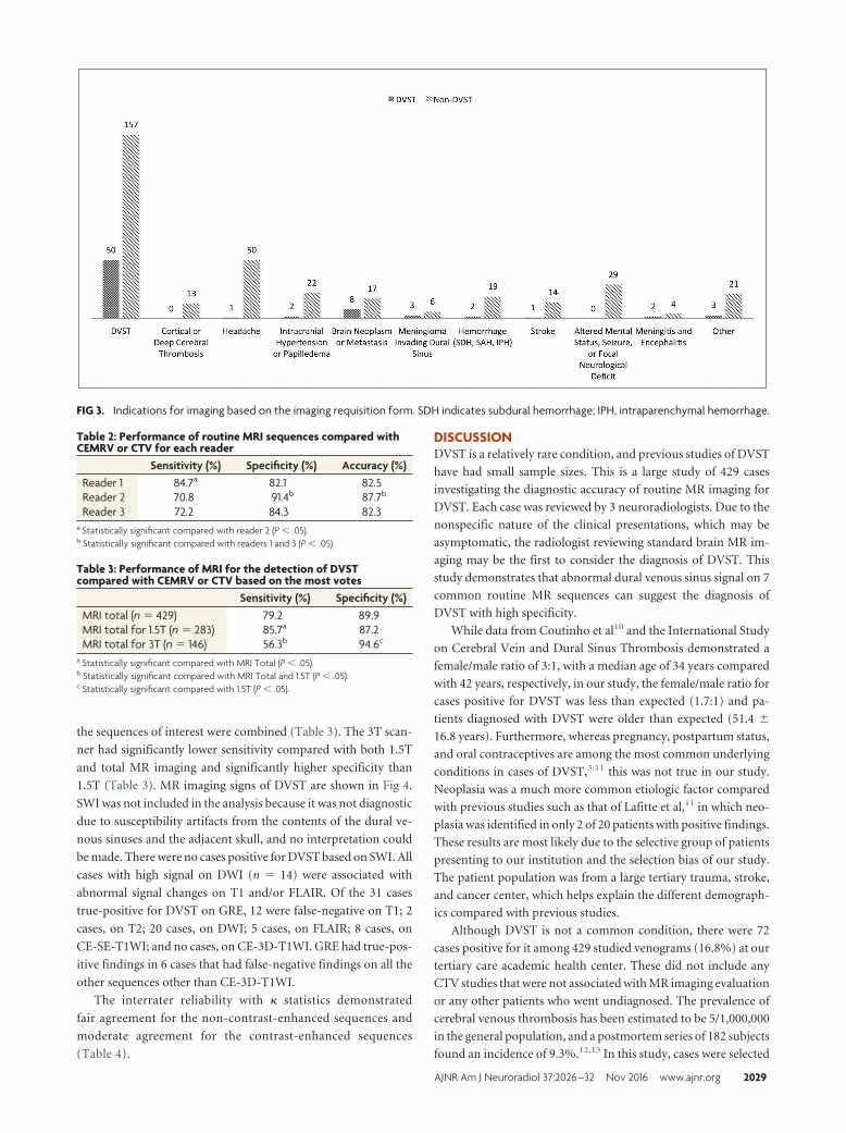
The most commonly stated clinical indication for imaging was
suspicion of DVST (Fig 3). For the cases positive for DVST, 69.4%
had suspicion of DVST indicated, so 30.6% of positive cases were
clinically unexpected DVST. Of the total 207 cases imaged with
the explicit indication of “rule out DVST,” 50 (24.2%) were pos-
itive for it.
Performance CharacteristicsWhen we compared the performance characteristics of the 3 read-
ers, reader 1 had significantly higher sensitivity than reader 2, and
reader 2 had significantly higher specificity and accuracy than
both readers 1 and 3 (Table 2). The most accurate reader (87.7%)
had higher specificity (91.4%) at the expense of lower sensitivity
(70.8%).
Routine MR imaging had an overall sensitivity of 79.2% and
specificity of 89.9% based on the most votes of the 3 readers when
FIG 2. Etiology or underlying condition based on electronic medical records. SDH indicates subdural hemorrhage; IPH, intraparenchymalhemorrhage; DAVF, dural arteriovenous fistula.
Table 1: Patient characteristicsDVST (n = 72) Non-DVST (n = 357) Total (n = 429)
Age (yr) (mean) 51.4 � 16.8 45.8 � 17.0 46.7 � 17.1Female (No.) (%) 45 (62.5) 239 (66.9) 284 (66.2)3T MRI (No.) (%) 16 (22.2) 130 (36.4) 146 (34.0)
2028 Patel Nov 2016 www.ajnr.org
the sequences of interest were combined (Table 3). The 3T scan-
ner had significantly lower sensitivity compared with both 1.5T
and total MR imaging and significantly higher specificity than
1.5T (Table 3). MR imaging signs of DVST are shown in Fig 4.
SWI was not included in the analysis because it was not diagnostic
due to susceptibility artifacts from the contents of the dural ve-
nous sinuses and the adjacent skull, and no interpretation could
be made. There were no cases positive for DVST based on SWI. All
cases with high signal on DWI (n � 14) were associated with
abnormal signal changes on T1 and/or FLAIR. Of the 31 cases
true-positive for DVST on GRE, 12 were false-negative on T1; 2
cases, on T2; 20 cases, on DWI; 5 cases, on FLAIR; 8 cases, on
CE-SE-T1WI; and no cases, on CE-3D-T1WI. GRE had true-pos-
itive findings in 6 cases that had false-negative findings on all the
other sequences other than CE-3D-T1WI.
The interrater reliability with � statistics demonstrated
fair agreement for the non-contrast-enhanced sequences and
moderate agreement for the contrast-enhanced sequences
(Table 4).
DISCUSSIONDVST is a relatively rare condition, and previous studies of DVST
have had small sample sizes. This is a large study of 429 cases
investigating the diagnostic accuracy of routine MR imaging for
DVST. Each case was reviewed by 3 neuroradiologists. Due to the
nonspecific nature of the clinical presentations, which may be
asymptomatic, the radiologist reviewing standard brain MR im-
aging may be the first to consider the diagnosis of DVST. This
study demonstrates that abnormal dural venous sinus signal on 7
common routine MR sequences can suggest the diagnosis of
DVST with high specificity.
While data from Coutinho et al10 and the International Study
on Cerebral Vein and Dural Sinus Thrombosis demonstrated a
female/male ratio of 3:1, with a median age of 34 years compared
with 42 years, respectively, in our study, the female/male ratio for
cases positive for DVST was less than expected (1.7:1) and pa-
tients diagnosed with DVST were older than expected (51.4 �
16.8 years). Furthermore, whereas pregnancy, postpartum status,
and oral contraceptives are among the most common underlying
conditions in cases of DVST,3,11 this was not true in our study.
Neoplasia was a much more common etiologic factor compared
with previous studies such as that of Lafitte et al,11 in which neo-
plasia was identified in only 2 of 20 patients with positive findings.
These results are most likely due to the selective group of patients
presenting to our institution and the selection bias of our study.
The patient population was from a large tertiary trauma, stroke,
and cancer center, which helps explain the different demograph-
ics compared with previous studies.
Although DVST is not a common condition, there were 72
cases positive for it among 429 studied venograms (16.8%) at our
tertiary care academic health center. These did not include any
CTV studies that were not associated with MR imaging evaluation
or any other patients who went undiagnosed. The prevalence of
cerebral venous thrombosis has been estimated to be 5/1,000,000
in the general population, and a postmortem series of 182 subjects
found an incidence of 9.3%.12,13 In this study, cases were selected
FIG 3. Indications for imaging based on the imaging requisition form. SDH indicates subdural hemorrhage; IPH, intraparenchymal hemorrhage.
Table 2: Performance of routine MRI sequences compared withCEMRV or CTV for each reader
Sensitivity (%) Specificity (%) Accuracy (%)Reader 1 84.7a 82.1 82.5Reader 2 70.8 91.4b 87.7b
Reader 3 72.2 84.3 82.3a Statistically significant compared with reader 2 (P � .05).b Statistically significant compared with readers 1 and 3 (P � .05).
Table 3: Performance of MRI for the detection of DVSTcompared with CEMRV or CTV based on the most votes
Sensitivity (%) Specificity (%)MRI total (n � 429) 79.2 89.9MRI total for 1.5T (n � 283) 85.7a 87.2MRI total for 3T (n � 146) 56.3b 94.6c
a Statistically significant compared with MRI Total (P � .05).b Statistically significant compared with MRI Total and 1.5T (P � .05).c Statistically significant compared with 1.5T (P � .05).
AJNR Am J Neuroradiol 37:2026 –32 Nov 2016 www.ajnr.org 2029

from patients who had CTV or CEMRV, which puts them at a
substantially higher pretest probability of having DVST than the
general population, and this does not represent the prevalence of
the disease. Therefore, positive predictive value, negative predic-
tive value, and accuracy were not reported. While this omission
limits the external validity of the study, the performance charac-
teristics are relevant to those in similar populations in tertiary care
centers.
Tang et al14 found that DVST was indicated on the imaging
requisitions in only 33% of MR imaging studies positive for
thrombosis,6 compared with our study in which 69.4% of cases
positive for DVST had “suspected DVST” on their imaging req-
uisitions. Some other indications for venography such as head-
ache, intracranial hypertension, and meningioma invading the
dural venous sinuses may be the cause or the effect of DVST.
Thus, 30.6% of cases positive for DVST were found in patients
FIG 4. MR imaging signs of dural venous sinus thrombosis from multiple cases. A, Hyperintense signal in the right transverse sinus on sagittal T1.B, Loss of flow void in the right transverse sinus on axial T2. C, Hyperintense signal in the right transverse sinus on FLAIR. D, Hyperintense signalin the right transverse sinus on DWI. E, Blooming artifacts in the right transverse sinus on GRE. F, Filling defect in the right transverse/sigmoid sinuson CE-SE-T1WI. G, Filling defect in the right transverse/sigmoid sinus on CE-3D-T1WI. H, Filling defect in the right transverse/sigmoid sinus onCTV. I, Filling defect in the right transverse/sigmoid sinus on MIP-CEMRV.
2030 Patel Nov 2016 www.ajnr.org

without clinical suspicion of it, highlighting the importance of
routine MR imaging signs of DVST in unexpected situations.
The overall sensitivity of MR imaging (79.2%) was slightly
lower than that in previously reported studies. Lafitte et al11 re-
ported that in 20 patients with clinically suspected DVST con-
firmed by digital subtraction angiography, MR imaging that in-
cluded spin-echo T1- and T2-weighted imaging was 90% sensitive
in diagnosing DVST, and �80% sensitivity was found in most
studies. This finding can be explained, in part, by the high sample
volume in our study, which is likely closer to the real performance
of the sequence of interest in the diagnosis of DVST in samples
from a tertiary care center.
The performance characteristics in this study are in keeping
with the findings of Saindane et al,7 who demonstrated that filling
defects on CE-3D-T1WI had a sensitivity of 67% and a specificity
of 100% for DVST compared with MRV. Sari et al8 demon-
strated that immediate CE-3D-T1WI had a sensitivity of
92.5%, specificity of 100%, and accuracy of 98.3% compared
with their reference standard. They proposed that delayed ac-
quisition of CE-3D-T1WI after contrast injection can lead to
contrast enhancement of the thrombus, which lowers the sen-
sitivity of the sequence.8
The reported sensitivity for hyperintense signal on DWI for
intraluminal thrombus has been highly variable in the literature,
ranging from 4% to 40%.2 Our results corroborate the study by
Favrole et al,15 which noted that a high DWI signal is always found
with concomitant signal changes on T1 and/or FLAIR sequences;
thus, it may not provide much diagnostic value.
GRE identified cases that were false-negative for DVST on all
of the other sequences other than CE-3D-T1WI, providing addi-
tional diagnostic value as part of a standard brain MR imaging.
These results are consistent with the findings of Ihn et al,16 who
demonstrated that GRE was the only sequence that was positive
for DVST in all 11 DVST cases in a study that included T1, T2,
FLAIR, and CE-SE-T1WI. Altinkaya et al17 have shown that GRE
has high diagnostic performance for acute and subacute superior
sagittal sinus and deep and cortical vein thrombosis, but not for
transverse and sigmoid sinus thrombosis due to susceptibility ar-
tifacts from the skull base.
Multiple reasons have been proposed for the low sensitivity of
MR imaging sequences for DVST. Hinman and Provenzale18 re-
ported that though in most cases, the diagnosis of DVST can be
made on routine MR imaging on the basis of the loss of a normal
flow void with abnormal signal intensity within the dural venous
sinus, during the first 5 days after formation, an acute thrombus
can mimic a normal flow void on spin-echo T2-weighted imaging
and may be isointense with gray matter on spin-echo T1-weighted
imaging.16,19 In this case, the hypointense signal on T2 can be
mistaken for patency. In the subacute stage, the thrombus be-
comes progressively hyperintense on both spin-echo T1- and T2-
weighted imaging.11 Enhancement of a chronic thrombus can
resemble normal flow.7,20,21 In the chronic stage, contrast-en-
hanced T1-weighted images of DVST can enhance to resemble a
patent sinus due to capillary channels or partial recanalization.6,7
Routine MR images need to be evaluated with caution to avoid
false-negative interpretations.
In our study, the specificity for DVST was high; this finding
indicates a low number of false-positive interpretations. False-
positives can occur if there is slow blood flow due to sinus hyp-
oplasia, which can create a high signal that resembles a thrombus.
This pitfall can be avoided by using multiple sequences and planes
of section.22 Filling defects caused by arachnoid granulations or
other anatomic material in the sinuses may also resemble throm-
bus, but signal isointense to CSF and morphology can help differ-
entiate them.6,23
In terms of interrater reliability, Ferro et al24 showed that with
a sample size of 40 cases, interrater reliability for DVST varies
from moderate to excellent, � range of 0.59 –1, when comparing
pairs of raters. In this study, for the individual MR images, there
was considerable variability because � statistics ranged from 0.28
to 0.42. The interrater reliability for overall thrombosis diagnosis
was only moderate at 0.50, which reflects the difficulty in inter-
preting the signs suggestive of DVST. Individual radiologists each
have their own propensity to make the diagnosis, which may ex-
plain why the performance characteristics of the 3 readers had
significant differences.
LimitationsAlthough readers were blinded to clinical history and whether
venography was completed, readers were focused on searching for
DVST, and the sample was chosen on the basis of patients who
had undergone CEMRV or CTV in a tertiary care center. These
features substantially increased the pretest probability of DVST
compared with a random sample and promoted selection bias.
This high prevalence would falsely increase the accuracy of MR
imaging for DVST among the 3 readers. The accuracy of the read-
ers was only reported to demonstrate individual differences, but it
is not generalizable to daily practice for the reasons described
above. Readers had access to some or all of the MR images (T1, T2
and/or FLAIR, GRE, DWI, SWI, CE-SE-T1WI, and/or CE-3D-
T1WI), which would confound individual sequence test parame-
ters. The neuroradiologists may have used secondary clues to the
diagnosis other than those of interest such as hemorrhage, edema,
or infarction to help them. The study included 18 patients with
idiopathic intracranial hypertension, which is known to have ab-
normal transverse sinus morphology and flow; however, none of
these patients were positive for DVST. In addition, the age of the
thrombosis was not taken into account. The signal characteristics
are dependent on the age of the thrombosis, and Idbaih et al25
have shown that the sensitivity of routine MR images changes as
the thrombus evolves.
Table 4: Interrater reliability for each MRI sequence individuallyand combined using the � coefficient
� Agreementa
Sagittal T1 (n � 418) 0.28 FairAxial T2 (n � 71) 0.34 FairAxial GRE (n � 420) 0.40 FairAxial FLAIR (n � 417) 0.34 FairAxial DWI (n � 421) 0.33 FairAxial CE-SE-T1WI (n � 241) 0.42 ModerateAxial CE-3D-T1WI (n � 199) 0.41 ModerateMRI total (n � 429) 0.50 Moderate
a Agreement was considered slight if � values were 0 – 0.20; fair if, 0.21– 0.40; moder-ate if, 0.41– 0.60; substantial if, 0.61– 0.80; and almost perfect if, 0.81–1.
AJNR Am J Neuroradiol 37:2026 –32 Nov 2016 www.ajnr.org 2031

CONCLUSIONSAbnormal dural venous sinus signal on routine MR images can
suggest DVST with high specificity and moderate interrater reli-
ability in high-risk patients. In patients with primary or metastatic
brain cancer, these signs need to be taken very seriously, and this
is particularly important in patients in whom there is no clinical
suspicion of DVST.
Disclosures: Richard I. Aviv—UNRELATED: Grants/Grants Pending: Canadian Insti-tutes of Health Research*; Other: Biogen Foundation fellowship.* Pejman JabehdarMaralani—UNRELATED: Grants/Grants Pending: Radiological Society of NorthAmerica,* Brain Tumor Foundation of Canada,* Comments: Both grants are relatedto a brain tumor project and not related to the submitted work. *Money paid to theinstitution.
REFERENCES1. Stam J. Thrombosis of the cerebral veins and sinuses. N Engl J Med
2005; 352:1791–98 CrossRef Medline2. Linn J, Bruckmann H. Cerebral venous and dural sinus thrombosis:
state-of-the-art imaging. Clin Neuroradiol 2010;20:25–37 CrossRefMedline
3. Ferro JM, Canhao P, Stam J, et al; ISCVT Investigators. Prognosis ofcerebral vein and dural sinus thrombosis: results of the Interna-tional Study on Cerebral Vein and Dural Sinus Thrombosis(ISCVT). Stroke 2004;35:664 –70 Medline
4. Ferro JM, Canhao P, Stam J, et al; ISCVT Investigators. Delay in thediagnosis of cerebral vein and dural sinus thrombosis: influence onoutcome. Stroke 2009;40:3133–38 CrossRef Medline
5. Khandelwal N1, Agarwal A, Kochhar R, et al. Comparison of CTvenography with MR venography in cerebral sinovenous thrombo-sis. AJR Am J Roentgenol 2006;187:637– 43 Medline
6. Provenzale JM, Kranz PG. Dural sinus thrombosis: sources of errorin image interpretation. AJR Am J Roentgenol 2011;196:23–31CrossRef Medline
7. Saindane AM, Mitchell BC, Kang J, et al. Performance of spin-echoand gradient-echo T1-weighted sequences for evaluation of duralvenous sinus thrombosis and stenosis. AJR Am J Roentgenol 2013;201:162– 69 CrossRef Medline
8. Sari S, Verim S, Hamcan S, et al. MRI diagnosis of dural sinus-cortical venous thrombosis: immediate post-contrast 3D GRET1-weighted imaging versus unenhanced MR venography andconventional MR sequences. Clin Neurol Neurosurg 2015;134:44 –54 CrossRef Medline
9. Altman DG. Practical Statistics for Medical Research. London: Chap-man and Hall; 1991
10. Coutinho JM, Ferro JM, Canhao P, et al. Cerebral venous and sinusthrombosis in women. Stroke 2009;40:2356 – 61 CrossRef Medline
11. Lafitte F, Boukobza M, Guichard JP, et al. MRI and MRA for diagno-
sis and follow-up of cerebral venous thrombosis (CVT). Clin Radiol1997;52:672–79 CrossRef Medline
12. Towbin A. The syndrome of latent cerebral venous thrombosis: itsfrequency and relation to age and congestive heart failure. Stroke1973;4:419 –30 CrossRef Medline
13. Saposnik G, Barinagarrementeria F, Brown RD Jr, et al; AmericanHeart Association Stroke Council and the Council on Epidemiologyand Prevention. Diagnosis and management of cerebral venousthrombosis: a statement for healthcare professionals from theAmerican Heart Association/American Stroke Association. Stroke2011;42:1158 –92 CrossRef Medline
14. Tang PH, Chai J, Chan YH, et al. Superior sagittal sinus thrombosis:subtle signs on neuroimaging. Ann Acad Med Singapore 2008;37:397– 401 Medline
15. Favrole P, Guichard JP, Crassard I, et al. Diffusion-weighted imagingof intravascular clots in cerebral venous thrombosis. Stroke 2004;35:99 –103 Medline
16. Ihn YK, Jung WS, Hwang SS. The value of T2*-weighted gradient-echo MRI for the diagnosis of cerebral venous sinus thrombosis.Clin Imaging 2013;37:446 – 450 CrossRef Medline
17. Altinkaya N, Demir S, Alkan O, et al. Diagnostic value of T2*-weighted gradient-echo MRI for segmental evaluation in cerebralvenous sinus thrombosis. Clin Imaging 2015;39:15–19 CrossRefMedline
18. Hinman JM, Provenzale JM. Hypointense thrombus on T2-weighted MR imaging: a potential pitfall in the diagnosis of duralsinus thrombosis. Eur J Radiol 2002;41:147–52 CrossRef Medline
19. Dormont D, Anxionnat R, Evrard, et al. MRI in cerebral venousthrombosis. J Neuroradiol 1994;21:81–99 Medline
20. Dormont D, Sag K, Biondi A, et al. Gadolinium-enhanced MR ofchronic dural sinus thrombosis. AJNR Am J Neuroradiol 1995;16:1347–52 Medline
21. Leach JL, Wolujewicz M, Strub WM. Partially recanalized chronicdural sinus thrombosis: findings on MR imaging, time-of-flightMR venography, and contrast-enhanced MR venography. AJNRAm J Neuroradiol 2007;28:782– 89 Medline
22. Mas JL, Meder JF, Meary E, et al. Magnetic resonance imaging inlateral sinus hypoplasia and thrombosis. Stroke 1990;21:1350 –56Medline
23. Leach JL, Jones BV, Tomsick TA, et al. Normal appearance of arach-noid granulations on contrast-enhanced CT and MR of the brain:differentiation from dural sinus disease. AJNR Am J Neuroradiol1996;17:1523–32 Medline
24. Ferro JM, Morgado C, Sousa R, et al. Interobserver agreement in themagnetic resonance location of cerebral vein and dural sinusthrombosis. Eur J Neurol 2007;14:353–56 CrossRef Medline
25. Idbaih A, Boukobza M, Crassard I, et al. MRI of clot in cerebralvenous thrombosis: high diagnostic value of susceptibility-weighted images. Stroke 2006;37:991–95 CrossRef Medline
2032 Patel Nov 2016 www.ajnr.org