Diagnostic Laparoscopy for Pelvic Disorders
37
Diagnostic Laparoscop y for Pelvic Disorders Venice June 2005 George S. Ferzli MD, FACS George E. Khoury, MD
-
Upload
george-s-ferzli -
Category
Health & Medicine
-
view
3.102 -
download
2
Transcript of Diagnostic Laparoscopy for Pelvic Disorders

Diagnostic Laparoscopyfor Pelvic Disorders
VeniceJune 2005
George S. Ferzli MD, FACSGeorge E. Khoury, MD

Differential Diagnosis:
Carter and Soper Chronic Pelvic Pain. Women’s Health in Primary Care, September 2000; vol. 3, 649-661.
Patient history and a physical exam
provide a base for diagnosis of
pelvic disorders. Categories include:
Gynecologic
Gastrointestinal
Urologic
Musculoskeletal
Surgical
Vascular

Gynecologic Disorders: Chronic pelvic pain
Ectopic pregnancy
Pelvic inflammatory disease
Endometriosis
Adhesions
Ovary: cysts, torsion
Fallopian tube: torsion,
salpingitis
Uterus: fibroid, leiomyomata
Pelvic congestion syndrome
Infertility

The role of laparoscopy as a diagnostic tool in chronic pelvic pain. Fred M Howard MS, MDBaillieÁ re's Clinical Obstetrics and Gynaecology Vol. 14, No. 3, pp. 467-494, 2000.
> 40% of gynaecological diagnostic laparoscopies are done for CPP
Combining the results of published series of laparoscopies for CPP shows that : No visible pathology is detected in 35% (range . 3±92%) of patients Endometriosis is diagnosed in 33% (range . 2±80%) Adhesive disease is found in 24% (range . 0± 52%)
A negative laparoscopy is not synonymous with no diagnosis or no disease
A meticulously performed negative laparoscopy means that a woman does not have endometriosis-associated or adhesion-associated pain
Gynecologic: Chronic Pelvic Pain

The role of laparoscopy in the management of pelvic pain in women of reproductive ageMaria Grazia Porpora, MD FERTILITY AND STERILITY, Vol. 6M, No. 5, November 1997.
The role of laparoscopy in women with chronic pelvic pain is more controversial and limited, but abnormal laparoscopic findings are detected in approximately 60% of those who have undergone a multidisciplinary investigation and received a tentative clinical diagnosis.
Gynecologic: Chronic Pelvic Pain

Gynecologic: Ectopic pregnancy
Photograph courtesy of Dr. Syed
Laparoscopy should be regarded as a therapeutic rather than diagnostic tool for suspected ectopic pregnancy.
Transvaginal ultrasound has replaced laparoscopy for diagnosis of ectopic pregnancy.
Laparoscopy has diagnostic role if probable tubal pregnancy or diagnosis in doubt.
Large uncontrolled studies have demonstrated that > 80% of ectopic pregnancies can be managed laparoscopically
The most commonly used procedures at laparoscopy are salpingectomy and salpingotomy.
The role of laparoscopy in the management of ectopic pregnancy. Martin Christopher Sowter, MD, MRCOGa, Jonathan Frappell, FRCS, FRCOG Reviews in Gynaecological Practice #2 (2002) 73-82

Gynecologic: PIDClinical diagnosis of PID is often difficult especially when symptoms are mild, as frequently when the primary organism is C. trachomatis.
Laparoscopy is the gold standard for the diagnosis of PID – should be used when diagnosis is uncertain, especially in young women for whom the preservation of fertility is important.
Sellors et al. reported that only by resorting to diagnostic laparoscopy were they able to demonstrate that PID was the cause of acute pelvic pain in 46% of a group of 95 women.
Laparoscopy should be considered for patients who have not responded to antibiotic therapy within 48 to 72 hours.
The role of laparoscopy in the management of pelvic pain in women of reproductive age. Maria Grazia Porpora, M.D. FERTILITY AND STERILITY Vol. 6M, No. 5, November 1997.

Gynecologic: Tubo-ovarian Abscess
Most commonly isolated pathogens from a tubo-ovarian abscess are C. trachomatis and peptostreptococci.
At laparoscopy the peritoneal cavity (pelvis and abdomen) is inspected carefully.
The surgical steps include adhesiolysis, aspiration of the abscess cavity, dissection and excision of necrotic tissue, tubal lavage, and irrigation of the peritoneal cavity before completion of the procedure.
Laparoscopic surgery combined with adequate broad-spectrum antibiotic therapy has proven successful in the treatment of more than 95% of patients.

Gynecologic: Endometriosis
Endometriosis is a histologically-defined disease :“the presence of ectopic tissue which possesses the histological structure and function of the uterine mucosa”. ( Sampson 1921)
Laparoscopy has largely replaced laparotomy as the diagnostic procedure for any patient suspected of having endometriosis.
Based on visual appearance endometriosis are classified as atypical (red, yellow, white or clear) or typical (black-brown, black or puckered black stellate) lesions.
The role of laparoscopy as a diagnostic tool in chronic pelvic pain. Fred M Howard MS, MD BaillieÁ re's Clinical Obstetrics and Gynaecology Vol. 14, No. 3, pp. 467-494, 2000.

The role of laparoscopy as a diagnostic tool in chronic pelvic pain. Fred M Howard MS, MD BaillieÁ re's Clinical Obstetrics and Gynaecology Vol. 14, No. 3, pp. 467-494, 2000.
Brownish lesion on the ovaryWhite fibrotic lesion on the uterosacral ligament
Peritoneal pocket of endometriosis Red stellate lesions in the cul-de-sac
Endometriosis presents with a variety of appearances that may make visual diagnosis difficult and inaccurate.

The role of laparoscopy as a diagnostic tool in chronic pelvic pain. Fred M Howard MS, MD BaillieÁ re's Clinical Obstetrics and Gynaecology Vol. 14, No. 3, pp. 467-494, 2000.
Visually misdiagnosed
as endometriosis: Haemangiomas, old suture, ovarian
carcinoma, residual carbon deposits from prior surgery, ectopic pregnancy, …
Histological confirmation of visually diagnosed endometriosis ranges from 9-90%, depending on characteristics of the lesions
Apparent classic endometriotic lesion that was actually a suture granuloma.
Gynecologic: Endometriosis

The role of laparoscopy as a diagnostic tool in chronic pelvic pain. Fred M Howard MS, MD BaillieÁ re's Clinical Obstetrics and Gynaecology Vol. 14, No. 3, pp. 467-494, 2000.
Laparoscopic evaluation requires detailed visualization of all sites of endometriosis:
A study of 716 women with endometriosis found these anatomical distributions: cul-de-sac and uterosacrals ( 69%) ovaries ( 45%) ovarian fossae ( 33%)and uterovesical fold ( 24% ) Chocolate fluid from needle puncture
of a suspected occult endometrioma.
Gynecologic: Endometriosis
ovaries (all surfaces) ovarian fossae pelvic peritoneum (cul-de-sac, periureteral, bladder peritoneum)
uterine ligaments sigmoid colon appendix fallopian tubes rectovaginal septum

Gynecologic: Adhesions
Pre-operative history of PID, endometriosis, perforated appendix, prior surgery or inflammatory bowel disease.
Presently the only definitive way to diagnose adhesions is by surgical visualization usually via laparoscopy instead of laparotomy.
Laparoscopic studies reveal adhesions on average in 24% of CPP patients and 17% of non-CPP patients.
The role of laparoscopy as a diagnostic tool in chronic pelvic pain. Fred M Howard MS, MD BaillieÁ re's Clinical Obstetrics and Gynaecology Vol. 14, No. 3, pp. 467-494, 2000.

Gynecologic: Adenomyosis
Endometrial cells penetrate the myometrium causing either localized (adenomyoma) or diffuse overgrowth.
Adenomyomas that penetrate the uterine cavity become submucosal tumors.
An enlarged uterus from adenomyosis is often misdiagnosed as being from fibroids
The role of laparoscopy as a diagnostic tool in chronic pelvic pain. Fred M Howard MS, MD BaillieÁ re's Clinical Obstetrics and Gynaecology Vol. 14, No. 3, pp. 467-494, 2000.
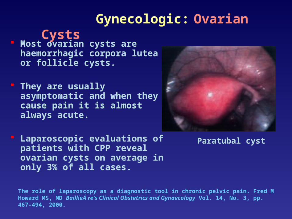
Most ovarian cysts are haemorrhagic corpora lutea or follicle cysts.
They are usually asymptomatic and when they cause pain it is almost always acute.
Laparoscopic evaluations of patients with CPP reveal ovarian cysts on average in only 3% of all cases.
Paratubal cyst
Gynecologic: Ovarian Cysts

The role of laparoscopy as a diagnostic tool in chronic pelvic pain. Fred M Howard MS, MD BaillieÁ re's Clinical Obstetrics and Gynaecology Vol. 14, No. 3, pp. 467-494, 2000.
Even when the surgeon is ‘certain’ that the ovary is benign, it is essential that tissue be sent for histological evaluation.
Open the cyst and inspect the lining for papillary structures or excrescences.
If these are noted, then a laparotomy should be done .
Gynecologic: Ovarian Cysts
Gynecologic: Ovarian Cysts
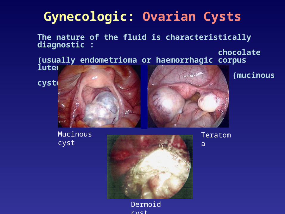
The nature of the fluid is characteristically diagnostic : chocolate (usually endometrioma or haemorrhagic corpus luteum) sebaceous (teratoma), or mucinous (mucinous cystoma).
Mucinous cyst Teratoma
Dermoid cyst

Gynecologic: Adnexal TorsionA rare gynecologic emergency that nearly always occurs unilaterally.
Common causes are benign ovarian tumors and cysts; malignant processes are rare.
Relapse or bilateral adnexal torsion can cause sterility interfering with fertility.
In 30% of the patients, there is torsion of a normal adnexa, while the majority of the cases are associated with ovarian pathology.
Adnexal torsion in very young girls: diagnostic pitfalls. Marieke Emontsa, Heleen Doornewaardb, J.(Co’tje) F. Admiraala,* European Journal of Obstetrics & Gynecology and Reproductive Biology 116 (2004) 207-210.
Gynecologic: Adnexal Torsion
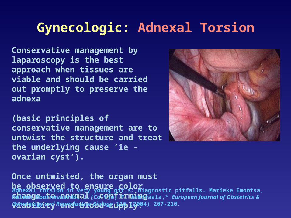
Conservative management by laparoscopy is the best approach when tissues are viable and should be carried out promptly to preserve the adnexa
(basic principles of conservative management are to untwist the structure and treat the underlying cause ‘ie - ovarian cyst’). Once untwisted, the organ must be observed to ensure color change to normal, confirming viability and blood supply.
Adnexal torsion in very young girls: diagnostic pitfalls. Marieke Emontsa, Heleen Doornewaardb, J.(Co’tje) F. Admiraala,* European Journal of Obstetrics & Gynecology and Reproductive Biology 116 (2004) 207-210.

Gynecologic: Endosalpingiosis
Endosalpingiosis is the presence of fallopian tubal glandular epithelium in an ectopic location. Visually it appears as white to yellow, opaque or translucent, punctate, cystic lesions.
Endosalpingiosis is generally not recognized by gynaecologists at the time of laparoscopic evaluation or is misdiagnosed as endometriosis.
The role of laparoscopy as a diagnostic tool in chronic pelvic pain. Fred M Howard MS, MD BaillieÁ re's Clinical Obstetrics and Gynaecology Vol. 14, No. 3, pp. 467-494, 2000

Gynecologic: Leiomyomata (fibroids)

The role of laparoscopy as a diagnostic tool in chronic pelvic pain. Fred M Howard MS, MD BaillieÁ re's Clinical Obstetrics and Gynaecology Vol. 14, No. 3, pp. 467-494, 2000.
Retrograde ovarian venography and transuterine pelvic venography demonstrate increased ovarian or uterine venous diameters, venous stasis and venous congestion and are the recognized techniques of diagnosis (not laparoscopy) of pelvic congestion syndrome.
Laparoscopy can be used to visualize pelvic varicosities by decreasing intra-abdominal pressure and gradually placing the patient in a reverse Trendelenburg position.
Treatment options for pelvic varicosities remain controversial and include ligation of abnormal veins, salpingo-oophrectomy, or hysterectomy, all of which can be performed laparoscopically.
Dilated ovarian veins
Gynecologic: Pelvic Congestion Syndrome

The role of laparoscopy as a diagnostic tool in chronic pelvic pain. Fred M Howard MS, MD BaillieÁ re's Clinical Obstetrics and Gynaecology Vol. 14, No. 3, pp. 467-494, 2000.
Post-operative peritoneal cyst (form of adhesive disease) occurs in patients with prior surgical procedures (may rarely occur in women with previous PID / no prior surgery).
Symptomatic post-operative peritoneal cysts are rare (apparently 16 published cases), are reported 6 weeks to 13 years after surgery and interestingly, only in women.
Ovarian remnant syndrome: presence of painful, histologically documented ovarian tissue in a patient who has undergone a previous bilateral oophorectomy.
Small ovarian remnant, without cyst formation, on the right pelvic sidewall in a patient with a prior right salpingoophorectomy.
Laparoscopy in Women with no Uterus and no Ovaries

Infertility

Gastrointestinal (Chronic or Recurring):
Irritable bowel syndrome
Chronic appendicitis
IBD (Crohn’s)
Diverticulosis
Diverticulitis
Meckel’s diverticulum
Tail gut cyst

Gastrointestinal: IBS
Approximately one third of women with chronic pelvic pain have irritable bowel syndrome (IBS)
IBS is characterized by abdominal pain and bowel symptoms such as bloating, urgency, diarrhea and constipation
IBS is associated with gynecologic problems such as endometriosis, dyspareunia, and dysmenorrhea
IBS is most prevalent during menstruating years – exacerbated during menstruation
Recognition and treatment of irritable bowel syndrome among women with chronic pelvic pain. Rachel E. Williams, PhD, Katherine E. Hartmann, MD, PhD American Journal of Obstetrics and Gynecology (2005) 192, 761-7.

Gastrointestinal: Acute Appendicitis
Diagnostic accuracy in young men is ±95% – in fertile women it varies from 55%-65%
Older age (>70 years) lowers diagnostic accuracy to ± 66% for men and women
Traditionally, a normal-appearing appendix is removed after a laparotomy in the right fossa.
Diagnostic laparoscopy offers the possibility of sparing a normal-appearing appendix.
Introducing diagnostic laparoscopy for patients with suspected acute appendicitis. AC Moberg, A Montgomery Surg Endosc (2000) 14: 942–947.

Chronic Appendicitis Diagnosed Preoperatively as an Ovarian Dermoid. Jo T VanWinter, MD and Derek A Beyer, MD J Pediatr Adolesc Gynecol (2004) 17:403-406.
Results from partial obstruction and subsequent release of the appendix causing chronic or recurrent pain and inflammation in the right lower quadrant.
The incidence of recurrent and chronic forms of appendicitis has been estimated at 10% and 1%, respectively among patients undergoing appendectomy for right lower quadrant pain.
Recurrent and chronic appendicitis may be misdiagnosed as PID in sexually active female patients.
Chronic appendicitis with fibrosis. There are no neutrophils in the lumen to suggest an acute process.
Gastrointestinal: Chronic Appendicitis

Musculoskeletal / Myofascial:
Hernias (inguinal, femoral, umbilical,
incisional, spigelian)
Nerve entrapment (neuritis)

Hernias:
Direct inguinal hernia
Obturator hernia
Indirect inguinal hernia
Femoral hernia

The role of laparoscopy as a diagnostic tool in chronic pelvic pain. Fred M Howard MS, MD BaillieÁ re's Clinical Obstetrics and Gynaecology Vol. 14, No. 3, pp. 467-494, 2000.
Most common hernias are inguinal (indirect or direct) and femoral in men and women. Femoral hernias are more common in women than men accounting for 20% of all groin hernias found in women.
A study of women with CPP evaluated and treated using laparoscopy found that sciatic hernias were diagnosed in 20 (1.8%) of 1100 women.
A sciatic hernia is a protrusion of a peritoneal sac and contents through the greater or lesser sciatic foramen. Also called a ‘sacral sciatic’, ‘gluteal’, or ‘ischiatic’ hernia. Sciatic hernias are rare.
Indirect inguinal hernia
Laparoscopy and Hernias

Pediatric Hernias
The incidence of recurrent inguinal hernias in children is low (0% to 0.8%)
Laparoscopy accurately identifies the nature of the defect in children with recurrent groin hernias, detecting unsuspected contralateral indirect, direct, or femoral hernias in 44% of those undergoing laparoscopy.
The Role of Laparoscopy in the Management of Suspected Recurrent Pediatric Hernias.Jon Perlstein and Jeffrey J Du Bois Sacramento, California J Pediatr Surg 35:1205-1208

Nerve entrapment (neuritis) from mesh
Staplalgia
Adhesive disease
Surgical:

Urologic:
Undescended testicle
Node dissection for staging of prostate and testicular cancer
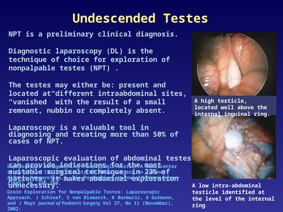
Undescended TestesNPT is a preliminary clinical diagnosis.
Diagnostic laparoscopy (DL) is the technique of choice for exploration of nonpalpable testes (NPT) .
The testes may either be: present and located at different intraabdominal sites, “vanished” with the result of a small remnant, nubbin or completely absent.
Laparoscopy is a valuable tool in diagnosing and treating more than 50% of cases of NPT.
Laparoscopic evaluation of abdominal testes can provide indications for the most suitable surgical technique; in 23% of patients, it makes abdominal exploration unnecessary.
Laparoscopic management of nonpalpable testes: A multicenter study of the Italian Society of Video Surgery in Infancy. Alfonso Papparellaa,*, Pio Parmeggiani Journal of Pediatric Surgery (2005) 40, 696-700.
Groin Exploration for Nonpalpable Testes: Laparoscopic Approach. J Schleef, S von Bismarck, K Burmucic, A Gutmann, and J Mayr Journal of Pediatric Surgery, Vol 37, No 11 (November), 2002:
A high testicle, located well above the internal inguinal ring.
A low intra-abdominal testicle identified at the level of the internal ring

RPLNDRPLND is an accurate staging tool providing important information to determine the need for chemotherapy. When performed properly,
RPLND eliminates the retroperitoneum as a site for relapse, which in turn provides emotional and psychological relief to the patient, and simplifies the follow-up protocol
Laparoscopic RPLND for the treatment of clinical Stage I NSGCT has evolved into an excellent alternative to traditional modes of therapy.
The procedure can replicate the advantages of open RPLND without the morbidity of a large incision.
Laparoscopic RPLND for clinical stage I nonseminomatous germ cell testicular cancer: current statusSam B. Bhayani, M.D., Mohamad E. Allaf, M.D., Louis R. Kavoussi, M.D.*Urologic Oncology: Seminars and Original Investigations 22 (2004) 145–148
Conclusion:
Laparoscopy provides a vital tool for diagnosing pelvic pain – it provides first hand visual comprehension of the problem as well as an immediate opportunity to continue with therapeutic surgical correction.