Diagnostic approach to hereditary renal hypouricemia Ivan Sebesta
Upload
mohamed-zaitounCategory
view
628download
0
Genito-Urinary System
Renal Cystic Diseases

Mohamed Zaitoun
Assistant Lecturer-Diagnostic Radiology Department , Zagazig University Hospitals
EgyptFINR (Fellowship of Interventional
Neuroradiology)[email protected]


Knowing as much as possible about your enemy precedes successful battle
and learning about the disease process precedes successful management

Renal Cystic Diseasesa) Renal Dysplasiab) Polycystic Diseasec) Cortical Cystsd) Medullary Cystse) Miscellaneous Intrarenal Cystsf) Extraparenchymal Renal Cysts

a) Renal Dysplasia :1-Multicystic Dysplastic Kidney (MCDK)2-Focal & Segmental Cystic Dysplasia3-Multiple Cysts Associated With Lower
Urinary Tract Obstruction
1-Multicystic Dysplastic Kidney (MCDK) :a) Incidenceb) Radiographic Features

a) Incidence :-Collection of large noncommunicating cysts
separated by fibrous tissue, there is no functioning renal parenchyma
-Develops in utero and the diagnosis is often made either antenatally or in the early neonatal period
-There may be a predisposition for the left kidney , a slightly higher incidence in males for unilateral MDCK and a higher incidence in females for bilateral MCDK

b) Radiographic Features :-Cystic renal mass with no excretory
function-Hypertrophy of contralateral kidney-Thick echogenic fibrous septa between
cysts-Atretic ureter-Absence of renal artery

Left kidney has multiple cysts without connection between them and without residual normal parenchyma. Sp = fetal spine


2-Focal & Segmental Cystic Dysplasia :-Segmental dysplasia is seen in children
with duplex (duplicated) kidney (incomplete fusion of upper and lower poles)

7-day-old girl with probable segmental dysplasia of left kidney, partial sagittal ultrasound view of left kidney shows partially cystic, partially echogenic mass (calipers), no substantial change was found during follow-up examinations

3-Multiple Cysts Associated With Lower Urinary Tract Obstruction :
-Usually with posterior urethral valve in males

b) Polycystic Disease :1-Autosomal Recessive PKD2-Autosomal Dominant PKD


1-Autosomal Recessive PKD :a) Incidenceb) Typesc) Associationsd) Radiographic Features
a) Incidence :-Autosomal recessive polycystic kidney
disease is one of the commonest inheritable infantile cystic renal diseases, but is far less common than autosomal dominant polycystic disease (ADPKD) which affects adults

b) Types :1-Antenatal Form : 90%-Oligohydramnios in utero-75% have death within 24 hours of delivery2-Neonatal Form :-Minimal hepatic fibrosis3-Infantile Form :-Moderate periportal fibrosis 4-Juvenile Form :-Gross hepatic fibrosis-Portal hypertension with splenomegaly and portosystemic
varices

c) Associations :-Caroli disease -Congenital hepatic fibrosis

d) Radiographic Features :1-Kidneys :-Enlarged hyperechoic kidneys (hallmark)-Cysts of 1 to 2 mm are seen2-U/S in Utero :-Nonvisualization of urine in bladder-Enlarged hyperechoic kidneys-Oligohydramnios (nonfunctioning kidneys)

3-Lungs :-Pulmonary hypoplasia (because of external
compression)-Pneumothorax4-Liver :-Hepatic fibrosis-Portal venous hypertension

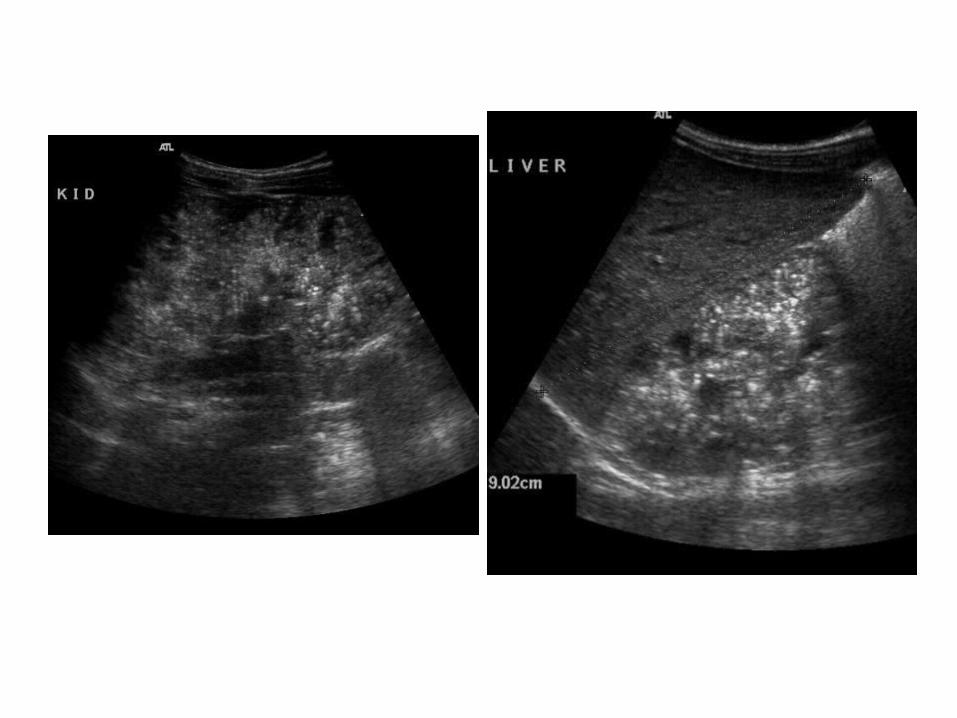
Fetal kidneys in ARPKD, (a) Longitudinal US scan of the left kidney in a 3rd-trimester fetus shows an enlarged, echogenic kidney (arrows) with loss of corticomedullary differentiation, note the paucity of amniotic fluid, indicating oligohydramnios, (b) Transverse US scan of a 26-week-old fetus shows enlarged kidneys (arrows) that are more echogenic than the liver (*)


ARPKD with caroli disease

ARPKD in a 13-year-old boy who was diagnosed as an infant, (a) US of the liver demonstrates the “central dot sign” (arrowhead), which represents the portal vein and hepatic artery branches completely surrounded by a dilated intrahepatic bile duct, (b) CT shows periportal hypoattenuation representing periportal fibrosis and edema (white arrow), cystic biliary dilatation with the central dot sign (white arrowhead), and splenomegaly due to portal hypertension, ascites is noted, note the expanded medullae with delayed transit of contrast material into the tubules of a segment of the upper pole of the right kidney (black arrowhead), the hypoattenuating focus in the spleen represents a splenic infarct (black arrow)

Respiratory distress in a newborn due to ARPKD with the oligohydramnios sequence, chest and abdominal radiograph demonstrates bilateral flank masses from enlarged kidneys that displace gas-filled bowel loops centrally (*), the lungs are small with a bell-shaped thorax and left pneumothorax (arrowhead) related to pulmonary hypoplasia

2-Autosomal Dominant PKD :a) Incidenceb) Associationsc) Radiographic Features
a) Incidence :-Referred to as adult polycystic kidney disease-Autosomal dominant polycystic kidney disease is
one of the most common serious hereditary disease
-The kidneys are normal at birth and with time develop multiple cysts , at the age of 30 years approximately 68% of patients will have visible cysts by ultrasound

b) Associations :-Hepatic cysts , 70%-Intracranial berry aneurysm , 20%-Cysts in pancreas and spleen , <5%-Hypertension-Colonic diverticulosis

c) Radiographic Features :-Kidneys are enlarged and contain innumerable
cysts-Simple renal cysts will appear anechoic with well
defined imperceptible walls and posterior acoustic enhancement, cysts with hemorrhage or infection will demonstrate echogenic material within the cyst
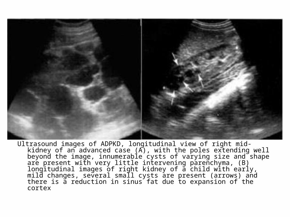
Ultrasound images of ADPKD, longitudinal view of right mid-kidney of an advanced case (A), with the poles extending well beyond the image, innumerable cysts of varying size and shape are present with very little intervening parenchyma, (B) longitudinal images of right kidney of a child with early, mild changes, several small cysts are present (arrows) and there is a reduction in sinus fat due to expansion of the cortex


-CT : Simple cysts appear as rounded structures with near water attenuation (HU ~ 0) , the wall are very thin and regular, and are often imperceptible , cysts which have had internal complications may be hyperattenuating with internal non-enhancing septations and / or calcifications


With hepatic cysts

(a) Coronal T2 shows enlargement of both kidneys with multiple expansile renal cysts (arrows), as well as multiple hepatic cysts (arrowheads), (b) Axial T2 fat-saturated obtained in another patient shows similar enlargement of the kidneys, with multiple cysts (arrows)

c) Cortical Cysts :1-Simple Cyst2-Complicated Cyst3-Multilocular Cystic Nephroma4-Syndromes Associated With Cysts5-End Stage Renal Disease &
Haemodyalisis

1-Simple Cyst :-Are benign and fluid filled-There incidence increases with advancing age
and they are found in at least 50% of people > 50 years of age
-U/S :*Anechoic *Acoustic enhancement*Sharply defined , imperceptible , smooth wall*Round or ovoid shape


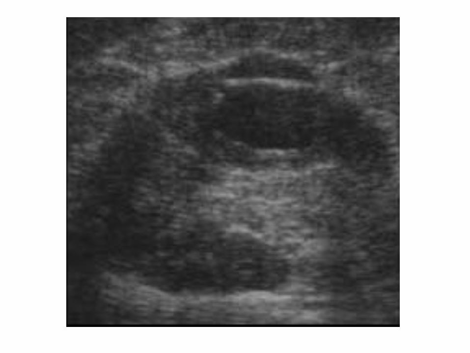
2-Complicated Cyst :-Are those that don’t meet the strict criteria of a simple
renal cyst-These include cysts containing :a) Internal echoesb) Septationsc) Calcificationd) Perceptible defined wall and mural nodularitya) Internal echoes :*Internal echoes within a cyst are usually the result of
hemorrhage or infection


b) Septations :*May be seen within a renal cyst and they
often occur following hemorrhage , infection and percutaneous aspiration


c) Calcification :*Calcification of the renal cysts may be fine
and linear or amorphous and thick


d) Perceptible defined thickened wall or mural nodularity :
*Essentially excludes a diagnosis of benign cyst , these lesions will all require surgical removal to exclude malignancy


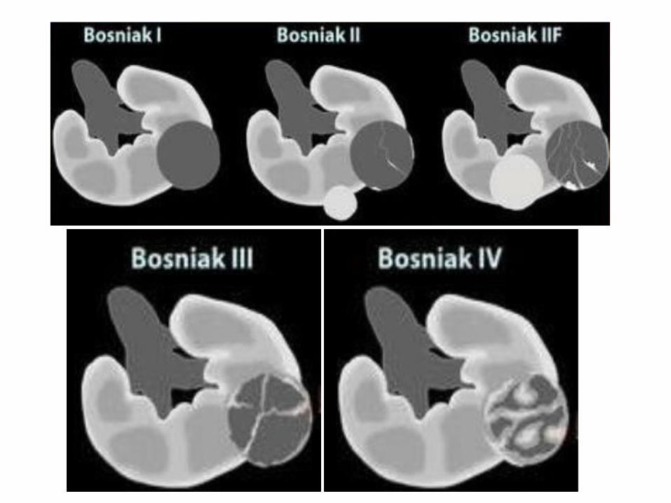
-Bosniak Classification :*Type 1 : Simple cyst*Type 2 : Minimally complicated cyst-Thin septation , calcium in wall*Type 2F : Minimally complicated cyst-Increased number of septa, minimally thickened
with nodular or thick calcifications*Type 3 : Intermediate lesion-Multiple thick septa , internal echos , mural
nodules*Type 4 : Clearly malignant-Solid mass component

Type 1
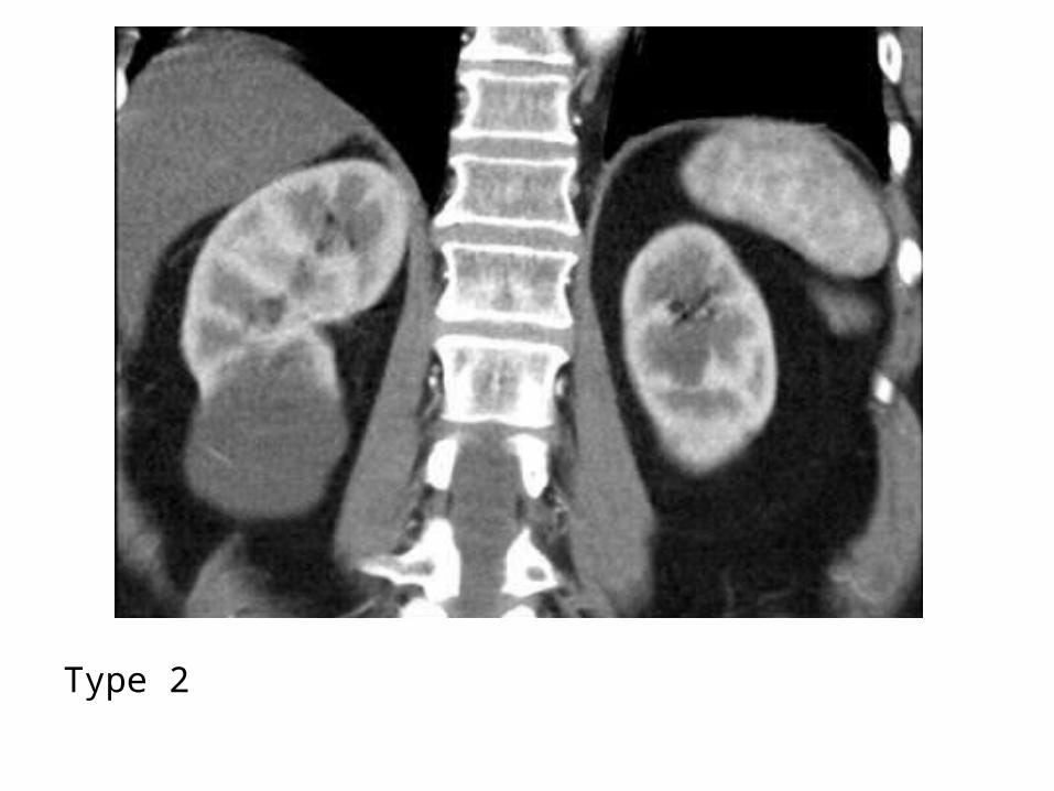
Type 2
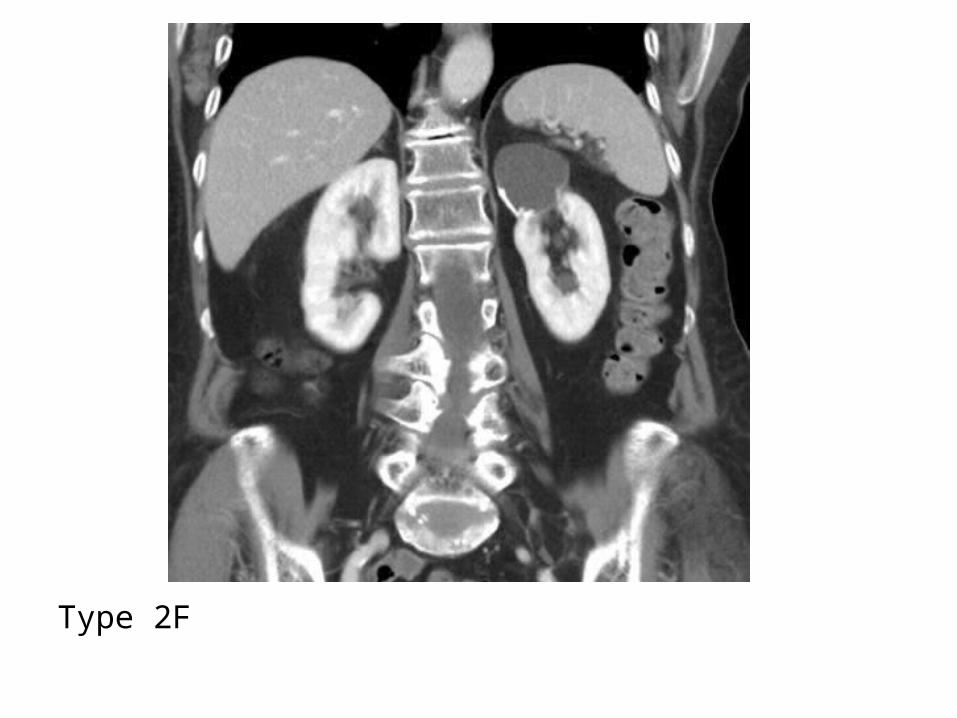
Type 2F
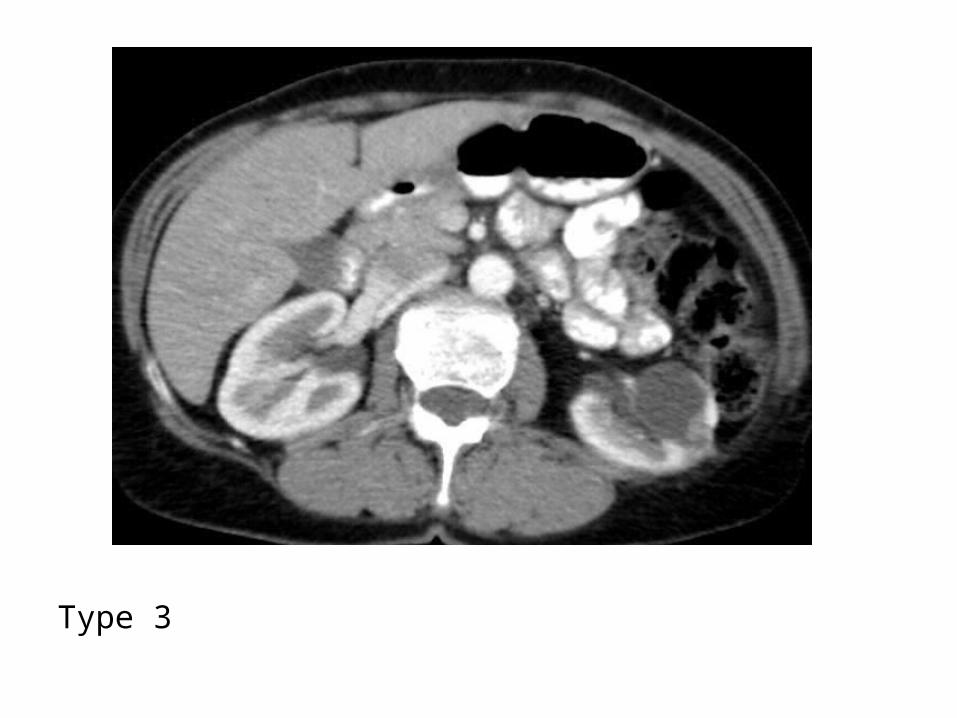
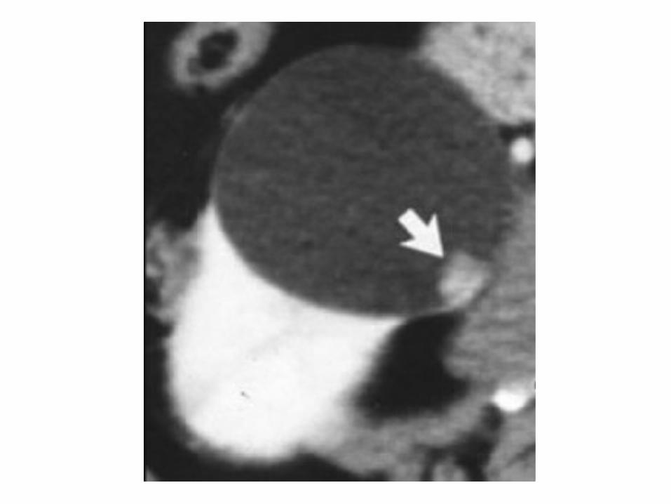
Type 3

Type 4

3-Multilocular Cystic Nephroma (MLCN) :a) Incidenceb) Radiographic Features

a) Incidence :-Congenital renal lesion characterized by
large (>10 cm) cystic spaces-It occurs in children ages 2 months to 4
years with 75% male predilection and in adults >40 years of age with 95% female predilection
-In Differential Diagnosis of Pediatric renal masses

b) Radiographic Features :-CT+C shows a well-defined intrarenal
multilocular mass that compresses or displaces the adjacent renal parenchyma , the septations enhance but the cysts do not
-Thick septae, nodules or a large solid component suggest Wilms tumor with cystic degeneration

Sagittal US image of the left renal fossa demonstrates a multicystic mass with variably sized cystic components and thin echogenic septa


4-Syndromes Associated With Cysts :-VHL disease-Tuberous Sclerosis-Zellweger’s Syndrome-Turner’s Syndrome-Triosomy 13 & 18

Von Hippel-Lindau disease, CT+C depicts multiple cysts in both kidneys (arrows) and an enhancing mass in the right kidney (arrowhead), the mass has attenuation similar to that in soft tissue, a finding suggestive of clear cell renal cell carcinoma

Tuberous sclerosis complex, CT+C shows multiple cysts in both kidneys (arrowheads) in a patient with diagnosed tuberous sclerosis

Neonate with Zellweger syndrome, (a) Ultrasound image of kidney in 1-day-old boy after birth shows multiple bilateral subcapsular cysts in diffusely echogenic kidneys and no corticomedullary differentiation, (b) MR image of trunk shows diffuse subcapsular cysts suggesting glomerulocystic kidneys

5-End Stage Renal Disease & Haemodyalisis :
-8-13 % in patients with renal failure not on dialysis
-> 90 % of patents on dialysis after 5 years-At least 3-5 cysts in each kidney

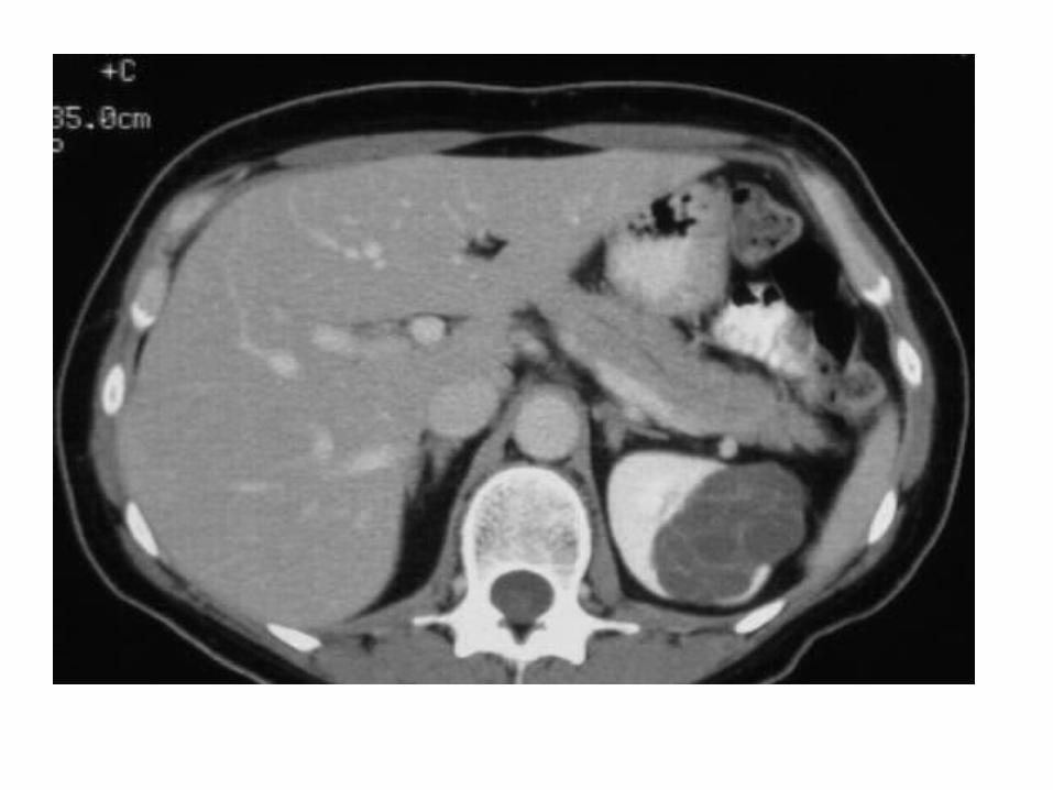
d) Medullary Cysts :1-Medullary Sponge Kidney2-Medullary Cystic Disease3-Calyceal Cyst (Diverticulum)

1-Medullary Sponge Kidney :-Bilateral in 60-80 %-Multiple small mainly pyramidal cysts-Medullary nephrocalcinosis occurs in the
majority of cases (80%)


2-Medullary Cystic Disease :-Belongs to group of pediatric cystic renal
diseases -Normal to small kidneys with multiple small
(1.5cm>) medullary cysts (sometimes cysts are too small to visualize)
-Loss of CMD and increased parenchymal echogenicity

3-Calyceal Cyst (Diverticulum) :-Small usually solitary cyst communicating
via an isthmus with the fornix of a calyx


e) Miscellaneous Intrarenal Cysts :1-Traumatic2-Inflammatory3-Neoplastic4-Cystic Hamartoma

1-Traumatic :-Intrarenal hematoma2-Inflammatory :-T.B.-Calculus Disease-Hydatid

3-Neoplastic :-Cystic Degeneration of a carcinoma-5% of renal cell carcinomas are cystic4-Cystic Hamartoma :-Usually large with thick capsule and
septations

f) Extraparenchymal Renal Cysts :1-Parapelvic Cyst2-Perinephric Cyst

1-Parapelvic Cyst :-Located in or near the hilum but doesn’t
communicate with the renal pelvis and therefore doesn’t opacify during uropgraphy


2-Perinephric Cyst :-Beneath the capsule or between the
capsule and perinephric fat-2ry to trauma , obstruction or replacement
of a hematoma