Diagnostic Findings of Sclerosing Mesenteritis and the...
4
The Indonesian Journal of Gastroenterology, Hepatology and Digestive Endoscopy 122 CASE REPORT Diagnostic Findings of Sclerosing Mesenteritis and the Disease Correlations with Caecal Adenocarcinoma Rabbinu Rangga Pribadi*, Murdani Abdullah*, Rizka Puteri Iskandar**, Velma Herwanto**, Okto Dewantoro**, I Wayan Murna Yonathan***, Arman Adel Abdullah***, Ening Krisnuhoni****, Diah Rini Handjari**** *Division of Gastroenterology, Department of Internal Medicine, Faculty of Medicine Universitas Indonesia, Cipto Mangunkusumo National General Hospital, Jakarta, Indonesia **Department of Internal Medicine, Faculty of Medicine, Universitas Indonesia Cipto Mangunkusumo National General Hospital, Jakarta, Indonesia ***Department of Radiology, Faculty of Medicine, Universitas Indonesia Cipto Mangunkusumo National General Hospital, Jakarta, Indonesia ****Department of Anatomical Pathology, Faculty of Medicine, Universitas Indonesia Cipto Mangunkusumo National General Hospital, Jakarta, Indonesia Corresponding author: Murdani Abdullah. Division of Gastroenterology, Department of Internal Medicine, Universitas Indonesia, Cipto Mangunkusumo National General Hospital. Jl. Diponegoro No. 71 Jakarta Indonesia. Phone: +62-213153957. E-mail: [email protected] ABSTRACT Sclerosing mesenteritis (SM) is a rare disease with non-specific clinical manifestations and should be supported by radiological examination and confirmed by histopathological evaluation. Its relationship with cancer especially caecal adenocarcinoma is still unclear. This case report describes a young man who was diagnosed as having SM and poorly-differentiated caecal adenocarcinoma. Keywords: sclerosing mesenteritis, caecal adenocarcinoma ABSTRAK Sclerosing mesenteritis (SM) merupakan penyakit langka dengan manifestasi klinis tidak spesifik. Diagnosis SM didukung dengan pemeriksaan radiologi dan ditegakkan dengan evaluasi histopatologi. Hubungannya dengan kanker terutama adenokarsinoma sekum masih belum jelas. Laporan kasus ini memaparkan lelaki muda dengan diagnosis SM dan adenokarsinoma sekum berdiferensiasi buruk. Kata kunci: sclerosing mesenteritis, adenokarsinoma sekum INTRODUCTION Sclerosing mesenteritis is an uncommon inflammatory disease of the intestinal mesenteric fat tissue. 1-4 The etiology remains unknown and hypothesized to be associated with previous abdominal surgery, abdominal trauma, autoimmune disease, drugs, cancer, ischemia, and infection. 5-7 The prevalence is 0.16-7.8%. Sclerosing mesenteritis is also reported in 0.6% of over than 7,000 patients undergoing abdominal computed tomography (CT) scan for other indications. 8 Clinical manifestations of sclerosing mesenteritis (SM) are not specific, varying from asymptomatic to
-
Upload
hoangnguyet -
Category
Documents
-
view
214 -
download
0
Transcript of Diagnostic Findings of Sclerosing Mesenteritis and the...
The Indonesian Journal of Gastroenterology, Hepatology and Digestive Endoscopy122
CASE REPORT
Diagnostic Findings of Sclerosing Mesenteritis and the Disease Correlations with Caecal Adenocarcinoma
Rabbinu Rangga Pribadi*, Murdani Abdullah*, Rizka Puteri Iskandar**, Velma Herwanto**, Okto Dewantoro**, I Wayan Murna Yonathan***,
Arman Adel Abdullah***, Ening Krisnuhoni****, Diah Rini Handjari****
*Division of Gastroenterology, Department of Internal Medicine, Faculty of Medicine Universitas Indonesia, Cipto Mangunkusumo National General Hospital, Jakarta, Indonesia **Department of Internal Medicine, Faculty of Medicine, Universitas Indonesia
Cipto Mangunkusumo National General Hospital, Jakarta, Indonesia ***Department of Radiology, Faculty of Medicine, Universitas Indonesia
Cipto Mangunkusumo National General Hospital, Jakarta, Indonesia ****Department of Anatomical Pathology, Faculty of Medicine, Universitas Indonesia
Cipto Mangunkusumo National General Hospital, Jakarta, Indonesia
Corresponding author: Murdani Abdullah. Division of Gastroenterology, Department of Internal Medicine, Universitas Indonesia, Cipto Mangunkusumo National General Hospital. Jl. Diponegoro No. 71 Jakarta Indonesia. Phone: +62-213153957. E-mail: [email protected]
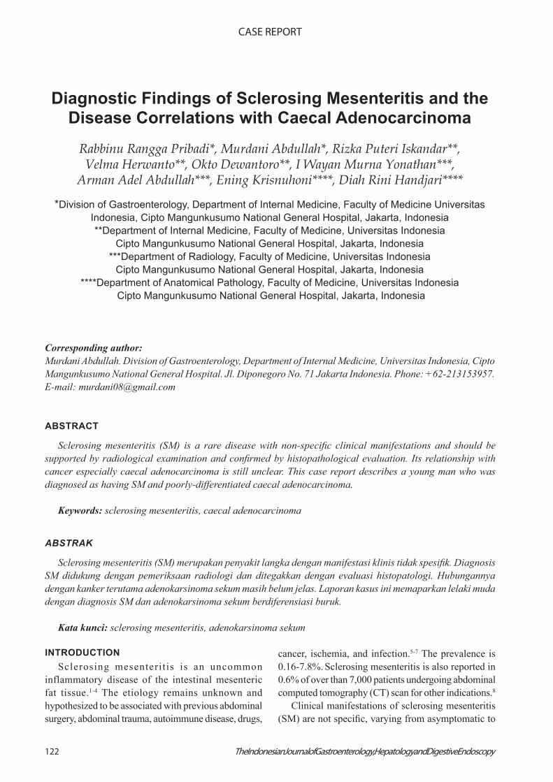
ABSTRACT
Sclerosing mesenteritis (SM) is a rare disease with non-specific clinical manifestations and should be supported by radiological examination and confirmed by histopathological evaluation. Its relationship with cancer especially caecal adenocarcinoma is still unclear. This case report describes a young man who was diagnosed as having SM and poorly-differentiated caecal adenocarcinoma.
Keywords: sclerosing mesenteritis, caecal adenocarcinoma
ABSTRAK
Sclerosing mesenteritis (SM) merupakan penyakit langka dengan manifestasi klinis tidak spesifik. Diagnosis SM didukung dengan pemeriksaan radiologi dan ditegakkan dengan evaluasi histopatologi. Hubungannya dengan kanker terutama adenokarsinoma sekum masih belum jelas. Laporan kasus ini memaparkan lelaki muda dengan diagnosis SM dan adenokarsinoma sekum berdiferensiasi buruk.
Kata kunci: sclerosing mesenteritis, adenokarsinoma sekum
INTRODUCTIONSclerosing mesenteritis is an uncommon
inflammatory disease of the intestinal mesenteric fat tissue.1-4 The etiology remains unknown and hypothesized to be associated with previous abdominal surgery, abdominal trauma, autoimmune disease, drugs,
cancer, ischemia, and infection.5-7 The prevalence is 0.16-7.8%. Sclerosing mesenteritis is also reported in 0.6% of over than 7,000 patients undergoing abdominal computed tomography (CT) scan for other indications.8
Clinical manifestations of sclerosing mesenteritis (SM) are not specific, varying from asymptomatic to

Volume 18, Number 2, August 2017 123
Diagnostic Findings of Sclerosing Mesenteritis and the Disease Correlations with Caecal Adenocarcinoma
abdominal pain, bloating, nausea, vomiting, weight loss, constipation, diarrhea, and fever.9,10 Diagnosis is not always conclusive on clinical findings and radiological examinations only. Definitive diagnosis is confirmed by histopathological evaluation.11
In approximately 69% of patients with SM, there are some concurrent malignancies such as lymphoma, urogenital cancer, and gastrointestinal cancer.12 In case series by Vlachos, et al12, 2 of 5 patients had colon malignancies (caecal and sigmoid colon cancer). Contrary to those reports, other literatures describe that the prevalence of cancer in SM patients is not different with the general population. The hypothesis that supports SM as a paraneoplastic syndrome is still controversial.7
This is a case of a male patient with the diagnosis of SM and poorly-differentiated caecal adenocarcinoma. The importance of SM diagnostic findings and relationship of SM with caecal adenocarcinoma will be highlighted. As far as we know, it is the first reported case in Indonesia.
CASE ILLUSTRATION
A 45-year-old male was referred to our hospital with a chief complaint of continuous vomiting since 11 days before admission. He had been complaining of nausea, vomiting, early satiety, and vague abdominal discomfort for the last six months. He was given antacids and ranitidine with no improvement. Four months ago he underwent an abdominal CT scan, and the result showed intestinal and peritoneal TB. He refused colonoscopy at the time. Laparoscopic biopsy result was suggestive of peritoneal TB. He was given anti-TB drugs started four months before admission. He consumed anti-TB drugs for only two months without any improvements.
Since two months before admission, he had been vomiting more frequently. He also had non-bloody diarrhea alternating with constipation. He had had progressive symptoms and signs of intestinal obstruction and underwent laparotomy at another hospital. After one month of hospitalization, he was discharged. Eleven days before admission, he was rehospitalized due to worsening continuous vomiting and obstipation. He had involuntary weight loss as much as 30 kg within the last four months.
Physical examination upon admission showed that he was fully alert with a body mass index of 14.7 kg/m2. Nasogastric tube (NGT) was inserted, producing greenish-colored fluid followed by coffee-
ground appearance fluid. Abdominal examination revealed doughnut-like consistency, dull to percussion, decreased abdominal skin turgor and decreased bowel sounds. Other physical examinations were within normal limits.
Laboratory examination upon admission showed leucocytosis, and decreased albumin levels. Carcinoembryonic antigen (CEA) was 10.55 ng/mL (normal value 0-4.6 ng/mL). Previous abdominal CT scan performed in another hospital showed fluid collection surrounding the liver, spleen, bilateral paracolic, intestinal, pelvic cavity, with mild enhancement in peripheral area. It also showed adhesion and inflammation of the small bowel beginning from duodenojejunal junction until distal ileum, transverse colon, and rectosigmoid colon, causing luminal narrowing and dilatation of duodenum and gaster. Previous abdominal paracentesis showed inconclusive results.
Patient was admitted to our hospital and underwent several examinations. Anterograde double balloon enteroscopy (DBE) revealed erosive gastropathy with gastric mucosal edema and multiple adhesions at the jejunum. Nasojejunal feeding tube (NJFT) and NGT were inserted for nutritional access and decompression, respectively. His repeated abdominal CT scan on the sixth day of current hospitalization showed ascites, thickening of bowel wall and peritoneum without any signs of ileus.
Figure 1. Coronal contrast-enhanced abdominal CT scan from current hospitalization showing thickening of bowel wall, ascites, and thickening of peritoneum
The initial results of omentum biopsy from another hospital showed non-specific granulomatous inflammation without any signs of malignancy.

The Indonesian Journal of Gastroenterology, Hepatology and Digestive Endoscopy124
Rabbinu Rangga Pribadi, Murdani Abdullah*, Rizka Puteri Iskandar et al.
However the specimen was reevaluated in our hospital and showed SM with no signs of malignancy.
DISCUSSION
Clinical manifestations of SM are not specific. Based on study by Plasencia et al abdominal discomfort was reported in two patients, weight loss in four patients, nausea in five patients, vomiting in six patients, abdominal pain in five patients. Another study by Akram et al showed that 10% were asymptomatic, 70% had abdominal pain, 26% had bloating, 25% had diarrhea, 23% had weight loss, 21% had nausea and vomiting, 16% had loss of appetite, 15% had constipation, 6% had fever, and 3% had night sweats.5,13 On physical examination, the study showed normal abdominal examination in 51% patients, abdominal tenderness in 24% patients, abdominal mass in 15% patients, and chylous ascites in 14% patients.13
This patient had nausea, vomiting, early satiety, and vague abdominal discomfort. Later on, he also complained of diarrhea, constipation, weight loss, and signs of abdominal tenderness and intestinal obstruction. He had no fever and night sweats. Generally, the clinical findings of this patient were in concordance with those described in the studies.
The CT scan findings of SM showed increased mesenterium attenuation to a solid soft tissue mass. Preservation of fat around the mesenteric vessels which is recognized as “fat ring sign” and also calcification may be discovered. Significant mesenteric vessels involvement could disturb the bowel blood vessel flow and cause bowel wall thickening due to ischemia and ascites.14
The patient’s abdominal CT scan showed no fat ring sign nor calcification. However, there were thickening of the small bowel wall and peritoneum. Ascites was also identified. Some of the patient’s abdominal CT scan findings were in concordance with the literatures.
Definitive diagnosis of SM requires biopsy and histopathological evaluation. Sclerosing mesenteritis has been described in three phases. The first phase is fat necrosis or mesenteric lipodystrophy. It is followed by the second phase, mesenteric panniculitis which is related to inflammatory process. The third phase is retractile mesenteritis which is marked by fibrosis with mesenteric retraction and shortening.15
Evaluation of the biopsy specimen from the omentum in our hospital revealed fibrotic tissue, adipose tissue, inflammatory cells, congestive vessels without specific infection or malignancy. It was consistent with SM in the third phase.
After the thorough explanations regarding the diagnostic findings in the above paragraphs, another issue of SM emerges. As illustrated in the case, our
Figure 2. A Hematoxyllin-eosin (HE) staining of omentum specimen (40x magnifying power) showing adipose tissue, fibrotic tissue, inflammatory cells, congestive vessels and absence of malignant cells consistent with sclerosing mesenteritis
Colonoscopy was also performed to evaluate the possibility of cancer. The procedure revealed that the entire colon was edematous and hyperemic and the hematoxyllin-eosin (HE) staining of the specimen taken from caecum showed poorly-differentiated caecal adenocarcinoma.
Figure 3. A Hematoxyllin-Eosin (HE) staining of caecum specimen (40x magnifying power) showing trabecular tumor cells with pleomorphic nuclei, prominent nucleoli, eosinophilic cytoplasm consistent with undifferentiated caecal adenocarcinoma
He was given partial parenteral nutrition, intravenous fluid, liquid diet via NJFT, and methyl prednisolone 62.5 mg twice daily intravenously, which was tapered down to 16 mg daily for three weeks. The response was inadequate and it was decided to do a laparotomy. The result was frozen abdomen and the surgery was halted. Afterwards, he suffered septic shock due to hospital acquired pneumonia and passed away.

Volume 18, Number 2, August 2017 125
Diagnostic Findings of Sclerosing Mesenteritis and the Disease Correlations with Caecal Adenocarcinoma
patient suffered from both SM and poorly differentiated caecal adenocarcinoma. Question arises whether it was a cause-effect relationship or just a coincidence.
Some studies were designed to answer the question. A research conducted by Gogebakan et al.1 showed an interesting fact. They studied 56 patients with SM and malignancies compared to 150 patients with malignancies and no SM (the control group). From those subjects, there were 12 (15.6%) colorectal cancer (CRC) patients in SM group and 26 (17.1%) CRC patients in control group. The analysis showed non-significant differences of CRC prevalence between those two groups (p = 0.852).1 The limitations of the study were the small number of SM and CRC cases and also the reliance of the SM diagnosis on radiological examination only and not by histopathology as the gold standard. Another study by van Putte-Katier et al.6 contradicts the previous research. They studied 46 SM patients with cancer and 87 malignancies patients without SM (the control group). There were 8 (17.4%) CRC patients in SM group and 18 (20.7%) CRC patients in control group. The prevalence of CRC was significantly lower in SM group compared to control group. The limitation of their study was the absence of histopathological proof of SM.6 Both studies did not specifically describe the location of the CRC (whether it is caecal cancer or not). As for know, the relationship between SM and caecal adenocarcinoma remains unclear. Further studies are needed to clarify this issue.
In conclusion, sclerosing mesenteritis is a rare mesenteric inflammatory disorder, which still has vague correlations with cancer, particularly caecal adenocarcinoma. Clinical findings of SM are not specific and the diagnosis should be supported by radiological investigation, and confirmed by histopathological examination.
REFERENCES1. Gogebakan O, Albrecht T, Osterhoff MA, Reimann A. Is
mesenteric panniculitis truely a paraneoplastic phenomenon? A matched pair analysis. Eur J Rad 2013;82:1853-9.
2. Hussein MRA, Abdelwahed SR. Mesenteric panniculitis: an update. Expert Rev Gastroenterol Hepatol 2015;9:67-8.
3. Scheer F, Spunar P, Wiggermann P, Wissgott C, Andresen R. Mesenteric panniculitis (MP) in CT – a predictor of malignancy? Fortschr Rontgenstr 2016; DOI:10.1055/s-0042-110100.
4. Pinheiro F, Rego A, Araujo-Filho I. Mesenteric panniculitis in the elderly – update on diagnostic and therapeutic approach. Intern J Surg Med 2016;2:127-33.
5. Plasencia LD, Ballester LR, Fernandez EMLT, Morales AH, Pallares AC, Siverio NH. Mesenteric panniculitis: experience in our center. Rev Esp Enferm Dig 2007;99:291-7.
6. Van Putte-Katier N, van Bommel EFH, Elgersma OE, Hendriksz TR. Mesenteric panniculitis: prevalence. Clinicoradiological presentation and 5-year follow-up. Br J Radiol 2014;87:20140451.
7. Badet N, Sailley N, Briquez C, Paquette B, Vuitton L, Delabrousse E. Mesenteric panniculitis: still an ambiguous condition. Diagnostic and Interventional Imaging 2015;96:251-7.
8. Irami AF, Cesar de CG, de Oliveira SA Jr, de Carvalho AV, Cordeiro JA, Chaves BMF, et al. Sclerosing mesenteritis-update on diagnostic and therapeutic approach. Transl Biomed 2016;7:1.
9. Umale NP, Tiwari SJ, Yadgire A. Sclerosing mesenteritis – a rare cause of abdominal pain and intra-abdominal mass: a case report. Int J Res Med Sci 2015;3:2863-6.
10. Graham A, Harvin G. Sclerosing mesenteritis: a rare cause of small bowel obstruction. Case Rep Gastroenterol 2016;10:63-7.
11. Devi PG, Prasanna MR, Venkatachalam TSP, Philip NP. Sclerosing mesenteritis: an unusual cause of intestinal obstruction. Int J Adv Med Health Res 2015;2:102-5.
12. Vlachos K, Archontovasilis F, Falidas E, Mathioulakis S, Konstandoudakis S, Villias C. Sclerosing mesenteritis: diverse clinical presentations and dissimilar treatment options. A case series and review of literature. Arch Intern Med 2011;4:17.
13. Akram S, Pardi DS, Schaffner JA, Smyrk TC. Sclerosing mesenteritis: clinical features, treatment, and outcome in ninety-two patients. Clin Gastroenterol Hepatol 2007;5:589-96
14. Horton KM, Lawler LP, Fishman EK. CT findings in sclerosing mesenteritis (panniculitis): spectrum of disease. Radio Graphics 2003;23:1561-7.
15. Rees JR, Burgess P. Benign mesenteric lipodystrophy presenting as low abdominal pain: a case report. Journal of Medical Case Reports 2010;4:119.