diagnosis presenting - Postgraduate Medical Journaldiagnosis. They are here described together with...
8
Postgraduate Medical Journal (April 1977) 53, 204-211 The differential diagnosis of the short-limbed dwarfs presenting at birth R. N. MUKHERJI M.R.C.P., D.C.H. P. D. Moss F.R.C.P., D.C.H. Department of Paediatrics, Royal Infirmary, Blackburn, Lancashire Summary Attention is drawn to the fact that in a number of types of short-limbed dwarfism a precise diagnosis can be made in the neonatal period. Examples are given and the prognostic and genetic implications are dis- cussed. It is important to be able to advise parents of the likely outlook for the infant and of the genetic implication. Early diagnosis is therefore not merely an academic exercise. Introduction When Rathbun (1948) first described neonatal hypophosphatasia he discussed three conditions of differential diagnosis. These consisted of osteogenesis imperfecta, achondroplasia and renal hyperpara- thyroidism. Since then a number of other conditions have been defined on a clinical, radiological and genetic basis (Tables 1 and 2). Such conditions are not excessively rare if sought. Ryan and Kozlowski (1971) found nineteen cases of bone dysplasia in consecutive radiographs of 109 infants dead on delivery. When a case of neonatal hypophospha- tasia occurred in the authors' unit they were able to include thirteen conditions in the differential diagnosis. They are here described together with prognosis and genetic implications. Achondroplasia Achondroplasia is the prototype of true bone dysplasia (Rubin, 1963) and it is the best described form of neonatal dwarfism. Its incidence per one million people was found to be fifteen in the United States (Potter and Coverstone, 1948), twenty-three in Denmark (Morch, 1941) and 283 in Northern Ireland (Stevenson, 1957). It is inherited as an autosomal dominant but over 90%/ of cases are fresh mutations especially with elderly parents (Barr and Forfar, 1973). Harris and Patton (1971) in their series found that the mutation rate was not greater than 1 in 100,000. Its incidence in twins is extremely rare. Clinically the condition may be recognizable at birth but often it is not, as the features of proximal limb shortening, large head, depressed nasal bridge and prominent forehead may be poorly developed. Harris and Patton (1971) reviewed seventeen still- born or early neonatal deaths originally diagnosed as achondroplastics and showed that in ten of these cases the diagnosis should have been thanatophoric dwarfism. This supports the view that achondro- plasia has been over diagnosed at birth and that the severely affected cases are, in fact, some other type of lethal neonatal dwarfism. Achondrogenesis This was first described under the title of anosteo- genesis (Parenti, 1936) as it was thought to be a variety of osteogenesis imperfecta. Fraccaro (1952) first used the term 'achondrogenesis' because of marked retardation of ossification. Parental con- sanguinity (Saldino, 1971) and multiple affected siblings (Houston, Awen and Kent, 1972) have been reported. The role of teratogenic agents is speculative as a case of achondrogenesis following maternal thalidomide ingestion has been described (Jurczok and Schollmeyer, 1962). Hydramnios is a frequent complication of the pregnancy and achondrogenic infants are usually premature and stillborn. It is a rare condition and is characterized at birth by extremely short limbs, large head, swollen abdomen, poor ossification of bones generally and essentially no neck (Fig. 1). Harris, Patton and Barson (1972) described two siblings under the title of 'pseudo- achondrogenesis with fractures' which is representa- tive of a different type of achondrogenesis and resembling the siblings described by Houston et al. (1972). This particular variety is now known as 'Harris and Houston' type of achondrogenesis (Fig. 2). The presence of fractures is the point of differentiation between the two types of this condi- tion. Asphyxiating thoracic dystrophy Jeune, Beraud and Carron (1955) first described this condition. It is characterized by extemely short ribs, variable degree of shortening of limbs and fingers and occasionally by polydactyly. The thorax is narrow and immobile. Hereditary nephritis leading to death has been reported (Shokeir, Houston and Awen, 1971). copyright. on February 20, 2020 by guest. Protected by http://pmj.bmj.com/ Postgrad Med J: first published as 10.1136/pgmj.53.618.204 on 1 April 1977. Downloaded from
Transcript of diagnosis presenting - Postgraduate Medical Journaldiagnosis. They are here described together with...
Postgraduate Medical Journal (April 1977) 53, 204-211
The differential diagnosis of the short-limbed dwarfs presenting at birth
R. N. MUKHERJIM.R.C.P., D.C.H.
P. D. MossF.R.C.P., D.C.H.
Department of Paediatrics, Royal Infirmary, Blackburn, Lancashire
SummaryAttention is drawn to the fact that in a number oftypes of short-limbed dwarfism a precise diagnosis canbe made in the neonatal period. Examples are givenand the prognostic and genetic implications are dis-cussed. It is important to be able to advise parents ofthe likely outlook for the infant and of the geneticimplication. Early diagnosis is therefore not merelyan academic exercise.
IntroductionWhen Rathbun (1948) first described neonatal
hypophosphatasia he discussed three conditions ofdifferential diagnosis. These consisted of osteogenesisimperfecta, achondroplasia and renal hyperpara-thyroidism. Since then a number of other conditionshave been defined on a clinical, radiological andgenetic basis (Tables 1 and 2). Such conditions arenot excessively rare if sought. Ryan and Kozlowski(1971) found nineteen cases of bone dysplasia inconsecutive radiographs of 109 infants dead ondelivery. When a case of neonatal hypophospha-tasia occurred in the authors' unit they were ableto include thirteen conditions in the differentialdiagnosis. They are here described together withprognosis and genetic implications.
AchondroplasiaAchondroplasia is the prototype of true bone
dysplasia (Rubin, 1963) and it is the best describedform of neonatal dwarfism. Its incidence per onemillion people was found to be fifteen in the UnitedStates (Potter and Coverstone, 1948), twenty-threein Denmark (Morch, 1941) and 283 in NorthernIreland (Stevenson, 1957). It is inherited as anautosomal dominant but over 90%/ of cases arefresh mutations especially with elderly parents (Barrand Forfar, 1973). Harris and Patton (1971) in theirseries found that the mutation rate was not greaterthan 1 in 100,000. Its incidence in twins is extremelyrare.
Clinically the condition may be recognizable atbirth but often it is not, as the features of proximallimb shortening, large head, depressed nasal bridgeand prominent forehead may be poorly developed.
Harris and Patton (1971) reviewed seventeen still-born or early neonatal deaths originally diagnosedas achondroplastics and showed that in ten of thesecases the diagnosis should have been thanatophoricdwarfism. This supports the view that achondro-plasia has been over diagnosed at birth and that theseverely affected cases are, in fact, some other typeof lethal neonatal dwarfism.
AchondrogenesisThis was first described under the title of anosteo-
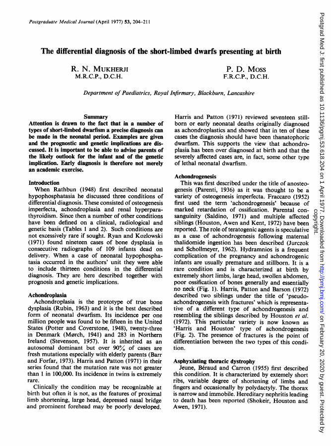
genesis (Parenti, 1936) as it was thought to be avariety of osteogenesis imperfecta. Fraccaro (1952)first used the term 'achondrogenesis' because ofmarked retardation of ossification. Parental con-sanguinity (Saldino, 1971) and multiple affectedsiblings (Houston, Awen and Kent, 1972) have beenreported. The role of teratogenic agents is speculativeas a case of achondrogenesis following maternalthalidomide ingestion has been described (Jurczokand Schollmeyer, 1962). Hydramnios is a frequentcomplication of the pregnancy and achondrogenicinfants are usually premature and stillborn. It is arare condition and is characterized at birth byextremely short limbs, large head, swollen abdomen,poor ossification of bones generally and essentiallyno neck (Fig. 1). Harris, Patton and Barson (1972)described two siblings under the title of 'pseudo-achondrogenesis with fractures' which is representa-tive of a different type of achondrogenesis andresembling the siblings described by Houston et al.(1972). This particular variety is now known as'Harris and Houston' type of achondrogenesis(Fig. 2). The presence of fractures is the point ofdifferentiation between the two types of this condi-tion.
Asphyxiating thoracic dystrophyJeune, Beraud and Carron (1955) first described
this condition. It is characterized by extemely shortribs, variable degree of shortening of limbs andfingers and occasionally by polydactyly. The thoraxis narrow and immobile. Hereditary nephritis leadingto death has been reported (Shokeir, Houston andAwen, 1971).
copyright. on F
ebruary 20, 2020 by guest. Protected by
http://pmj.bm
j.com/
Postgrad M
ed J: first published as 10.1136/pgmj.53.618.204 on 1 A
pril 1977. Dow
nloaded from

Diagnosis of short-limbed dwarfs at birth 205
TABLE 1. Clinical differences
Part of limbs AssociatedType Family history Antenatal involved abnormalities Inheritance
Achondroplasia (mutant) - - Proximal Enlarged head, de- Autosomalpressed nasal bridge, dominanthands and feet shortand broad
Achondrogenesis Parental consan- Hydramnios Both proximal Large head, essentially Autosomalguinity stillbirth and distal no neck, swollen recessive
abdomen
Asphyxiating thoracic - - Distal Extremely short ribs, Autosomaldistal narrow and immobile recessive
thorax, polydactyly,hereditary nephritis
Chondrodysplasia punctata - - Proximal and Flexion contracture of Autosomaldistal joints, cataracts, recessive
ectodermal changes,mental deficiency
Chondro-ectodermal - - Distal Thoracic dystrophy, Autosomaldysplasia (Ellis-Van polydactyly, con- recessiveCreveld syndrome) genital heart, ecto-
dermal changes
Diastrophic dwarf - - Distal Marked talipes equino- Autosomalvarus, resemblesarthrogryposis, 'hitch-hiker' thumb
Homozygous achondro- Both parents -Proximal Large head, depressed Autosomalplasia achondroplastic nasal bridge, frontal dominant
bossing, early death
Hypophosphatasia Parental consan- X-ray diagnostic Proximal and Soft skull bones, Autosomalguinity distal marked angulation recessive
and deformities oflong bones
Lymphopenic agamma- - - Proximal Features of achondro- Not knownglobulinaemia with short- plasia, lymphopenia,limbed dwarfism agammaglobulin-
aemia, early death
Metatrophic dwarf - - Proximal and Long narrow thorax, Autosomaldistal caudal appendage, recessive
swollen joints
Osteogenesis imperfecta Blue sclera X-ray diagnostic Proximal and Soft skull, blue sclera Autosomaldistal dominant
recessive
Spondylo-epiphyseal - - Proximal and Myopia, retinal detach- Autosomaldysplasia congenita distal ment, cataract, cleft dominant
palate
Thanatophoric dwarf Parental consan- Hydramnios, Proximal Redundant skin folds, Autosomalguinity weak fetal move- limbs abducted and recessive
ments, stillbirth, externally rotated,X-ray diagnostic large head with frontal
prominence
copyright. on F
ebruary 20, 2020 by guest. Protected by
http://pmj.bm
j.com/
Postgrad M
ed J: first published as 10.1136/pgmj.53.618.204 on 1 A
pril 1977. Dow
nloaded from

206 R. N. Mukherji and P. D. Moss
TABLE 2. Radiographic differences
Type Spine Limbs Pelivs Thorax, ribs Skull
Achondroplasia Narrow inter- Proximal bones Narrow sacrum Thorax long and Large vault,(mutant) pedicular distance short, ball and and sciatic notch narrow small base and
of lower lum- socket appear- foramen magnumbar vertebrae ance of meta-
physes
Achondrogenesis Poorly mineralized Marked shorten- Poorly mineralized Short ribs, clavicles Poorly mineral-and ossified ing, poorly disproportionately ized, otherwise
mineralized long normal
Asphyxiating thoracic Normal Variable short- Hypoplastic iliac Extremely short Normaldystrophy ening hook-shaped horizontal ribs
sciatic notch
Chondrodysplasia Punctate calcifica- Both proximal and -Normal Craniostenosispunctata tions of inter- distal shortening,
vertebral discs puncatate calcifi-cations of jointcapsules andepiphyses
Chondro-ectodermal Normal Short distal bones, Reduced height of Short ribs Normaldysplasia (Ellis-Van hypoplastic bonesCreveld syndrome) terminal phalanges,
carpal and tarsalsfused into solidmasses
Diastrophic dwarf Hypoplastic Hypoplastic first Acetabular dys- Long thorax Normalcervical verte- metacarpal plasiabrae
Homozygous Flattening of Markedly short Small iliacs Short ribs Large, small baseachondroplasia vertebrae
Hypophosphatasia Normal Appearance of Normal Marked costo- Little or no bonesevere rickets chondral bead- material
ing
Metatrophic dwarf Flat vertebrae Metaphyses mark- Crescent-shaped Long thorax, Normaledly flared, iliac crests short ribsenlarged andirregular
Osteogenesis imper- Shallow vertebrae Thick-thin bones, Normal Deformities Poorly ossifiedfecta stunted, deformed
Spondylo-epiphyseal Hypoplastic odon- Short, bowing of Small iliac wings Bell-shaped thorax Normaldisplasia congenita toid femora
Thanatophoric dwarf Flat vertebrae, Markedly short, Short iliacs Short ribs, wide Enlarged, shortdeep discs bowing of femur anterior ends base, premature
fusion ofcoronal andlamboidalsutures
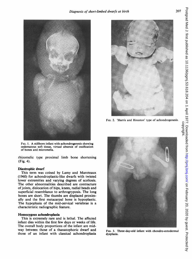
Chondro-ectodermal dysplasiaThis condition was described in 1940 by Ellis
and Van Creveld. It is a complex dysplasia in whichskeletal defects are associated with ectodermalanomalies affecting hair, teeth and nails, and con-genital heart disease is common. The shortening oflimbs in this condition is the reversal of achondro-plasia in that it is the distal parts of the limbs which
are shortened rather than the proximal (Fig. 3) andpolydactyly of fingers and toes is commonly found.
Chondrodysplasia punctataThese infants are recognizable at birth as dwarfs
with short limbs. Radiologically punctate calcifica-tions are found in the epiphyses, joint capsules,trachea, larynx, hyoid, intervertebral discs and in
copyright. on F
ebruary 20, 2020 by guest. Protected by
http://pmj.bm
j.com/
Postgrad M
ed J: first published as 10.1136/pgmj.53.618.204 on 1 A
pril 1977. Dow
nloaded from
Diagnosis of short-limbed dwarfs at birth 207
Z:::A .....
FIG. 1. A stillborn infant with achondrogenesis showingoedematous soft tissue, virtual absence of ossificationof bones and micromelia.
rhizomelic type proximal limb bone shortening(Fig. 4).
Diastrophic dwarfThis term was coined by Lamy and Maroteaux
(1960) for achondroplastic-like dwarfs with twistedlower extremities and varying degrees of scoliosis.The other abnormalities described are contractureofjoints, dislocation of hips, knees, radial heads andsuperficial resemblance to arthrogryposis. The longbones are short. The thumbs are displaced proxim-ally and the first metacarpal bone is hypoplastic.The hypoplasia of the mid-cervical vertebrae is acharacteristic radiographic feature.
Homozygous achondroplasiaThis is extremely rare and is lethal. The affected
infant dies within the first few days or weeks of life.The overall body proportions of the infant are mid-way between those of a thanatophoric dwarf andthose of an infant with classical achondroplasia
i
FIG. 2. 'Harris and Houston' type of achondrogenesis.
FIG. 3. Three-day-old infant with chondro-ectodermaldysplasia.
dyslaia copyright. on F
ebruary 20, 2020 by guest. Protected by
http://pmj.bm
j.com/
Postgrad M
ed J: first published as 10.1136/pgmj.53.618.204 on 1 A
pril 1977. Dow
nloaded from

208 R. N. Mukherji and P. D. Moss
ii..iill;·::·.
c
··:·.;·i L:
··'··
;.g.tii::
:.
:::."::.:.:::"li'·I::8··'· "
iL·
FIG. 4. A newborn infant with rhizomelic type of chon-drodysplasia punctata.
(Fig. 5). The clinical diagnosis is made withoutdifficulty as a homozygous achondroplast is the off-spring of two achondroplastic parents.
HypophosphatasiaThis is an autosomal recessive condition.The term 'hypophosphatasia' was used for the
first time by Rathbun (1948) to designate an osteo-blastic condition with low serum alkaline phospha-tase. There are three salient features-abnormalmineralization of bone, increased urinary excretionof phosphoethanolamine and diminished alkalinephosphatase activity. Fraser (1957) described threedifferent categories depending on the age of onset:newborn, infants and children. The newborn type isthe most severe (Figs 6 and 7) and carries the worstprognosis. Hypercalcaemia is a common complica-tion presumably due to accumulation of calcium inthe blood as it fails to be incorporated into bonebecause of absent or low alkaline phosphataseactivity. Dent (1956) maintained that the hyper-calcaemia in hypophosphatasia was due to hyper-sensitivity to vitamin D. This hypercalcaemia canbe complicated by nephrocalcinosis which can con-tribute to some of the fatal outcome.
ii:··
ilil,qS.Og%Va.E.jl .a%[Bli.B.ii.e.;;.l:l,II;::. ':j·;;
ljZ
ft.c.r=;·ii
: ·.··
" '':'
·:::
.i:
i·
..i.r··:··-'·Q
.··:
··: ;. '3'-
.::,:
PE; . '.JSAl
FIG. 5. Two-day-old infant with homozygous achon-droplasia.
Lymphopenic agammaglobulinaemia with short-limbeddwarfismMcKusick and Cross (1966) first described a
patient with Swiss-type agammaglobulinaemia anda skeletal disorder resembling achondroplasia butthey thought the sketetal disorder had no relation-ship to the agammaglobulinaemia. A few monthslater Davis (1966) described a newborn female infantwith achondroplasia and Swiss-type agammaglo-bulinaemia. He concluded that because of the rarityof both conditions their concurrence was due togenetic association and not simply to chance. Thesubsequent recognition of isolated cases of thisnature (Fulginiti et al., 1967; Alexander and Dunbar,1968) have given support to Davis's assumption.
Metatrophic dwarfThis term 'metatrophic' (which means 'variable')
copyright. on F
ebruary 20, 2020 by guest. Protected by
http://pmj.bm
j.com/
Postgrad M
ed J: first published as 10.1136/pgmj.53.618.204 on 1 A
pril 1977. Dow
nloaded from

·:i··
:.i
":i·:
::·:'::
:··.: I:·:'.li"'iiiliHEBPII':
:::::lril..ii.EE.ii ·iii.....i ....··:···:i,I······:::::iii.aii:::i'·r:'·',:· ···'i..iii·iiilliiiii :·:i
':''L:i·: .aii*.'..i.!.;i.f.'s.B.JB.sl.' ':'' ··i:l.·rb.. :i ·.
·::i:;ii. .:iiHii'·:. ''··s ;i: ::·:·::.·: "· :·i:
r.;··- ···:*.Jg -YF.·.,...?..:R,?::i:;l(.
':'·.":i::::8..n?8.%$1.&2.88f$i.:·: :;i::ii·:
·::·: i.i. .:·:::
i,!!l4:SIII.YI IBr.KBLti.sllll(lPI.ad/bL.'...BBlls$llsl 1II:'i':i·
··::i··::I:::·ll;pstl8..L.i.i.1%41..1e.-sl. · : ·:.':··i:i
·ii:,iii
FIG. 6. Day-old infant with neonatal hypophosphatasiashowing large head, narrow thorax, bowing of legs andcutaneous dimples on outer aspects of legs.
.."it
*t·.*--A
FIG. 7. Day-old infant with neonatal hypophosphatasiashowing demineralization simulating severe rickets.
i:"· ..~
::....
FIG. 8. Metatrophic dwarf showing expansion of allmetaphyses, dumb-bell shaped long bones and flatteningof vertebrae.
i'·:·'
··-.
.
·.d.'·;:iti
·:ii:l··.
··w::
FIG. 9. Osteogenesis imperfecta congenita.
copyright. on F
ebruary 20, 2020 by guest. Protected by
http://pmj.bm
j.com/
Postgrad M
ed J: first published as 10.1136/pgmj.53.618.204 on 1 A
pril 1977. Dow
nloaded from

210 R. N. Mukherji and P. D. Moss
in:-
FIG. 10. Spondylo-epiphyseal dysplasia congenita show-ing shortening of ribs and long bones, flattening ofthoracic vertebrae posteriorly and hypoplastic odontoid.
was defined by Maroteaux, Spranger and Wiedemann(1966) for a condition characterized by variation inthe clinical picture with age. Initially they super-ficially resemble achondroplastics with limbs shorterthan trunk; later, the proportions reverse with moreshortening of the trunk than limbs and bear resem-blance to Morquio's disease. The skeletal radio-graphs show expansion of all the metaphyses,shortening of long bones which become dumb-bellshaped and flattening of vertebral bodies (Fig. 8).
Osteogenesis imperfecta congenitaThis condition in the majority of cases results
from sporadic mutation and is inherited as auto-somal dominant. The severe neonatal type indicates arecessive mode of inheritance. It is characterized bymultiple fractures of long bones which consequentlybecome deformed, angulated and short (Fig. 9).
Spondylo-epiphyseal dysplasia congenitaThis condition was first described by Spranger and
Wiedemann (1966). It selectively affects vertebrae,epiphyses of long bones and occasionally adjacent
.P'% '·
s·
..·*
··;:····
:::::
.. II*sx.. .
. -----······· "* .·-*
.:.:
FIG. 11. Thanatophoric dwarf.
metaphyses. The associated clinical abnormalitiesare: severe myopia, retinal detachment, cataracts,deafness and cleft palate. Roaf, Longmore andForrester (1967) reported four cases of a syndromecomprising bone dysplasia, retinal detachment anddeafness. These were probably cases of spondylo-epiphyseal dysplasia. There are six types of spondylo-epiphyseal dysplasia distinguishable on clinical,genetic and radiological grounds. It is inappro-priate to discuss all here. The skeletal radiograph(Fig. 10) shows the abnormalities that are found inthe congenital variety.
Thanatophoric dwarfMaroteaux, Lamy and Robert (1967) first reported
this condition which is the commonest cause ofneonatal deaths amongst short-limbed dwarfs. Thetitle means 'death bearing'. Harris and Patton (1971)reviewed seventeen stillborn and early neonataldeaths originally diagnosed as achondroplasia andshowed that in ten of these the diagnosis should havebeen thanatophoric dwarf. The clinical appearance ischaracteristic (Fig. 11) and consists of very short
copyright. on F
ebruary 20, 2020 by guest. Protected by
http://pmj.bm
j.com/
Postgrad M
ed J: first published as 10.1136/pgmj.53.618.204 on 1 A
pril 1977. Dow
nloaded from

Diagnosis of short-limbed dwarfs at birth 211
limbs, redundant skin folds, limbs held in abductionand external rotation, a large head and depressednasal bridge.
ConclusionsIt is now possible to make an early and precise
diagnosis in a number of different types of short-limbed dwarfs. This is of importance for prognosisand genetic counselling.
AcknowledgmentsThe authors are grateful to Dr R. Harris and the Journal of
Clinical Genetics for permission to reproduce two figures(Figs 2 and 11). They gratefully acknowledge permission fromDr R. M. Saldino for the illustrations (Figs 1, 3, 4, 5, 8, 10)obtained from Medical Radiography, and photographypublished by Radiography Markets Divisions, EastmanKodak Company.The authors would like to thank Mrs M. E. Hunt for her
invaluable help in typing the manuscript.
ReferencesALEXANDER, W.J. & DUNBAR, J.S. (1968) Unusual bone
changes in thymic alymphoplasia. Annals of Radiology,11, 389.
BARR D.G.D. & FORFAR, J.O. (1973) Textbook of Paedia-trics. Churchill Livingstone, London and Edinburgh.
DAVIS, J.A. (1966) A case of Swiss type of agammaglo-bulinaemia and achondroplasia. British Medical Journal,2, 1371.
DENT, C.E. (1956) Bone structure and metabolism. In: CibaFoundation Symposium, p. 266. Churchill, London.
ELLIS, R.W.B. & VAN CREVELD, S. (1940) Syndrome charac-terized by ectodermal dysplasia, polydactyly, chondro-dysplasia and congenital morbus cordis: report of 3 cases.Archives of Disease in Childhood, 15, 65.
FRACCARO, M. (1952) Contributo allo studio delle malattiedel mesenchima osteopoitico: l'achondrogenesi. Foliahereditaria et pathologica (Milano) 1, 190.
FRASER, D. (1957) Hypophosphatasia. American Journal ofMedicine, 730.
FULGINITI, V.A., HATHAWAY, W.E., PEARLMAN, D.S. &KEMP, C.H. (1967) Agammaglobulinaemia and achon-droplasia. British Medical Journal, 2, 1371.
HARRIS, R. & PATTON, J.T. (1971) Achondroplasia andthanatophoric dwarfism in newborn. Clinical Genetics, 2,61.
HARRIS, R., PATTON, J.T. & BARSON, A.J. (1972) Pseudo-achondrogenesis with fractures. Clinical Genetics, 3, 435.
HOUSTON, C.S., AWEN, C.F. & KENT, H.P. (1972) Fatal
neonatal dwarfism. Journal of the Canadian Association ofRadiologists, 23, 45.
JEUNE, M., BERAUD, C. & CARRON, R. (1955) Dystrophiethoracique asphyxiante de caractere familial. Archivesfrancaises de pediatrie, 12, 886.
JEUNE, M., BERAUD, C. & CARRON, R. (1962) Zur frage desgehauften Auftretens von Extremitatenmissbildungen beiNeugeborenen. Geburtshilfe und Frauenheilkunde, 22,400.
JURCZOK, F. & SCHOLLMEYER, R. (1962) Zur Frage desgehauften Auftretens von Extremitatenmissbildungen beiNeugeborenen, Geburtshilfe und Frauenheilkunde, 22,400.
LAMY, M. & MAROTEAUX, P. (1960) Diastrophic nanism.Presse Medicale, 68, 1977.
McKusIcK, V.A. & CROSS, H.E. (1966) Ataxia telangiectasiaand Swiss type agammaglobulinaemia. Journal of theAmerican Medical Association, 195, 739.
MAROTEAUX, P., LAMY, M. & ROBERT, J.M. (1967) Lenanisme thanatopore. Presse Medicale, 75, 2519.
MAROTEAUX, P., SPRANGER, J. & WIEDEMANN, H.R. (1966)Der metatropische Zwergwuchs. Archiv fur Kinderheil-kunde, 173, 211.
MORCH, E.T. (1941) Chondrodystrophic Dwarfs in Denmark.Ejnar Munksgaard, Copenhagen.
PARENTI, G.C. (1936) La anosteogenesi (una varieta dellaosteogenesi imperfetta). Pathologica. Genova, 28, 447.
POTTER, E.L. & COVERSTONE, V.A. (1948) Chondrodystrophyfoetalis. American Journal of Obstetrics and Gynecology,56, 790.
RATHBUN, J.C. (1948) 'Hypophosphatasia'. New develop-mental anomaly. American Journal of Diseases of Children,75, 822.
ROAF, R., LONGMORE, J.B. & FORRESTER, R.M. (1967) Achildhood syndrome of bone dysplasia, retinal detachmentand deafness. Developmental Medicine and Child Neurology,9, 464.
RUBIN, P. (1963) Achondroplasia versus pseudoachondro-plasia. Radiologic Clinics of North America, 1, 621.
RYAN, J. & KOZLOWSKI, K. (1971) Australian RadiationRecords, 15, 213.
SALDINO, R.M. (1971) Lethal short limbed dwarfism: achon-drogenesis and thanatophoric dwarfism. American Journalof Roentgenology, 112, 185.
SHOKEIR, M.H.K., HOUSTON, C.S. & AWEN, C.F. (1971)Asphyxiating thoracic chondrodystrophy. Association withrenal disease and evidence for possible heterozygous ex-pression. Journal of Medical Genetics, 8, 107.
SPRANGER, J. & WIEDEMAN, H.R. (1966) Dysplasia spondylo-epiphysaria congenita. Helvetica paediatrica acta, 21, 598.
STEVENSON, A.C. (1957) Achondroplasia: an account of thecondition in Northern Ireland. American Journal ofHuman Genetics, 9, 8.
copyright. on F
ebruary 20, 2020 by guest. Protected by
http://pmj.bm
j.com/
Postgrad M
ed J: first published as 10.1136/pgmj.53.618.204 on 1 A
pril 1977. Dow
nloaded from