Diagnosis - PeaceHealth · Borowitz et al (1993) AJCP 100:534-40. Steltzer et al (1993) Ann NY Acad...
57
Diagnosis Greg Wolgamot, MD PhD
Transcript of Diagnosis - PeaceHealth · Borowitz et al (1993) AJCP 100:534-40. Steltzer et al (1993) Ann NY Acad...
Diagnosis
Greg Wolgamot, MD PhD

Workup of leukemia:
1.Blood
2.Lymph node
3.Marrow
4.Prognostic indicators


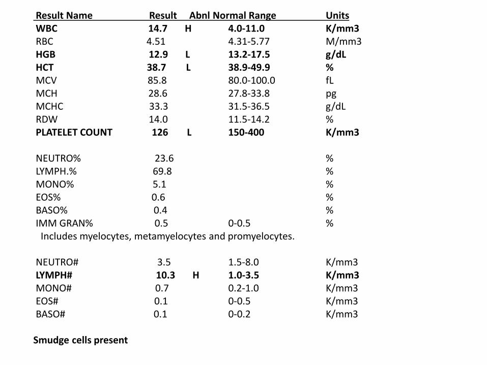
Result Name Result Abnl Normal Range Units WBC 14.7 H 4.0-11.0 K/mm3 RBC 4.51 4.31-5.77 M/mm3 HGB 12.9 L 13.2-17.5 g/dL HCT 38.7 L 38.9-49.9 % MCV 85.8 80.0-100.0 fL MCH 28.6 27.8-33.8 pg MCHC 33.3 31.5-36.5 g/dL RDW 14.0 11.5-14.2 % PLATELET COUNT 126 L 150-400 K/mm3 NEUTRO% 23.6 % LYMPH.% 69.8 % MONO% 5.1 % EOS% 0.6 % BASO% 0.4 % IMM GRAN% 0.5 0-0.5 % Includes myelocytes, metamyelocytes and promyelocytes. NEUTRO# 3.5 1.5-8.0 K/mm3 LYMPH# 10.3 H 1.0-3.5 K/mm3 MONO# 0.7 0.2-1.0 K/mm3 EOS# 0.1 0-0.5 K/mm3 BASO# 0.1 0-0.2 K/mm3 Smudge cells present


PATHOLOGIST INTERPRETATION: Lymphocytosis suggestive of chronic lymphocytic leukemia Flow cytometry would be contributory for further workup John W. Hoyt, MD Pathologist
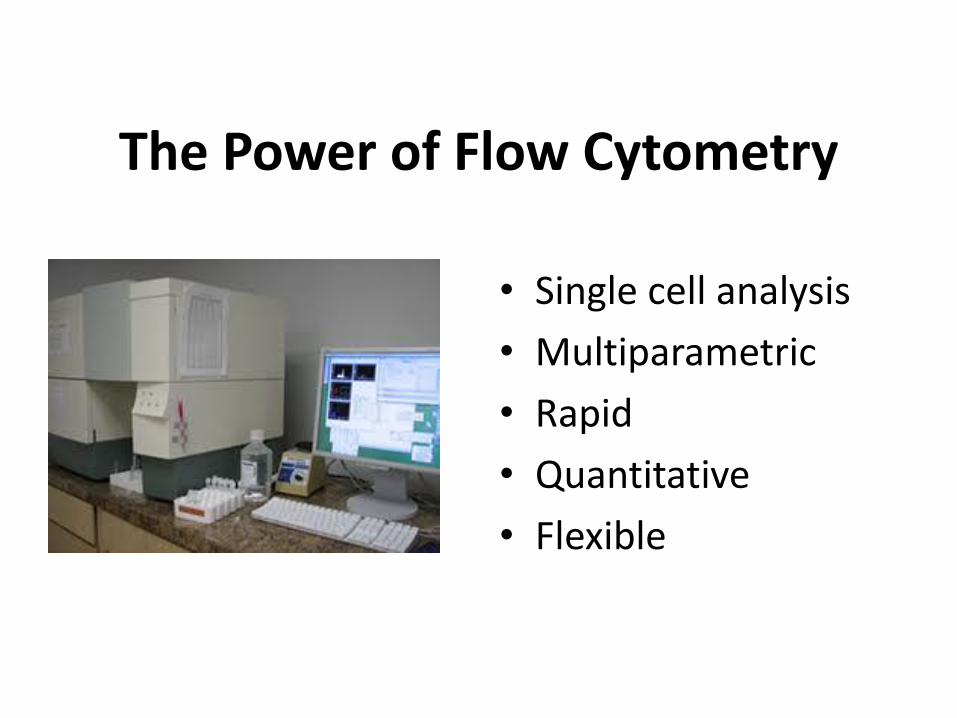
The Power of Flow Cytometry
• Single cell analysis
• Multiparametric
• Rapid
• Quantitative
• Flexible
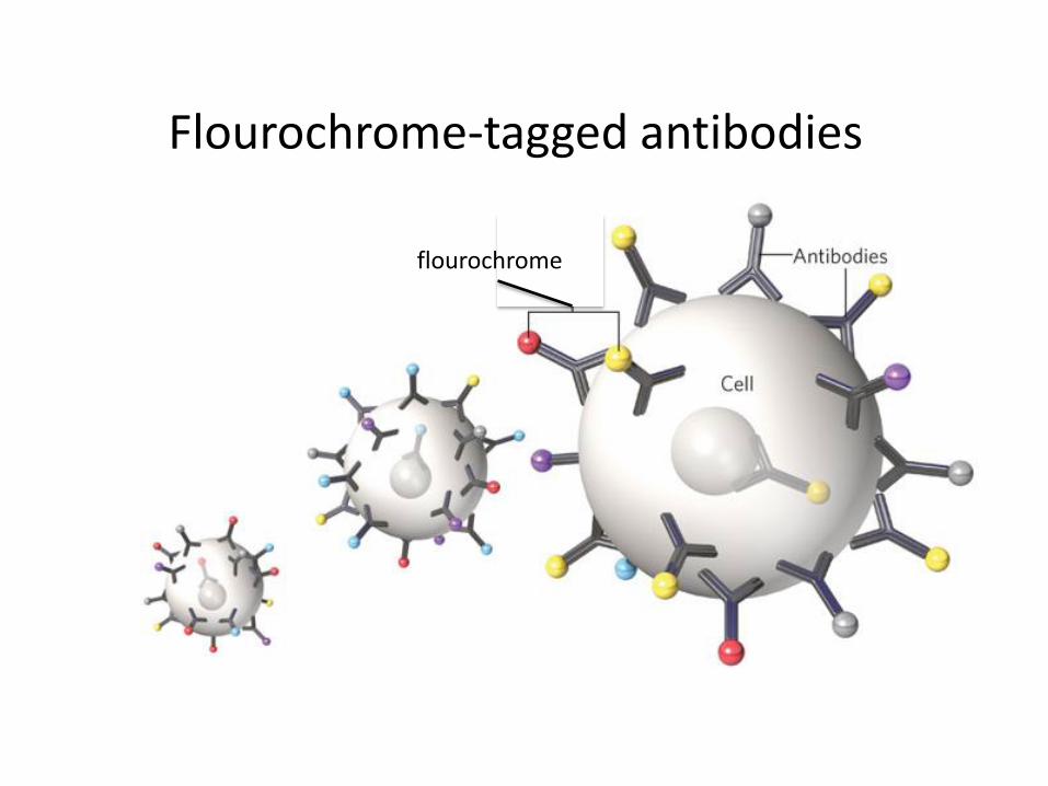
Flourochrome-tagged antibodies
flourochrome
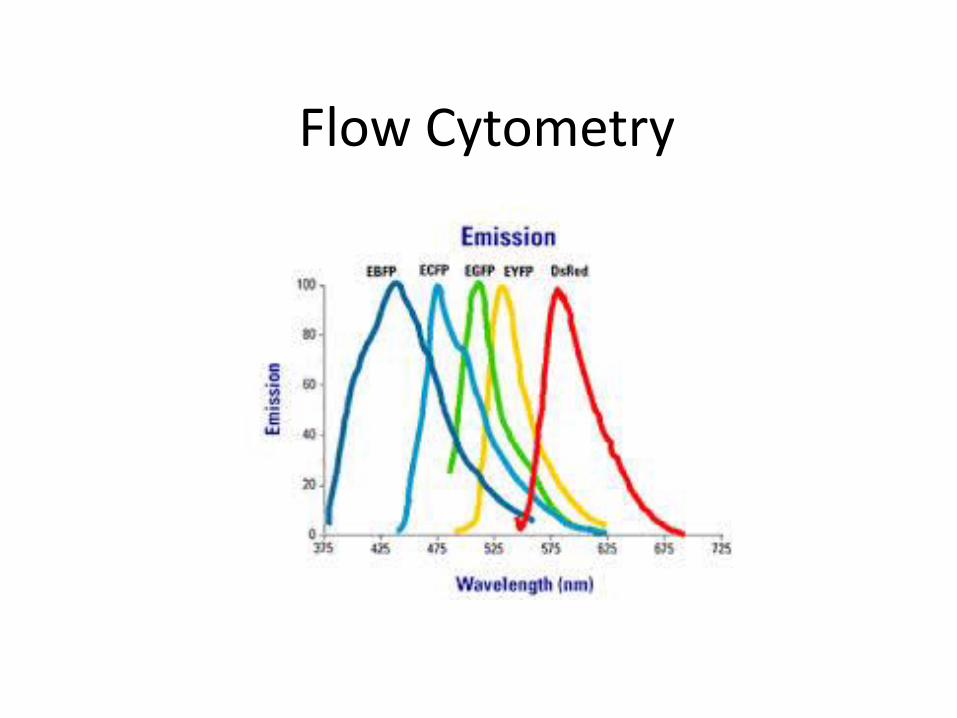
Flow Cytometry

Flow Cell

Flow Cytometry


Flow Cytometry

CD45/SS
Borowitz et al (1993) AJCP 100:534-40. Steltzer et al (1993) Ann NY Acad Sci 667:265-280

Normal Granulocytic Maturation
Wood and Borowitz (2006) Henry’s Laboratory Methods
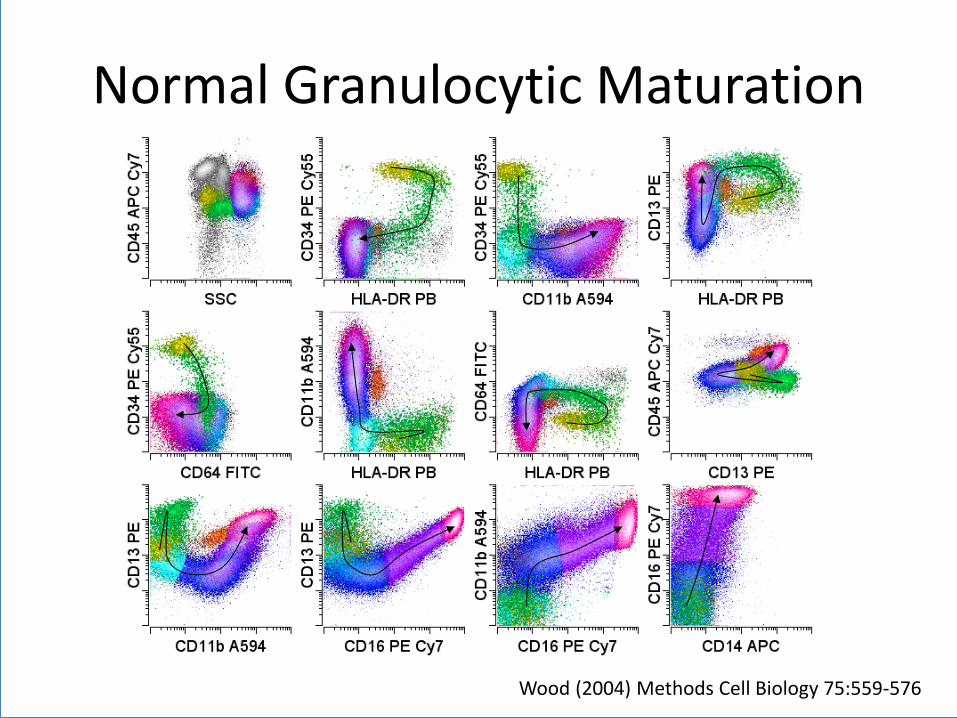
Normal Granulocytic Maturation
Wood (2004) Methods Cell Biology 75:559-576

Normal B cell Maturation
Wood and Borowitz (2006) Henry’s Laboratory Medicine

Abnormal population identification
• Normal – Antigens expressed in consistent and
reproducible patterns with maturation
• Neoplastic – Increased or decreased normal antigens
– Asynchronous maturational expression
– Aberrant antigen expression
– Homogeneous expression
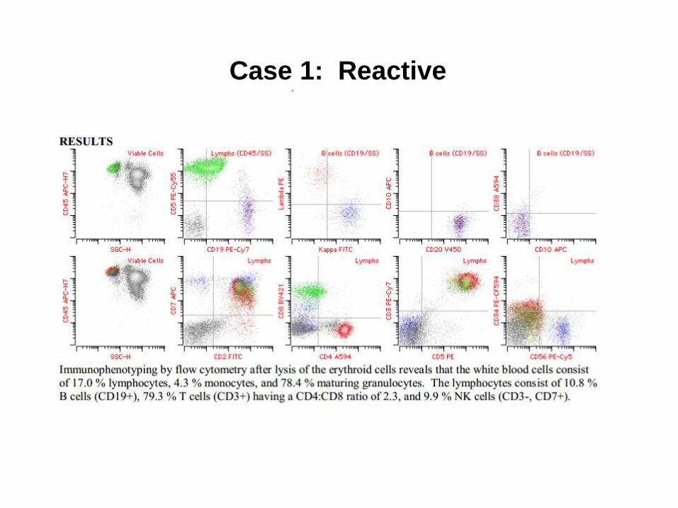
Case 1: Reactive

Case 3: CLL

WOLGAMOT Lymphoma
WOLGAMOT
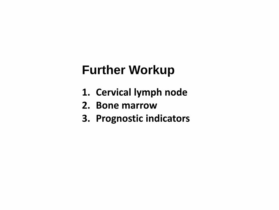
1. Cervical lymph node 2. Bone marrow 3. Prognostic indicators
Further Workup

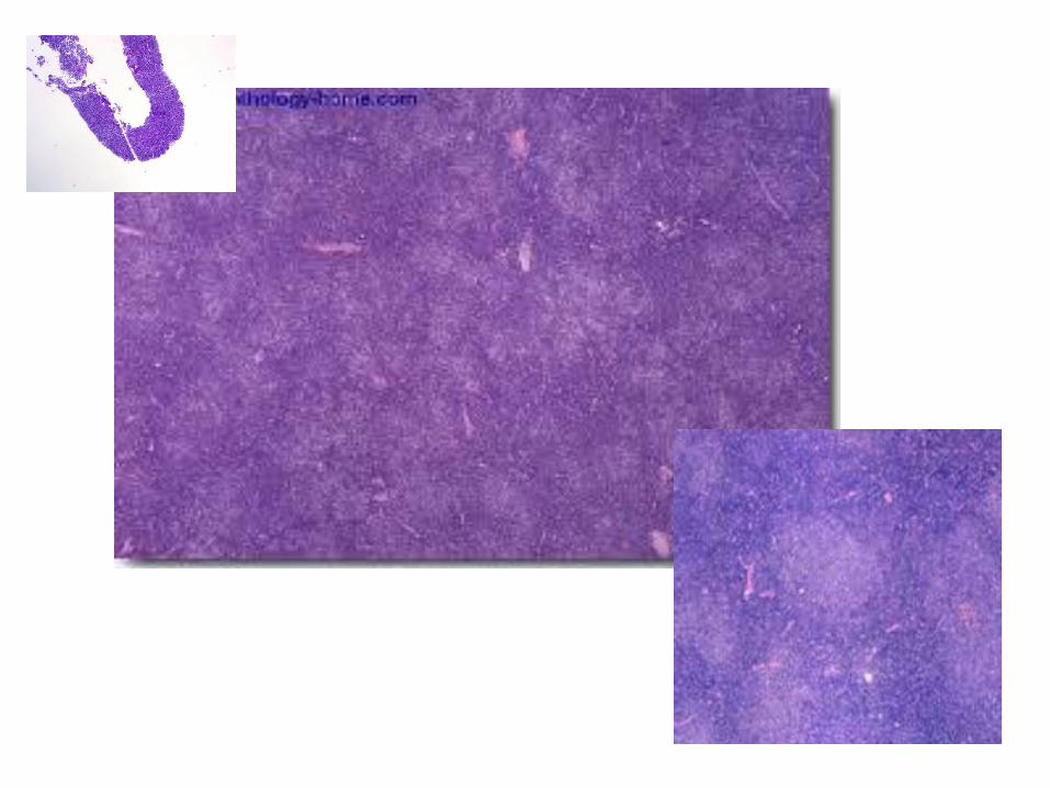
Core biopsy of cervical lymph node
Case 3


Case 3: CLL
Mutated CLL = good prognosis Unmutated CLL = poor prognosis

FINAL DIAGNOSIS: Cervical lymph nodes, core biopsy: Chronic lymphocytic leukemia/small lymphocytic lymphoma (CLL/SLL). Camilla T. Allen, MD Pathologist
Case 3: CLL
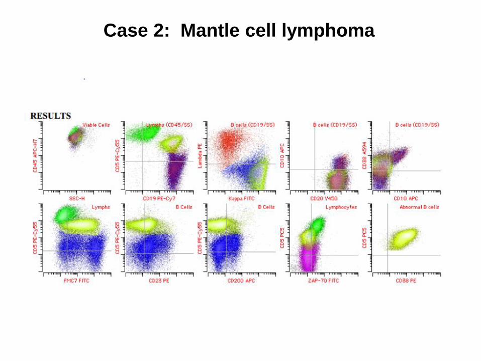
Case 2: Mantle cell lymphoma

Bone marrow aspirate


Case 3: CLL
Result Name Result Abnl Normal Range Units WBC 14.7 H 4.0-11.0 K/mm3 RBC 4.51 4.31-5.77 M/mm3 HGB 12.9 L 13.2-17.5 g/dL HCT 38.7 L 38.9-49.9 % MCV 85.8 80.0-100.0 fL MCH 28.6 27.8-33.8 pg MCHC 33.3 31.5-36.5 g/dL RDW 14.0 11.5-14.2 % PLATELET COUNT 126 L 150-400 K/mm3 NEUTRO% 23.6 % LYMPH.% 69.8 % MONO% 5.1 % EOS% 0.6 % BASO% 0.4 % IMM GRAN% 0.5 0-0.5 % Includes myelocytes, metamyelocytes and promyelocytes. NEUTRO# 3.5 1.5-8.0 K/mm3 LYMPH# 10.3 H 1.0-3.5 K/mm3 MONO# 0.7 0.2-1.0 K/mm3 EOS# 0.1 0-0.5 K/mm3 BASO# 0.1 0-0.2 K/mm3 Smudge cells present

CLL Abnormality Prognosis Very High risk High risk low risk Very low risk
10 year survival ------> 29% 37% 57% same as controls
del 17p13.1 (p53) DCI FISH poor; chemo resistance; consider BMT X
p53 mutation poor; chemo resistance X
BIRC3 very poor; chemo resistance, mutually exclusive to p53 X
ZAP70 expression poor X
CD38 expression poor X
NOTCH associated with Richter's X
del 11q22.3 DCI FISH intermediate risk. Bulky nodes, faster growth, unmutated IgH, requires alkylating drugs (cytoxan, bendamustine) X
SF3B1 poor. Resistance to fludarabine X
trisomy 12 DCI FISH good; low risk. If no NOTCH mutation, low risk X, if NOTCH X, if no NOTCH
IgH mutation present good (unmutated is poor) X
normal cytogenetics good X
del 13q14.3/13q34 DCI FISH good, if isolated X
13p if isolated 13p, good; very low risk X, if isolated
t(14;19) ?
t(2;14) ?
Prognostic Indicators

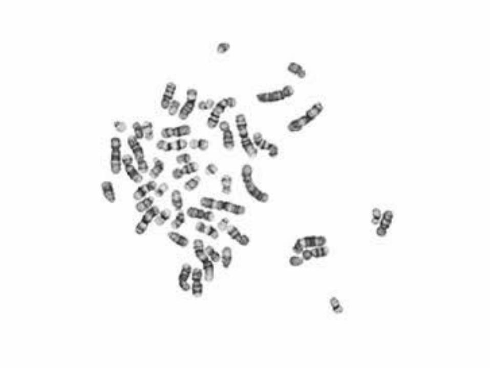
Cytogenetics & Molecular Studies
1. Cytogenetics 2. FISH (flourescence in situ hybridization)

Case 1: Would be normal

Case 2: Mantle cell lymphoma

Case 3: CLL

CLL Abnormality Prognosis Very High risk High risk low risk Very low risk
10 year survival ------> 29% 37% 57% same as controls
del 17p13.1 (p53) DCI FISH poor; chemo resistance; consider BMT X
p53 mutation poor; chemo resistance X
BIRC3 very poor; chemo resistance, mutually exclusive to p53 X
ZAP70 expression poor X
CD38 expression poor X
NOTCH associated with Richter's X
del 11q22.3 DCI FISH intermediate risk. Bulky nodes, faster growth, unmutated IgH, requires alkylating drugs (cytoxan, bendamustine) X
SF3B1 poor. Resistance to fludarabine X
trisomy 12 DCI FISH good; low risk. If no NOTCH mutation, low risk X, if NOTCH X, if no NOTCH
IgH mutation present good (unmutated is poor) X
normal cytogenetics good X
del 13q14.3/13q34 DCI FISH good, if isolated X
13p if isolated 13p, good; very low risk X, if isolated
t(14;19) ?
t(2;14) ?
Prognostic Indicators

1) Probe a specific sequence of DNA or RNA
2) Visualize ‘in situ’ – within the context of tissue
3) Can be performed on interphase cells
4) Can be performed on non-living/fixed cells
In Situ Hybridization (ISH)
Key features:

In Situ Hybridization (ISH)
A C G T A A A G T A C G T A A A G T G G T A G T . . . . . . . .
A A G T A G T T C C C T
1) Target (DNA or RNA)
2) Probe (DNA)
3) Detection method S35 S35 S35 * * *
(radiolabel) (fluorochrome: FISH) FITC Peroxidase or phosphatase (enzyme: CISH)
. . . . . . . .
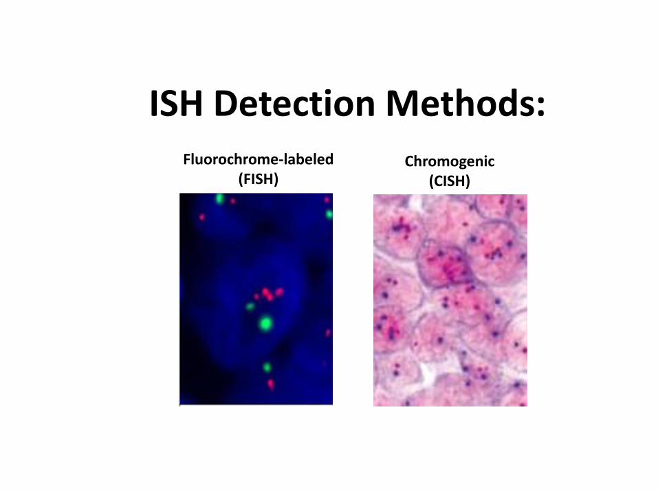
ISH Detection Methods: Fluorochrome-labeled
(FISH) Chromogenic
(CISH)

Uses of In Situ Hybridization
Chromosome enumeration
Locus-specific copy number alterations
Translocation detection
Detection of specific transcripts (RNA)
Detection of foreign DNA/RNA
Trisomy 21 by FISH HER2 amplification by FISH
CISH for kappa and lambda light chain mRNA
t(11;14) translocation by single-fusion FISH
Detection of EBV RNA by CISH

Case 2: Mantle cell lymphoma

Fusion Probes vs. Break-Apart Probes for Translocation Detection
Target two loci Identifies both partners
involved in a translocation
Target one locus Useful when there are
numerous potential partner genes

Case 2: Mantle cell lymphoma
Fusion probes

Case 3: CLL

CLL Abnormality Prognosis Very High risk High risk low risk Very low risk
10 year survival ------> 29% 37% 57% same as controls
del 17p13.1 (p53) DCI FISH poor; chemo resistance; consider BMT X
p53 mutation poor; chemo resistance X
BIRC3 very poor; chemo resistance, mutually exclusive to p53 X
ZAP70 expression poor X
CD38 expression poor X
NOTCH associated with Richter's X
del 11q22.3 DCI FISH intermediate risk. Bulky nodes, faster growth, unmutated IgH, requires alkylating drugs (cytoxan, bendamustine) X
SF3B1 poor. Resistance to fludarabine X
trisomy 12 DCI FISH good; low risk. If no NOTCH mutation, low risk X, if NOTCH X, if no NOTCH
IgH mutation present good (unmutated is poor) X
normal cytogenetics good X
del 13q14.3/13q34 DCI FISH good, if isolated X
13p if isolated 13p, good; very low risk X, if isolated
t(14;19) ?
t(2;14) ?
Prognostic Indicators

Question 2: FISH: 1. Include both osteichthyes and chondrichthyes. 2. Is a molecular technique that can detect
specific sequences of DNA.
3. Allows classification of leukemias based on the proteins the cells express.

WOLGAMOT Lymphoma
WOLGAMOT

WOLGAMOT Lymphoma
WOLGAMOT

Ryan Fortna , MD PhD NW Pathology Brent Wood, MD PhD U of Washington
Cancer Program at SJH
Special thanks to: