Diagnosis of peste des petits ruminants infection in … · Vihol PD, Patel JM (2015) Diagnosis of...
6
Veterinary World, EISSN: 2231-0916 Available at www.veterinaryworld.org/Vol.8/April-2015/2.pdf Veterinary World, EISSN: 2231-0916 443 RESEARCH ARTICLE Open Access Diagnosis of peste des petits ruminants infection in small ruminants through in‑house developed Indirect ELISA: Practical considerations K. K. Sharma 1 , D. P. Kshirsagar 2 , I. H. Kalyani 1 , D. R. Patel 1 , P. D. Vihol 3 and J. M. Patel 3 1.Department of Veterinary Microbiology, Vanbandhu College of Veterinary Science and Animal Husbandry, Navsari Agricultural University, Navsari, Gujarat, India; 2. Department of Veterinary Public Health and Epidemiology, Vanbandhu College of Veterinary Science and Animal Husbandry, Navsari Agricultural University, Navsari, Gujarat, India; 3. Department of Veterinary Pathology, Vanbandhu College of Veterinary Science and Animal Husbandry, Navsari Agricultural University, Navsari, Gujarat, India. Corresponding author: D. P. Kshirsagar, e-mail: [email protected], KKS: [email protected], IHK: [email protected], DRP: [email protected], PDV: [email protected], JMP: [email protected] Received: 26-11-2014, Revised: 25-02-2015, Accepted: 05-03-2015, Published online: 07-04-2015 doi: 10.14202/vetworld.2015.443-448. How to cite this article: Sharma KK, Kshirsagar DP, Kalyani IH, Patel DR, Vihol PD, Patel JM (2015) Diagnosis of peste des petits ruminants infection in small ruminants through in-house developed Indirect ELISA: Practical considerations, Veterinary World 8(4): 443-448. Abstract Aim: The work was conducted to diagnose peste des petits ruminants (PPR) outbreak through an in house developed indirect ELISA (thereafter referred as iELISA) its comparison with other available diagnostic tests and description of practical considerations in its development, utility and limitations. Materials and Methods: An outbreak resembled to PPR occurred in two different places of southern Gujarat viz. Vapi and Navsari, affecting 622 animals, including both goat (n = 476) and sheep (n = 146). Animals displayed the typical signs of PPR at Vapi; however diarrhea was the inconsistent feature in animals of Navsari. The affection caused morbidity of 100% and mortality were 73.68% (n = 392/532) and 56.67% (n = 51/90) in Vapi and Navsari outbreaks, respectively. Relevant ante mortem and post mortem samples were collected from representative animals. At the outset of the epidemic no kit was available with us, so agar gel immunodiffusion (AGID) was carried out and a commercial ELISA (cELISA) kit was ordered for making diagnosis through antibody demonstration. Meanwhile, an iELISA was developed in house using PPR vaccine as antigen and protein G conjugated HRPO antibody as detector. Histopathology and results of sandwich ELISA were also used to diagnose PPR virus (PPRV) in the outbreak. Results: The iELISA developed had detected PPRV antibodies in 22/24 samples (91.66%). Significant difference was observed in disease sensitivity pattern of two species by Chi‑square test. While AGID failed to detect antibodies in any sample. Results were reconfirmed by comparing with commercially available cELISA kit. Conclusion: PPR is an economically important disease and for the rapid diagnosis of PPR the in house developed antibody capture iELISA can be a suitable cost effective alternative. Keywords: agar gel immunodiffusion, competitive ELISA, Goat, indirect ELISA, peste des petits ruminants. Introduction Peste des petits ruminants (PPR) is translated as “plague of small ruminants”, which is caused by an ss negative sense ribonucleic acid containing enveloped virus PPR virus (PPRV) which is member of genus morbillivirus of family Paramyxoviridae [1]. This is an office international des epizootics (OIE) “list A” disease characterized by respiratory, lymphatic, and alimentary tract affection as the name indicates; it causes heavy morbidity and mortality in small rumi‑ nants like goat and sheep [2]. Now, this disease has established as endemic in India and prevalent in almost every part of the country where goat and sheep flocks are reared. Sheep and goat husbandry is restricted to poor and marginal farmers of India as the entry of the virus and resultant mor‑ bidity and mortality pose havoc on the livelihood of affected farmers. It has been estimated that this disease alone causes economic loss of 1800 million Indian rupees (approximately US$ 39 million) per year [3]. In endemic areas, PPR is considered to be one of the main constraints to improve productivity of small ruminants [4]. Though clinical signs are suggestive of disease, but clinical picture warrants differentiation of infection from many diseases, particularly with cap‑ rine contagious pleuropneumonia and hemorrhagic septicemia [2], which is possible through certain laboratory‑based microbiological tests. PPRV infec‑ tion can be diagnosed through precipitation tests like Agar gel immunodiffusion (AGID), counter immuno‑ electrophoresis, ELISA (antigen detecting sandwich ELISA (sELISA) or antibody detecting cELISA), polymerase chain reaction (PCR) includes reverse transcription PCR or qRTPCR, cell culture, and virus neutralization test (VNT) [2,5]. All of the methods mentioned above have their own merit and demerits. Precipitation tests are though easy to perform, but they lack the sensitivity and specificity. PCR and cell culture‑based methods are very costly and technically demanding, again cell culture methods, including are very time‑consuming, which cannot commensurate Copyright: The authors. This article is an open access article licensed under the terms of the Creative Commons Attributin License (http://creative commons.org/licenses/by/2.0) which permits unrestricted use, distribution and reproduction in any medium, provided the work is properly cited.
Transcript of Diagnosis of peste des petits ruminants infection in … · Vihol PD, Patel JM (2015) Diagnosis of...
Veterinary World, EISSN: 2231-0916Available at www.veterinaryworld.org/Vol.8/April-2015/2.pdf
Veterinary World, EISSN: 2231-0916 443
RESEARCH ARTICLEOpen Access
Diagnosis of peste des petits ruminants infection in small ruminants through in‑house developed Indirect ELISA: Practical considerations
K. K. Sharma1, D. P. Kshirsagar2, I. H. Kalyani1, D. R. Patel1, P. D. Vihol3 and J. M. Patel3
1. Department of Veterinary Microbiology, Vanbandhu College of Veterinary Science and Animal Husbandry, NavsariAgricultural University, Navsari, Gujarat, India; 2. Department of Veterinary Public Health and Epidemiology,
Vanbandhu College of Veterinary Science and Animal Husbandry, Navsari Agricultural University, Navsari, Gujarat, India; 3. Department of Veterinary Pathology, Vanbandhu College of Veterinary Science and Animal Husbandry, Navsari
Agricultural University, Navsari, Gujarat, India.Corresponding author: D. P. Kshirsagar, e-mail: [email protected], KKS: [email protected], IHK: [email protected], DRP: [email protected], PDV: [email protected], JMP: [email protected]
Received: 26-11-2014, Revised: 25-02-2015, Accepted: 05-03-2015, Published online: 07-04-2015
doi: 10.14202/vetworld.2015.443-448. How to cite this article: Sharma KK, Kshirsagar DP, Kalyani IH, Patel DR, Vihol PD, Patel JM (2015) Diagnosis of peste des petits ruminants infection in small ruminants through in-house developed Indirect ELISA: Practical considerations, Veterinary World 8(4): 443-448.
AbstractAim: The work was conducted to diagnose peste des petits ruminants (PPR) outbreak through an in house developed indirect ELISA (thereafter referred as iELISA) its comparison with other available diagnostic tests and description of practical considerations in its development, utility and limitations.
Materials and Methods: An outbreak resembled to PPR occurred in two different places of southern Gujarat viz. Vapi and Navsari, affecting 622 animals, including both goat (n = 476) and sheep (n = 146). Animals displayed the typical signs of PPR at Vapi; however diarrhea was the inconsistent feature in animals of Navsari. The affection caused morbidity of 100% and mortality were 73.68% (n = 392/532) and 56.67% (n = 51/90) in Vapi and Navsari outbreaks, respectively. Relevant ante mortem and post mortem samples were collected from representative animals. At the outset of the epidemic no kit was available with us, so agar gel immunodiffusion (AGID) was carried out and a commercial ELISA (cELISA) kit was ordered for making diagnosis through antibody demonstration. Meanwhile, an iELISA was developed in house using PPR vaccine as antigen and protein G conjugated HRPO antibody as detector. Histopathology and results of sandwich ELISA were also used to diagnose PPR virus (PPRV) in the outbreak.
Results: The iELISA developed had detected PPRV antibodies in 22/24 samples (91.66%). Significant difference was observed in disease sensitivity pattern of two species by Chi‑square test. While AGID failed to detect antibodies in any sample. Results were reconfirmed by comparing with commercially available cELISA kit.
Conclusion: PPR is an economically important disease and for the rapid diagnosis of PPR the in house developed antibody capture iELISA can be a suitable cost effective alternative.
Keywords: agar gel immunodiffusion, competitive ELISA, Goat, indirect ELISA, peste des petits ruminants.
Introduction
Peste des petits ruminants (PPR) is translated as “plague of small ruminants”, which is caused by an ss negative sense ribonucleic acid containing enveloped virus PPR virus (PPRV) which is member of genus morbillivirus of family Paramyxoviridae [1]. This is an office international des epizootics (OIE) “list A” disease characterized by respiratory, lymphatic, and alimentary tract affection as the name indicates; it causes heavy morbidity and mortality in small rumi‑nants like goat and sheep [2].
Now, this disease has established as endemic in India and prevalent in almost every part of the country where goat and sheep flocks are reared. Sheep and goat husbandry is restricted to poor and marginal farmers of India as the entry of the virus and resultant mor‑bidity and mortality pose havoc on the livelihood of affected farmers. It has been estimated that this disease
alone causes economic loss of 1800 million Indian rupees (approximately US$ 39 million) per year [3]. In endemic areas, PPR is considered to be one of the main constraints to improve productivity of small ruminants [4]. Though clinical signs are suggestive of disease, but clinical picture warrants differentiation of infection from many diseases, particularly with cap‑rine contagious pleuropneumonia and hemorrhagic septicemia [2], which is possible through certain laboratory‑based microbiological tests. PPRV infec‑tion can be diagnosed through precipitation tests like Agar gel immunodiffusion (AGID), counter immuno‑electrophoresis, ELISA (antigen detecting sandwich ELISA (sELISA) or antibody detecting cELISA), polymerase chain reaction (PCR) includes reverse transcription PCR or qRTPCR, cell culture, and virus neutralization test (VNT) [2,5]. All of the methods mentioned above have their own merit and demerits. Precipitation tests are though easy to perform, but they lack the sensitivity and specificity. PCR and cell culture‑based methods are very costly and technically demanding, again cell culture methods, including are very time‑consuming, which cannot commensurate
Copyright: The authors. This article is an open access article licensed under the terms of the Creative Commons Attributin License (http://creative commons.org/licenses/by/2.0) which permits unrestricted use, distribution and reproduction in any medium, provided the work is properly cited.
Available at www.veterinaryworld.org/Vol.8/April-2015/2.pdf
Veterinary World, EISSN: 2231-0916 444
for field based diagnosis of the acute infections like PPR. ELISA is though not free from limitation but bet‑ter suited among the candidate tests [6,7]. Two types of ELISA has been employed by various workers for PPR diagnosis; Antigen capturing (sELISA) for anti‑gen detection [8‑10] and monoclonal antibody‑based competitive ELISA (cELISA) for antibody detec‑tion [7,11,12]. Both the types of ELISA are available commercially, but in secondary set up laboratories under tropical conditions, like ours, the cost, shelf life of kit due to deterioration of its heat labile components are major hurdles to remain equip all the time for PPR diagnosis. Whereas, acuteness, associated morbidity, and mortality, as well as poor economic condition of animal owners, warrant rapid diagnosis.
Similar conditions have been described by Balamurgan et al. [1], where author suggested going for in house developed Indirect ELISA (iELISA), which is well comparable with commercial cELISA system or VNT. Hence, when an outbreak suggestive of PPR had been investigated, it was confirmed by detection of PPR antibodies in sera of affected ani‑mals through an in house developed antibody capture iELISA and results were reconfirmed by comparing with commercially available cELISA kit.
The present communication description is given regarding considerations encountered during its devel‑opment and thereafter disease confirmation through it.Materials and MethodsEthical approval
As per CPCSEA guidelines, study involving clinical samples does not require approval of Institute Animal Ethics Committee and the authors were per‑mitted by animal owners for sampling.Location, animals and sampling
An outbreak of disease resembled to PPR was occurred in different flocks of small ruminants, maintained for subsistence purpose, in two southern towns of Gujarat state of India viz. Navsari and Vapi, in February ‑ March months. Though, this region is known for high humidity and heavy rainfall but that time climate was like to typical springs of India. Where temperature remains around 30°C with moder‑ate humidity and no rainfall was recorded at that time.
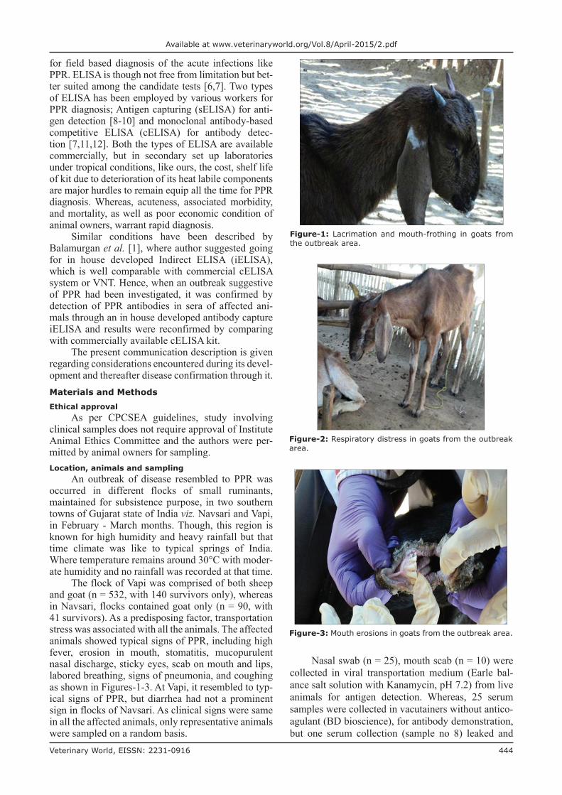
The flock of Vapi was comprised of both sheep and goat (n = 532, with 140 survivors only), whereas in Navsari, flocks contained goat only (n = 90, with 41 survivors). As a predisposing factor, transportation stress was associated with all the animals. The affected animals showed typical signs of PPR, including high fever, erosion in mouth, stomatitis, mucopurulent nasal discharge, sticky eyes, scab on mouth and lips, labored breathing, signs of pneumonia, and coughing as shown in Figures‑1‑3. At Vapi, it resembled to typ‑ical signs of PPR, but diarrhea had not a prominent sign in flocks of Navsari. As clinical signs were same in all the affected animals, only representative animals were sampled on a random basis.
Nasal swab (n = 25), mouth scab (n = 10) were collected in viral transportation medium (Earle bal‑ance salt solution with Kanamycin, pH 7.2) from live animals for antigen detection. Whereas, 25 serum samples were collected in vacutainers without antico‑agulant (BD bioscience), for antibody demonstration, but one serum collection (sample no 8) leaked and
Figure‑1: Lacrimation and mouth-frothing in goats from the outbreak area.
Figure‑2: Respiratory distress in goats from the outbreak area.
Figure‑3: Mouth erosions in goats from the outbreak area.

Available at www.veterinaryworld.org/Vol.8/April2015/2.pdf
Veterinary World, EISSN: 2231-0916 445
could not be processed further. Necropsies were also carried out and samples were collected for histopatho‑logical examination. Utmost aseptic precautions were exercised during sample collection and were trans‑ported to the laboratory on ice. These were reached to laboratory within 3 h of collection.Tests carried out for diagnosis
OIE manual [2] guidance was taken in to refer‑ence and decided to carry out AGID as well as anti‑body demonstration ELISA at our laboratory. While at the same time samples were also sent to National Institute of Veterinary epidemiology and Disease Informatics (NIVEDI), Bangalore for antigen detec‑tion and confirmation of our results. Hence, a com‑mercial competitive ELISA kit was ordered (IDvet, cELISA kit), meanwhile an iELISA had been devel‑oped with available resources using PPR vaccine (pro‑cured from Indian Veterinary research Institute) as antigen.AGID test for antibody demonstration
The test was carried out to detect antibodies against PPR virus as per Khan et al. [13] and OIE manual [2]. Briefly, 1% agarose suspension was made and poured in 6 ml quantity in 35 mm Petri plates. Wells were cut with cardboard template which had been supplied for 35 mm Petri plate with some other commercial AGID kit. PPR vaccine was used antigen source and serum of 45 days vaccinated goat was used as positive control with phosphate buffered saline as a negative control. Samples were placed in alternate wells and development of precipitation line within 72 h was to be recorded as a positive result. Test was declared negative if no visible line developed after acetic acid washing (5% glacial acetic acid for 5 min).Antibody capture iELISA
A total of 24 samples were tested with an in house developed iELISA considering earlier works [1,14]. Two 96 well flat bottom maxisorp microtiter plate (Nunc, Denmark) were coated with very high amount of vaccine antigen, where vaccine vial was reconstituted to 100 doses in 1 ml carbonate bicar‑bonate buffer (pH‑9.4). 100 µl of suspension was put in each well. Each plate was kept overnight at 4°C at static condition. Next morning unbound antigen was washed with washing buffer (0.025% Tween‑PBS, three washing of 3 min each). The wells were blocked with 200 µl 3% bovine serum albumin (BSA) in wash‑ing buffer and washed again. Serum samples in three dilutions, i.e., 1:10, 1:20 and 1:40 were applied to each well in duplicate. Earlier, we thought to apply positive control of AGID as a positive control, but we used positive and negative controls of commercial c‑ELISA kit. After 1 h incubation at room temperature with shaking at 300 rpm, unbound antibodies were washed. Then, G protein conjugated antibodies with HRPO (supplied along with sheep and goat Brucella antibody detection kit by NIVEDI Bangalore) was used as detector antibodies. The color reaction was
observed using outpatient department tablets (5 mg for 10 ml distilled water, Thermo Fischer scientific) with 3% H2O2. After stopping with 1M H2SO4, absor‑bance was recorded using 492 nm filter on ELISA reader (Ms Tecan corp). A positive‑negative (P/N) ratio 2 or above [1] was considered positive upon the linearity of negative controls.Competitive ELISA (c ELISA)
As commercial cELISA kit (M/S IDvet, France) had been received meanwhile, laboratory confirma‑tion of iELISA results and disease diagnosis was made through it. The test was carried out as per manufacturer instructions. Briefly, Antigen coated wells were applied with serum samples (1:2 diluted) and incubated for 45 min at room temperature, then plates were washed thrice with washing buffer. Then, HRPO conjugated antibodies directed against nucleoprotein of virus was applied to wells and again incubated for 30 min to cover unbound antigens. Washing was done again and TMB substrate solution was incubated for 15 min and reading was taken at 450 nm on ELISA reader after inclusion of stopping solution (Multisakn Ex, Thermo Corp).Antigen detection and histopathology
Nasal swabs, triturated mouth swabs, and tissue samples (with sterile PBS in sterile pastel and mortar) were cleared by centrifugation at 5000 rpm for 10 min and supernatants were sent to NIVEDI, Bangalore. The results received were compared and used to con‑firm the disease. The histopathological examination was carried out.Statistical analysis
Chi square test was applied as per the procedures of Snedecor and Cochran [15] to determine the sig‑nificance of difference in mortality pattern between sheep and goat by PPRV infection p<0.05 and degree of freedom = 1.ResultsClinical picture and statistic
The clinical signs were strongly indicative of PPR in two outbreaks, with morbidity of 100% and mortality were 73.68% (n = 392/532) and 56.67% (n = 51/90) in Vapi and Navsari outbreaks, respectively. Further, partitioning could be made in Vapi outbreak, where 81.60% (n = 315/386) goat and 52.74% (77/146) sheep were died. Significant differ‑ence was observed in disease sensitivity pattern of two species (Chi‑square value = 30.49 at p<0.05, df = 1). Summary of outbreak is described in Table‑1.AGID and iELISA
The one prerequisite of applying antibody demonstration test is that there must be a negative his‑tory of vaccination and these animals should not be vaccinated with PPR vaccine. Of the 24 sera sample tested by AGID, all showed negative results for the presence of precipitation line; therefore, this test was not found useful to apply for this purpose.
Available at www.veterinaryworld.org/Vol.8/April-2015/2.pdf
Veterinary World, EISSN: 2231-0916 446
iELISA and cELISAInitially three dilutions, 1:10, 1:20, and 1:40
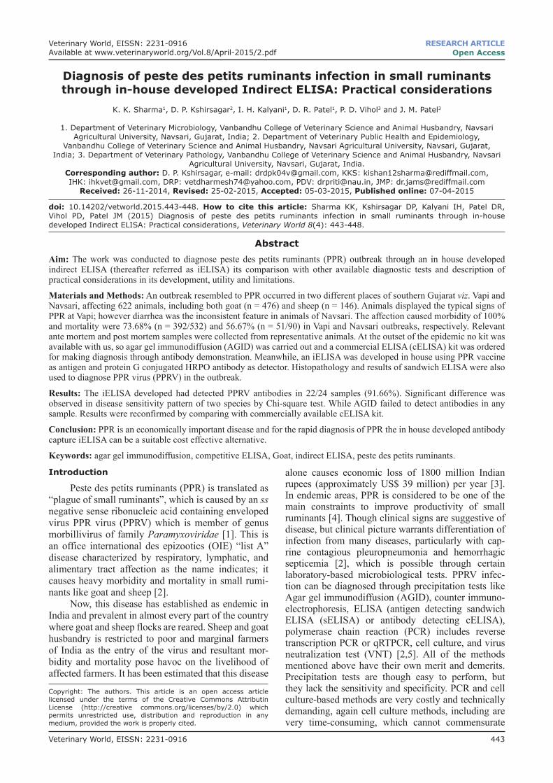
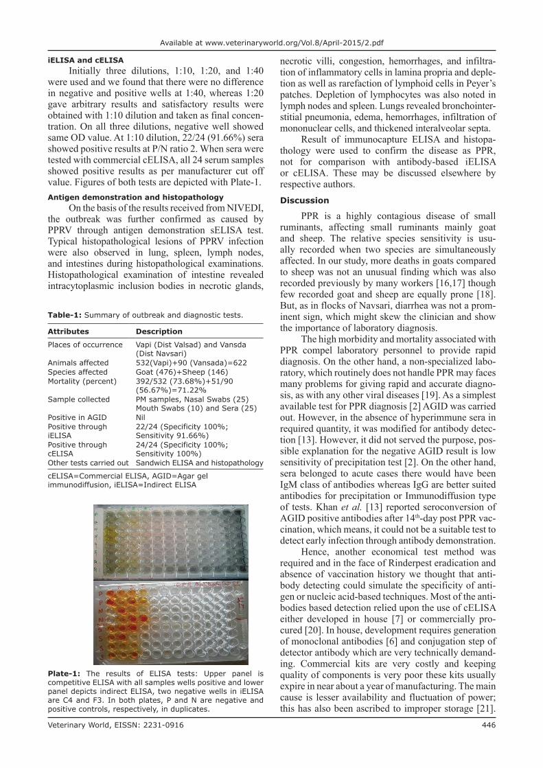
were used and we found that there were no difference in negative and positive wells at 1:40, whereas 1:20 gave arbitrary results and satisfactory results were obtained with 1:10 dilution and taken as final concen‑tration. On all three dilutions, negative well showed same OD value. At 1:10 dilution, 22/24 (91.66%) sera showed positive results at P/N ratio 2. When sera were tested with commercial cELISA, all 24 serum samples showed positive results as per manufacturer cut off value. Figures of both tests are depicted with Plate‑1.Antigen demonstration and histopathology
On the basis of the results received from NIVEDI, the outbreak was further confirmed as caused by PPRV through antigen demonstration sELISA test. Typical histopathological lesions of PPRV infection were also observed in lung, spleen, lymph nodes, and intestines during histopathological examinations. Histopathological examination of intestine revealed intracytoplasmic inclusion bodies in necrotic glands,
necrotic villi, congestion, hemorrhages, and infiltra‑tion of inflammatory cells in lamina propria and deple‑tion as well as rarefaction of lymphoid cells in Peyer’s patches. Depletion of lymphocytes was also noted in lymph nodes and spleen. Lungs revealed bronchointer‑stitial pneumonia, edema, hemorrhages, infiltration of mononuclear cells, and thickened interalveolar septa.
Result of immunocapture ELISA and histopa‑thology were used to confirm the disease as PPR, not for comparison with antibody‑based iELISA or cELISA. These may be discussed elsewhere by respective authors.Discussion
PPR is a highly contagious disease of small ruminants, affecting small ruminants mainly goat and sheep. The relative species sensitivity is usu‑ally recorded when two species are simultaneously affected. In our study, more deaths in goats compared to sheep was not an unusual finding which was also recorded previously by many workers [16,17] though few recorded goat and sheep are equally prone [18]. But, as in flocks of Navsari, diarrhea was not a prom‑inent sign, which might skew the clinician and show the importance of laboratory diagnosis.
The high morbidity and mortality associated with PPR compel laboratory personnel to provide rapid diagnosis. On the other hand, a non‑specialized labo‑ratory, which routinely does not handle PPR may faces many problems for giving rapid and accurate diagno‑sis, as with any other viral diseases [19]. As a simplest available test for PPR diagnosis [2] AGID was carried out. However, in the absence of hyperimmune sera in required quantity, it was modified for antibody detec‑tion [13]. However, it did not served the purpose, pos‑sible explanation for the negative AGID result is low sensitivity of precipitation test [2]. On the other hand, sera belonged to acute cases there would have been IgM class of antibodies whereas IgG are better suited antibodies for precipitation or Immunodiffusion type of tests. Khan et al. [13] reported seroconversion of AGID positive antibodies after 14th‑day post PPR vac‑cination, which means, it could not be a suitable test to detect early infection through antibody demonstration.
Hence, another economical test method was required and in the face of Rinderpest eradication and absence of vaccination history we thought that anti‑body detecting could simulate the specificity of anti‑gen or nucleic acid‑based techniques. Most of the anti‑bodies based detection relied upon the use of cELISA either developed in house [7] or commercially pro‑cured [20]. In house, development requires generation of monoclonal antibodies [6] and conjugation step of detector antibody which are very technically demand‑ing. Commercial kits are very costly and keeping quality of components is very poor these kits usually expire in near about a year of manufacturing. The main cause is lesser availability and fluctuation of power; this has also been ascribed to improper storage [21].
Plate‑1: The results of ELISA tests: Upper panel is competitive ELISA with all samples wells positive and lower panel depicts indirect ELISA, two negative wells in iELISA are C4 and F3. In both plates, P and N are negative and positive controls, respectively, in duplicates.
Table‑1: Summary of outbreak and diagnostic tests.
Attributes Description
Places of occurrence Vapi (Dist Valsad) and Vansda (Dist Navsari)
Animals affected 532(Vapi)+90 (Vansada)=622Species affected Goat (476)+Sheep (146)Mortality (percent) 392/532 (73.68%)+51/90
(56.67%)=71.22%Sample collected PM samples, Nasal Swabs (25)
Mouth Swabs (10) and Sera (25)Positive in AGID NilPositive through iELISA
22/24 (Specificity 100%; Sensitivity 91.66%)
Positive through cELISA
24/24 (Specificity 100%; Sensitivity 100%)
Other tests carried out Sandwich ELISA and histopathology
cELISA=Commercial ELISA, AGID=Agar gel immunodiffusion, iELISA=Indirect ELISA

Available at www.veterinaryworld.org/Vol.8/April2015/2.pdf
Veterinary World, EISSN: 2231-0916 447
An indirect ELISA can be a suitable alternative for labor intensive and technically demanding VNT [7] and costly cELISA [1].
Regarding the development of iELISA test some modifications were applied to suit early detection of disease in the absence of validated components. As rapid results were required, instead of using checker‑board for antigen and antibody dilutions, we applied concentrated form of antigen to cover most of the sites of wells; further blocking was done with 3% BSA to check the false positive results. Last modifi‑cation was the use of Protein G conjugated detector antibody, which helped us to screen goat and sheep sera simultaneously; otherwise use of anti‑spe‑cies antibodies necessitated two separate tests. The idea of use of applying protein G conjugated was obtained from Neilson et al. [14]. Protein G is a Staphylococcus aureus protein and bind strongly with Fc portion of the antibody. Rather, it can be applied to detect antibody of any species with isotype detection can be switched by use of protein A or protein G.
iELISA so developed could detect serum diluted to 1:10, recently, Truong et al. [22] attempted simi‑lar test with antigen derived from Vero cell culture and reported use of 1:50 as initial dilution where they get detectable IgG on 8th day post infection. The dif‑ference is again due to the early collection of serum, which might contain low IgG level which remained undetectable at higher dilutions. However iELISA, unlike AGID can detect early disease. The iELISA proved specific in comparison of cELISA, but could not detect two serum samples as positive which was detected by commercial cELISA. The similar results have been described by Balamurgan et al. [1], where they reported 95.09 and 100% specificity whereas 90.01% and 80% sensitivity against cELISA and VNT, respectively. The false negative results may require correction of lower limit of detection or else there may be interference by large sized IgM in bind‑ing with antigen. On the application level, all the pos‑itive samples may be declared positive, but negative sample need to be reconfirmed with more sensitive test. As the test shown similar OD value for negative control, false positive should not be a problem.Conclusions
PPR is a disease of high morbidity and mor‑tality that affect small ruminants reared by poor and marginal farmers of India. The clinical picture may vary and diarrhea may not be a prominent sign. As most of the flocks are unvaccinated against PPR virus, so an economical antibody‑based test may serve the purpose of PPR diagnosis. Though, AGID was found unsuitable but an in house iELISA was proved equally specific with commercial cELISA, but showed few false negative results. sELISA and histopathological examination proved useful adjuncts for the final dec‑laration of the outbreak as PPR.
Authors’ Contributions
IHK and KKS designed the study. KKS, DPK, and DRP collected the samples and performed the experiments. PDV and JMP carried out necropsy and evaluated gross and histopathological changes. KKS and DPK analyzed the data. KKS, DPK, and IHK drafted and revised the manuscript. All authors read and approved the final manuscript.Acknowledgments
The authors are thankful to Dr. V. Balamurugan, National Institute of Veterinary Epidemiology and Disease Informatics (NIVEDI), Bangalore for providing confirmatory diagnosis of PPR by antigen detection. The authors are grateful to the Dean, College of Veterinary Science and Animal Husbandry, Navsari (Gujarat) for providing the facilities and fund under the Budget head: 12049 to pursue this work.Competing Interests
The authors declare that they have no competing interests.References1. Balamurugan, V., Singh, R.P., Saravanan, P., Sen, A.,
Sarkar, J., Sahay, B., Rasool, T.J. and Singh, R.K. (2007) Development of an indirect ELISA for the detection of antibodies against peste des petits ruminants’ virus in small ruminants. Vet. Res. Commun., 31(3): 355‑364.
2. OIE, World Organization for Animal Health. (2013) Peste des petites ruminatum. Manual of Diagnostic Tests and Vaccines for Terrestrial Animals. 6th ed., Vol. 1, Ch. 2.7.11. Office International Des Epizootics, Paris, France.
3. Singh, R.P. (2011) Control strategies for peste des petits ruminants in small ruminants of India. Rev. Sci. Tech., 30(3): 879‑887.
4. Chauhan, H.C., Chandel, B.S., Kher, H.N., Dadawala, A.I. and Agrawal, S.M. (2009) Pesti des petits ruminant’s virus infection in animals. Vet. World, 2(4): 150‑155.
5. Balamurugan, V., Hemadri, D., Gajendragad, M.R., Singh, R.K. and Rahman, H. (2014) Diagnosis and con‑trol of peste des petits ruminants: A comprehensive review. Indian J. Virol. 25:39‑56.
6. Singh, R.P., Sreenivasa, B.P., Dhar, P., Shah, L.C. and Bandyopadhyay, S.K. (2004) Development of a monoclonal antibody based competitive‑ELISA for detection and titra‑tion of antibodies to Peste des petits ruminants (PPR) virus. Vet. Microbiol., 98: 3‑15.
7. Choi, K.S., Nah, J.J., Ko, Y.J., Kang, S.Y. and Jo, N.I. (2005) Rapid competitive enzyme‑Linked immunosorbent assay for detection of antibodies to peste des petits ruminants virus. Clin. Diagn. Lab. Immunol., 12(4): 542‑547.
8. Libeau, G., Prehaud, C., Lancelot, R., Colas, F., Guerre, L., Bishop, D.H. and Diallo, A., (1995) Development of a com‑petitive ELISA for detecting antibodies to the peste des petits ruminant’s virus using a recombinant nucleoprotein. Res. Vet. Sci., 58(1): 50‑55.
9. Singh, R.P., Bandyopadhyay, S.K., Sreenivasa, B.P. and Dhar, P. (2004) Production and characterization of mono‑clonal antibodies to Peste des petits ruminants (PPR) virus. Vet. Res. Commun., 28(7): 623‑639.
10. Couacy‑Hymann, E., Bodjo, S.C., Koffi, M.Y., Kouakou, C. and Danho, T. (2009) The early detection of peste‑des‑pe‑tits‑ruminants (PPR) virus antigens and nucleic acid from experimentally infected goats using RT‑PCR and

Available at www.veterinaryworld.org/Vol.8/April-2015/2.pdf
Veterinary World, EISSN: 2231-0916 448
immunocapture ELISA techniques. Res. Vet. Sci., 87(2): 332‑335.
11. Singh, R.P., Sreenivasa, B.P., Dhar, P. and Bandyopadhyay, S.K. (2004) Development of a sand‑wich‑ELISA for the diagnosis of peste des petits ruminants (PPR) infection in small ruminants using anti – Nucleocapsid protein monoclonal antibody. Arch. Virol., 149(11): 2155‑2170.
12. Zhang, G.A., Zeng, J.C., Zhu, Y.B., Dong, S.B., Zhu, S.C., Yu, R.B., Duoji, C.C., Lei, Z.A. and Li, Z.B. (2012) Development of an indirect ELISA with artificially synthe‑sized N protein of PPR virus. Intervirology, 55(1): 12‑20.
13. Khan, S.B., Sadique, U., Khan, F.A., Ahmad, I., Rehman, Z.U., Khan, M.A., Idrees, M. and Shah, Z. (2013) Factors affecting efficacy and immu‑nological response of peste des petits ruminants (PPR) vac‑cines in small ruminants. J. Anim. Plant Sci., 23(1): 43‑46.
14. Nielsen, K., Smith, P., Yu, W., Nicoletti, P., Elzer, P., Robles, C., Bermudez, R., Renteria, T., Moreno, F., Ruiz, A., Massengill, C., Muenks, Q., Jurgersen, G., Tollersrud, T., Samartino, L., Conde, S., Forbes, L., Gall, D., Perez, B., Rojas, X. and Minas, A. (2005) Towards single screening test for brucellosis. Rev. Sci. Tech. official Int. Epizoot., 24(3): 1027‑1038.
15. Snedecor, G.W., Cochran, W.G. (1994) Statistical Methods. 8th ed. Iowa State University Press, Ames, Iowa, USA.
16. Kumar, P., Kumar, R., Sharma, A.K. and Tripathi, B.N. (2002) Pathology of peste des petits ruminants (PPR)
********
in goats and sheep: Spontaneous study. Indian J. Vet. Pathol., 26(2): 15‑18.
17. Purushothaman, V., Hemaltha, S., Ravikumar, G., Govindrajan, R., Sundar, N. and Kotteseswaran, A. (2014) An outbreak of peste des petits ruminant virus in small ani‑mals. Indian Vet. J., 83: 133‑136.
18. Bhaskar, S.R., Deshmukh, V.V., Chopade, N.A., Rautmare, S.S. and Aziz, A. (2011) Peste des petits rumi‑nants (PPR) outbreak in sheep and goats in Maharashtra: Laboratory confirmation by s‑ELISA (Mukteshwar) and Vero cell culture. Anim. Sci. Rep., 5 (2): 64‑68.
19. Murphy, F.A., Gibbs, E.P.J., Horzinek, M.C. and Studdert, M.J. (1999) Veterinary Virology. 3rd ed. Academic Press, San Diego, CA. p193‑224.
20. Madboli, A.A. and Ali, S.M. (2012) Histopathological and immunohistochemical studies on the female genital sys‑tem and some visceral organs in sheep and goat naturally infected by peste des petits ruminants virus. Glob. Vet., 9(6): 752‑760.
21. Mccullough, K.C., Sheshberadaran, H., Norrby, E., Obi, T.U. and Crowther, J.R. (1986) Monoclonal antibod‑ies against morbilliviruses. Rev. Sci. Tech. Official. Int. Epizoot., 5: 411‑427.
22. Truong, T., Boshra, H., Embury‑Hyatt, C., Nfon, C. and Gerdts, V. (2014) Peste des petits ruminants virus tissue tro‑pism and pathogenesis in sheep and goats following experi‑mental infection. PLoS One, 9(1):e87145.