Diagnosis and treatment of hypernatremia - Western University and... · Diagnosis and treatment of...
15
3 Diagnosis and treatment of hypernatremia Saif A. Muhsin, MBChB a , David B. Mount, MD, Associate Chief a, b, * a Renal Division, Brigham and Women's Hospital, Boston, MA, USA b Veterans Affairs Boston Healthcare System, Boston, MA, USA article info Article history: Available online 4 March 2016 Keywords: hypernatremia vasopressin osmoreceptor TRPV1 lithium diabetes insipidus Hypernatremia is defined as a serum sodium level above 145 mmol/L. It is a frequently encountered electrolyte disturbance in the hospital setting, with an unappreciated high mortality. Understanding hypernatremia requires a comprehension of body fluid compartments, as well as concepts of the preservation of normal body water balance. The human body maintains a normal osmolality between 280 and 295 mOsm/kg via Arginine Vaso- pressin (AVP), thirst, and the renal response to AVP; dysfunction of all three of these factors can cause hypernatremia. We review new developments in the pathophysiology of hypernatremia, in addi- tion to the differential diagnosis and management of this impor- tant electrolyte disorder. © 2016 Published by Elsevier Ltd. Introduction Water balance Hypernatremia is defined as an increase in the plasma Na þ concentration to >145 mM. Considerably less common than hyponatremia, hypernatremia is however associated with mortality rates of as much as 40e60%. Hypernatremia most commonly occurs in ICUs, mostly developing after admission, and has been associated with increased mortality and prolonged length of ICU stay [1]. A recent study showed that severity rather than duration of the hypernatremia following the ICU admission was associated with increased mortality and increased length of stay (40% and 28% increase, respectively) [2]. * Corresponding author. Renal Division, Brigham and Women's Hospital, Boston MA 02115, USA. E-mail address: [email protected] (D.B. Mount). Contents lists available at ScienceDirect Best Practice & Research Clinical Endocrinology & Metabolism journal homepage: www.elsevier.com/locate/beem http://dx.doi.org/10.1016/j.beem.2016.02.014 1521-690X/© 2016 Published by Elsevier Ltd. Best Practice & Research Clinical Endocrinology & Metabolism 30 (2016) 189e203
Transcript of Diagnosis and treatment of hypernatremia - Western University and... · Diagnosis and treatment of...

Best Practice & Research Clinical Endocrinology & Metabolism 30 (2016) 189e203
Contents lists available at ScienceDirect
Best Practice & Research ClinicalEndocrinology & Metabolism
journal homepage: www.elsevier .com/locate/beem
3
Diagnosis and treatment of hypernatremia
Saif A. Muhsin, MBChB a,David B. Mount, MD, Associate Chief a, b, *
a Renal Division, Brigham and Women's Hospital, Boston, MA, USAb Veterans Affairs Boston Healthcare System, Boston, MA, USA
a r t i c l e i n f o
Article history:Available online 4 March 2016
Keywords:hypernatremiavasopressinosmoreceptorTRPV1lithiumdiabetes insipidus
* Corresponding author. Renal Division, BrighamE-mail address: [email protected] (D.B. Mo
http://dx.doi.org/10.1016/j.beem.2016.02.0141521-690X/© 2016 Published by Elsevier Ltd.
Hypernatremia is defined as a serum sodium level above145 mmol/L. It is a frequently encountered electrolyte disturbancein the hospital setting, with an unappreciated high mortality.Understanding hypernatremia requires a comprehension of bodyfluid compartments, as well as concepts of the preservation ofnormal body water balance. The human body maintains a normalosmolality between 280 and 295 mOsm/kg via Arginine Vaso-pressin (AVP), thirst, and the renal response to AVP; dysfunction ofall three of these factors can cause hypernatremia. We review newdevelopments in the pathophysiology of hypernatremia, in addi-tion to the differential diagnosis and management of this impor-tant electrolyte disorder.
© 2016 Published by Elsevier Ltd.
Introduction
Water balance
Hypernatremia is defined as an increase in the plasma Naþ concentration to>145mM. Considerablyless common than hyponatremia, hypernatremia is however associatedwithmortality rates of asmuchas 40e60%. Hypernatremia most commonly occurs in ICUs, mostly developing after admission, and hasbeen associated with increased mortality and prolonged length of ICU stay [1]. A recent study showedthat severity rather than duration of the hypernatremia following the ICU admission was associatedwith increased mortality and increased length of stay (40% and 28% increase, respectively) [2].
and Women's Hospital, Boston MA 02115, USA.unt).

S.A. Muhsin, D.B. Mount / Best Practice & Research Clinical Endocrinology & Metabolism 30 (2016) 189e203190
Secondary analysis of a recent prospective study in the ICU showed that almost 50% of pre-dialysispatients with acute kidney injury had a dysnatremia, mainly hypernatremia, and that there was anincrease in mortality especially with severe hypernatremia (serum sodium �156) compared to nor-monatremic patients (89.1% versus 64.6% respectively) [3]. Preoperative hypernatremia is also asso-ciated with increased perioperative 30-day morbidity and mortality [4].
Understanding hypernatremia requires a comprehension of the main body fluid compartments aswell as an appreciation of the basic concepts of maintenance of normal body water balance. Total bodywater (TBW) is a key physiological term in this context. TBW has been estimated to be about 60% ofbody weight in men and 50% in women; this notably is a simplified estimate. TBW is further dividedinto two main compartments, an extracellular fluid (ECF) and an intracellular fluid (ICF) compartment.The ECF compartment includes plasma, interstitial and lymph fluid, connective tissue and bone,transcellular fluid within body cavities, and adipose tissue [5].
Tonicity refers to the behavior of cell volume in a given solution and represents the action ofeffective osmoles across a membrane. Cellular volume expands when cells are bathed in relativelyhypotonic solutions and contracts when bathed in relatively hypertonic solutions, due to movement ofwater in and out of the cell respectively to eventually reach a steady state tonicity. Effective or activeosmoles include sodium (and associated anions) and glucose in the extracellular compartment,whereas the ionic osmotic driver in the intracellular compartment is primarily potassium (and asso-ciated anions). On the other hand, osmolality represents the sum of both effective and ineffectiveosmoles in any 1 kg of body fluid. Ineffective osmoles, typically urea and alcohol [6] can cross freelyacross cell membranes and hence do not generally alter cellular volume. Osmolality is a poor indicatorof tonicity given the presence of these ineffective osmoles. While effects of tonicity on cellular sizecannot be measured directly, serum sodium can serve as a useful surrogate for tonicity in all bodycompartments at steady state.
Hypertonicity (dehydration) refers to the loss of total-body water such that cellular volume con-tracts, whereas volume depletion is a term used to signify loss of extracellular fluid volume. These twodistinct conditions have different clinical features as well as different therapeutic responses [6,7].
Osmoreceptors and thirst
Vasopressin secretion, thirst, and the renal response to vasopressin collaborate to maintain normalhuman body fluid osmolality between 280 and 295 mOsm/kg. Thirst and vasopressin secretion areunder the control of osmoreceptor neurons within the central nervous system (CNS) (see Fig. 1). Classiccanine experiments performed in the 1940s, correlating the effect on urine output of carotid infusion ofvarious osmolytes, led to the postulation of a central “osmoreceptor” [8]. The primary “osmostat”within the CNS is encompassed within the organum vasculosum of the lamina terminalis (OVLT); thissmall periventricular region lacks a bloodebrain barrier, allowing for direct sensing of the osmolality ofcirculating blood. Osmoreceptive neurons are however widely distributed within the CNS, such thatvasopressin (AVP) release and thirst are controlled by overlapping osmosensitive neural networks[9e12]. Osmosensitive neurons are thus found in the subfornical organ (SFO) and the nucleus tractussolitarii, centers which help integrate regulation of circulating osmolality with that of related phe-nomena, such as extracellular fluid volume [9,10,12] (see Fig. 1).
Osmosensitive neurons from the supraoptic nucleus differ dramatically from hippocampal neurons,in that they demonstrate exaggerated changes in cell volume during cell shrinkage (hypertonic media)or cell swelling (hypotonicmedia) [13]. In hippocampal neurons, cell swelling evokes a rapid regulatoryvolume decrease (RVD) response, whereas cell shrinkage evokes a regulatory volume increase (RVI)response. In consequence, if external tonicity is slowly increased or decreased these RVD and RVImechanisms are sufficient to prevent any change in the cell volume of hippocampal neurons; incontrast, osmosensitive neurons exhibit considerable changes in cell volume during such osmoticramps [13]. This relative lack of volume regulatory mechanisms maximizes the mechanical effect ofextracellular tonicity and generates an ideal osmotic sensor.
Osmosensitive neurons depolarize after cell shrinkage induced by exposure to hypertonic stimuli,with a marked increase in neuronal spike discharges; the associated current is due to activation of anonselective cation channel [14], with five-fold higher permeability for Ca2þ over Naþ [15]. Hypotonic

Fig. 1. Osmoregulatory circuits in the mammalian nervous system. Sagittal illustration of the rat brain, in which the relativepositions of relevant structures and nuclei have been compressed into a single plane. Neurons and pathways are color-coded todistinguish osmosensory, integrative and effector areas. Vasopressin (AVP) is synthesized in magnocellular neurons within thesupraoptic (SON) and paraventricular (PVN) nuclei of the hypothalamus; the distal axons of these neurons project to the posteriorpituitary (PP) from which AVP is released into the circulation. ACC, anterior cingulate cortex; AP, area postrema; DRG, dorsal rootganglion; IML, intermediolateral nucleus; INS, insula; MnPO, median preoptic nucleus; NTS, nucleus tractus solitarius; OVLT,organum vasculosum laminae terminalis; PAG, periaqueductal grey; PBN, parabrachial nucleus; PP, posterior pituitary; PVN, para-ventricular nucleus; SFO, subfornical organ; SN, sympathetic nerve; SON, supraoptic nucleus; SpN, splanchnic nerve; THAL, thal-amus; VLM, ventrolateral medulla. (With permission from Bourque CW, “Central mechanisms of osmosensation and systemicosmoregulation”, Nat Rev Neurosci 2008; 9:519e31).
S.A. Muhsin, D.B. Mount / Best Practice & Research Clinical Endocrinology & Metabolism 30 (2016) 189e203 191
stimuli in turn hyperpolarize the cells and abolish spike discharges [14]. Depolarization and spikedischarges, in the absence of hypertonicity, can also be evoked by suction-induced changes in cellvolume during whole-cell voltage recording, suggesting involvement of a stretch-inactivated cationchannel [14].
Mechanosensitive, stretch-inactivated cation channels, linked to the cytoskeleton [16], are thoughtbe key components of the osmoreceptor complex. The TRPV1 channel (transient receptor potentialvanilloid channel 1) appears to be a critical component of the mechanosensitive osmoreceptor, withloss of osmoreceptive neuronal depolarization and neuronal activation after hypertonic stimuli inTRPV1 �/� mice [17,18]. Specifically, an N-terminal splice variant of TRPV1 has been implicated in thisprocess, with detectable expression of TRPV1 C-terminal exons by RT-PCR in neurons from the SONwithout detectable expression of N-terminal exons; these AVP-positive neurons also stain positivewitha C-terminal TRPV1 antibody, suggesting the involvement of an N-terminal splice-form. More recently,the relevant alternatively spliced isoform (TRPV1dn) has been cloned and characterized; TRPV1dn hasan alternative start codon with a truncated N-terminus [19]. TRPV1dn encodes a shrinkage-activatedchannel and can rescue the phenotype of osmoreceptor neurons from TRPV1 �/� mice [19]. Theswelling-activated TRPV4 channel is also expressed in osmoreceptor neurons, where it may play aninhibitory role, limiting the thirst response in hypotonicity and perhaps downregulating osmotic-
S.A. Muhsin, D.B. Mount / Best Practice & Research Clinical Endocrinology & Metabolism 30 (2016) 189e203192
induced AVP release; however, there are substantial differences in the reported phenotypes of TRPV4knockout mice [20,21], such that the exact role of TRPV4 is still controversial.
At the neuronal network level, OVLT and adjacent circumventricular regions collaborate to regulatewater intake and AVP release, in a number of different species [22,23] (see Fig.1). In humans, functionalmagnetic resonance imaging (fMRI) studies have revealed thirst-associated activation of the anteriorwall of the third ventricle, encompassing the OVLT, in subjects treated with a rapid infusion of hy-pertonic saline [24]. In sheep, ablation of the OVLT or SFO alone does not affect osmotic-induceddrinking; combined ablation of both regions is more effective, but still only partially effective. Com-plete abolition of thirst is however seen in sheep after combined ablation of the OVLT, the adjacentmedian preoptic nucleus (MnPO), and much of the SFO [25]. Similar observations can be made inrespect to AVP release, in that combined ablation of the OVLT, SFO, andMnPO is required to fully abolishosmotic-induced release of AVP; notably, “non-osmotic” stimuli such as hemorrhage and fever are stilleffective in inducing AVP release in these animals [23].
Classically, the onset of thirst, defined as the conscious need for water, was considered to have athreshold of ~295 mOsm/kg, i.e. ~10 mMosm/kg above that for AVP release [26]. However, more recentstudies using semi-quantitative visual analog scales to assess thirst suggest that the osmotic threshold isvery close to that of AVP release, with a steady increase in the intensity of thirst as osmolality increasesabove this threshold [27]. Thirst and AVP release share a potent “off” response to drinking, with a rapiddrop that precedes any change in circulating osmolality. Teleologically, this reflex response serves toprevent over-hydration [27]. Peripheral osmoreceptors in the oropharynx, upper GI tract, and/or portalvein are postulated to sense the rapid change in local osmolality during drinking, via TRPV4 channels[28], and relay the information back through the vagus nerve and splanchnic nerves [12].
As with AVP release (see below), thirst is stimulated by hypovolemia, although this requires a deficitof 8e10% in plasma volume, versus the 1e2% increase in tonicity that is sufficient to stimulate osmoticthirst [29]. Angiotensin is a particularly potent dipsogenic agent, particularly when infused directly intothe brain or, more recently, overproduced in the SFO in transgenic mice [30]. The neuronal effects ofangiotensin-II are evidently required for hypovolemic thirst, but not osmotic thirst [31].
Vasopressin
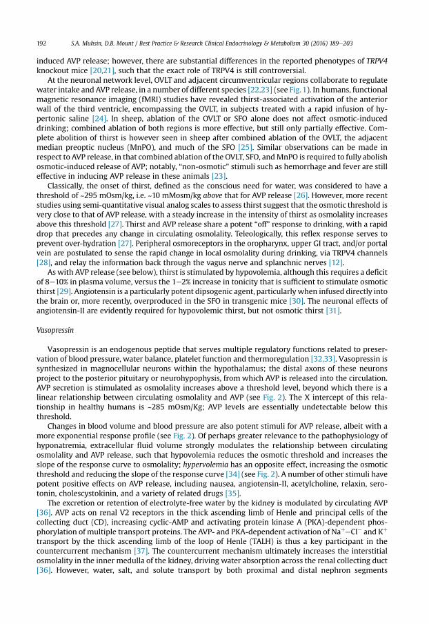
Vasopressin is an endogenous peptide that serves multiple regulatory functions related to preser-vation of blood pressure, water balance, platelet function and thermoregulation [32,33]. Vasopressin issynthesized in magnocellular neurons within the hypothalamus; the distal axons of these neuronsproject to the posterior pituitary or neurohypophysis, fromwhich AVP is released into the circulation.AVP secretion is stimulated as osmolality increases above a threshold level, beyond which there is alinear relationship between circulating osmolality and AVP (see Fig. 2). The X intercept of this rela-tionship in healthy humans is ~285 mOsm/Kg; AVP levels are essentially undetectable below thisthreshold.
Changes in blood volume and blood pressure are also potent stimuli for AVP release, albeit with amore exponential response profile (see Fig. 2). Of perhaps greater relevance to the pathophysiology ofhyponatremia, extracellular fluid volume strongly modulates the relationship between circulatingosmolality and AVP release, such that hypovolemia reduces the osmotic threshold and increases theslope of the response curve to osmolality; hypervolemia has an opposite effect, increasing the osmoticthreshold and reducing the slope of the response curve [34] (see Fig. 2). A number of other stimuli havepotent positive effects on AVP release, including nausea, angiotensin-II, acetylcholine, relaxin, sero-tonin, cholescystokinin, and a variety of related drugs [35].
The excretion or retention of electrolyte-free water by the kidney is modulated by circulating AVP[36]. AVP acts on renal V2 receptors in the thick ascending limb of Henle and principal cells of thecollecting duct (CD), increasing cyclic-AMP and activating protein kinase A (PKA)-dependent phos-phorylation of multiple transport proteins. The AVP- and PKA-dependent activation of NaþeCl� and Kþ
transport by the thick ascending limb of the loop of Henle (TALH) is thus a key participant in thecountercurrent mechanism [37]. The countercurrent mechanism ultimately increases the interstitialosmolality in the inner medulla of the kidney, driving water absorption across the renal collecting duct[36]. However, water, salt, and solute transport by both proximal and distal nephron segments

Fig. 2. A) A comparison of the response of circulating vasopressin to hemodynamic and osmotic stimulation in healthy adults. Theshaded area represents the reference range of plasma arginine vasopressin under normal conditions of hydration with plasmaosmolality varying from 284 to 293 mosmol/kg. (From Baylis PH, “Osmoregulation and control of vasopressin secretion in healthyhumans”, Am J Physiol 1987; 253:R671-8, with permission). B) The influence of hemodynamic status on osmotic stimulation ofvasopressin release in healthy adults. The heavy oblique line in the center depicts the relationship of plasma vasopressin toosmolality in normovolemic, normotensive subjects. Lighter lines to the left or right depict the relationship when blood volume and/or pressure are acutely decreased or increased by the different percentages indicated in the center circles. (From Robertson GL et al.,“The osmoregulation of vasopressin,” Kidney Int 1976; 10:25e37).
S.A. Muhsin, D.B. Mount / Best Practice & Research Clinical Endocrinology & Metabolism 30 (2016) 189e203 193
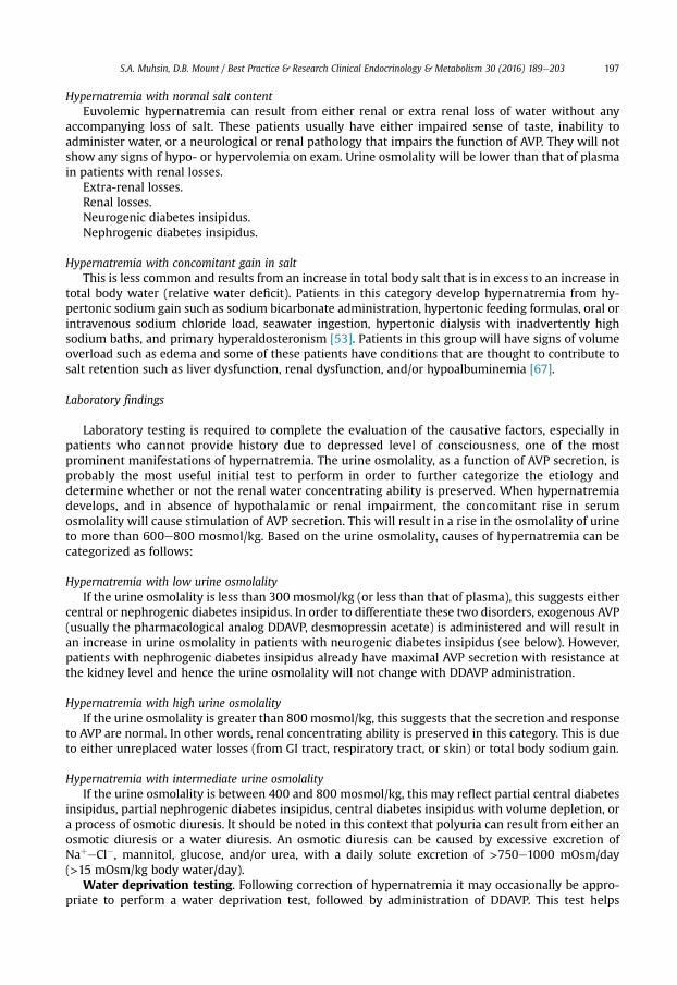
participates in the renal concentrating mechanism. Water transport across apical and basolateralaquaporin-1water channels in the descending thin limb of the loop of Henle is thus involved [38,39], asis passive absorption of NaþeCl� by the thin ascending limb, via apical and basolateral CLC-K1 chloridechannels and paracellular Naþ transport [40,41] (see Fig. 3). Renal urea transport in turn playsimportant roles in the generation of the medullary osmotic gradient and the ability to excrete solute-free water under conditions of both high and low protein intake [36] (see Fig. 3).
AVP-induced, PKA-dependent phosphorylation of the aquaporin-2 water channel in principal cellsstimulates the insertion of active water channels into the lumen of the collecting duct, resulting intransepithelial water absorption down the medullary osmotic gradient. Under “anti-diuretic” condi-tions, with increased circulating AVP, the kidney reabsorbs water filtered by the glomerulus,

Fig. 3. The renal concentrating mechanism. Water, salt, and solute transport by both proximal and distal nephron segmentsparticipates in the renal concentrating mechanism (see text for details). Diagram showing the location of the major transportproteins involved; a loop of Henle is depicted on the left, collecting duct on the right. UT, urea transporter; AQP, aquaporin; NKCC2,NaeKe2Cl cotransporter; ROMK, renal outer medullary K channel; CLC-K1, chloride channel. (With permission from Sands JM,“Molecular approaches to urea transporters,” J Am Soc Nephrol. 13(11):2795e806, 2002).
S.A. Muhsin, D.B. Mount / Best Practice & Research Clinical Endocrinology & Metabolism 30 (2016) 189e203194
equilibrating the osmolality across the collecting duct epithelium to excrete a hypertonic, “concen-trated” urine (osmolality of up to 1200 mOsm/kg). In the absence of circulating AVP, insertion ofaquaporin-2 channels andwater absorption across the collecting duct is essentially abolished, resultingin secretion of a hypotonic, dilute urine (osmolality as low as 30e50 mOsm/kg). Abnormalities in this“final common pathway” are involved in most disorders of water homeostasis, e.g., a reduced or absentinsertion of active aquaporin-2 water channels into themembrane of principal cells in both central andnephrogenic diabetes insipidus.
Cerebral adaptation to hypernatremia
Hypernatremia increases osmolality of the ECF, generating an osmotic gradient between the ECFand ICF, an efflux of intracellular water, and cellular shrinkage. Initially, hypernatremia results in areduced brain volume, which is reversed by cerebrospinal fluid movement into the brain with asubsequent increase in the interstitial volume [42,43], in addition to cellular uptake of solutes by thecell as part of the RVI response [42,44,45]. This RVI process initially involves the uptake of inorganicions (Naþ, Kþ, and Cl�) via transporters such as NKCC1 [46], followed by a more delayed accumulationof organic osmolytes, primarily myo-inositol, the amino acids glutamine, glutamate, and taurine[44,45,47,48]. Some of the relevant osmolytes transporters are induced in neurons by the osmo-sensitive transcription factor TonEBP [49], whereas the slow induction of osmolytes transporters inoligodendrocytes and glia likely occurs through transcriptional mechanisms that are independent ofTonEBP [48].

S.A. Muhsin, D.B. Mount / Best Practice & Research Clinical Endocrinology & Metabolism 30 (2016) 189e203 195
Etiology
Hypernatremia is usually the result of a combined water and electrolyte deficit, with losses of H2Oin excess of Naþ. This imbalance can also result from net water loss, which can be pure water or hy-potonic fluid loss, or less frequently from a gain of hypertonic sodium. In order for the hypernatremia tobe sustained, there needs to be a defect in the thirst mechanism or a lack of access to water. Notably,hypernatremia in the ICU is the predominant presentation, with the most common causes being a lackof provision of sufficient freewater, hypertonic sodium loading that occurs during volume resuscitationor hypertonic bicarbonate administration [50], suboptimal daily free water provision, and impairedrenal water conservation due to acute kidney injury and/or diuretic therapy. In the out-of-hospitalsetting, hypernatremia is most commonly associated with free water losses from non-renal sites[51,52]. The etiologies of hypernatremia are as follows [53]:
Hypernatremia from net water loss
This is the most common cause of hypernatremia and can be further subcategorized into renal andnon-renal losses.
Renal water lossesNeurogenic or central diabetes insipidus: this can result from traumatic brain injury, space
occupying lesions, or infections. Mutations in the AVP gene are associated with hereditary neurogenicdiabetes insipidus (Chapter *).
Nephrogenic diabetes insipidus: this can result from renal dysfunction, electrolyte perturbationssuch as hypercalcemia or hypokalemia, or medication effects such as lithium, foscarnet, amphotericin,vasopressin receptor antagonists, demeclocycline, methoxyflurane. Hereditary causes include loss-of-function mutations in the V2 vasopressin receptor gene, the aquaporin-2 gene, or aquaporin-1 [54](Chapter**).
Lithium is a particularly common cause of acquired nephrogenic DI (NDI). Lithium causes NDIvia direct inhibition of renal glycogen synthase kinase-3 (GSK3), a kinase thought to be the phar-macological target of lithium in psychiatric disease; renal GSK3 is required for the response ofprincipal cells to AVP [55]. Lithium also induces the expression of COX2 in the renal medulla [56];COX2-derived prostaglandins inhibit AVP-stimulated salt transport by the thick ascending limb [37]and AVP-stimulated water transport by the collecting duct [57], thus exacerbating lithium-associated polyuria. The entry of lithium through the amiloride-sensitive Naþ channel ENaC isrequired for the effect of the drug on principal cells [58,59], such that combined therapy withinlithium and amiloride can mitigate lithium-associated NDI [60]. However, lithium causes chronictubulointerstitial scarring and chronic kidney disease after prolonged therapy, such that patientsmay have a persistent NDI long after stopping the drug, with a reduced therapeutic benefit fromamiloride [61].
Renal losses: loop diuretics, osmotic diuresis.
Non renal water lossesHypodypsia: affected individuals demonstrate a lack of thirst despite hypertonicity. A variety of
infiltrative, neoplastic, vascular, congenital, and traumatic processes in this circumventricular regioncan be associated with abnormalities in thirst and AVP release. Patients with this “adipsic” or“essential” hypernatremia generally exhibit combined defects in both AVP release and thirst [62]. Insome cases, however, thirst is impaired but not AVP release [62], underscoring the functional redun-dancy and/or plasticity of the osmosensitive neuronal network; alternatively, the intrinsic osmo-sensitivity of the magnocellular neurons that synthesize and secrete AVP may preserve a residualosmotic-induced AVP release [23].
Unreplaced insensible losses from the respiratory system.Cutaneous losses: sweating, burns.Gastrointestinal losses: vomiting, diarrhea, nasogastric drainage, enterocutaneous fistula. Diarrhea
is the most common gastrointestinal cause of hypernatremia. Notably, osmotic diarrhea and viral

S.A. Muhsin, D.B. Mount / Best Practice & Research Clinical Endocrinology & Metabolism 30 (2016) 189e203196
gastroenteritides typically generate stools with Naþ and Kþ <100 mM, thus leading to water loss andhypernatremia; in contrast, secretory diarrhea typically results in isotonic stool and thus hypovolemiaþ/� hypovolemic hyponatremia.
Hypernatremia from hypertonic sodium gain
Sodium bicarbonate infusion. Ampules of sodium bicarbonate are approximately twice as hyper-tonic as hypertoic saline, such that administration of undiluted ampules leads to marked increases inserum sodium [50].
Feeding.Oral salt intake.Sea water ingestion.Hypertonic enemas.Hypertonic dialysis.Primary hyperaldosteronism.Cushing's syndrome.
Diagnosis
Thorough history, clinical examination and laboratory testing help establish the etiology ofhypernatremia.
History and physical examination
The history is essential to suggest the possible cause for the hypernatremia and also guidemanagement. Patients who have sustained a traumatic brain injury might suffer from central dia-betes insipidus, whereas those with a psychiatric illness with current or prior lithium treatment aremore likely to have a nephrogenic diabetes insipidus. History is also crucial in determining thechronicity of hypernatremia, as this will be helpful in making management decisions. Hyper-natremia that is thought to have developed within the previous 48 h is considered acute, whilepatients who have had symptoms for more than 48 h or those with unknown time of start ofsymptoms are considered to have chronic hypernatremia. Patients with acute hypernatremia willusually have more prominent symptoms than patients in whom hypernatremia develops over alonger period of time. These symptoms can include lethargy, weakness, and irritability and mayadvance to seizures and coma [63,64]. The sudden shrinkage of brain cells in acute hypernatremiamay lead to parenchymal or subarachnoid hemorrhages and/or subdural hematomas; however,these vascular complications are primarily encountered in pediatric and neonatal patients. Osmoticdamage to muscle membranes can also lead to hypernatremic rhabdomyolysis [65]. Chronichypernatremia will have less prominent symptoms, due primarily to the accumulation of intracel-lular osmolytes within the CNS.
Determining the patient's volume status is one of the most important steps in management ofhypernatremia. The main task is to identify whether there has been an accompanying loss of salt,through history and physical examination. Following the initial assessment, the patients can becategorized into one of the following three groups depending on their volume status:
Hypernatremia with concomitant loss of saltPatients with hypovolemic hypernatremia will have loss of both water and salt but with relatively
larger losses of water. The losses are usually from either the kidneys or from the gastrointestinal tract.The presenting symptoms and signs will be those of hypovolemia such as tachycardia, orthostatichypotension. Measurement of the urine sodium is useful to differentiate these two, as it will be lowwhen there is loss of sodium from the gastrointestinal tract reflecting an intact renal water conservingability. Notably, this physiology is not clear-cut, given the phenomenon of “dehydration-inducednatriuresis”, wherein increases in renal medullary tonicity cause a natriuresis to protect againstworsening hypertonicity [66].
S.A. Muhsin, D.B. Mount / Best Practice & Research Clinical Endocrinology & Metabolism 30 (2016) 189e203 197
Hypernatremia with normal salt contentEuvolemic hypernatremia can result from either renal or extra renal loss of water without any
accompanying loss of salt. These patients usually have either impaired sense of taste, inability toadminister water, or a neurological or renal pathology that impairs the function of AVP. They will notshow any signs of hypo- or hypervolemia on exam. Urine osmolality will be lower than that of plasmain patients with renal losses.
Extra-renal losses.Renal losses.Neurogenic diabetes insipidus.Nephrogenic diabetes insipidus.
Hypernatremia with concomitant gain in saltThis is less common and results from an increase in total body salt that is in excess to an increase in
total body water (relative water deficit). Patients in this category develop hypernatremia from hy-pertonic sodium gain such as sodium bicarbonate administration, hypertonic feeding formulas, oral orintravenous sodium chloride load, seawater ingestion, hypertonic dialysis with inadvertently highsodium baths, and primary hyperaldosteronism [53]. Patients in this group will have signs of volumeoverload such as edema and some of these patients have conditions that are thought to contribute tosalt retention such as liver dysfunction, renal dysfunction, and/or hypoalbuminemia [67].
Laboratory findings
Laboratory testing is required to complete the evaluation of the causative factors, especially inpatients who cannot provide history due to depressed level of consciousness, one of the mostprominent manifestations of hypernatremia. The urine osmolality, as a function of AVP secretion, isprobably the most useful initial test to perform in order to further categorize the etiology anddetermine whether or not the renal water concentrating ability is preserved. When hypernatremiadevelops, and in absence of hypothalamic or renal impairment, the concomitant rise in serumosmolality will cause stimulation of AVP secretion. This will result in a rise in the osmolality of urineto more than 600e800 mosmol/kg. Based on the urine osmolality, causes of hypernatremia can becategorized as follows:
Hypernatremia with low urine osmolalityIf the urine osmolality is less than 300 mosmol/kg (or less than that of plasma), this suggests either
central or nephrogenic diabetes insipidus. In order to differentiate these two disorders, exogenous AVP(usually the pharmacological analog DDAVP, desmopressin acetate) is administered and will result inan increase in urine osmolality in patients with neurogenic diabetes insipidus (see below). However,patients with nephrogenic diabetes insipidus already have maximal AVP secretion with resistance atthe kidney level and hence the urine osmolality will not change with DDAVP administration.
Hypernatremia with high urine osmolalityIf the urine osmolality is greater than 800 mosmol/kg, this suggests that the secretion and response
to AVP are normal. In other words, renal concentrating ability is preserved in this category. This is dueto either unreplaced water losses (from GI tract, respiratory tract, or skin) or total body sodium gain.
Hypernatremia with intermediate urine osmolalityIf the urine osmolality is between 400 and 800 mosmol/kg, this may reflect partial central diabetes
insipidus, partial nephrogenic diabetes insipidus, central diabetes insipidus with volume depletion, ora process of osmotic diuresis. It should be noted in this context that polyuria can result from either anosmotic diuresis or a water diuresis. An osmotic diuresis can be caused by excessive excretion ofNaþeCl�, mannitol, glucose, and/or urea, with a daily solute excretion of >750e1000 mOsm/day(>15 mOsm/kg body water/day).
Water deprivation testing. Following correction of hypernatremia it may occasionally be appro-priate to perform a water deprivation test, followed by administration of DDAVP. This test helps

S.A. Muhsin, D.B. Mount / Best Practice & Research Clinical Endocrinology & Metabolism 30 (2016) 189e203198
determine whether an inappropriate water diuresis is caused by central DI or NDI. The patient shouldbe water restricted beginning in the early morning, with careful monitoring of vital signs, weight, andhourly urine output; overnight water deprivation of patients with diabetes insipidus is unsafe andclinically inappropriate, given the potential for severe hypernatremia. Water deprivation is alsoinappropriate in patients who already have hypernatremia, wherein AVP secretion should already beactivated. The serum Naþ concentration - more accurate and more immediately available than serumosmolality e should be monitored hourly during water deprivation. A baseline AVP sample should bedrawn at the beginning of the test, with a second sample drawn once the serum Naþ reaches148e150 mEq/L. At this point a single 2 mg dose of the V2 vasopressin receptor agonist DDAVP can beadministered, followed by ongoing measurement of urine output, serum Naþ concentration, in addi-tion to urine and serum osmolality.
During water deprivation testing patients with nephrogenic DI will fail to respond to DDAVP, with aurine osmolality that increases by<50% or<150mOsm/kg from baseline, in combinationwith a normalor high circulating AVP level; patients with central DI will respond to DDAVP, with a reduced circulatingAVP. Patients may exhibit a partial response to DDAVP, with a >50% rise in urine osmolality thatnonetheless fails to reach 800 mOsm/kg; the level of circulating AVP will help differentiate the un-derlying cause, i.e., nephrogenic versus central DI. Patients with “partial NDI” can thus achieve urineosmolalities of 500e600 mOsm/kg after DDAVP treatment, but will not maximally concentrate theirurine to 800 mOsm/Kg or higher.
Treatment
The treatment of hypernatremia requires a comprehensive understanding of the predisposingmechanism. The most common form of hypernatremia is that due to water loss with impaired thirstmechanism or inability to administer water. Management is targeted towards treating the incitingfactor and correcting the hyperosmolality [53]. A stepwise approach is helpful in order to addressseveral considerations and can be summarized by the following points:
Identify and initiate treatment for the predisposing factor
Addressing the inciting factor is key in preventing further loss of water or hypertonic sodium gain.As mentioned in the “diagnosis” section, careful history, clinical examination to determine the volumestatus and measurement of urine sodium and osmolality will help further elucidate the etiology.Treatment might entail, for example, withholding loop diuretics, administering insulin in the case ofhyperglycemia, treating vomiting or diarrhea, withholding/adjusting hypertonic tube feeds, oradministering DDAVP for central diabetes insipidus. A key principle however, is that treatment of thecause of the hypernatremia should only be attempted once “eunatremia” has been established throughadequate and ongoing free water administration.
Determine whether the hypernatremia is acute or chronic
Attention should be made as to whether the hypernatremia has developed within the preceding48 h as the rate of correction can possibly be liberalized in patients with acute hypernatremia especiallyif they are presenting with symptoms. Patients with chronic hypernatremia require slower rates ofcorrection to avoid cerebral edema. Further details on the rate of correction are mentioned below.
Determine the amount of fluid to be replaced
Assess the need for volume resuscitationDetermining the patient's volume status is one of the most important steps in management of
hypernatremia. The main task here is to identify whether there has been an accompanying loss of salt,through history and physical examination as explained above. Patients with hypovolemia on presen-tation should be resuscitated with 0.9% sodium chloride regardless of the serum sodium level untiltheir vital signs are normal.

S.A. Muhsin, D.B. Mount / Best Practice & Research Clinical Endocrinology & Metabolism 30 (2016) 189e203 199
Calculate the water deficitThe mainstay of therapy for hypernatremia is to administer fluids that are dilute relative to plasma
in order to replace the water deficit. This requires the estimation of the amount of water that has beenlost. This can be simplified into the following equations by Adrogu�e and Madias [53].
Formula (1):
Water deficit ¼ TBW� ðplasma½Naþ �=140� 1Þ¼ ð0:4� 0:5Þ � lean body weight� ðplasma ½Naþ �=140� 1Þ (1)
Formula (2):
Change in Na ¼ ðinfusate Naþ�serum NaÞ=ðTBWþ 1Þ (2)
Assess for any ongoing losses that need to be replacedWater replacement should address the total body water deficit in addition to any ongoing losses of
water. Obligatory water losses from stool and sweat vary but are estimated to be 30e40 mL/h. Inaddition to insensible water output, ongoing water losses in the urine need to be accounted for whendeciding the infusion rate of the replacement fluid and this provides for the most accurate estimate ofthe sodium level resulting from a given amount of infusate [51]. The amount of pure water lost in urinecan be determined by calculating the electrolyte free water clearance (EFWC) [68] using the followingequation:
EFWC ¼ urine volume� ð1� ½urine Naþ urine K�=serum NaÞ ½42�:
If a patient is making 75 mL/h and his urinary sodium and potassium levels are summed to 128mEq/L with a serum sodium level 160mEq/L, the electrolyte freewater clearance for this patient equals75 mL/h � (1� [128 mEq/L/160 mEq/L]), which is 15 mL/h. In other words, in addition to the waterdeficit that will need to be replaced, an additional 15mL per hour of water will have to be administeredin order to reach the target sodium level.
Select the type and rate of replacement solution
The choice of replacement solution to be given and the infusion rate are important factors to avoidovercorrection of the hypernatremia [69]. Overcorrection of hypernatremia is associated withincreased risk of cerebral edema, due to the CNS response to hypertonicity. Classically, the recom-mendation is to replace the calculated free water deficit over 48 h. Notably, the plasma Naþ concen-tration should be corrected by no more than 10 mM/day, which may take longer than 48 h in patientswith severe hypernatremia (>160 mM). A rare exception is patients with acute hypernatremia (<48 h)due to sodium loading, who can safely be corrected rapidly at a rate of 1 mM/h. As in management ofhyponatremia, frequent measurement of serum Naþ is critical for monitoring the response to therapyand adjusting the rate or choice of intravenous fluid.
It should be noted that there have been a number of studies revealing that slower rates of correctionand persistent hypernatremia are associated with an increased risk of death. Multivariate analysis of aretrospective study of 131 patients hospitalized with severe hypernatremia thus showed that a slowerrate of correction of hypernatremia was an independent predictor of 30-day mortality [hazards ratio(HR), 3.85; P < 0.0001] [70]. Another recent retrospective study assessing outcomes in the emergencydepartment of 82 patients with hypernatremia (serum sodium, �150) in the emergency room showedthat slower rate of sodium correction, rather than the initial severity of the hyperosmorality wasassociated with an increased risk of death during hospitalization (HR, 10.29; P < 0.001) [71]. Except forhypovolemic patients requiring resuscitation with 0.9% sodium chloride, all other patients shouldreceive either 0.45% sodium chloride or 5% dextrose water infusions to replace the water deficit andongoing fluid losses.

S.A. Muhsin, D.B. Mount / Best Practice & Research Clinical Endocrinology & Metabolism 30 (2016) 189e203200
Other therapies
Patients with hypervolemic hypernatremia present a therapeutic challenge as the volumeexpansion in these patients inhibits the release of AVP, thereby promoting water excretion in theurine. Cessation of the inciting factors and administration of water is usually the initial step intherapy. Treatment with loop diuretics would enhance more aquaresis relative to natriuresis therebyexacerbating the hypertonicity. Infusing dextrose 5% water would address the hypertonicity butworsen the volume overload state. Simultaneous use of intravenous dextrose 5% water and loopdiuretics can however be used to lower the serum sodium in addition to achieving a net negativetotal body water balance. Nguyen and Kurtz derived an equation to determine the amount ofdextrose 5% water required to lower the serum sodium to a target level while maintaining a set netnegative water balance [72].
Hemodialysis has been used for treatment of hypernatremia, especially in cases where hydrationwith or without diuresis have failed to bring the sodium level down to the desired target or in patientsin whom there are indications for renal replacement therapy [73,74]. Continuous renal replacementtherapy has been reported to be useful in situations of hypernatremia and congestive heart failure [75].A recent retrospective cohort study evaluating 95 patients in the intensive care unit with acute severehypernatremia revealed that continuous veno-venous hemofiltration led to a greater reduction inserum sodium and was associated with an improved 28-day survival rate compared with conventionaltherapy using calculation of water deficit and administration of intravenous hypotonic fluids (34.8% vs.8.7% respectively, P.0.002) [76].
Specific treatment for neurogenic and nephrogenic diabetes insipidus
The main treatment modality for patients with central diabetes insipidus is to supplement anti-diuretic hormone. This is usually given in the form of DDAVP, a synthetic analog with a longer half-lifeandmostly V2 receptor agonism, thus with a dominance of antidiuretic rather than pressor effects [77].DDAVP can be administered by intravenous, subcutaneous, or intranasal routes.
The use of thiazide diuretics in diabetes insipidus has been shown to decrease urine volume andincrease urine osmolality [78]. The mechanism of the paradoxical antidiuretic effect of thiazides indiabetes insipidus is thought to be related to volume contraction, leading to an increase in proximaltubular reabsorption of water and sodium thereby decreasing distal delivery of water and subsequentexcretion [79,80]. However, it has been shown that volume repletion does not abrogate the antidiureticeffect of thiazides [81]. Thiazides also directly increase water absorption in perfused collecting ducts[82], even in the absence of circulating AVP. In an animal model of lithium-associated NDI, thiazidesincrease expression of aquaporin-2 [83]. Overall, however, we find very limited clinical utility forthiazides in acquired nephrogenic DI, wherein increased oral free water intake is usually sufficient toprevent hypernatremia and where patients often have chronic kidney disease (CKD) with greatersusceptibility to additive diuretic-induced renal insufficiency.
The entry of lithium through the amiloride-sensitive Naþ channel ENaC is required for the effect ofthe drug on principal cells [58,59], such that combined therapy within lithium and amiloride canmitigate lithium-associated NDI [60]. Concomitant therapy with amiloride is thus an attractive optionin themanagement of patients treated with lithium, however this approach has not gainedwidespreadacceptance.
Renal prostaglandins have been shown to play an important role in the pathogenesis of lithium-induced nephrogenic diabetes insipidus. Physiologically, renal prostaglandins exert antagonistic ef-fects on vasopressin mediated osmotic water flow [57]. Animal studies have shown that lithium in-duces the expression of cyclooxegenase 2 (COX2) in the medullary interstitial cell, via the inhibition ofglycogen synthase kinase-3beta (GSK-3beta), leading to increased levels of urinary prostaglandin E2[56]. COX2 inhibition resulted in a significant reduction in lithium-induced polyuria in this model [56].Clinical effects for NSAIDs or COX2 inhibitors have also been reported in humans lithium-associatedNDI. Again, however, as with thiazides our enthusiasm for COX2 inhibition in lithium-associated DIis minimal, since most patients have associated CKD and most patients can accommodate their defectby increased free water intake.

S.A. Muhsin, D.B. Mount / Best Practice & Research Clinical Endocrinology & Metabolism 30 (2016) 189e203 201
References
[1] Lindner G, Funk GC, Schwarz C, et al. Hypernatremia in the critically ill is an independent risk factor for mortality. Am JKidney Dis 2007;50:952e7.
*[2] Waite MD, Fuhrman SA, Badawi O, et al. Intensive care unit-acquired hypernatremia is an independent predictor ofincreased mortality and length of stay. J Crit Care 2013;28:405e12.
[3] Mendes RS, Soares M, Valente C, et al. Predialysis hypernatremia is a prognostic marker in acute kidney injury in need ofrenal replacement therapy. J Crit Care 2015;30:982e7.
[4] Leung AA, McAlister FA, Finlayson SR, et al. Preoperative hypernatremia predicts increased perioperative morbidity andmortality. Am J Med 2013;126:877e86.
[5] Bhave G, Neilson EG. Body fluid dynamics: back to the future. J Am Soc Nephrol 2011;22:2166e81.[6] Bhave G, Neilson EG. Volume depletion versus dehydration: how understanding the difference can guide therapy. Am J
Kidney Dis 2011;58:302e9.[7] Mount DB. Fluid and electrolyte disturbances. In: Loscalzo J, editor. Harrison's principles of internal medicine. 19 ed.
McGraw Hill; 2015. p. 295e312.[8] Verney EB. The antidiuretic hormone and the factors which determine its release. Proc R Soc Lond B Biol Sci 1947;135:
25e106.[9] McKinley MJ, Denton DA, Oldfield BJ, et al. Water intake and the neural correlates of the consciousness of thirst. Semin
Nephrol 2006;26:249e57.[10] Bourque CW, Ciura S, Trudel E, et al. Neurophysiological characterisation of osmosensitive neurons. Exp Physiol 2007;92:
499e505.[11] Sewards TV, Sewards MA. The awareness of thirst: proposed neural correlates. Conscious Cogn 2000;9:463e87.[12] Bourque CW. Central mechanisms of osmosensation and systemic osmoregulation. Nat Rev Neurosci 2008;9:519e31.[13] Zhang Z, Bourque CW. Osmometry in osmosensory neurons. Nat Neurosci 2003;6:1021e2.[14] Oliet SH, Bourque CW. Mechanosensitive channels transduce osmosensitivity in supraoptic neurons. Nature 1993;364:
341e3.[15] Zhang Z, Bourque CW. Calcium permeability and flux through osmosensory transduction channels of isolated rat su-
praoptic nucleus neurons. Eur J Neurosci 2006;23:1491e500.[16] Prager-Khoutorsky M, Bourque CW. Mechanical basis of osmosensory transduction in magnocellular neurosecretory
neurones of the rat supraoptic nucleus. J Neuroendocrinol 2015;27:507e15.[17] Ciura S, Bourque CW. Transient receptor potential vanilloid 1 is required for intrinsic osmoreception in organum vas-
culosum lamina terminalis neurons and for normal thirst responses to systemic hyperosmolality. J Neurosci 2006;26:9069e75.
[18] Sharif Naeini R, Witty MF, Seguela P, et al. An N-terminal variant of Trpv1 channel is required for osmosensory trans-duction. Nat Neurosci 2006;9:93e8.
*[19] Zaelzer C, Hua P, Prager-Khoutorsky M, et al. DeltaN-TRPV1: a molecular co-detector of body temperature and osmoticstress. Cell Rep 2015;13:23e30.
*[20] Liedtke W, Friedman JM. Abnormal osmotic regulation in trpv4-/- mice. Proc Natl Acad Sci U. S. A 2003;100:13698e703.[21] Mizuno A, Matsumoto N, Imai M, et al. Impaired osmotic sensation in mice lacking TRPV4. Am J Physiol Cell Physiol 2003;
285:C96e101.[22] McKinley MJ, Cairns MJ, Denton DA, et al. Physiological and pathophysiological influences on thirst. Physiol Behav 2004;
81:795e803.[23] McKinley MJ, Mathai ML, McAllen RM, et al. Vasopressin secretion: osmotic and hormonal regulation by the lamina
terminalis. J Neuroendocrinol 2004;16:340e7.[24] Egan G, Silk T, Zamarripa F, et al. Neural correlates of the emergence of consciousness of thirst. Proc Natl Acad Sci U. S. A
2003;100:15241e6.[25] McKinley MJ, Mathai ML, Pennington G, et al. Effect of individual or combined ablation of the nuclear groups of the
lamina terminalis on water drinking in sheep. Am J Physiol 1999;276:R673e83.[26] Robertson GL. Abnormalities of thirst regulation. Kidney Int 1984;25:460e9.[27] McKenna K, Thompson C. Osmoregulation in clinical disorders of thirst appreciation. Clin Endocrinol (Oxf) 1998;49:
139e52.*[28] Lechner SG, Markworth S, Poole K, et al. The molecular and cellular identity of peripheral osmoreceptors. Neuron 2011;
69:332e44.[29] Fitzsimons JT. Angiotensin, thirst, and sodium appetite. Physiol Rev 1998;78:583e686.*[30] Sakai K, Agassandian K, Morimoto S, et al. Local production of angiotensin II in the subfornical organ causes elevated
drinking. J Clin Invest 2007;117:1088e95.[31] McKinley MJ, Walker LL, Alexiou T, et al. Osmoregulatory fluid intake but not hypovolemic thirst is intact in mice lacking
angiotensin. Am J Physiol Regul Integr Comp Physiol 2008;294:R1533e43.[32] Ishikawa SE, Schrier RW. Pathophysiological roles of arginine vasopressin and aquaporin-2 in impaired water excretion.
Clin Endocrinol (Oxf) 2003;58:1e17.*[33] Schrier RW. Vasopressin and aquaporin 2 in clinical disorders of water homeostasis. Semin Nephrol 2008;28:289e96.[34] Robertson GL, Aycinena P, Zerbe RL. Neurogenic disorders of osmoregulation. Am J Med 1982;72:339e53.[35] Verbalis JG, Berl T. Disorders of water balance. In: Brenner BM, editor. The kidney. 8th ed. Philadelphia, PA: Saunders;
2008. p. 459e546.[36] Sands JM, Layton HE. Advances in understanding the urine-concentrating mechanism. Annu Rev Physiol 2014;76:
387e409.[37] Mount DB. Thick ascending limb of the loop of Henle. Annu Rev Physiol 2014;9:1974e86.[38] Ma T, Yang B, Gillespie A, et al. Severely impaired urinary concentrating ability in transgenic mice lacking aquaporin-1
water channels. J Biol Chem 1998;273:4296e9.

S.A. Muhsin, D.B. Mount / Best Practice & Research Clinical Endocrinology & Metabolism 30 (2016) 189e203202
[39] Chou CL, Knepper MA, Hoek AN, et al. Reduced water permeability and altered ultrastructure in thin descending limb ofHenle in aquaporin-1 null mice. J Clin Invest 1999;103:491e6.
[40] Matsumura Y, Uchida S, Kondo Y, et al. Overt nephrogenic diabetes insipidus in mice lacking the CLC-K1 chloride channel.Nat Genet 1999;21:95e8.
[41] Liu W, Morimoto T, Kondo Y, et al. Analysis of NaCl transport in thin ascending limb of Henle's loop in CLC-K1 null mice.Am J Physiol Ren Physiol 2002;282:F451e7.
[42] Strange K. Regulation of solute and water balance and cell volume in the central nervous system. J Am Soc Nephrol 1992;3:12e27.
[43] Pullen RG, DePasquale M, Cserr HF. Bulk flow of cerebrospinal fluid into brain in response to acute hyperosmolality. Am JPhysiol 1987;253:F538e45.
*[44] Heilig CW, Stromski ME, Blumenfeld JD, et al. Characterization of the major brain osmolytes that accumulate in salt-loaded rats. Am J Physiol 1989;257:F1108e16.
*[45] Lien YH, Shapiro JI, Chan L. Effects of hypernatremia on organic brain osmoles. J Clin Invest 1990;85:1427e35.[46] O'Neill WC. Physiological significance of volume-regulatory transporters. Am J Physiol 1999;276:C995e1011.[47] Paredes A, McManus M, Kwon HM, et al. Osmoregulation of Na(þ)-inositol cotransporter activity and mRNA levels in
brain glial cells. Am J Physiol 1992;263:C1282e8.[48] Maallem S, Mutin M, Gonzalez-Gonzalez IM, et al. Selective tonicity-induced expression of the neutral amino-acid
transporter SNAT2 in oligodendrocytes in rat brain following systemic hypertonicity. Neuroscience 2008;153:95e107.
[49] Loyher ML, Mutin M, Woo SK, et al. Transcription factor tonicity-responsive enhancer-binding protein (TonEBP) whichtransactivates osmoprotective genes is expressed and upregulated following acute systemic hypertonicity in neurons inbrain. Neuroscience 2004;124:89e104.
[50] Blumberg A, Weidmann P, Shaw S, et al. Effect of various therapeutic approaches on plasma potassium and majorregulating factors in terminal renal failure. Am J Med 1988;85:507e12.
*[51] Lindner G, Schwarz C, Kneidinger N, et al. Can we really predict the change in serum sodium levels? an analysis ofcurrently proposed formulae in hypernatraemic patients. Nephrol Dial Transpl 2008;23:3501e8.
[52] Palevsky PM, Bhagrath R, Greenberg A. Hypernatremia in hospitalized patients. Ann Intern Med 1996;124:197e203.[53] Adrogue HJ, Madias NE. Hypernatremia. N Engl J Med 2000;342:1493e9.[54] King LS, Choi M, Fernandez PC, et al. Defective urinary-concentrating ability due to a complete deficiency of aquaporin-1.
N Engl J Med 2001;345:175e9.[55] Rao R, Patel S, Hao C, et al. GSK3beta mediates renal response to vasopressin by modulating adenylate cyclase activity.
J Am Soc Nephrol 2010;21:428e37.[56] Rao R, Zhang MZ, Zhao M, et al. Lithium treatment inhibits renal GSK-3 activity and promotes cyclooxygenase 2-
dependent polyuria. Am J Physiol Ren Physiol 2005;288:F642e9.[57] Stokes JB. Integrated actions of renal medullary prostaglandins in the control of water excretion. Am J Physiol 1981;240:
F471e80.[58] Kortenoeven ML, Li Y, Shaw S, et al. Amiloride blocks lithium entry through the sodium channel thereby attenuating the
resultant nephrogenic diabetes insipidus. Kidney Int 2009;76:44e53.[59] Christensen BM, Zuber AM, Loffing J, et al. alphaENaC-mediated lithium absorption promotes nephrogenic diabetes
insipidus. J Am Soc Nephrol 2011;22:253e61.*[60] Bedford JJ, Weggery S, Ellis G, et al. Lithium-induced nephrogenic diabetes insipidus: renal effects of amiloride. Clin J Am
Soc Nephrol 2008;3:1324e31.[61] Batlle DC, von Riotte AB, Gaviria M, et al. Amelioration of polyuria by amiloride in patients receiving long-term lithium
therapy. N Engl J Med 1985;312:408e14.[62] Baylis PH, Thompson CJ. Osmoregulation of vasopressin secretion and thirst in health and disease. Clin Endocrinol (Oxf)
1988;29:549e76.[63] Arieff AI, Guisado R. Effects on the central nervous system of hypernatremic and hyponatremic states. Kidney Int 1976;10:
104e16.[64] Snyder NA, Feigal DW, Arieff AI. Hypernatremia in elderly patients. A heterogeneous, morbid, and iatrogenic entity. Ann
Intern Med 1987;107:309e19.[65] Denman JP. Hypernatraemia and rhabdomyolysis. Med J Aust 2007;187:527e8.[66] Chen S, Grigsby CL, Law CS, et al. Tonicity-dependent induction of Sgk1 expression has a potential role in dehydration-
induced natriuresis in rodents. J Clin Invest 2009;119:1647e58.[67] Kahn T. Hypernatremia with edema. Arch Intern Med 1999;159:93e8.[68] Rose BD. New approach to disturbances in the plasma sodium concentration. Am J Med 1986;81:1033e40.[69] Kumar S, Berl T. Sodium. Lancet 1998;352:220e8.[70] Alshayeb HM, Showkat A, Babar F, et al. Severe hypernatremia correction rate and mortality in hospitalized patients. Am J
Med Sci 2011;341:356e60.[71] Bataille S, Baralla C, Torro D, et al. Undercorrection of hypernatremia is frequent and associated with mortality. BMC
Nephrol 2014;15:37.[72] Nguyen MK, Kurtz I. Correction of hypervolaemic hypernatraemia by inducing negative Naþ and Kþ balance in excess of
negative water balance: a new quantitative approach. Nephrol Dial Transpl 2008;23:2223e7.[73] Choi JH, Lee HS, Kim SM, et al. Paranoid Adipsia-induced severe hypernatremia and uremia treated with hemodialysis.
Electrolyte Blood Press 2013;11:29e32.[74] Nur S, Khan Y, Nur S, et al. Hypernatremia: correction rate and hemodialysis. Case Rep Med 2014;2014:736073.[75] Park HS, Hong YA, Kim HG, et al. Usefulness of continuous renal replacement therapy for correcting hypernatremia in a
patient with severe congestive heart failure. Hemodial Int 2012;16:559e63.[76] Ma F, Bai M, Li Y, et al. Continuous venovenous hemofiltration (CVVH) versus conventional treatment for acute severe
hypernatremia in critically ill patients: a retrospective study. Shock 2015;44:445e51.[77] Richardson DW, Robinson AG. Desmopressin. Ann Intern Med 1985;103:228e39.

S.A. Muhsin, D.B. Mount / Best Practice & Research Clinical Endocrinology & Metabolism 30 (2016) 189e203 203
[78] Crawford JD, Kennedy GC. Chlorothiazid in diabetes insipidus. Nature 1959;183:891e2.[79] Shirley DG, Walter SJ, Laycock JF. The antidiuretic effect of chronic hydrochlorothiazide treatment in rats with diabetes
insipidus: renal mechanisms. Clin Sci (Lond) 1982;63:533e8.[80] Shirley DG, Walter SJ, Thomsen K. A comparison of micropuncture and lithium clearance methods in the assessment of
renal tubular function in rats with diabetes insipidus. Pflugers Arch 1983;399:266e70.[81] Spannow J, Thomsen K, Petersen JS, et al. Influence of renal nerves and sodium balance on the acute antidiuretic effect of
bendroflumethiazide in rats with diabetes insipidus. J Pharmacol Exp Ther 1997;282:1155e62.[82] Cesar KR, Magaldi AJ. Thiazide induces water absorption in the inner medullary collecting duct of normal and Brattleboro
rats. Am J Physiol 1999;277:F756e60.[83] Kim GH, Lee JW, Oh YK, et al. Antidiuretic effect of hydrochlorothiazide in lithium-induced nephrogenic diabetes
insipidus is associated with upregulation of aquaporin-2, Na-Cl co-transporter, and epithelial sodium channel. J Am SocNephrol 2004;15:2836e43.