DIAGNOSIS AND SUCCESSFUL LAPAROSCOPIC...
17
DIAGNOSIS AND SUCCESSFUL LAPAROSCOPIC MANAGEMENT OF PREGNANCY IN A RUDIMENTARY HORN: AN INTERESTING CASE REPORT INTRODUCTION Mullerian duct anomalies have been reported to occur in 3.2% of women (Simon et al., 1991). They can be diagnosed at different stages of life. The presentation can vary from a mass resulting from an obstructed mullerian system in an infant (mucocolpos), primary amenorrhea, cyclical pain with mass per abdomen (hematocolpos), to infertility or bad obstetric history (Raga et al 1997., Golan et al., 1989 Johansen et al., 1983). A unicornuate uterus is one such anomaly which results from the failure of lateral fusion of the malformed duct with the contralateral duct. The malformed duct presents as a rudimentary horn which may or may not consist of a functional endometrial cavity. The presence of functional endometrium in a non- communicating rudimentary horn may further complicate the condition by causing cryptomenorrhea, hematocolpos or retrograde menstruation leading to pelvic endometriosis. Pregnancy in a non- communicating rudimentary horn is a rare form of ectopic gestation and its incidence is between 1/100,000 to 1/140,000 pregnancies (Heinonen, 1997). The only possible explanation of this phenomenon is by transperitoneal migration of sperm, ovum or zygote. The rudimentary horn pregnancy (RHP) usually culminates in rupture of the pregnant horn. Unlike other ectopic gestations, rupture is usually seen in the second or third trimester because of the thick uterine horn musculature. It leads to life threatening intra-peritoneal bleeding and hemorrhagic shock. Therefore, early pre-rupture diagnosis is of utmost importance for the maternal well being. Here, we report a case of RHP diagnosed sonographically leading to successful laparoscopic management. CASE REPORT A 34 year old lady came to our hospital in January 2009 with a history of 35 days amenorrhea. Her urine pregnancy test was positive and she wanted the present pregnancy to be terminated. As per her obstetric history, she had previous two missed abortions at 60-65 days of pregnancy evacuated by dilatation and curettage. The third pregnancy resulted in a successful caesarean delivery at term. Per operative findings at that time confirmed the presence of a unicornuate uterus with a right rudimentary horn. She had no relevant medical history. The transvaginal sonography (TVS) revealed a good endometrial reaction in the non communicating rudimentary horn while the uterus demonstrated a thin endometrium with a corresponding corpus luteum of the right ovary. Serum beta hCG was advised for the patient and the value was 7694.2mIU/ml. A provisional diagnosis of pregnancy in the right rudimentary horn was made. She was advised stringent follow up at 40 days to visualize a sac in the rudimentary horn or an ectopic pregnancy. But the patient was unwilling for any follow up and left the hospital. She subsequently came for review after 10 days at 45 days of amenorrhea when the TVS showed a gestational sac with yolk sac and cardiac activity in the right horn. A ring of myometrium was seen surrounding the gestation sac and there was no continuity observed between the endometrium lining the gestational sac with that of the unicornuate uterus. These two sonographic features further supported our pre operative diagnosis of rudimentary horn pregnancy (RHP). The patient was counseled and laparoscopy was done. The per- operative findings confirmed the presence of an enlarged and congested right rudimentary horn with an ectopic gestation (Fig 1 & 2). The pregnant horn was attached to the uterus with a fibrotic band. Bilateral adnexae appeared normal with the presence of a corpus luteum in the right ovary (Fig 2). Laparoscopic excision of the pregnant horn with ipsilateral salpingo-oopherectomy was performed with the help of harmonic scalpel and bipolar electrocautery (Fig 3 & Fig 4). The excised specimen was sent for histopathological examination which confirmed the diagnosis of a RHP. The patient recovered well and post operative period was uneventful.
Transcript of DIAGNOSIS AND SUCCESSFUL LAPAROSCOPIC...
DIAGNOSIS AND SUCCESSFUL LAPAROSCOPIC MANAGEMENT OF PREGNANCY IN A RUDIMENTARY HORN: AN INTERESTING CASE REPORT INTRODUCTION Mullerian duct anomalies have been reported to occur in 3.2% of women (Simon et al., 1991). They can be
diagnosed at different stages of life. The presentation can vary from a mass resulting from an obstructed
mullerian system in an infant (mucocolpos), primary amenorrhea, cyclical pain with mass per abdomen
(hematocolpos), to infertility or bad obstetric history (Raga et al 1997., Golan et al., 1989 Johansen et al.,
1983). A unicornuate uterus is one such anomaly which results from the failure of lateral fusion of the
malformed duct with the contralateral duct. The malformed duct presents as a rudimentary horn which may
or may not consist of a functional endometrial cavity. The presence of functional endometrium in a non-
communicating rudimentary horn may further complicate the condition by causing cryptomenorrhea,
hematocolpos or retrograde menstruation leading to pelvic endometriosis.
Pregnancy in a non- communicating rudimentary horn is a rare form of ectopic gestation and its incidence
is between 1/100,000 to 1/140,000 pregnancies (Heinonen, 1997). The only possible explanation of this
phenomenon is by transperitoneal migration of sperm, ovum or zygote. The rudimentary horn pregnancy
(RHP) usually culminates in rupture of the pregnant horn. Unlike other ectopic gestations, rupture is
usually seen in the second or third trimester because of the thick uterine horn musculature. It leads to life
threatening intra-peritoneal bleeding and hemorrhagic shock. Therefore, early pre-rupture diagnosis is of
utmost importance for the maternal well being. Here, we report a case of RHP diagnosed sonographically
leading to successful laparoscopic management.
CASE REPORT A 34 year old lady came to our hospital in January 2009 with a history of 35 days amenorrhea. Her urine
pregnancy test was positive and she wanted the present pregnancy to be terminated. As per her obstetric
history, she had previous two missed abortions at 60-65 days of pregnancy evacuated by dilatation and
curettage. The third pregnancy resulted in a successful caesarean delivery at term. Per operative findings at
that time confirmed the presence of a unicornuate uterus with a right rudimentary horn. She had no
relevant medical history.
The transvaginal sonography (TVS) revealed a good endometrial reaction in the non communicating
rudimentary horn while the uterus demonstrated a thin endometrium with a corresponding corpus luteum
of the right ovary. Serum beta hCG was advised for the patient and the value was 7694.2mIU/ml. A
provisional diagnosis of pregnancy in the right rudimentary horn was made. She was advised stringent
follow up at 40 days to visualize a sac in the rudimentary horn or an ectopic pregnancy. But the patient was
unwilling for any follow up and left the hospital. She subsequently came for review after 10 days at 45
days of amenorrhea when the TVS showed a gestational sac with yolk sac and cardiac activity in the right
horn. A ring of myometrium was seen surrounding the gestation sac and there was no continuity observed
between the endometrium lining the gestational sac with that of the unicornuate uterus. These two
sonographic features further supported our pre operative diagnosis of rudimentary horn pregnancy (RHP).
The patient was counseled and laparoscopy was done.
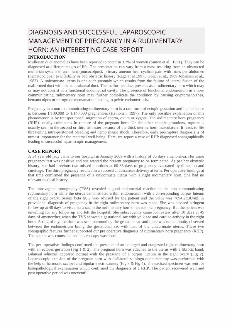
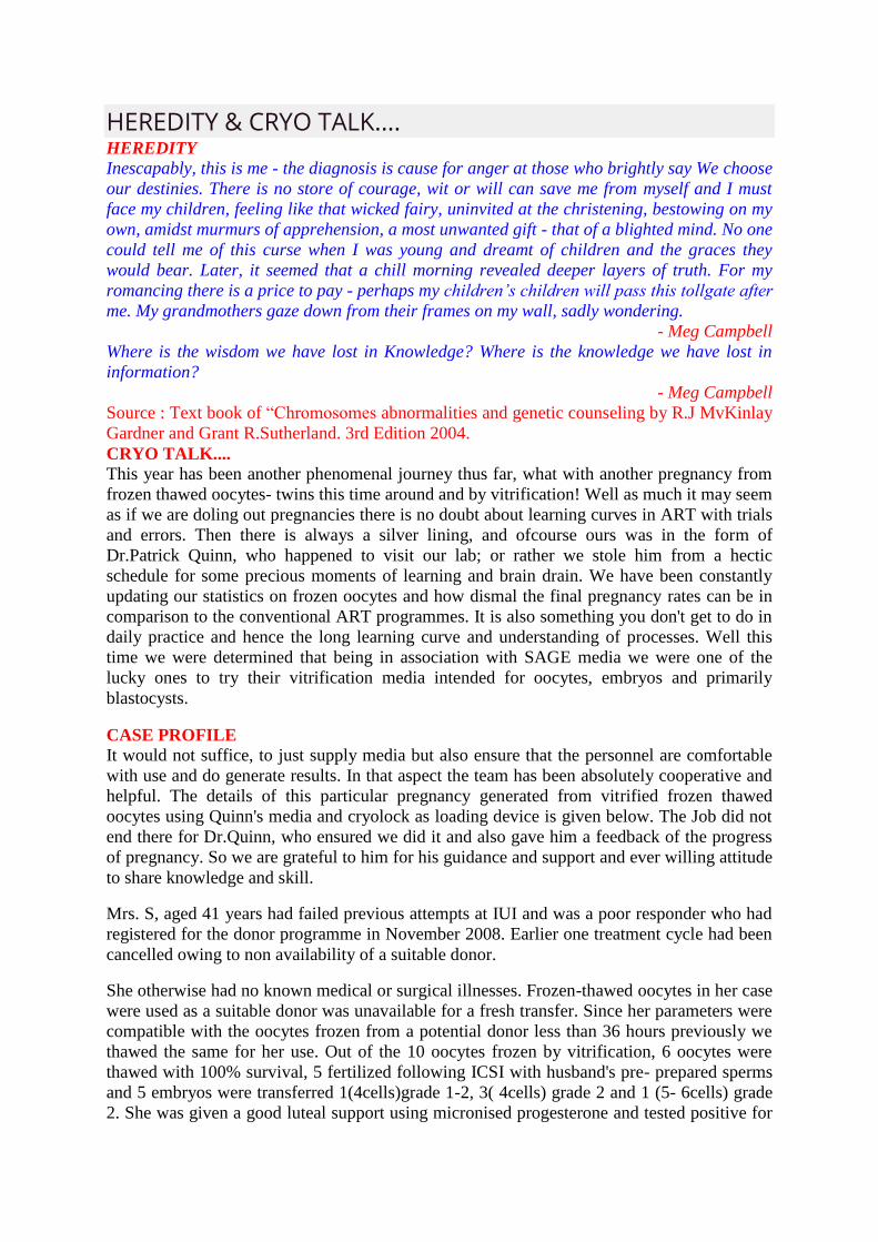
The per- operative findings confirmed the presence of an enlarged and congested right rudimentary horn
with an ectopic gestation (Fig 1 & 2). The pregnant horn was attached to the uterus with a fibrotic band.
Bilateral adnexae appeared normal with the presence of a corpus luteum in the right ovary (Fig 2).
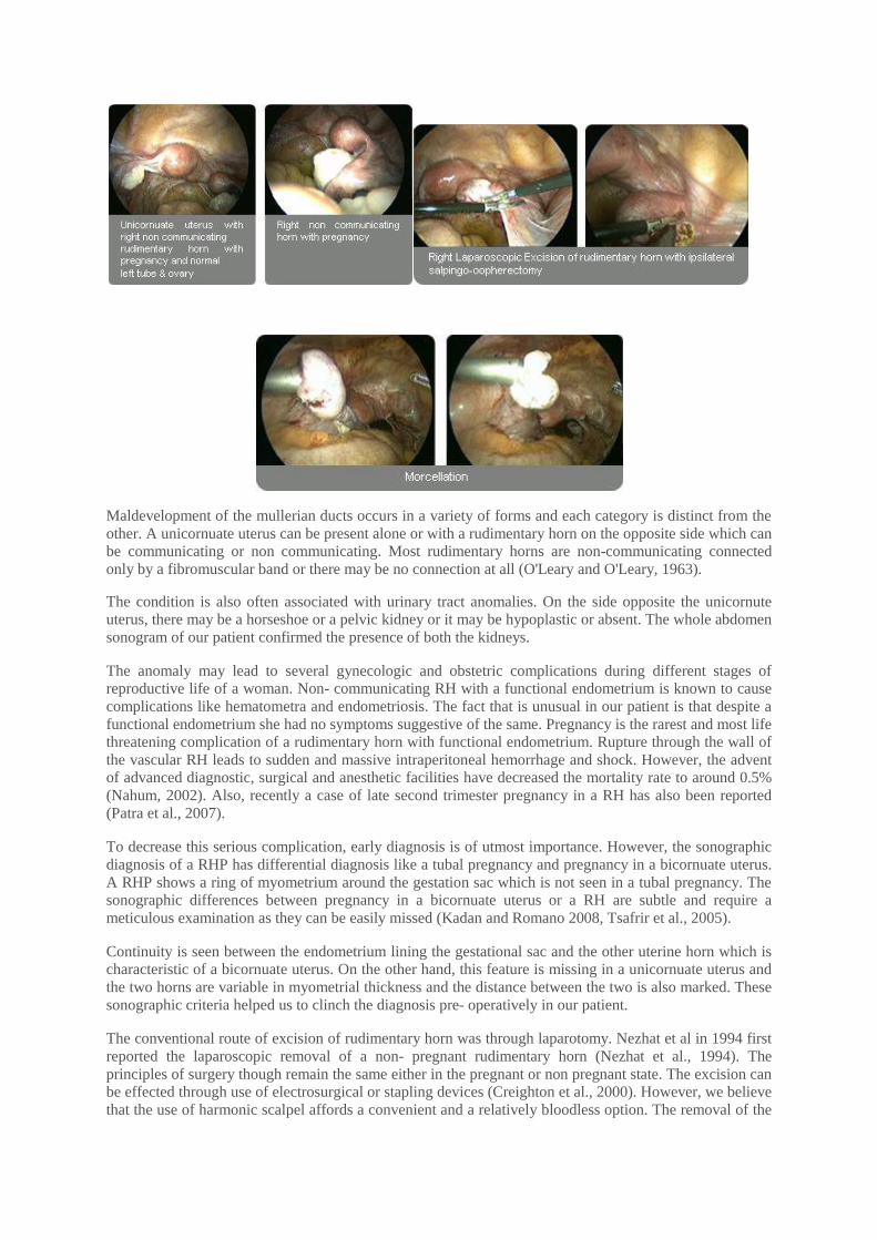
Laparoscopic excision of the pregnant horn with ipsilateral salpingo-oopherectomy was performed with
the help of harmonic scalpel and bipolar electrocautery (Fig 3 & Fig 4). The excised specimen was sent for
histopathological examination which confirmed the diagnosis of a RHP. The patient recovered well and
post operative period was uneventful.
Maldevelopment of the mullerian ducts occurs in a variety of forms and each category is distinct from the
other. A unicornuate uterus can be present alone or with a rudimentary horn on the opposite side which can
be communicating or non communicating. Most rudimentary horns are non-communicating connected
only by a fibromuscular band or there may be no connection at all (O'Leary and O'Leary, 1963).
The condition is also often associated with urinary tract anomalies. On the side opposite the unicornute
uterus, there may be a horseshoe or a pelvic kidney or it may be hypoplastic or absent. The whole abdomen
sonogram of our patient confirmed the presence of both the kidneys.
The anomaly may lead to several gynecologic and obstetric complications during different stages of
reproductive life of a woman. Non- communicating RH with a functional endometrium is known to cause
complications like hematometra and endometriosis. The fact that is unusual in our patient is that despite a
functional endometrium she had no symptoms suggestive of the same. Pregnancy is the rarest and most life
threatening complication of a rudimentary horn with functional endometrium. Rupture through the wall of
the vascular RH leads to sudden and massive intraperitoneal hemorrhage and shock. However, the advent
of advanced diagnostic, surgical and anesthetic facilities have decreased the mortality rate to around 0.5%
(Nahum, 2002). Also, recently a case of late second trimester pregnancy in a RH has also been reported
(Patra et al., 2007).
To decrease this serious complication, early diagnosis is of utmost importance. However, the sonographic
diagnosis of a RHP has differential diagnosis like a tubal pregnancy and pregnancy in a bicornuate uterus.
A RHP shows a ring of myometrium around the gestation sac which is not seen in a tubal pregnancy. The
sonographic differences between pregnancy in a bicornuate uterus or a RH are subtle and require a
meticulous examination as they can be easily missed (Kadan and Romano 2008, Tsafrir et al., 2005).
Continuity is seen between the endometrium lining the gestational sac and the other uterine horn which is
characteristic of a bicornuate uterus. On the other hand, this feature is missing in a unicornuate uterus and
the two horns are variable in myometrial thickness and the distance between the two is also marked. These
sonographic criteria helped us to clinch the diagnosis pre- operatively in our patient.
The conventional route of excision of rudimentary horn was through laparotomy. Nezhat et al in 1994 first
reported the laparoscopic removal of a non- pregnant rudimentary horn (Nezhat et al., 1994). The
principles of surgery though remain the same either in the pregnant or non pregnant state. The excision can
be effected through use of electrosurgical or stapling devices (Creighton et al., 2000). However, we believe
that the use of harmonic scalpel affords a convenient and a relatively bloodless option. The removal of the
uterine horn with the help of a morcellator is quick and also eliminates the need of an extra colpotomy
incision or extension of the suprapubic incision.
Rudimentary horn pregnancy is a rare and serious condition and every attempt should be made to diagnose
the condition at the earliest. To the best of our knowledge, this case is one of the few where the
preoperative sonographic diagnosis of the same was made before rupture and managed laparoscopically.
The meticulous examination of a patient suspected with rudimentary horn pregnancy using the non
invasive modality of ultrasound can go a long way in the early diagnosis and management.
REFERENCES 1. Simon C, Martinez L, Pardo F, et al. 1991. Mullerian defects in women with normal reproductive
outcome. Fertility and Sterility. 56,1192-3.
2. Raga F, Bauset C, Remohi J, Bonilla- Musoles F, Simon C, Pellicer A. 1997. Reproductive impact of
congenital Mullerian anomalies. Human Reproduction 12, 2277 81.
3. Golan A, Langer R, Bukovsky I, Caspi E 1989. Congenital anomalies of the Mullerian system. Fertility
and Sterility 51,74755.
4. Johansen K. 1983. Pregnancy in a rudimentary horn. Obstetrics Gynecology 3, 61:565-7
5. Heinonen PK 1997. Unicornuate uterusand rudimentary horn. Fertility and Sterility 68, 224 30.
6. O'Leary JL, O'Leary JA. 1963. Rudimentary Horn Pregnancy. Obstetrics Gynecology 22, 371-5.
7. Nahum GG 2002. Rudimentary uterine horn pregnancy. The 20th-century worldwide experience of 588
cases. The Journal of reproductive medicine. 47,151-63.
8. Patra S, Puri M, Trivedi SS, Yadav R, Bali J 2007.Unruptured term pregnancy with a live fetus with
placenta percreta in a non- communicating rudimentary horn. Congenitalanomalies, 47: 156-7.
9. Kadan Y, Romano S 2008. Rudimentary horn pregnancy diagnosed by ultrasound and treated by
laparoscopy--a case report and review of the literature. Journal of minimally invasive gynecology 15, 527-
30.
10. Tsafrir A, Rojansky N, Sela HY, Gomori JM, Nadjari M 2005. Rudimentary horn pregnancy: first-
trimester prerupture sonographic diagnosis and confirmation by magnetic resonance imaging. Journal of
ultrasound in medicine 24, 219-23.
11. Nezhat F, Nezhat C, Bess O, Nezhat CH 1994. Laparoscopic amputation of a noncommunicating
rudimentary horn after a hysteroscopic diagnosis: a case study. Surgical laparoscopy & endoscopy 2, 1556.
Dr. Priya Selvaraj MD MNAMS MCE
Dr. Gunjan Singh MD DNB MRCOG (I)
Dr. Megha Agrawal MS G G Hospital
KARYOTYPE POLYMORPHISM AND SIGNIFICANCE IN INFERTILITY There are certain influences which have been documented to have an effect on the genetic conduct of a
conception be it natural or assisted. These are namely consanguinity, inheritable 30% disorders, sex linked
disorders, hormonal imbalance and environmental factors. The end result of any of these could be repeated
miscarriages, phenotypic abnormalities, metabolic syndromes or carcinogenesis. In an assisted conception
unit we attempt to overcome these by adequate counseling for options like PGD and selective embryo
transfers, donor programmes, weight loss and dietary rectification, removal of environmental hazards and
adoption. Given all this, let us take the situation of idiopathic infertility where there are no known causes
actually preventing conception, but requiring assistance of some form.
Can there be the existence of a chromosomal element either in the form of inversions, deletions and
translocations in these otherwise normal couples with no other demonstrable causes of infertility? If so,
what influence does it have on fertilization and conception? Can this be implicated as one of the reasons
for not conceiving? Literature has revealed that in patients who have simple fertilization failure deemed as
idiopathic, the proportion of women with chromosomal abnormalities was higher in comparison to their
male counterparts, about 6.4%.
When a significant male factor was present, the incidence of chromosomal abnormalities in women was
2.5%. Stern et al noted the rate of balanced reciprocal and robertsonian carriers to be 3% in those who had
failed more than 10 attempts at embryo transfers (1) . In yet another series, the incidence of non autosomal
translocation carriers in women who failed IVF attempts more than 15 times was 8% (Raziel et al 2002 )
(2). While a fraction of cases of male infertility do have presence of Y chromosome microdeletions yet
another group have normal karyotypes. However there is an increased sperm aneuploidy/diploidy rate with
sex chromosomes being most affected.
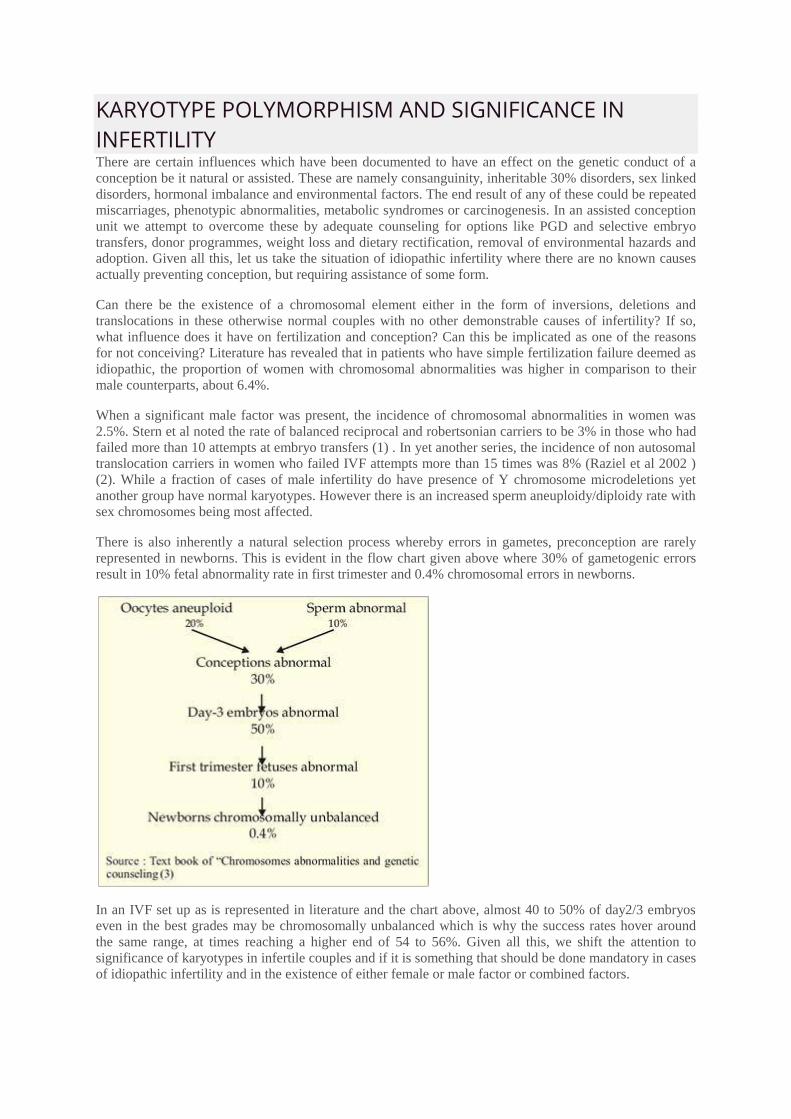
There is also inherently a natural selection process whereby errors in gametes, preconception are rarely
represented in newborns. This is evident in the flow chart given above where 30% of gametogenic errors
result in 10% fetal abnormality rate in first trimester and 0.4% chromosomal errors in newborns.
In an IVF set up as is represented in literature and the chart above, almost 40 to 50% of day2/3 embryos
even in the best grades may be chromosomally unbalanced which is why the success rates hover around
the same range, at times reaching a higher end of 54 to 56%. Given all this, we shift the attention to
significance of karyotypes in infertile couples and if it is something that should be done mandatory in cases
of idiopathic infertility and in the existence of either female or male factor or combined factors.

We are in the initial stages of our study in this regard, where 9 men of idiopathic infertility, demonstrated
karyotype polymorphism namely interstitial deletions of long arm of Y chromosome (Yqh), translocations
and increase in heterochromatin or satellite on either long or short arms of autosomes or Y chromosome.
These men had normal semen parameters but whose wives had not conceived at all. It is known that
translocations are associated with infertility, miscarriage and fetal abnormalities but how many of these
men are prone to contribute to the above mishaps? What are the risks in conception and transmission?
According to vegetti et al (2000), men who fathered children with abnormal karyotypes through ICSI did
not demonstrate a higher rate of aneuploidy when tested by FISH in comparison to men with similar male
factor but showed a higher incidence when compared to normal population.
At our centre, the incidence of either partner demonstrating a karyotype deviation is 8.51% while both
partners is 1.31%. A comment of normal variation needs to be ascertained as opposed to the suggestion for
prenatal fetal Karyotyping. The outcomes for the above mentioned karyotypes are available for three
couples where pregnancies are in the first trimester with no demonstrable anomalies thus far.
It remains to be seen as to how many of these couples would be influenced by a karyotype in achieving a
normal conception. We have started to collect our data and are curious about polymorphisms involving one
or both partners. According to literature those involving chromosome 9 are associated with infertility while
it has been rarely reported for 21. We seem to be dealing with more cases involving the latter. This was
written to spark off ideas and hopefully we should be able to publish good data. We also need to look at a
control group to actually determine the significance-namely couples who have conceived with karyotypic
polymorphism and their outcomes.
REFERENCES 1. Stern C., Pertile, M., Norris, H, L. And Baker, H.W.G. (1999). Chromosome translocations in couples
with in-vitro fertilization implantation failure. Hum.Repord., 14, 2097-2101.
2. Raziel, A., Friedler,S., Schachter, M., Kasterstein, E., Strassburger, D. and Ron-El, R. Increased
frequency of female partner chromosomal abnormalities in patients with high order implantation failure
after in vitro fertilization. Fertil.Steril., 78, 515-519.
3. Text book of “Chromosomes abnormalities and genetic counseling by R.J MvKinlay Gardner and Grant
R.Sutherland. 3rd Edition 2004.
4. Vegetti, W., Van asche, E., Frias, A., Verheyan G., Bianchi, M.M., Bondulle, M., Liebaers, I. and Van
Steirtegham, A. 2000. Corelation between semen parameters and sperm aneuploidy rates investigated by
fluorenscense in-situ hybridization in infertile men. Hum.Reprod.15, 351-365.
Dr. Priya Selvaraj MD MNAMS MCE
G G Hospital
HEREDITY & CRYO TALK.... HEREDITY Inescapably, this is me - the diagnosis is cause for anger at those who brightly say We choose
our destinies. There is no store of courage, wit or will can save me from myself and I must
face my children, feeling like that wicked fairy, uninvited at the christening, bestowing on my
own, amidst murmurs of apprehension, a most unwanted gift - that of a blighted mind. No one
could tell me of this curse when I was young and dreamt of children and the graces they
would bear. Later, it seemed that a chill morning revealed deeper layers of truth. For my
romancing there is a price to pay - perhaps my children’s children will pass this tollgate after
me. My grandmothers gaze down from their frames on my wall, sadly wondering.
- Meg Campbell
Where is the wisdom we have lost in Knowledge? Where is the knowledge we have lost in
information?
- Meg Campbell
Source : Text book of “Chromosomes abnormalities and genetic counseling by R.J MvKinlay
Gardner and Grant R.Sutherland. 3rd Edition 2004.
CRYO TALK.... This year has been another phenomenal journey thus far, what with another pregnancy from
frozen thawed oocytes- twins this time around and by vitrification! Well as much it may seem
as if we are doling out pregnancies there is no doubt about learning curves in ART with trials
and errors. Then there is always a silver lining, and ofcourse ours was in the form of
Dr.Patrick Quinn, who happened to visit our lab; or rather we stole him from a hectic
schedule for some precious moments of learning and brain drain. We have been constantly
updating our statistics on frozen oocytes and how dismal the final pregnancy rates can be in
comparison to the conventional ART programmes. It is also something you don't get to do in
daily practice and hence the long learning curve and understanding of processes. Well this
time we were determined that being in association with SAGE media we were one of the
lucky ones to try their vitrification media intended for oocytes, embryos and primarily
blastocysts.
CASE PROFILE It would not suffice, to just supply media but also ensure that the personnel are comfortable
with use and do generate results. In that aspect the team has been absolutely cooperative and
helpful. The details of this particular pregnancy generated from vitrified frozen thawed
oocytes using Quinn's media and cryolock as loading device is given below. The Job did not
end there for Dr.Quinn, who ensured we did it and also gave him a feedback of the progress
of pregnancy. So we are grateful to him for his guidance and support and ever willing attitude
to share knowledge and skill.
Mrs. S, aged 41 years had failed previous attempts at IUI and was a poor responder who had
registered for the donor programme in November 2008. Earlier one treatment cycle had been
cancelled owing to non availability of a suitable donor.
She otherwise had no known medical or surgical illnesses. Frozen-thawed oocytes in her case
were used as a suitable donor was unavailable for a fresh transfer. Since her parameters were
compatible with the oocytes frozen from a potential donor less than 36 hours previously we
thawed the same for her use. Out of the 10 oocytes frozen by vitrification, 6 oocytes were
thawed with 100% survival, 5 fertilized following ICSI with husband's pre- prepared sperms
and 5 embryos were transferred 1(4cells)grade 1-2, 3( 4cells) grade 2 and 1 (5- 6cells) grade
2. She was given a good luteal support using micronised progesterone and tested positive for
pregnancy on 31/03/09. It was a triplet gestation on scan which eventually underwent
spontaneous reduction to twins. The first trimester progressed well with initial screening
turning out to be normal. However she became a hypertensive and was treated with
conventional antihypertensives used in pregnancy
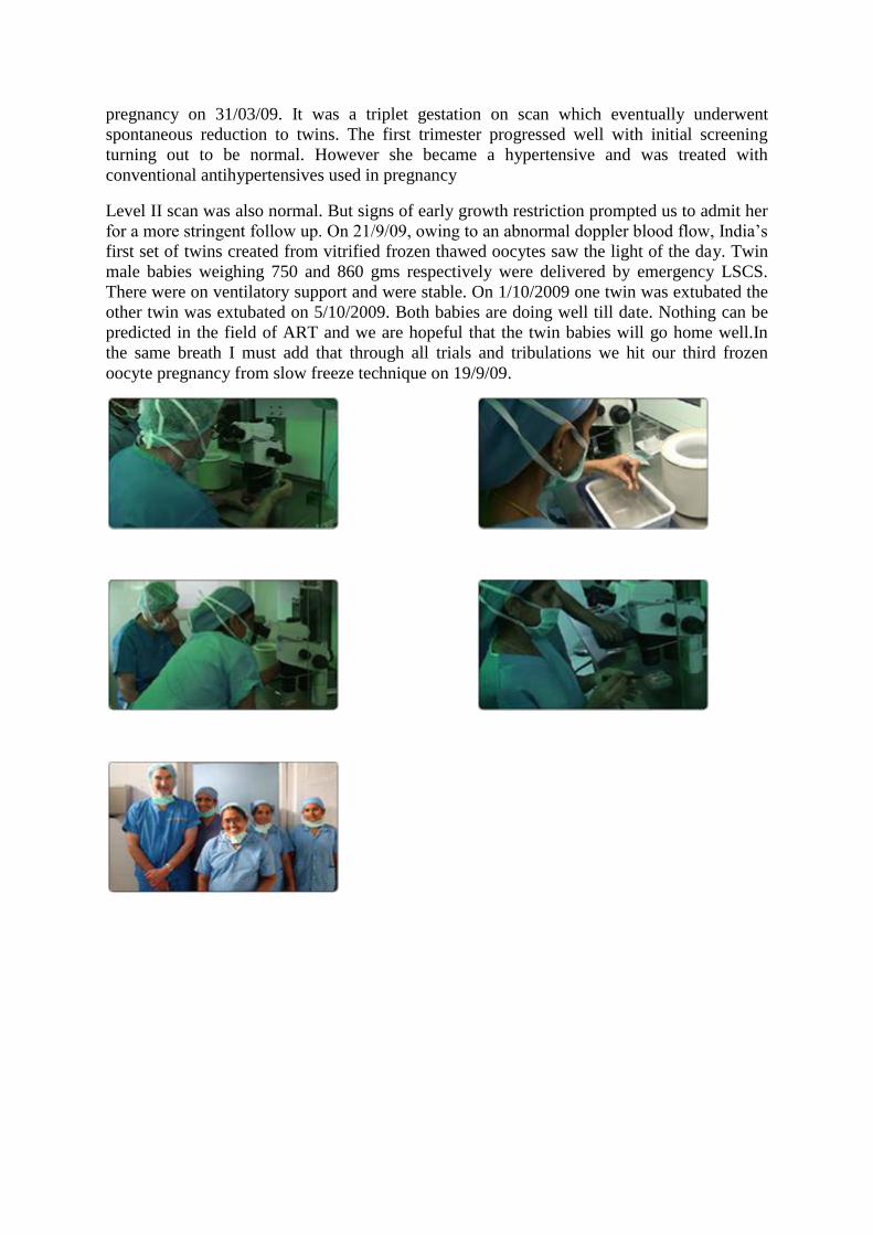
Level II scan was also normal. But signs of early growth restriction prompted us to admit her
for a more stringent follow up. On 21/9/09, owing to an abnormal doppler blood flow, India’s
first set of twins created from vitrified frozen thawed oocytes saw the light of the day. Twin
male babies weighing 750 and 860 gms respectively were delivered by emergency LSCS.
There were on ventilatory support and were stable. On 1/10/2009 one twin was extubated the
other twin was extubated on 5/10/2009. Both babies are doing well till date. Nothing can be
predicted in the field of ART and we are hopeful that the twin babies will go home well.In
the same breath I must add that through all trials and tribulations we hit our third frozen
oocyte pregnancy from slow freeze technique on 19/9/09.

CARE FOR A SPA RX? Welcome to the year's first newsletter and this time we Well as much as we may all want to head to one ...
the SPA (Single Port Access) that we are actually talking about is a boon in the field of laparoscopic
surgery. What with robotics and minimal access taking over conventional skilled surgeons and multiport
entries. Yes, we are talking about single port access surgery which has the following advantages.
How this works? 1. Lower blood loss 2. Less pain
2. Faster recovery time ( 3-4 days)
3. Less scarring
4. Lower infection rate
5. Lower rate of complications
Conventional laparoscopic approach uses a main viewing port and 2-3 working access ports. In SPA the
umbilical port is the lone entry through a single incision. Introduction of a triport or quadriport, allows the
surgeon to perform standard laparoscopic procedures through multi instrument accesses fitted in the
umbilical port.
South India’s First SPA Surgery Dr.Deepu Rajkamal Selvaraj - 2009 First SPA Hysterectomy - 21/8/2009 First SPA Varicocelectomy -
11/9/2009
World First SPA Surgeries First SPA Cholecystectomy - May 2007 - Dr. Paul Gurcillo - (USA) First Hysterectomy - July 2007 - Dr.
Kate O Hanlan - (USA)

DIET AND PCOS & PARENT TALK DIET AND PCOS Until recently, diet was not thought of as an important adjunct in the treatment or management of PCOS.
However, the recent discovery regarding the role of insulin resistance plays an important part in
understanding the need for dietary intervention. Suggestive research also indicates that being overweight,
and obese are associated mostly with this condition and hence the need for a nutritional recommendation to
maintain a healthy lifestyle and improve the symptoms of this condition. A balanced diet is one that
comprises all the five food groups (cereals, pulses and legumes, fruits and vegetables, milk and meat
products and fats, oils and sugars) in required quantities based on age, activity and body composition As an
Indian one can easily balance his/her food by including all these groups in their daily food intake. In our
day to day life we find that most often both lunch and dinner is usually balanced, hence one has to
concentrate on the breakfast as this meal kick- starts the day and also provides one-third of an individual's
metabolic and nutrient requirement. Since no single food group has all the nutrients that can nourish our
body it is important to have variety of healthy options among the food groups to ensure the balance. A
balanced diet coupled with moderate regular physical activity is the simple equation to a healthy life. A
well balanced diet can actually show a marked difference in weight management but better still a deeper
understanding about each nutrient would provide more insight into the diet r e q u i r e d f o r P C O . High
intake of carbohydrates, especially the refined ones (i.e. Sugar, white flour and its products - semolina,
sooji, white bread, white rice, etc.) will quickly convert to sugar and cause elevated levels of insulin. Since
high levels of insulin is one of the causative factors in these women, its better to avoid refined
carbohydrates and instead include whole cereals like wheat, ragi, jowar, bajra etc. Sugar can be included in
moderation depending on the sugar levels of the individual. Wise method of including carbohydrates
would be by maintaining a combination method with protein, fat, vitamins, minerals, fibre and water.
Moderate inclusion of protein in the form of pure proteins (Skimmed fat off) would be the ideal play as
these foods have low glycemic index and also provide bulk in the diet which cuts on the carb percentage.
Care should be taken to include protein without fat as this combination is more prevalent among Indian
foods (i.e. Milk, chicken, fish, egg etc) Either the food itself comes in the combination or the type of
preparation (deep frying) adds to this effect. Hence vegetarian source of proteins such as legumes
contribute more to managing PCOS.
A diet rich in saturated fats (ghee, butter, animal fat etc) can contribute to PCOS whereas unsaturated fats
(vegetable oils, fish oils, almonds, and flaxseed) can help to modify the syndrome process. Since essential
fatty acids play a major role in our metabolism embarking on a zero fat diet does not help. Right
combination and balance of saturated and unsaturated will do the needful.
Vitamins like vitamin C helps in fat oxidation whereas certain other vitamins are essential to keep the
metabolic fire burning hence going low on these can lead
to weight gain contributing to PCOS. Certain minerals like
calcium and iron are required during PCOS as it can also
help in fertility. Vitamins also serve as antioxidants
helping in efficient excretion of the metabolic wastes and
toxins and in weight loss. The best source would be fruits
(in moderation) and vegetables. Non nutrients like fibre
and water also contribute to the satiety value of the system
and hence indirectly help in PCOS, fibre especially the
soluble fiber can help in relieving constipation which can
occur due to reduced calorie intake.
Good water intake can contribute to proper metabolism in
the body for utilization of sugars by the system
contributing to insulin efficiency
This is also not a temporary diet it is one that you will
need to follow long term! Therefore you will need to
make it as healthy and tasty aspossible. Remember any diet works best if coupled with some kind of
physical activity especially the one that is in long run and enjoyable!
Number of studies indicates that physical activity is an essential factor in managing PCOS - and the
resultant weight loss increases fertility as well as adds to a healthy lifestyle. Visceral fat, the fat that
surrounds organs, is best lost through a program of diet and exercise. While weight loss is affected by diet
alone, or by exercise alone, studies have shown that belly fat (visceral) is affected by the combination of
both. Women with PCOS, as a rule, are unable to lose weight, and this very factor makes them targets of
diabetes and heart disease, both of which are often accompanying diseases of PCOS. Exercise is very
effective in improving the metabolic processes which in turn has a pronounced the effect on heart, chronic
fatigue syndrome, and even in some forms of cancers. A moderate exercise program not only has physical
benefits, but also improves mental outlook. Exercise promotes the propagation of endorphins, which in
turn balance the mood and emotions. Since depression is also an accompanying feature of PCOS, exercise
can benefit greatly in this regard as well. Starting an exercise program can sound like a difficult task, but
just remember that your main goal is to meet the basic physical activity recommendations: 30 minutes of
moderate-intensity physical activity at least five days per week, or vigorous-intensity activity at least three
days per week, and strength training two to three times per week. Choose activities that appeal to you and
will make exercise fun (cycling, swimming, aerobics, dancing etc). Walking is a great, easy way to do
moderate-intensity physical activity.
In conclusion, we know that weight loss plays a key role in decreasing insulin resistance in obese women.
It appears that a lower glycemic diet may play an important role in helping to control insulin levels as well
as promoting weight loss. However if weight loss is not required in certain women, the concept of
balanced diet is still essential for weight maintenance and general well being.
- Mrs. Veena Sekar
Nutritionist, G G Hospital
PARENT TALK Respected Doctor,
I am writing this letter in enormous gratitude for bringing in my life boundless pleasure. I am D r K a n i
m o z h i m a r r i e d t o Dr Ashok Orthopaedician working in Withybush Hospital Haverfordwest South Wales UK.
After marriage I was issueless for 7 long years and even after a few trials clinically in the UK I was not
able to conceive. Having heard about the great service rendered by you I came over to GG Hospital in September2006. Immediately you and Dr Priya took me under your care.
Due to fantastic care and dedication to each and every case and bestowing great personal attention to the different circumstances of the individual cases and their needs I was able to conceive. Throughout my
confinement I received excellent attention to each and every small setbacks and finally gave birth to a
chubby little boy in May 2007. I and my husband very gratefully remember you, Dr Priya, Dr Vijaya and all the dedicated staff of GG
Hospital on the occasion of his 2nd birthday. I am enclosing one of his photos .
we hope and pray to God that your great service and pioneering work to continue and benefit many many
families and bring everlasting joy and cheer unto them.
With highest regards
Dr Kanimozhi
Dr Ashok FRCS(Glasgow)

ART BABES...
NAME : BRAMMI JEYAKUMARAN
PROCEDURE : IVF - ET
DOB : 01-10-1998 & 11 YEARS
CLASS : 6th std
HOBBIES: learning vocal / music /classical dance
AMBITION :: To become a doctor Maintains a good academic record an proficiency in dance.
Participated in state level “spell bee” nation level bright scholar”, international level ‘ma millan’ exams
and won prizes.
She has performed in jaya tv, ‘thakadimi tha ‘jackpot’ as also in programmes of kalaignar t chutti tv, and
raj tv.She has joined the group of her guru to perfor at ida, at the natyanjali, kumbakonam and oth temple
programmesShe has won many prizes in cometition
Hobbies are drawing, painting and singing
NAME : Annapoorna
PROCEDURE : IVF - ET
DOB : 11.08.1998
CLASS : 6th std
HOBBIES : Reading books /cycling /Watching TV
EXTRA ACTIVITIES : Learning vocal/Music/Classical dance
AMBITION :: To Become an scientist

Our 1st frozen oocyte baby Turned One .. ..On 27/8/09
PRESS CLIPS....

HOT CHOCOLATES ! A group of graduates, well established in their careers, were discussing their lives at a class reunion. They
decided to go visit their old university professor, now retired, who was always an inspiration to them. During their visit, the conversation turned to complaints about stress in their work, lives and relationships.
Offering his guests hot chocolate, the professor went into the kitchen and returned with a large pot of hot chocolate and an assortment of cups. Some cups were porcelain, glass, crystal, some plain looking, some
expensive, some exquisite. He invited each to help themselves to the hot chocolate.
When they all had a cup of hot chocolate in hand, the professor shared his thoughts. “Notice that all the
nice looking, expensive cups were taken, leaving behind the plain and cheap ones.”
“While it is normal for you to want only the best for yourselves, that is the source of your problems and
stress.”
“The cup that you are drinking from adds nothing to the quality of the hot chocolate. In most cases it is just more expensive and in some cases even hides what we drink.”
“What each of you really wanted was hot chocolate. You did not want the cup . . . but you consciously went
for the best cups.” “And soon, you began to eye one another's cups.”
“Now friends, please consider this . . . “Life is the hot chocolate . . . your job, money and position in
society are the cups.”
“They are just tools to hold and contain life.” “The cup you have does not define, nor does it change, the quality of life You are living.”
“Sometimes, by concentrating only on the cup, we fail to enjoy the hot chocolate God has provided us.”
Always remember this . . . . God brews the hot chocolate, He does not choose the cup.
The happiest people don't have the best of everything.
They just make the best of everything that they have!! Live simply . . . Love generously . . . Care deeply . . .
Speak kindly . . . Leave the rest to God.
~ and remember ~
The richest person is not the one who has the most, but the one who needs the least.
Enjoy your hot chocolate!!

MONTHLY VARIATIONS IN ART PREGNANCIES -(JANUARY 2009 -JUNE 2009)
ART Vs IUI : PREGNANCIES STATISTICS ( JANUARY 2009 -JUNE 2009)
TOTAL No. OF PREGNANCIES/ MONTHLY (JANUARY 2009 -JUNE 2009)
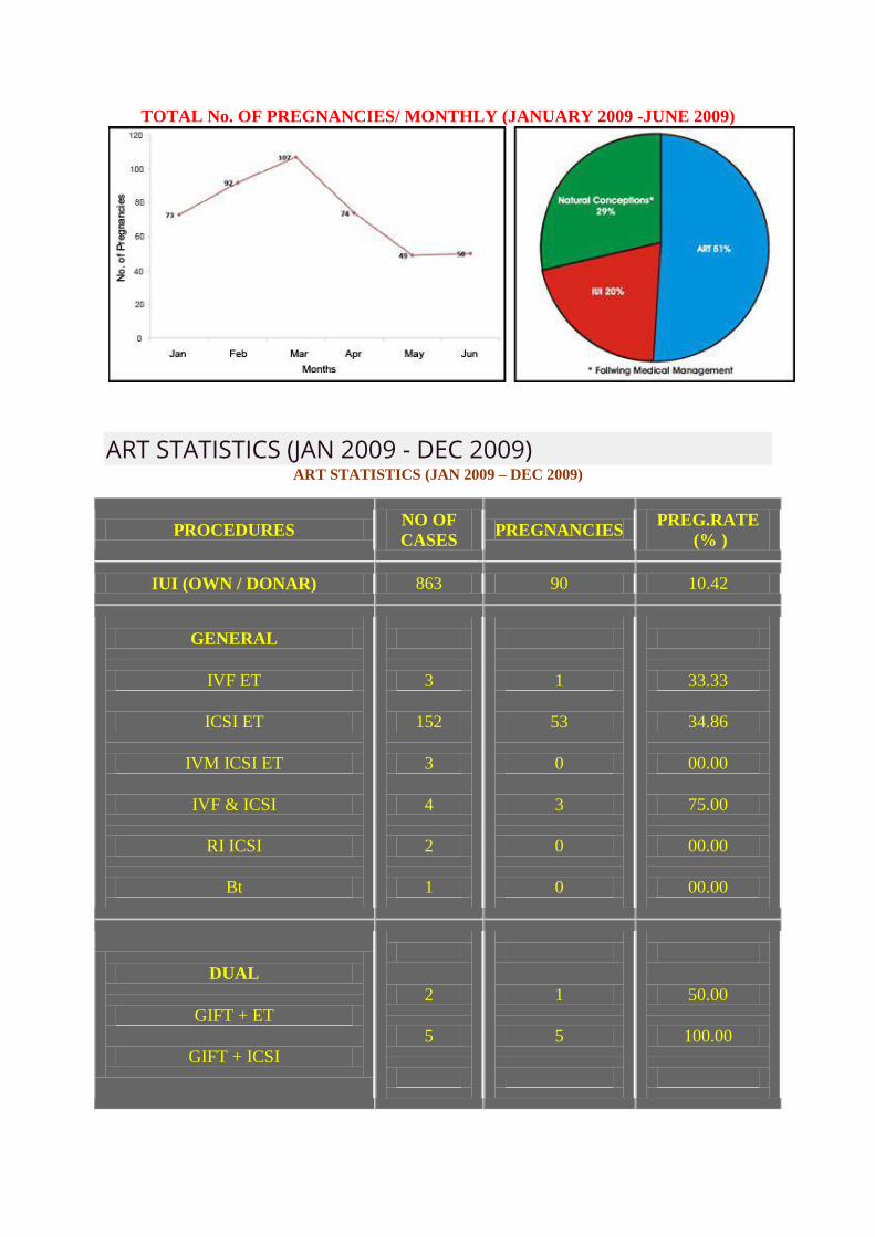
ART STATISTICS (JAN 2009 - DEC 2009) ART STATISTICS (JAN 2009 – DEC 2009)
PROCEDURES NO OF
CASES PREGNANCIES
PREG.RATE
(% )
IUI (OWN / DONAR) 863 90 10.42
GENERAL
IVF ET
ICSI ET
IVM ICSI ET
IVF & ICSI
RI ICSI
Bt
3
152
3
4
2
1
1
53
0
3
0
0
33.33
34.86
00.00
75.00
00.00
00.00
DUAL
GIFT + ET
GIFT + ICSI
2
5
1
5
50.00
100.00

FROZEN EMBRYOS
FROZEN CISI
FROZEN ICSI +IVF
FROZEN OOCYTE + ICSI
FROZEN PROST + ET
32
10
1
1
8
3
0
0
25.00
30.00
00.00
00.00
SEQUENTIAL TRANSFER (
OWN / DONOR )
DAY 2 AND DAY 5
TRANSFER
RUPTURE ET (OWN /
DONAR)
142
32
84
13
59.15
40.62
DONOR OOCYTE
PROGRAMME ( DOP )
IVF ET
ICSI ET
IVF & ICSI ET
FROZEN ET
OOCYTE THAWING – ICSI
ET
BT
GIFT
8
52
3
1
3
3
2
3
23
2
0
1
0
0
37.50
44.23
66.66
00.00
33.33
00.00
00.00
DUAL
GIFT + ET
GIFT + ICSI
1
4
0
2
00.00
50.00

DONOR EMBRYO
PROGRAMME
IVT ET
ICSI ET
FROZEN DET
BT
9
1
20
6
5
1
8
3
55.55
100.00
40.00
50.00
DUAL
GIFT + ET
2
0
00.00
OWN + DOP
IVF + ET
ICSI ET
1
6
1
4
100.00
66.6
OWN + DET
4
2
50.00
Total Number of pregnancies achieved : 3265
Total Number of patients delivered by ART : 1707
Total Number of babies delivered by ART : 2187
Total Number of ongoing pregnancies : 244
Total Number of Fetal wastages :1299
Lost in follow up : 15


![Preconception Laparoscopic Cervical Cerclage: The ... · 1965 [5], trans-abdominal cervical cerclage prior to pregnancy has emerged as a safe and effective intervention for improving](https://static.fdocuments.in/doc/165x107/5f8851cdb723447a244bb50e/preconception-laparoscopic-cervical-cerclage-the-1965-5-trans-abdominal.jpg)