Diagnosis and management of upper gastrointestinal...
26
1 Last saved: 5/27/11 Diagnosis and management of upper gastrointestinal bleeding Thad Wilkins, M.D., Department of Family Medicine, Georgia Health Sciences University Naiman Khan, M.D., Department of Family Medicine, Georgia Health Sciences University Akash Nabh, M.D., Department of Medicine, Georgia Health Sciences University Robert R. Schade, M.D., Department of Medicine, Georgia Health Sciences University Author Information Thad Wilkins, M.D. Associate Professor Department of Family Medicine 1120 15 th Street, HB-4032 Georgia’s Health Sciences University Augusta, GA 30912 Contact information Phone: 706-721-8018 Email: [email protected] Abstract word count: 241 (150-250) Manuscript word count: 1592 (1500-1800) Number of references: 33 (10-35) Number of tables: 5 Number of figures: 6
Transcript of Diagnosis and management of upper gastrointestinal...

1 Last saved: 5/27/11
Diagnosis and management of upper gastrointestinal bleeding
Thad Wilkins, M.D., Department of Family Medicine, Georgia Health Sciences University
Naiman Khan, M.D., Department of Family Medicine, Georgia Health Sciences University
Akash Nabh, M.D., Department of Medicine, Georgia Health Sciences University
Robert R. Schade, M.D., Department of Medicine, Georgia Health Sciences University
Author Information
Thad Wilkins, M.D.
Associate Professor
Department of Family Medicine
1120 15th Street, HB-4032
Georgia’s Health Sciences University
Augusta, GA 30912
Contact information
Phone: 706-721-8018
Email: [email protected]
Abstract word count: 241 (150-250)
Manuscript word count: 1592 (1500-1800)
Number of references: 33 (10-35)
Number of tables: 5
Number of figures: 6

2 Last saved: 5/27/11
Abbreviations:
Upper gastrointestinal bleeding (UGIB)
Nonsteroidal anti-inflammatory drug (NSAID)
Odds ratio (OR)
Peptic ulcer disease (PUD)
Peptic ulcer bleeding (PUB)
Helicobacter pylori (H. Pylori)

3 Last saved: 5/27/11
Abstract
Upper gastrointestinal bleeding (UGIB) causes significant morbidity and mortality in the United
States. Bleeding due to peptic ulcer disease is the most common cause of UGIB and is
associated with NSAID use and the relatively high prevalence of H. Pylori infection. Patients
with portal hypertension and cirrhosis may hemorrhage from varices. Rapid assessment and
resuscitation should precede diagnostic evaluation. Two large caliber peripheral catheters or a
central venous line should be inserted for intravenous access and crystalloid intravenous fluids
initiated. Initial laboratory tests should include: hemoglobin, hematocrit, platelet count,
prothrombin time, partial thromboplastin time, INR, creatinine, BUN, type and cross match.
Blood transfusion should be administered to patients with a hemoglobin level less than 7 g/dL.
Patients can be risk stratified based on clinical assessment and endoscopic findings. Early
endoscopy (within 24 hours of presentation) is recommended in most patients, allowing rapid
diagnosis and application of endoscopic therapies including injection with epinephrine, thermal
therapy, application of clips, and banding. Endoscopic therapy results in reduced morbidity,
decreased hospital stay, decreased risk of recurrent bleeding, and the need for surgery.
Although administration of proton pump inhibitors does not decrease mortality, risk for
rebleeding, or surgery, they downstage the severity of the bleeding lesion and reduce the need
for endoscopic therapy. Despite successful endoscopic therapy, rebleeding can occur in 10-
20% of patients and a second attempt at endoscopic therapy is recommended. Arteriography or
surgery may be needed if there is persistent and severe bleeding.

4 Last saved: 5/27/11
<LH>Background
In the US in 2004, non-variceal upper gastrointestinal bleeding (UGIB) resulted in
400,000 hospital admissions per year, costing more than $2 billion annually (1). UGIB is
associated with increasing nonsteroidal anti-inflammatory drug (NSAID) usage and the high
prevalence (64%) of Helicobacter pylori (H. Pylori) infections in patients with peptic ulcer
bleeding (PUB) (2). UGIB is twice as common in men as in women and increases in prevalence
with age (3). Despite advances in therapy, the in-hospital mortality from UGIB remains high
(13%) and rebleeding is common (15%) (4, 5). This review article focuses on acute non-
variceal UGIB in adults although we briefly review management of variceal bleeding.
<LH> Pathogenesis
UGIB includes hemorrhage originating from the esophagus to the ligament of Treitz.
PUB is the main cause of UGIB (61%), and in the US, duodenal ulcers are more common than
gastric ulcers (6). Hospitalizations for PUB decreased by 30% from 1996 to 2006, and in 2006
the inpatient mortality from PUB was 2.7% (6). Patients with gastric ulcers (55.1%) are
hospitalized more commonly than patients with duodenal ulcers (38.5%), but the mortality rate
for duodenal ulcers (3.7%) is higher than that of gastric ulcers (2.1%) (6). Duodenal ulcers are
more likely to erode into large vessels causing more severe bleeding. Table 1 lists common
causes of UGIB. In a meta-analysis of 16 studies of 1633 participants taking NSAIDs, both H.
Pylori (odds ratio (OR) (1.7)) and NSAID use (OR 4.8) increase the risk of UGIB, and this risk
increases when both factors are present (OR 6.1) (7). H. Pylori adheres to the gastric
epithelium and renders the underlying mucosa more vulnerable to damage by producing
enzymes and toxins (8), and affecting gastrin levels and acid output. The risk of UGIB varies
based on the type of NSAID (9), (Table 2).
<LH> Diagnosis

5 Last saved: 5/27/11
Rapid assessment and resuscitation should precede the diagnostic evaluation in
unstable patients with severe bleeding. Some patients may require intubation to decrease risk
for aspiration. All patients with active bleeding resulting in hemodynamic instability should be
admitted to an intensive care unit for resuscitation and close observation. Two large caliber
peripheral catheters or a central venous line should be inserted for intravenous access and
crystalloid fluids initiated. Other measures include monitoring urine output, obtaining
electrocardiogram, and continuous telemetry monitoring. The physician should consider
transferring a patient with significant UGIB to a tertiary medical center based on local expertise
and the availability of facilities. Patients admitted primarily for UGIB have a lower mortality
compared to patients admitted for other reasons who have a subsequent UGIB during their
hospitalization (4, 10). See Figure 1 for an algorithmic approach to UGIB.
<SH> History and physical examination
Important historical information includes: presence of abdominal pain, vomiting,
dysphagia, black tarry stools, bright red blood per rectum, hematemesis, and chest pain.
Medication use should be elicited, especially prior use of clopidogrel, warfarin, NSAIDs, aspirin,
selective serotonin reuptake inhibitors (SSRIs), or corticosteroids since treatment with these
medications increase the risk for UGIB (11-13). SSRIs inhibit platelet aggregation and are
associated with UGIB; the concurrent use of NSAIDs or aspirin with SSRIs further increases the
risk of UGIB (13). The physician should ascertain prior history of peptic ulcer disease (PUD),
prior episode of UGIB, history of cirrhosis, and alcohol or illicit drug use. The physician should
also inquire about other comorbid conditions that may affect outcomes such as prior abdominal
surgery, diabetes mellitus, coronary artery disease, chronic renal or liver disease, or chronic
obstructive pulmonary disease.

6 Last saved: 5/27/11
Blood pressure and pulse rate may be normal. If bleeding is severe, patients may be
hypotensive or tachycardic or may exhibit orthostatic hypotension. Physical examination
should assess for tenderness, guarding, rebound, prior surgical scars, or sequela of chronic
liver disease. Rectal examination should be performed and stool color assessed, e.g. for
melena or bright red blood. Stool specimen should be collected for occult blood testing.
Initial laboratory tests should include: hemoglobin, hematocrit, platelet count,
prothrombin time, partial thromboplastin time, INR, BUN, creatinine, and type and crossmatch.
Patients with active bleeding and a coagulopathy or thrombocytopenia should be considered for
transfusion with fresh frozen plasma and platelets, respectively (4). Blood transfusions should
be administered to patients with hemoglobin of less than 7 g/dL and hemoglobin maintained at 9
g/dL (4, 14).
<SH> Nasogastric tube lavage
Evaluation should focus on determining whether the bleed is from an upper or a lower
gastrointestinal source. Nasogastric lavage has a low sensitivity and poor negative likelihood
ratio for UGIB in patients with melena or hematochezia (15, 16). However, a positive
nasogastric tube lavage that yields blood or coffee-ground like material confirms the diagnosis
of UGIB and predicts that bleeding is caused by a high-risk lesion (15, 16).
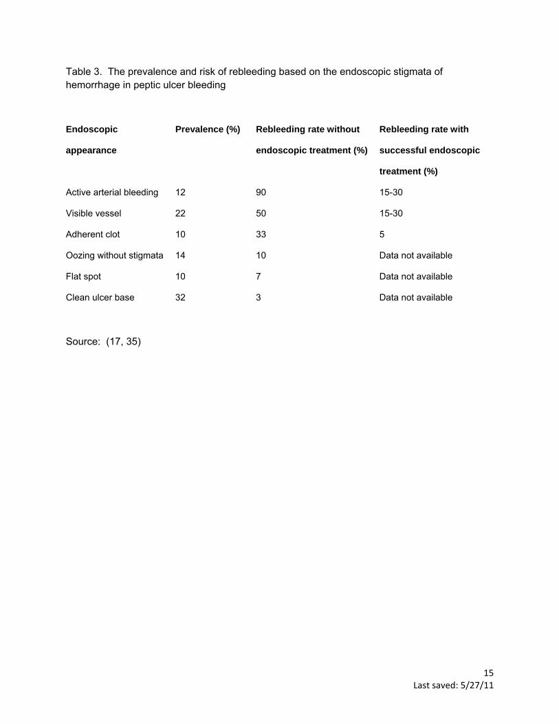
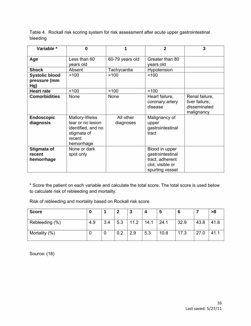
<SH>Risk stratification
Risk stratification is based on clinical assessment and endoscopic findings. Clinical
assessment includes age, presence of shock, systolic blood pressure, heart rate, and comorbid
conditions. Mortality increases with increasing co-morbid conditions and age (17). Endoscopic
findings include the cause of the bleeding and stigmata of recent hemorrhage, see Table 3. The

7 Last saved: 5/27/11
Rockall risk scoring system uses a combination of clinical and endoscopic findings to predict the
risk of rebleeding and mortality, (table 4) (18).
<LH> Treatment
Early upper endoscopy (within 24 hours of presentation) is recommended in most
patients with UGIB as it confirms the diagnosis and allows for targeted endoscopic treatment,
resulting in reduced morbidity, hospital stay, risk of recurrent bleeding, and need for surgery (4).
Figures 2-6 show examples of endoscopic findings. Although prokinetic agents to evacuate the
stomach are not recommended (19), gastric lavage should be considered. One of the benefits
of gastric lavage is to clear the stomach of blood, increasing the success of endoscopic
localization of the source of bleeding. Endoscopic therapies include injection with epinephrine,
thermal application, application of clips, and banding. A Cochrane review of 18 studies of PUB
including 1868 participants found that adding an additional endoscopic treatment after
epinephrine injection significantly reduced rebleeding rates from 18.5% to 10% and reduced
mortality from 4.7% to 2.5% (20). Low risk patients, e.g. clean ulcer base with PUB can be
safely discharged on the same day as endoscopy (4). Most patients with high-risk PUB
stigmata, e.g. active arterial bleeding, visible vessel, or adherent clot should be hospitalized for
at least 72 hours with intravenous PPI therapy after endoscopic hemostasis since most
rebleeding occurs in this time frame (4).
Although a systematic review of 6 randomized controlled trials of 2223 participants found
no statistically significant differences in mortality, rebleeding, or surgery between patients
receiving proton pump inhibitors (PPIs) and control treatment (placebo or H2 receptor
antagonists) (21). Patients treated with PPI therapy compared to control treatment had
significantly reduced stigmata of recent hemorrhage, e.g. active arterial bleeding, visible vessel,
or adherent clot (37.2% compared to 46.5%, OR 0.67) and reduced need for endoscopic

8 Last saved: 5/27/11
therapy (8.6% compared to 11.7%, OR 0.68) (21). In a randomized controlled trial of 767
patients with PUB randomly assigned to intravenous PPI therapy or placebo, fewer patients
receiving intravenous PPI therapy (5.9%) had recurrent bleeding within 72 hours than those
receiving placebo (10.3%) (P= 0.026) (22). The difference in bleeding recurrence remains
significant at 7 days and 30 days (P= 0.010) (22). All patients admitted with significant UGIB
should be started on intravenous PPI therapy until confirmation of the cause of bleeding at
endoscopy (4). Use of H2 receptor antagonists is not recommended for patients with UGIB.
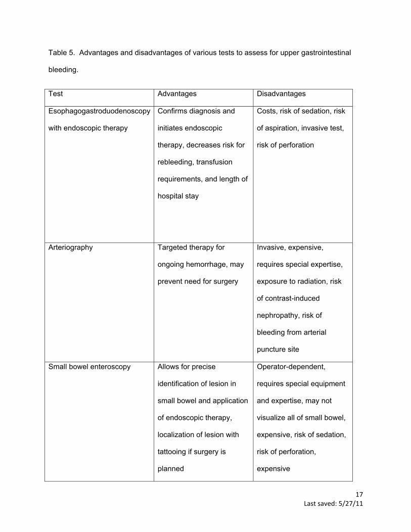
<SH>Recurrent hemorrhage
Rebleeding after successful endoscopic therapy occurs in 10-20% of patients. The risk
of rebleeding and mortality can be calculated with a clinical decision rule (18, 23). If rebleeding
occurs, a second attempt at endoscopic therapy is recommended. In patients determined to be
at high-risk for rebleeding, scheduled repeat endoscopy may reduce the rebleeding rate and be
cost effective (4). However, a routine second-look endoscopy the next day is not recommended
(4). Arteriography with embolization usually precedes surgical therapy since both are equally
effective in treating patients with persistent bleeding (24). Surgical therapy is usually
recommended if therapeutic methods including endoscopy and arteriography with embolization
have failed to control the bleeding or if interventional radiology expertise is not available after
failed endoscopic attempt. Surgical therapy is also indicated in patients with recurrent
hemorrhage or hemodynamic instability despite fluid resuscitation and blood transfusion. In
patients in whom no cause of UGIB was identified, small bowel evaluation with enteroscopy or
video capsule endoscopy should be considered to evaluate for a small bowel source of the
bleeding. Table 5 lists advantages and disadvantages of common tests used to diagnose
UGIB.
<LH>Prevention

9 Last saved: 5/27/11
H. Pylori and NSAIDs are the major causes of PUB in the US and preventive strategies
should focus on these etiologies. Smoking and alcohol use impairs ulcer healing and patients
should be counseled about smoking cessation and moderation of alcohol use. A systematic
review of 41 randomized controlled trials of patients on NSAIDs found that double dose H2
receptor antagonist (RR 0.44) and PPIs (RR =0.40) significantly reduced the risk of PUB (25).
In patients with a history of PUB, aspirin, clopidogrel, and NSAIDs should be avoided if possible.
In patients on aspirin who develop PUB, aspirin therapy with PPI therapy should be restarted as
soon as the risk for cardiovascular complication is thought to outweigh the risk of rebleeding (1).
A Cochrane review of 7 studies of 578 patients concluded that eradication of H. Pylori infection
in patients with PUB reduces the long-term rate of rebleeding (2.9%) compared with patients in
the non-eradication group (20%) (NNT = 7) (26). In patients with PUB associated with H. Pylori
eradication is essential and should be confirmed by urea breath test, stool antigen test, or the
biopsy urease test. A repeat upper endoscopy in 8-12 weeks is recommended for patients with
PUB secondary to gastric ulcers to assess for healing and to exclude malignancy and for
patients with severe esophagitis to exclude Barrett’s esophagus.
<LH> Variceal hemorrhage
Patients with cirrhosis should be screened with upper endoscopy to rule out varices (27).
If patients have no varices on the initial endoscopy, it should be repeated in 3 years (27).
Consider starting nonselective β-blockers, e.g. propranolol or nadolol in patients with varices to
reduce portal pressure and decrease the risk of future hemorrhage (27). In patients with a
history of varices who present with acute UGIB, upper endoscopy should be performed within
12 hours to confirm the diagnosis and to treat variceal hemorrhage (27). Endoscopic variceal
ligation is the preferred endoscopic treatment for esophageal variceal hemorrhage and is
superior to sclerotherapy (28). A review of 12 trials of 1241 patients found that broad-spectrum
antibiotics, e.g. ceftriaxone, norfloxacin, ciprofloxacin in patients with variceal hemorrhage

10 Last saved: 5/27/11
reduced overall mortality (RR 0.79) and risk of rebleeding (RR 0.53) (29). A Cochrane review of
21 trials of 2588 patients found no difference in mortality or risk of rebleeding with somatostatin
and its derivatives, e.g. octreotide in active variceal hemorrhage (30). Octreotide is often
administered to patients with variceal hemorrhage however its use is controversial. If octreotide
is used it should be initiated promptly in patients with variceal bleeding and continued for 3-5
days in conjunction with endoscopic therapy since this improves the immediate and 5 day
rebleeding rates (27). Salvage treatment with transjugular intrahepatic portosystemic stent-
shunt procedure or surgery should be considered especially for patients with gastric varices
when medical and endoscopic treatment fail to control bleeding (31). The Model for End-Stage
Liver Disease score should be calculated for prognosis and as a guide to decision-making
regarding liver transplant (32).

11 Last saved: 5/27/11
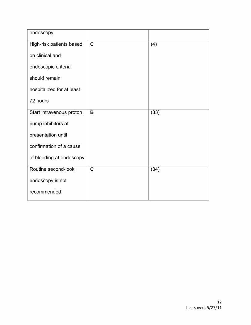
Key recommendations for practice.
Key Clinical
Recommendation
Evidence Rating References
Immediately evaluate and
initiate appropriate
resuscitation
C (4)
Transfuse patients with
hemoglobin level < 7.0
mg/L
C (4, 14)
Perform upper
endoscopy for the
evaluation of upper
gastrointestinal bleeding
within 24 hours of
presentation
C (4)
Low-risk patients with
acute peptic ulcer
bleeding based on
clinical and endoscopic
criteria may be
discharged promptly after
C (4)
12 Last saved: 5/27/11
endoscopy
High-risk patients based
on clinical and
endoscopic criteria
should remain
hospitalized for at least
72 hours
C (4)
Start intravenous proton
pump inhibitors at
presentation until
confirmation of a cause
of bleeding at endoscopy
B (33)
Routine second-look
endoscopy is not
recommended
C (34)

13 Last saved: 5/27/11
Table 1. Causes of upper gastrointestinal bleeding.
Diagnosis Distinguishing features Frequency (%)
Peptic ulcer disease Prior history of aspirin or
NSAID use associated
with abdominal pain,
food reduces pain,
nocturnal symptoms,
past history of peptic
ulcer disease or
Helicobacter pylori
62
Esophagogastric varices History of cirrhosis and
portal hypertension
6
Gastritis and duodenitis Same as peptic ulcer
disease above
8
Mallory-Weiss tears History of repeated
retching or vomiting
4
Gastrointestinal malignancy History of weight loss,
smoking, or alcohol
consumption, more
common in Asians
2
Arteriovenous
malformations
Painless bleeding in
older patients (>70 years
old), history of iron
deficiency anemia
10
Esophagitis or esophageal
ulcer
Heartburn, indigestion, or
dysphagia
Dieulafoy’s lesion More common in men,
painless bleeding
Other Variable
No identifiable source 8 Adapted from (3)

14 Last saved: 5/27/11
Table 2. The relative risk of upper gastrointestinal bleeding associated with use of nonsteroidal anti-inflammatory drugs (NSAIDs)
NSAID Relative risk of upper gastrointestinal bleeding
Ibuprofen 2.7
Diclofenac 4.0
Meloxicam 4.0
Naproxen 5.2
Indomethacin 5.3
Ketoprofen 5.7
Piroxicam 9.3
Ketorolac 14.0
Source: (9)
15 Last saved: 5/27/11
Table 3. The prevalence and risk of rebleeding based on the endoscopic stigmata of hemorrhage in peptic ulcer bleeding
Endoscopic
appearance
Prevalence (%) Rebleeding rate without
endoscopic treatment (%)
Rebleeding rate with
successful endoscopic
treatment (%)
Active arterial bleeding 12 90 15-30
Visible vessel 22 50 15-30
Adherent clot 10 33 5
Oozing without stigmata 14 10 Data not available
Flat spot 10 7 Data not available
Clean ulcer base 32 3 Data not available
Source: (17, 35)
16 Last saved: 5/27/11
Table 4. Rockall risk scoring system for risk assessment after acute upper gastrointestinal bleeding
Variable * 0 1 2 3
Age Less than 60 years old
60-79 years old Greater than 80 years old
Shock Absent Tachycardia Hypotension Systolic blood pressure (mm Hg)
>100 >100 <100
Heart rate <100 >100 >100 Comorbidities None None Heart failure,
coronary artery disease
Renal failure, liver failure, disseminated malignancy
Endoscopic diagnosis
Mallory-Weiss tear or no lesion identified, and no stigmata of recent hemorrhage
All other diagnoses
Malignancy of upper gastrointestinal tract
Stigmata of recent hemorrhage
None or dark spot only
Blood in upper gastrointestinal tract, adherent clot, visible or spurting vessel
* Score the patient on each variable and calculate the total score. The total score is used below to calculate risk of rebleeding and mortality.
Risk of rebleeding and mortality based on Rockall risk score
Score
0 1 2 3 4 5 6 7 >8
Rebleeding (%)
4.9 3.4 5.3 11.2 14.1 24.1 32.9 43.8 41.8
Mortality (%)
0 0 0.2 2.9 5.3 10.8 17.3 27.0 41.1
Source: (18)
17 Last saved: 5/27/11
Table 5. Advantages and disadvantages of various tests to assess for upper gastrointestinal
bleeding.
Test Advantages Disadvantages
Esophagogastroduodenoscopy
with endoscopic therapy
Confirms diagnosis and
initiates endoscopic
therapy, decreases risk for
rebleeding, transfusion
requirements, and length of
hospital stay
Costs, risk of sedation, risk
of aspiration, invasive test,
risk of perforation
Arteriography Targeted therapy for
ongoing hemorrhage, may
prevent need for surgery
Invasive, expensive,
requires special expertise,
exposure to radiation, risk
of contrast-induced
nephropathy, risk of
bleeding from arterial
puncture site
Small bowel enteroscopy Allows for precise
identification of lesion in
small bowel and application
of endoscopic therapy,
localization of lesion with
tattooing if surgery is
planned
Operator-dependent,
requires special equipment
and expertise, may not
visualize all of small bowel,
expensive, risk of sedation,
risk of perforation,
expensive

18 Last saved: 5/27/11
Capsule endoscopy No sedation required,
noninvasive test, allows
visualization of the entire
small bowel
Capsule retention may
occur, can miss lesions
because images are not
continuous, cannot perform
therapeutic maneuvers

F
Figure 1. An aalgorithm for mmanagement off acute upper ggastrointestinaal hemorrhage..
Last sav19
ved: 5/27/11
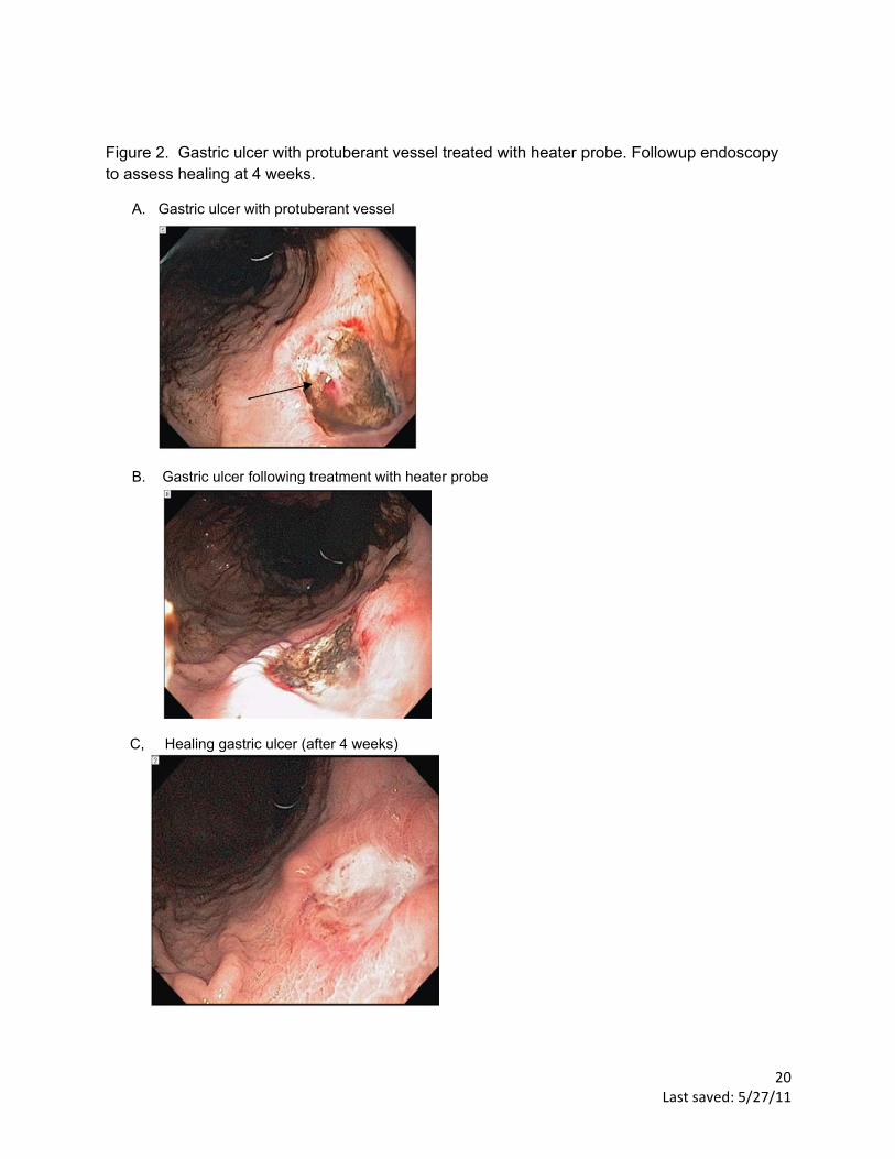
Figure 2.to assess
A. G
B. G
C, H
. Gastric ulcs healing at
Gastric ulcer w
Gastric ulcer f
Healing gastr
cer with protu4 weeks.
with protubera
following treat
ic ulcer (after
uberant vess
ant vessel
tment with he
r 4 weeks)
sel treated w
eater probe
with heater p
L
probe. Follow
ast saved: 5/2
wup endosco
20 27/11
opy

Figure 3.
. Duodenal ulcer with ad
dherent clot
Last saved: 5/221
27/11

Figure 4.
Ab
. Dieulafoy’s
A. Dieulafoyleeding
s lesion caus
y’s lesion ca
sing upper G
ausing uppe
GI bleeding
er GI
B. DDieulafoy’s le
L
esion after a
ast saved: 5/2
application o
22 27/11
of clip

Figure 5.
A. M
. Mallory-We
Mallory-Weiss
eis tear
tear
Last saved: 5/223
27/11

Figure 6.. Esophageaal varix caussing upper GGI bleeding aafter endosc
L
copic ligation
ast saved: 5/2
n
24 27/11

25 Last saved: 5/27/11
REFERENCES
1. Kanwal F, Barkun A, Gralnek IM, Asch SM, Kuipers EJ, Bardou M, et al. Measuring quality of care in patients with nonvariceal upper gastrointestinal hemorrhage: development of an explicit quality indicator set. Am J Gastroenterol. 2010 Aug;105(8):1710‐8. 2. Sanchez‐Delgado J, Gene E, Suarez D, Garcia‐Iglesias P, Brullet E, Gallach M, et al. Has H. pylori Prevalence in Bleeding Peptic Ulcer Been Underestimated[quest] A Meta‐Regression. Am J Gastroenterol. 2011. 3. Longstreth GF. Epidemiology of hospitalization for acute upper gastrointestinal hemorrhage: a population‐based study. Am J Gastroenterol. 1995 Feb;90(2):206‐10. 4. Barkun AN, Bardou M, Kuipers EJ, Sung J, Hunt RH, Martel M, et al. International consensus recommendations on the management of patients with nonvariceal upper gastrointestinal bleeding. Ann Intern Med. 2010 Jan 19;152(2):101‐13. 5. van Leerdam ME, Vreeburg EM, Rauws EA, Geraedts AA, Tijssen JG, Reitsma JB, et al. Acute upper GI bleeding: did anything change? Time trend analysis of incidence and outcome of acute upper GI bleeding between 1993/1994 and 2000. Am J Gastroenterol. 2003 Jul;98(7):1494‐9. 6. Wang YR, Richter JE, Dempsey DT. Trends and outcomes of hospitalizations for peptic ulcer disease in the United States, 1993 to 2006. Ann Surg. 2010 Jan;251(1):51‐8. 7. Huang JQ, Sridhar S, Hunt RH. Role of Helicobacter pylori infection and non‐steroidal anti‐inflammatory drugs in peptic‐ulcer disease: a meta‐analysis. Lancet. 2002 Jan 5;359(9300):14‐22. 8. Pajares JM. H. pylori infection: its role in chronic gastritis, carcinoma and peptic ulcer. Hepatogastroenterology. 1995 Nov‐Dec;42(6):827‐41. 9. Masso Gonzalez EL, Patrignani P, Tacconelli S, Garcia Rodriguez LA. Variability among nonsteroidal antiinflammatory drugs in risk of upper gastrointestinal bleeding. Arthritis Rheum. 2010 Jun;62(6):1592‐601. 10. Silverstein FE, Gilbert DA, Tedesco FJ, Buenger NK, Persing J. The national ASGE survey on upper gastrointestinal bleeding. II. Clinical prognostic factors. Gastrointest Endosc. 1981 May;27(2):80‐93. 11. Delaney JA, Opatrny L, Brophy JM, Suissa S. Drug drug interactions between antithrombotic medications and the risk of gastrointestinal bleeding. Cmaj. 2007 Aug 14;177(4):347‐51. 12. Hernandez‐Diaz S, Rodriguez LA. Steroids and risk of upper gastrointestinal complications. Am J Epidemiol. 2001 Jun 1;153(11):1089‐93. 13. Yuan Y, Tsoi K, Hunt RH. Selective serotonin reuptake inhibitors and risk of upper GI bleeding: confusion or confounding? Am J Med. 2006 Sep;119(9):719‐27. 14. Hebert PC, Wells G, Blajchman MA, Marshall J, Martin C, Pagliarello G, et al. A multicenter, randomized, controlled clinical trial of transfusion requirements in critical care. Transfusion Requirements in Critical Care Investigators, Canadian Critical Care Trials Group. N Engl J Med. 1999 Feb 11;340(6):409‐17. 15. Palamidessi N, Sinert R, Falzon L, Zehtabchi S. Nasogastric aspiration and lavage in emergency department patients with hematochezia or melena without hematemesis. Acad Emerg Med. 2010 Feb;17(2):126‐32. 16. Aljebreen AM, Fallone CA, Barkun AN. Nasogastric aspirate predicts high‐risk endoscopic lesions in patients with acute upper‐GI bleeding. Gastrointest Endosc. 2004 Feb;59(2):172‐8. 17. Zimmerman J, Siguencia J, Tsvang E, Beeri R, Arnon R. Predictors of mortality in patients admitted to hospital for acute upper gastrointestinal hemorrhage. Scand J Gastroenterol. 1995 Apr;30(4):327‐31. 18. Rockall TA, Logan RF, Devlin HB, Northfield TC. Risk assessment after acute upper gastrointestinal haemorrhage. Gut. 1996 Mar;38(3):316‐21.

26 Last saved: 5/27/11
19. Barkun AN, Bardou M, Martel M, Gralnek IM, Sung JJ. Prokinetics in acute upper GI bleeding: a meta‐analysis. Gastrointest Endosc. 2010 Dec;72(6):1138‐45. 20. Vergara M, Calvet X, Gisbert JP. Epinephrine injection versus epinephrine injection and a second endoscopic method in high risk bleeding ulcers. Cochrane Database Syst Rev. 2007(2):CD005584. 21. Sreedharan A, Martin J, Leontiadis GI, Dorward S, Howden CW, Forman D, et al. Proton pump inhibitor treatment initiated prior to endoscopic diagnosis in upper gastrointestinal bleeding. Cochrane Database Syst Rev. 2010(7):CD005415. 22. Sung JJ, Barkun A, Kuipers EJ, Mossner J, Jensen DM, Stuart R, et al. Intravenous esomeprazole for prevention of recurrent peptic ulcer bleeding: a randomized trial. Ann Intern Med. 2009 Apr 7;150(7):455‐64. 23. Song SY, Chung JB, Moon YM, Kang JK, Park IS. Comparison of the hemostatic effect of endoscopic injection with fibrin glue and hypertonic saline‐epinephrine for peptic ulcer bleeding: a prospective randomized trial. Endoscopy. 1997 Nov;29(9):827‐33. 24. Ripoll C, Banares R, Beceiro I, Menchen P, Catalina MV, Echenagusia A, et al. Comparison of transcatheter arterial embolization and surgery for treatment of bleeding peptic ulcer after endoscopic treatment failure. J Vasc Interv Radiol. 2004 May;15(5):447‐50. 25. Rostom A, Dube C, Wells G, Tugwell P, Welch V, Jolicoeur E, et al. Prevention of NSAID‐induced gastroduodenal ulcers. Cochrane Database Syst Rev. 2002(4):CD002296. 26. Gisbert JP, Khorrami S, Carballo F, Calvet X, Gene E, Dominguez‐Munoz JE. H. pylori eradication therapy vs. antisecretory non‐eradication therapy (with or without long‐term maintenance antisecretory therapy) for the prevention of recurrent bleeding from peptic ulcer. Cochrane Database Syst Rev. 2004(2):CD004062. 27. Garcia‐Tsao G, Sanyal AJ, Grace ND, Carey W. Prevention and management of gastroesophageal varices and variceal hemorrhage in cirrhosis. Hepatology. 2007 Sep;46(3):922‐38. 28. Laine L, Cook D. Endoscopic ligation compared with sclerotherapy for treatment of esophageal variceal bleeding. A meta‐analysis. Ann Intern Med. 1995 Aug 15;123(4):280‐7. 29. Chavez‐Tapia NC, Barrientos‐Gutierrez T, Tellez‐Avila FI, Soares‐Weiser K, Uribe M. Antibiotic prophylaxis for cirrhotic patients with upper gastrointestinal bleeding. Cochrane Database Syst Rev. 2010(9):CD002907. 30. Gotzsche PC, Hrobjartsson A. Somatostatin analogues for acute bleeding oesophageal varices. Cochrane Database Syst Rev. 2008(3):CD000193. 31. Rossle M, Haag K, Ochs A, Sellinger M, Noldge G, Perarnau JM, et al. The transjugular intrahepatic portosystemic stent‐shunt procedure for variceal bleeding. N Engl J Med. 1994 Jan 20;330(3):165‐71. 32. Kamath PS, Wiesner RH, Malinchoc M, Kremers W, Therneau TM, Kosberg CL, et al. A model to predict survival in patients with end‐stage liver disease. Hepatology. 2001 Feb;33(2):464‐70. 33. Lau JY, Leung WK, Wu JC, Chan FK, Wong VW, Chiu PW, et al. Omeprazole before endoscopy in patients with gastrointestinal bleeding. N Engl J Med. 2007 Apr 19;356(16):1631‐40. 34. Romagnuolo J. Routine second look endoscopy: ineffective, costly and potentially misleading. Can J Gastroenterol. 2004 Jun;18(6):401‐4. 35. Forrest JA, Finlayson ND, Shearman DJ. Endoscopy in gastrointestinal bleeding. Lancet. 1974 Aug 17;2(7877):394‐7.