Diagnosis and management of gastric dysplasia - KJIMkjim.org/upload/kjim-2016-021.pdf · Keywords:...
9
Copyright © 2016 The Korean Association of Internal Medicine This is an Open Access article distributed under the terms of the Creative Commons Attribution Non-Commercial License (http://creativecommons.org/licenses/ by-nc/3.0/) which permits unrestricted noncommercial use, distribution, and reproduction in any medium, provided the original work is properly cited. pISSN 1226-3303 eISSN 2005-6648 http://www.kjim.org REVIEW Korean J Intern Med 2016;31:201-209 http://dx.doi.org/10.3904/kjim.2016.021 INTRODUCTION Gastric cancer remains one of the most challenging ma- lignant diseases worldwide. Gastric dysplasia is a pre- cancerous lesion and the penultimate stage in gastric carcinogenesis, particularly the intestinal type, as hy- pothesized by Correa [1]. Therefore, identification, man- agement, and surveillance of such lesions are import- ant for early detection and prevention of gastric cancer. Nevertheless, the accurate diagnosis and management of this lesion remain controversial. Therefore, in this review, current knowledge of this lesion is discussed, together with relevant diagnostic and therapeutic strat- egies. DEFINITION The World Health Organization (WHO) defines dyspla- sia in the gastrointestinal system as the presence of his- tologically unequivocal neoplastic epithelium without evidence of tissue invasion [2]. Some confusion exists regarding the terms adenoma and dysplasia. Originally, adenoma was considered a raised circumscribed lesion, either sessile or pedunculated, in contrast to dysplasia, which was defined as a flat or depressed mucosa [3]. However, use of terms such as “flat adenoma” or “de- pressed adenoma” by some investigators has resulted in confusion. The WHO defines gastric adenomas as cir- cumscribed, polypoid lesions composed of tubular and/ or villous structures, lined by dysplastic epithelium [4]. There has been an effort to standardize the terminology of adenoma and dysplasia; adenoma has been defined as neoplastic circumscribed benign lesions unassociat- ed with underlying inflammation such as atrophic gas- tritis whether pedunculated, sessile, flat, or depressed. In contrast, dysplasia is defined as benign neoplastic lesions associated with underlying inflammation [5]. Department of Internal Medicine, Chungnam National University School of Medicine, Daejeon, Korea Gastric dysplasia is a neoplastic lesion and a precursor of gastric cancer. The Pa- dova, Vienna, and World Health Organization classifications were developed to overcome the discrepancies between Western and Japanese pathologic diagnoses and to provide a universally accepted classification of gastric epithelial neopla- sia. At present, the natural history of gastric dysplasia is unclear. Much evidence suggests that patients with high-grade dysplasia are at high risk of progression to carcinoma or synchronous carcinoma. Therefore, endoscopic resection is re- quired. Although patients with low-grade dysplasia have been reported to be at low risk of progression to carcinoma, due to the marked histologic discrepancies between forceps biopsy and endoscopic specimens, endoscopic resection for this lesion is recommended, particularly in the presence of other risk factors (large size; depressed gross type; surface erythema, unevenness, ulcer, or erosion; and tubulovillous or villous histology). Helicobacter pylori eradication in patients with dysplasia after endoscopic resection appear to reduce the incidence of metachro- nous lesions. Keywords: Intraepithelial neoplasia; Dysplasia; Adenoma; Stomach Diagnosis and management of gastric dysplasia Jae Kyu Sung Received : February 1, 2016 Accepted : February 4, 2016 Correspondence to Jae Kyu Sung, M.D. Department of Internal Medi- cine, Chungnam National Uni- versity School of Medicine, 282 Munhwa-ro, Jung-gu, Daejeon 35015, Korea Tel: +82-42-280-7186 Fax: +82-42-254-4553 E-mail: [email protected]
Transcript of Diagnosis and management of gastric dysplasia - KJIMkjim.org/upload/kjim-2016-021.pdf · Keywords:...
Copyright © 2016 The Korean Association of Internal MedicineThis is an Open Access article distributed under the terms of the Creative Commons Attribution Non-Commercial License (http://creativecommons.org/licenses/by-nc/3.0/) which permits unrestricted noncommercial use, distribution, and reproduction in any medium, provided the original work is properly cited.
pISSN 1226-3303eISSN 2005-6648
http://www.kjim.org
REVIEW
Korean J Intern Med 2016;31:201-209http://dx.doi.org/10.3904/kjim.2016.021
INTRODUCTION
Gastric cancer remains one of the most challenging ma-lignant diseases worldwide. Gastric dysplasia is a pre-cancerous lesion and the penultimate stage in gastric carcinogenesis, particularly the intestinal type, as hy-pothesized by Correa [1]. Therefore, identification, man-agement, and surveillance of such lesions are import-ant for early detection and prevention of gastric cancer. Nevertheless, the accurate diagnosis and management of this lesion remain controversial. Therefore, in this review, current knowledge of this lesion is discussed, together with relevant diagnostic and therapeutic strat-egies.
DEFINITION
The World Health Organization (WHO) defines dyspla-
sia in the gastrointestinal system as the presence of his-tologically unequivocal neoplastic epithelium without evidence of tissue invasion [2]. Some confusion exists regarding the terms adenoma and dysplasia. Originally, adenoma was considered a raised circumscribed lesion, either sessile or pedunculated, in contrast to dysplasia, which was defined as a flat or depressed mucosa [3]. However, use of terms such as “flat adenoma” or “de-pressed adenoma” by some investigators has resulted in confusion. The WHO defines gastric adenomas as cir-cumscribed, polypoid lesions composed of tubular and/or villous structures, lined by dysplastic epithelium [4]. There has been an effort to standardize the terminology of adenoma and dysplasia; adenoma has been defined as neoplastic circumscribed benign lesions unassociat-ed with underlying inflammation such as atrophic gas-tritis whether pedunculated, sessile, flat, or depressed. In contrast, dysplasia is defined as benign neoplastic lesions associated with underlying inflammation [5].
Department of Internal Medicine, Chungnam National University School of Medicine, Daejeon, Korea
Gastric dysplasia is a neoplastic lesion and a precursor of gastric cancer. The Pa-dova, Vienna, and World Health Organization classifications were developed to overcome the discrepancies between Western and Japanese pathologic diagnoses and to provide a universally accepted classification of gastric epithelial neopla-sia. At present, the natural history of gastric dysplasia is unclear. Much evidence suggests that patients with high-grade dysplasia are at high risk of progression to carcinoma or synchronous carcinoma. Therefore, endoscopic resection is re-quired. Although patients with low-grade dysplasia have been reported to be at low risk of progression to carcinoma, due to the marked histologic discrepancies between forceps biopsy and endoscopic specimens, endoscopic resection for this lesion is recommended, particularly in the presence of other risk factors (large size; depressed gross type; surface erythema, unevenness, ulcer, or erosion; and tubulovillous or villous histology). Helicobacter pylori eradication in patients with dysplasia after endoscopic resection appear to reduce the incidence of metachro-nous lesions.
Keywords: Intraepithelial neoplasia; Dysplasia; Adenoma; Stomach
Diagnosis and management of gastric dysplasiaJae Kyu Sung
Received : February 1, 2016Accepted : February 4, 2016
Correspondence toJae Kyu Sung, M.D.Department of Internal Medi-cine, Chungnam National Uni-versity School of Medicine, 282 Munhwa-ro, Jung-gu, Daejeon 35015, KoreaTel: +82-42-280-7186Fax: +82-42-254-4553E-mail: [email protected]
202 www.kjim.org
The Korean Journal of Internal Medicine Vol. 31, No. 2, March 2016
http://dx.doi.org/10.3904/kjim.2016.021
Currently, however, adenoma and dysplasia are used in-discriminately by most clinicians.
CLASSIFICATION SYSTEMS
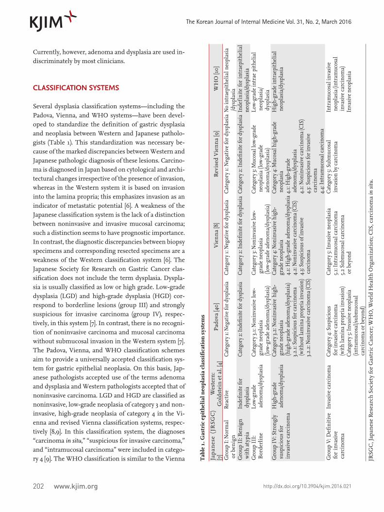
Several dysplasia classification systems—including the Padova, Vienna, and WHO systems—have been devel-oped to standardize the definition of gastric dysplasia and neoplasia between Western and Japanese patholo-gists (Table 1). This standardization was necessary be-cause of the marked discrepancies between Western and Japanese pathologic diagnosis of these lesions. Carcino-ma is diagnosed in Japan based on cytological and archi-tectural changes irrespective of the presence of invasion, whereas in the Western system it is based on invasion into the lamina propria; this emphasizes invasion as an indicator of metastatic potential [6]. A weakness of the Japanese classification system is the lack of a distinction between noninvasive and invasive mucosal carcinoma; such a distinction seems to have prognostic importance. In contrast, the diagnostic discrepancies between biopsy specimens and corresponding resected specimens are a weakness of the Western classification system [6]. The Japanese Society for Research on Gastric Cancer clas-sification does not include the term dysplasia. Dyspla-sia is usually classified as low or high grade. Low-grade dysplasia (LGD) and high-grade dysplasia (HGD) cor-respond to borderline lesions (group III) and strongly suspicious for invasive carcinoma (group IV), respec-tively, in this system [7]. In contrast, there is no recogni-tion of noninvasive carcinoma and mucosal carcinoma without submucosal invasion in the Western system [7]. The Padova, Vienna, and WHO classification schemes aim to provide a universally accepted classification sys-tem for gastric epithelial neoplasia. On this basis, Jap-anese pathologists accepted use of the terms adenoma and dysplasia and Western pathologists accepted that of noninvasive carcinoma. LGD and HGD are classified as noninvasive, low-grade neoplasia of category 3 and non-invasive, high-grade neoplasia of category 4 in the Vi-enna and revised Vienna classification systems, respec-tively [8,9]. In this classification system, the diagnoses “carcinoma in situ,” “suspicious for invasive carcinoma,” and “intramucosal carcinoma” were included in catego-ry 4 [9]. The WHO classification is similar to the Vienna T
able
1. G
astr
ic e
pith
elia
l neo
plas
ia c
lass
ifica
tion
sys
tem
s
Japa
nes
e (J
RSG
C)
[7]
Wes
tern
:G
olds
tein
et a
l. [4
]Pa
dova
[40]
Vie
nna
[8]
Rev
ised
Vie
nna
[9]
WH
O [1
0]
Gro
up I:
Nor
mal
o
r ben
ign
Reac
tive
Cate
gory
1: N
egat
ive f
or d
yspl
asia
Cate
gory
1: N
egat
ive f
or d
yspl
asia
Cat
egor
y 1:
Neg
ativ
e fo
r dys
plas
ia N
o in
trae
pith
elia
l neo
plas
ia /d
yspl
asia
Gro
up II
: Ben
ign
with
aty
pia
Inde
finite
for
dys
plas
iaCa
tego
ry 2:
Inde
finite
for d
yspl
asia
Cate
gory
2: I
ndefi
nite
for d
yspl
asia
Cate
gory
2: I
ndefi
nite
for d
yspl
asia
Ind
efin
ite fo
r in
trae
pith
elia
l ne
opla
sia/
dysp
lasi
aG
roup
III:
Bor
derl
ine
Low-
grad
e ad
enom
a/dy
spla
sia
Cate
gory
3.1:
Non
inva
sive
low-
gra
de n
eopl
asia
(low
-gra
de ad
enom
a/dy
spla
sia)
Cate
gory
3: N
onin
vasi
ve lo
w- g
rade
neo
plas
ia (l
ow-g
rade
aden
oma/
dysp
lasi
a)
Cate
gory
3: M
ucos
al lo
w-gr
ade
neo
plas
ia (l
ow-g
rade
aden
oma/
dysp
lasi
a)
Low-
grad
e int
rae
pith
elia
l n
eopl
asia
/ d
yspl
asia
Gro
up IV
: Str
ongl
y s
uspi
ciou
s for
inva
sive
car
cino
ma
Hig
h-gr
ade
aden
oma/
dysp
lasi
aCa
tego
ry 3.
2: N
onin
vasi
ve h
igh-
gra
de n
eopl
asia
(hig
h-gr
ade a
deno
ma/
dysp
lasi
a)3.2
.1: S
uspi
ciou
s for
car
cino
ma
(with
out l
amin
a pro
pria
inva
sion
)3.2
.2: N
onin
vasi
ve c
arci
nom
a (CI
S)
Cate
gory
4: N
onin
vasi
ve h
igh-
grad
e neo
plas
ia4.
1: H
igh-
grad
e ade
nom
a/dy
spla
sia
4.2:
Non
inva
sive
car
cino
ma (
CIS)
4.3:
Susp
icio
us o
f inv
asiv
e c
arci
nom
a
Cate
gory
4: M
ucos
al h
igh-
grad
e n
eopl
asia
4.1:
Hig
h-gr
ade
aden
oma/
dysp
lasi
a4.
2: N
onin
vasi
ve c
arci
nom
a (CI
S)4.
3: Su
spic
ious
for i
nvas
ive
car
cino
ma
4.4:
Intr
amuc
osal
car
cino
ma
Hig
h-gr
ade i
ntra
epith
elia
l n
eopl
asia
/dys
plas
ia
Gro
up V
: Defi
nitiv
e fo
r in
vasi
ve c
arci
nom
a
Inva
sive
car
cino
ma
Cate
gory
4: S
uspi
ciou
s fo
r inv
asiv
e car
cino
ma
(with
lam
ina p
ropr
ia in
vasi
on)
Cate
gory
5: In
vasi
ve n
eopl
asia
(int
ram
ucos
al/s
ubm
ucos
al c
arci
nom
a or b
eyon
d)
Cate
gory
5: In
vasi
ve n
eopl
asia
5.1
: Int
ram
ucos
al c
arci
nom
a5.2
Sub
muc
osal
car
cino
ma
or b
eyon
d
Cate
gory
5: S
ubm
ucos
al in
vasi
on b
y car
cino
ma
Intr
amuc
osal
inva
sive
neo
plas
ia (i
ntra
muc
osal
inva
sive
car
cino
ma)
Inva
sive
neo
plas
ia
JRSG
C, J
apan
ese
Res
earc
h So
ciet
y fo
r G
astr
ic C
ance
r; W
HO
, Wor
ld H
ealt
h O
rgan
izat
ion;
CIS
, car
cino
ma
in si
tu.
203
Sung JK. Gastric dysplasia
www.kjim.orghttp://dx.doi.org/10.3904/kjim.2016.021
classification; however, the term “dysplasia” is used syn-onymously with “intraepithelial neoplasia/dysplasia” [10]. Therefore, categories 3 and 4 in the revised Vienna classification system correspond to low-grade and high-grade intraepithelial neoplasia/dysplasia, respectively, in the WHO classification.
CLINICAL FEATURES AND NATURAL HISTORY
The prevalence of dysplasia is reported to be 0.5% to 3.75% in Western countries and 9% to 20% in regions with a high incidence of gastric adenocarcinoma, such as Colombia and China [11]. Patients with such lesions are predominantly male and are ~10 years younger than gastric cancer patients (61.35 years for gastric dysplasia vs. 70 years for gastric cancer) [12]. Gastric dysplasia can be found anywhere in the stomach, but most commonly in the antrum [13]. Most gastric dysplasia is discovered incidentally during screening endoscopic examinations.
Both LGD and HGD have the potential to progress to carcinoma. Therefore, predicting the risk of malignant transformation at diagnosis for these lesions is import-ant. However, the real risk of progression to cancer for dysplasia remains unclear. Indeed, well-defined, long-term follow-up studies, well-designed biopsy-sampling protocols, and obtaining informed patient consent can be problematic in clinical trials in terms of clarifying the natural history of gastric dysplasia. The risk of malig-nant change increases with the histological grade of the dysplasia. Previous studies have consistently demon-strated that patients with HGD are at high risk of pro-gression to carcinoma or synchronous carcinoma. The rate of malignant change of HGD has been reported to be range from 60% to 85% over a median interval of 4 to 48 months [14-20]. A recent nationwide cohort study, which demonstrated that ~25% of patients with HGD received a diagnosis of gastric cancer within 1 year of the initial diagnosis, confirmed the high risk of malig-nant change in HGD [21]. Compared to HGD, LGD has a lower risk of progression to carcinoma. LGD has been documented to regress in 38% to 75% and persist in 19% to 50% of cases [22]. Of LGD cases, 0% to 23% exhibit malignant change within a mean of 10 to 48 months [15,18,20,23,24]. Recent observational studies have con-firmed the low risk of malignant change in patients with
LDG (3% to 9%) [20,23].Gastric dysplasia has a high risk of synchronous car-
cinoma in other areas of the stomach [25]. Synchronous adenocarcinoma has been found in up to 30% of pa-tients with gastric dysplasia [26].
HISTOLOGIC DISCREPANCY: FORCEPS BIOPSY AND ENDOSCOPIC RESECTION
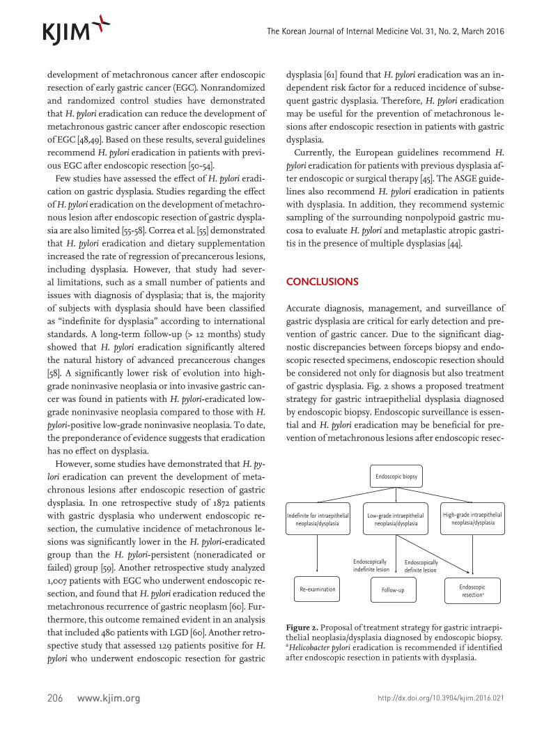
A concern regarding the accurate diagnosis of gastric dysplasia is that endoscopic forceps biopsy specimens are not representative of the entire lesion; therefore, significant discrepancies can be found between histo-logic diagnoses based on forceps biopsy and resected specimens (Fig. 1). A series of studies have reported that diagnosis of LGD by forceps biopsy could be upgraded to HGD or carcinoma. A recent meta-analysis [27] of 16 studies involving 3,303 patients with endoscopic forceps biopsy-proven gastric LGD lesions showed that 25% were diagnosed as advanced lesions, including gastric HGD (16.7%) and gastric carcinoma (6.9%) after endo-scopic resection. In other words, one out of four forceps biopsy-proven gastric LGDs might be underdiagnosed and should actually be HGD or even gastric carcinoma. This high rate of underdiagnosis indicates that merely a follow-up strategy is insufficient for patients with LGD.
Repeat endoscopic biopsy is a possible solution to this issue. However, this also has limitations, as demonstrat-ed by a 70.4% rate of histological concordance between repeat endoscopic biopsy and postendoscopic resection specimens [28].
Theoretically, obtaining a larger specimen using the large cup of jumbo biopsy forceps (open diameter of 8 mm) can increase diagnostic accuracy. However, a recent study reported that jumbo forceps biopsy specimens do not increase the concordance rate compared to conven-tional forceps (open diameter of 6.8 mm) specimens [29]. The authors instead recommended obtaining at least four endoscopic biopsy specimens to improve the his-tologic accuracy. This resulted in an increased concor-dance rate in LGD from 76.2% for the first endoscopic biopsy to 95.2% [29]. However, increasing the number or size of forceps biopsies can alter the neoplastic lesion and cause submucosal fibrosis, which can make endo-scopic resection problematic. Therefore, these strate-T
able
1. G
astr
ic e
pith
elia
l neo
plas
ia c
lass
ifica
tion
sys
tem
s
Japa
nes
e (J
RSG
C)
[7]
Wes
tern
:G
olds
tein
et a
l. [4
]Pa
dova
[40]
Vie
nna
[8]
Rev
ised
Vie
nna
[9]
WH
O [1
0]
Gro
up I:
Nor
mal
o
r ben
ign
Reac
tive
Cate
gory
1: N
egat
ive f
or d
yspl
asia
Cate
gory
1: N
egat
ive f
or d
yspl
asia
Cat
egor
y 1:
Neg
ativ
e fo
r dys
plas
ia N
o in
trae
pith
elia
l neo
plas
ia /d
yspl
asia
Gro
up II
: Ben
ign
with
aty
pia
Inde
finite
for
dys
plas
iaCa
tego
ry 2:
Inde
finite
for d
yspl
asia
Cate
gory
2: I
ndefi
nite
for d
yspl
asia
Cate
gory
2: I
ndefi
nite
for d
yspl
asia
Ind
efin
ite fo
r in
trae
pith
elia
l ne
opla
sia/
dysp
lasi
aG
roup
III:
Bor
derl
ine
Low-
grad
e ad
enom
a/dy
spla
sia
Cate
gory
3.1:
Non
inva
sive
low-
gra
de n
eopl
asia
(low
-gra
de ad
enom
a/dy
spla
sia)
Cate
gory
3: N
onin
vasi
ve lo
w- g
rade
neo
plas
ia (l
ow-g
rade
aden
oma/
dysp
lasi
a)
Cate
gory
3: M
ucos
al lo
w-gr
ade
neo
plas
ia (l
ow-g
rade
aden
oma/
dysp
lasi
a)
Low-
grad
e int
rae
pith
elia
l n
eopl
asia
/ d
yspl
asia
Gro
up IV
: Str
ongl
y s
uspi
ciou
s for
inva
sive
car
cino
ma
Hig
h-gr
ade
aden
oma/
dysp
lasi
aCa
tego
ry 3.
2: N
onin
vasi
ve h
igh-
gra
de n
eopl
asia
(hig
h-gr
ade a
deno
ma/
dysp
lasi
a)3.2
.1: S
uspi
ciou
s for
car
cino
ma
(with
out l
amin
a pro
pria
inva
sion
)3.2
.2: N
onin
vasi
ve c
arci
nom
a (CI
S)
Cate
gory
4: N
onin
vasi
ve h
igh-
grad
e neo
plas
ia4.
1: H
igh-
grad
e ade
nom
a/dy
spla
sia
4.2:
Non
inva
sive
car
cino
ma (
CIS)
4.3:
Susp
icio
us o
f inv
asiv
e c
arci
nom
a
Cate
gory
4: M
ucos
al h
igh-
grad
e n
eopl
asia
4.1:
Hig
h-gr
ade
aden
oma/
dysp
lasi
a4.
2: N
onin
vasi
ve c
arci
nom
a (CI
S)4.
3: Su
spic
ious
for i
nvas
ive
car
cino
ma
4.4:
Intr
amuc
osal
car
cino
ma
Hig
h-gr
ade i
ntra
epith
elia
l n
eopl
asia
/dys
plas
ia
Gro
up V
: Defi
nitiv
e fo
r in
vasi
ve c
arci
nom
a
Inva
sive
car
cino
ma
Cate
gory
4: S
uspi
ciou
s fo
r inv
asiv
e car
cino
ma
(with
lam
ina p
ropr
ia in
vasi
on)
Cate
gory
5: In
vasi
ve n
eopl
asia
(int
ram
ucos
al/s
ubm
ucos
al c
arci
nom
a or b
eyon
d)
Cate
gory
5: In
vasi
ve n
eopl
asia
5.1
: Int
ram
ucos
al c
arci
nom
a5.2
Sub
muc
osal
car
cino
ma
or b
eyon
d
Cate
gory
5: S
ubm
ucos
al in
vasi
on b
y car
cino
ma
Intr
amuc
osal
inva
sive
neo
plas
ia (i
ntra
muc
osal
inva
sive
car
cino
ma)
Inva
sive
neo
plas
ia
JRSG
C, J
apan
ese
Res
earc
h So
ciet
y fo
r G
astr
ic C
ance
r; W
HO
, Wor
ld H
ealt
h O
rgan
izat
ion;
CIS
, car
cino
ma
in si
tu.
204 www.kjim.org
The Korean Journal of Internal Medicine Vol. 31, No. 2, March 2016
http://dx.doi.org/10.3904/kjim.2016.021
gies to improve diagnostic accuracy have limitations in terms of clinical application.
It is important to determine the type of gastric LGD likely to be underdiagnosed. Various findings have been reported concerning this issue. It is generally accepted that the probability of malignant transformation of dys-plasia increases with lesion size. Adenoma 2 cm or more in diameter has been considered potentially malignant [30]. Several studies have confirmed that a lesion size ≥ 2 cm is an independent predictor of upgraded histology in LGD lesions. However, even small LGDs (< 2 cm) may
be upgraded to an HGD or carcinoma. One study indi-cated that ~20% of lesions with a diameter of less than 1 cm and about 35% of those with a diameter of 1 to 1.9 cm were HGD or carcinoma [31]. Other studies showed that forceps biopsy-proven gastric LGDs of ≥ 1 cm diameter were an independent risk factor for HGD or carcinoma [32,33].
The surface appearance of dysplasia has also been identified as a risk factor for upgrade to a diagnosis of HGD or carcinoma in gastric LGD after endoscopic re-section. Features associated with an upgraded diagnosis
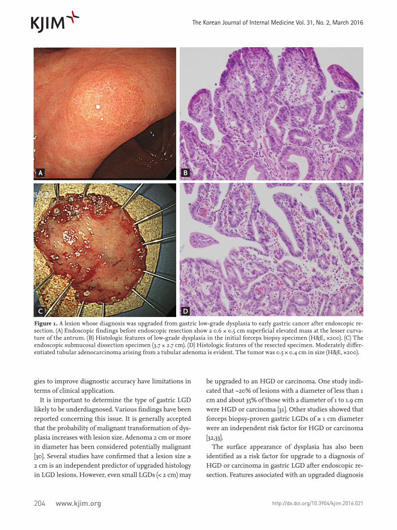
Figure 1. A lesion whose diagnosis was upgraded from gastric low-grade dysplasia to early gastric cancer after endoscopic re-section. (A) Endoscopic findings before endoscopic resection show a 0.6 × 0.5 cm superficial elevated mass at the lesser curva-ture of the antrum. (B) Histologic features of low-grade dysplasia in the initial forceps biopsy specimen (H&E, ×200). (C) The endoscopic submucosal dissection specimen (3.7 × 2.7 cm). (D) Histologic features of the resected specimen. Moderately differ-entiated tubular adenocarcinoma arising from a tubular adenoma is evident. The tumor was 0.5 × 0.4 cm in size (H&E, ×200).
A
C
B
D
205
Sung JK. Gastric dysplasia
www.kjim.orghttp://dx.doi.org/10.3904/kjim.2016.021
include depressed macroscopic type, surface erythema, surface unevenness (nodularity), and erosion or ulcer-ation [31,32,34-39]. The risk of malignancy is also related to the villosity of the growth pattern [33].
In summary, larger size; depressed gross type; a sur-face appearance with erythema, unevenness, ulcer, or erosion; and tubulovillous or villous histology on for-ceps biopsy specimens are predictive factors for an up-graded diagnosis in LGD patients following endoscopic resection. Therefore, it is recommended that an endo-scopic forceps biopsy-proven gastric LGD lesion with these predictive factors undergo endoscopic resection.
MANAGEMENT
Generally there is no controversy regarding the appro-priate management of HGD. Such lesions require en-doscopic resection due to the potential for progression to carcinoma and the coexistence of carcinoma. In con-trast, few definite guidelines regarding the management of LGD are available. Given the lower risk of malignant transformation, some investigators recommend annual endoscopic surveillance with rebiopsy for LGD [40,41], while others suggest that active resection is necessary because histological diagnosis based on forceps biopsy can be inaccurate due to sampling error, and the final diagnosis can be upgraded to HGD or even invasive car-cinoma after endoscopic resection [42]. Repeated endo-scopic examination with biopsies can impose a physical, psychological, and financial burden on the patient, al-though few studies of these issues have been reported [42]. In contrast, endoscopic resection is less invasive than surgical resection but also has a risk of complica-tions.
In the revised Vienna classification, endoscopic treat-ment or surgical local treatment was recommended for HGD [43]. In addition, endoscopic treatment or fol-low-up was recommended for LGD [43]. The choice of treatment is dependent on the size of the lesion; depth of invasion as assessed endoscopically, radiologically, or ultrasonographically; and other general factors (patient age and comorbidities) [43].
Several guidelines recommend endoscopic resection for gastric dysplasia. The most recent American Soci-ety for Gastrointestinal Endoscopy (ASGE) guidelines
[44] suggested that adenoma of any size should be re-moved endoscopically if possible. The British Society of Gastroenterology guidelines [13] also recommended complete removal of adenoma if safe to do so. The Eu-ropean guidelines [45] recommended that if LGD is di-agnosed endoscopically, endoscopic resection should be considered to obtain a more accurate histologic diagno-sis. Otherwise, endoscopically indefinite lesions should undergo follow-up within 1 year after diagnosis. In ad-dition, they recommended that endoscopic resection should be considered for patients with endoscopically defined HGD. If the lesion is endoscopically indistinct HGD, the guidelines recommend immediate endoscop-ic reassessment with extensive biopsy sampling and surveillance at 6- to 12-month intervals. Moreover, they highlighted that disappearance of dysplasia or its as-sumed disappearance as assessed by follow-up endo-scopic biopsies does not rule out the possible progres-sion to invasive cancer.
Finally, because there is a risk of synchronous carcino-ma in patients with gastric dysplasia, thorough evalua-tion of the entire stomach should be performed and any abnormalities biopsied. Endoscopic surveillance is also mandatory after resection to screen for metachronous lesions [13,46]. Concerning the surveillance schedule, the ASGE guidelines suggest surveillance endoscopy 1 year following resection of gastric dysplasia [44]. The British Society of Gastroenterology guidelines recommend en-doscopic follow-up 6 months after resection in patients with incompletely resected polyps or those with HGD, and 1 year after removal of other polyps [13].
HELICOBACTER PYLORI ERADICATION FOR PREVENTION OF METACHRONOUS LESIONS AFTER ENDOSCOPIC RESECTION OF GASTRIC DYSPLASIA
Previous studies have evaluated the effect of Helicobacter pylori eradication on preneoplastic lesions such as gas-tric atrophy and intestinal metaplasia. Conflicting re-sults have been reported regarding the reversibility of gastric atrophy and intestinal metaplasia after eradica-tion therapy. H. pylori eradication appears to improve atrophy but not intestinal metaplasia [47]. Another issue is whether H. pylori eradication reduces the subsequent
206 www.kjim.org
The Korean Journal of Internal Medicine Vol. 31, No. 2, March 2016
http://dx.doi.org/10.3904/kjim.2016.021
development of metachronous cancer after endoscopic resection of early gastric cancer (EGC). Nonrandomized and randomized control studies have demonstrated that H. pylori eradication can reduce the development of metachronous gastric cancer after endoscopic resection of EGC [48,49]. Based on these results, several guidelines recommend H. pylori eradication in patients with previ-ous EGC after endoscopic resection [50-54].
Few studies have assessed the effect of H. pylori eradi-cation on gastric dysplasia. Studies regarding the effect of H. pylori eradication on the development of metachro-nous lesion after endoscopic resection of gastric dyspla-sia are also limited [55-58]. Correa et al. [55] demonstrated that H. pylori eradication and dietary supplementation increased the rate of regression of precancerous lesions, including dysplasia. However, that study had sever-al limitations, such as a small number of patients and issues with diagnosis of dysplasia; that is, the majority of subjects with dysplasia should have been classified as “indefinite for dysplasia” according to international standards. A long-term follow-up (> 12 months) study showed that H. pylori eradication significantly altered the natural history of advanced precancerous changes [58]. A significantly lower risk of evolution into high-grade noninvasive neoplasia or into invasive gastric can-cer was found in patients with H. pylori-eradicated low-grade noninvasive neoplasia compared to those with H. pylori-positive low-grade noninvasive neoplasia. To date, the preponderance of evidence suggests that eradication has no effect on dysplasia.
However, some studies have demonstrated that H. py-lori eradication can prevent the development of meta-chronous lesions after endoscopic resection of gastric dysplasia. In one retrospective study of 1872 patients with gastric dysplasia who underwent endoscopic re-section, the cumulative incidence of metachronous le-sions was significantly lower in the H. pylori-eradicated group than the H. pylori-persistent (noneradicated or failed) group [59]. Another retrospective study analyzed 1,007 patients with EGC who underwent endoscopic re-section, and found that H. pylori eradication reduced the metachronous recurrence of gastric neoplasm [60]. Fur-thermore, this outcome remained evident in an analysis that included 480 patients with LGD [60]. Another retro-spective study that assessed 129 patients positive for H. pylori who underwent endoscopic resection for gastric
dysplasia [61] found that H. pylori eradication was an in-dependent risk factor for a reduced incidence of subse-quent gastric dysplasia. Therefore, H. pylori eradication may be useful for the prevention of metachronous le-sions after endoscopic resection in patients with gastric dysplasia.
Currently, the European guidelines recommend H. pylori eradication for patients with previous dysplasia af-ter endoscopic or surgical therapy [45]. The ASGE guide-lines also recommend H. pylori eradication in patients with dysplasia. In addition, they recommend systemic sampling of the surrounding nonpolypoid gastric mu-cosa to evaluate H. pylori and metaplastic atropic gastri-tis in the presence of multiple dysplasias [44].
CONCLUSIONS
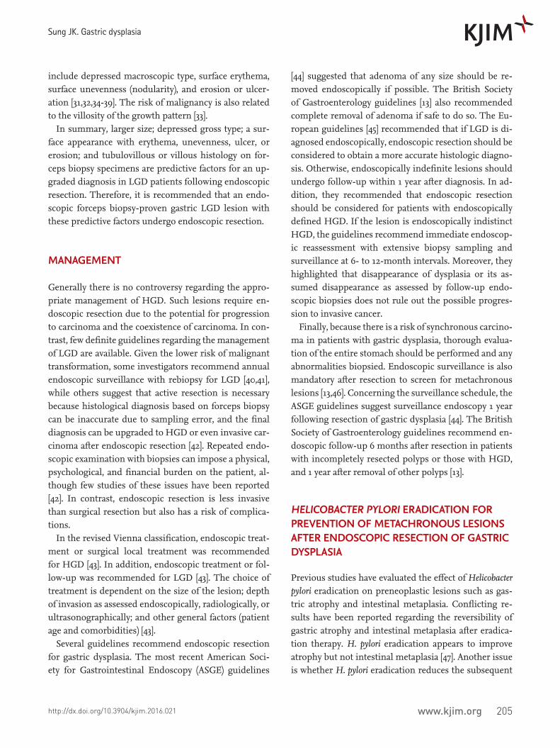
Accurate diagnosis, management, and surveillance of gastric dysplasia are critical for early detection and pre-vention of gastric cancer. Due to the significant diag-nostic discrepancies between forceps biopsy and endo-scopic resected specimens, endoscopic resection should be considered not only for diagnosis but also treatment of gastric dysplasia. Fig. 2 shows a proposed treatment strategy for gastric intraepithelial dysplasia diagnosed by endoscopic biopsy. Endoscopic surveillance is essen-tial and H. pylori eradication may be beneficial for pre-vention of metachronous lesions after endoscopic resec-
Indefinite for intraepithelial neoplasia/dysplasia
Low-grade intraepithelial neoplasia/dysplasia
High-grade intraepithelial neoplasia/dysplasia
Follow-up
Endoscopic biopsy
Endoscopic resectiona
Endoscopically indefinite lesion Endoscopically
definite lesion
Re-examination
Figure 2. Proposal of treatment strategy for gastric intraepi-thelial neoplasia/dysplasia diagnosed by endoscopic biopsy. aHelicobacter pylori eradication is recommended if identified after endoscopic resection in patients with dysplasia.
207
Sung JK. Gastric dysplasia
www.kjim.orghttp://dx.doi.org/10.3904/kjim.2016.021
tion in patients with gastric dysplasia.
Conflict of interestNo potential conflict of interest relevant to this article was reported.
REFERENCES
1. Correa P. A human model of gastric carcinogenesis. Can-cer Res 1988;48:3554-3560.
2. Morson BC, Sobin LH, Grundmann E, Johansen A, Nagayo T, Serck-Hanssen A. Precancerous conditions and epithelial dysplasia in the stomach. J Clin Pathol 1980;33:711-721.
3. Ming SC. Dysplasia of gastric epithelium. Front Gastro-intest Res 1979;4:164-172.
4. Goldstein NS, Lewin KJ. Gastric epithelial dysplasia and adenoma: historical review and histological criteria for grading. Hum Pathol 1997;28:127-133.
5. Lewin KJ. Nomenclature problems of gastrointestinal ep-ithelial neoplasia. Am J Surg Pathol 1998;22:1043-1047.
6. Schlemper RJ, Kato Y, Stolte M. Review of histological classifications of gastrointestinal epithelial neoplasia: differences in diagnosis of early carcinomas between Japanese and Western pathologists. J Gastroenterol 2001;36:445-456.
7. Schlemper RJ, Itabashi M, Kato Y, et al. Differences in di-agnostic criteria for gastric carcinoma between Japanese and western pathologists. Lancet 1997;349:1725-1729.
8. Schlemper RJ, Riddell RH, Kato Y, et al. The Vienna clas-sification of gastrointestinal epithelial neoplasia. Gut 2000;47:251-255.
9. Schlemper RJ, Kato Y, Stolte M. Diagnostic criteria for gastrointestinal carcinomas in Japan and Western coun-tries: proposal for a new classification system of gastro-intestinal epithelial neoplasia. J Gastroenterol Hepatol 2000;15 Suppl:G49-G57.
10. Yakirevich E, Resnick MB. Pathology of gastric cancer and its precursor lesions. Gastroenterol Clin North Am 2013;42:261-284.
11. Lauwers GY, Riddell RH. Gastric epithelial dysplasia. Gut 1999;45:784-790.
12. Setia N, Lauwers GY. Gastric dysplasia: update and practi-cal approach. Diagn Histopathol 2015;21:312-322.
13. Goddard AF, Badreldin R, Pritchard DM, Walker MM,
Warren B; British Society of Gastroenterology. The man-agement of gastric polyps. Gut 2010;59:1270-1276.
14. Lansdown M, Quirke P, Dixon MF, Axon AT, Johnston D. High grade dysplasia of the gastric mucosa: a marker for gastric carcinoma. Gut 1990;31:977-983.
15. Di Gregorio C, Morandi P, Fante R, De Gaetani C. Gas-tric dysplasia: a follow-up study. Am J Gastroenterol 1993;88:1714-1719.
16. Rugge M, Farinati F, Di Mario F, Baffa R, Valiante F, Car-din F. Gastric epithelial dysplasia: a prospective multi-center follow-up study from the Interdisciplinary Group on Gastric Epithelial Dysplasia. Hum Pathol 1991;22:1002-1008.
17. Saraga EP, Gardiol D, Costa J. Gastric dysplasia: a histo-logical follow-up study. Am J Surg Pathol 1987;11:788-796.
18. Fertitta AM, Comin U, Terruzzi V, et al. Clinical signifi-cance of gastric dysplasia: a multicenter follow-up study: Gastrointestinal Endoscopic Pathology Study Group. En-doscopy 1993;25:265-268.
19. Kokkola A, Haapiainen R, Laxen F, et al. Risk of gastric carcinoma in patients with mucosal dysplasia associated with atrophic gastritis: a follow up study. J Clin Pathol 1996;49:979-984.
20. Yamada H, Ikegami M, Shimoda T, Takagi N, Maruyama M. Long-term follow-up study of gastric adenoma/dys-plasia. Endoscopy 2004;36:390-396.
21. de Vries AC, van Grieken NC, Looman CW, et al. Gastric cancer risk in patients with premalignant gastric lesions: a nationwide cohort study in the Netherlands. Gastroen-terology 2008;134:945-952.
22. Srivastava A, Lauwers GY. Gastric epithelial dysplasia: the Western perspective. Dig Liver Dis 2008;40:641-649.
23. Rugge M, Cassaro M, Di Mario F, et al. The long term out-come of gastric non-invasive neoplasia. Gut 2003;52:1111-1116.
24. Park SY, Jeon SW, Jung MK, et al. Long-term follow-up study of gastric intraepithelial neoplasias: progression from low-grade dysplasia to invasive carcinoma. Eur J Gastroenterol Hepatol 2008;20:966-970.
25. Carmack SW, Genta RM, Graham DY, Lauwers GY. Management of gastric polyps: a pathology-based guide for gastroenterologists. Nat Rev Gastroenterol Hepatol 2009;6:331-341.
26. Abraham SC, Park SJ, Lee JH, Mugartegui L, Wu TT. Ge-netic alterations in gastric adenomas of intestinal and foveolar phenotypes. Mod Pathol 2003;16:786-795.
208 www.kjim.org
The Korean Journal of Internal Medicine Vol. 31, No. 2, March 2016
http://dx.doi.org/10.3904/kjim.2016.021
27. Zhao G, Xue M, Hu Y, Lai S, Chen S, Wang L. How com-monly is the diagnosis of gastric low grade dysplasia up-graded following endoscopic resection? A meta-analysis. PLoS One 2015;10:e0132699.
28. Nam KW, Song KS, Lee HY, et al. Spectrum of final patho-logical diagnosis of gastric adenoma after endoscopic resection. World J Gastroenterol 2011;17:5177-5183.
29. Jeon HK, Ryu HY, Cho MY, et al. A randomized trial to determine the diagnostic accuracy of conventional vs. jumbo forceps biopsy of gastric epithelial neoplasias be-fore endoscopic submucosal dissection: open-label study. Gastric Cancer 2014;17:661-668.
30. Tomasulo J. Gastric polyps. Histologic types and their re-lationship to gastric carcinoma. Cancer 1971;27:1346-1355.
31. Cho SJ, Choi IJ, Kim CG, et al. Risk of high-grade dys-plasia or carcinoma in gastric biopsy-proven low-grade dysplasia: an analysis using the Vienna classification. En-doscopy 2011;43:465-471.
32. Choi CW, Kim HW, Shin DH, et al. The risk factors for discrepancy after endoscopic submucosal dissection of gastric category 3 lesion (low grade dysplasia). Dig Dis Sci 2014;59:421-427.
33. Min BH, Kim KM, Kim ER, et al. Endoscopic and histo-pathological characteristics suggesting the presence of gastric mucosal high grade neoplasia foci in cases initial-ly diagnosed as gastric mucosal low grade neoplasia by forceps biopsy in Korea. J Gastroenterol 2011;46:17-24.
34. Kim MK, Jang JY, Kim JW, et al. Is lesion size an indepen-dent indication for endoscopic resection of biopsy-prov-en low-grade gastric dysplasia? Dig Dis Sci 2014;59:428-435.
35. Lim H, Jung HY, Park YS, et al. Discrepancy between en-doscopic forceps biopsy and endoscopic resection in gas-tric epithelial neoplasia. Surg Endosc 2014;28:1256-1262.
36. Won CS, Cho MY, Kim HS, et al. Upgrade of lesions initially diagnosed as low-grade gastric dysplasia upon forceps biopsy following endoscopic resection. Gut Liver 2011;5:187-193.
37. Lee CK, Chung IK, Lee SH, et al. Is endoscopic forceps biopsy enough for a definitive diagnosis of gastric epithe-lial neoplasia? J Gastroenterol Hepatol 2010;25:1507-1513.
38. Park DI, Rhee PL, Kim JE, et al. Risk factors suggesting malignant transformation of gastric adenoma: univariate and multivariate analysis. Endoscopy 2001;33:501-506.
39. Kim JH, Kim YJ, An J, et al. Endoscopic features sug-gesting gastric cancer in biopsy-proven gastric adeno-
ma with high-grade neoplasia. World J Gastroenterol 2014;20:12233-12240.
40. Rugge M, Nitti D, Farinati F, di Mario F, Genta RM. Non-invasive neoplasia of the stomach. Eur J Gastroen-terol Hepatol 2005;17:1191-1196.
41. Weinstein WM, Goldstein NS. Gastric dysplasia and its management. Gastroenterology 1994;107:1543-1545.
42. Nishida T, Tsutsui S, Kato M, et al. Treatment strategy for gastric non-invasive intraepithelial neoplasia diagnosed by endoscopic biopsy. World J Gastrointest Pathophysiol 2011;2:93-99.
43. Dixon MF. Gastrointestinal epithelial neoplasia: Vienna revisited. Gut 2002;51:130-131.
44. ASGE Standards of Practice Committee, Evans JA, Chan-drasekhara V, et al. The role of endoscopy in the manage-ment of premalignant and malignant conditions of the stomach. Gastrointest Endosc 2015;82:1-8.
45. Dinis-Ribeiro M, Areia M, de Vries AC, et al. Management of precancerous conditions and lesions in the stomach (MAPS): guideline from the European Society of Gas-trointestinal Endoscopy (ESGE), European Helicobacter Study Group (EHSG), European Society of Pathology (ESP), and the Sociedade Portuguesa de Endoscopia Di-gestiva (SPED). Endoscopy 2012;44:74-94.
46. Jang MY, Cho JW, Oh WG, et al. Clinicopathological characteristics of synchronous and metachronous gastric neoplasms after endoscopic submucosal dissection. Ko-rean J Intern Med 2013;28:687-693.
47. Choi IJ. Current evidence of effects of Helicobacter pylori eradication on prevention of gastric cancer. Korean J In-tern Med 2013;28:525-537.
48. Uemura N, Mukai T, Okamoto S, et al. Effect of Helico-bacter pylori eradication on subsequent development of cancer after endoscopic resection of early gastric cancer. Cancer Epidemiol Biomarkers Prev 1997;6:639-642.
49. Fukase K, Kato M, Kikuchi S, et al. Effect of eradication of Helicobacter pylori on incidence of metachronous gas-tric carcinoma after endoscopic resection of early gastric cancer: an open-label, randomised controlled trial. Lan-cet 2008;372:392-397.
50. Chey WD, Wong BC; Practice Parameters Committee of the American College of Gastroenterology. American Col-lege of Gastroenterology guideline on the management of Helicobacter pylori infection. Am J Gastroenterol 2007;102:1808-1825.
51. Malfertheiner P, Megraud F, O’Morain CA, et al. Manage-
209
Sung JK. Gastric dysplasia
www.kjim.orghttp://dx.doi.org/10.3904/kjim.2016.021
ment of Helicobacter pylori infection: the Maastricht IV/ Florence Consensus Report. Gut 2012;61:646-664.
52. Asaka M, Kato M, Takahashi S, et al. Guidelines for the management of Helicobacter pylori infection in Japan: 2009 revised edition. Helicobacter 2010;15:1-20.
53. Fock KM, Katelaris P, Sugano K, et al. Second Asia-Pacific Consensus Guidelines for Helicobacter pylori infection. J Gastroenterol Hepatol 2009;24:1587-1600.
54. Kim SG, Jung HK, Lee HL, et al. Guidelines for the di-agnosis and treatment of Helicobacter pylori infection in Korea, 2013 revised edition. Korean J Gastroenterol 2013;62:3-26.
55. Correa P, Fontham ET, Bravo JC, et al. Chemopreven-tion of gastric dysplasia: randomized trial of antioxidant supplements and anti-helicobacter pylori therapy. J Natl Cancer Inst 2000;92:1881-1888.
56. Mera R, Fontham ET, Bravo LE, et al. Long term follow up of patients treated for Helicobacter pylori infection. Gut 2005;54:1536-1540.
57. You WC, Brown LM, Zhang L, et al. Randomized dou-ble-blind factorial trial of three treatments to reduce the prevalence of precancerous gastric lesions. J Natl Cancer Inst 2006;98:974-983.
58. Rugge M, Russo VM, Guido M. Review article: what have we learnt from gastric biopsy? Aliment Pharmacol Ther 2003;17 Suppl 2:68-74.
59. Shin SH, Jung da H, Kim JH, et al. Helicobacter pylori eradication prevents metachronous gastric neoplasms after endoscopic resection of gastric dysplasia. PLoS One 2015;10:e0143257.
60. Bae SE, Jung HY, Kang J, et al. Effect of Helicobacter py-lori eradication on metachronous recurrence after endo-scopic resection of gastric neoplasm. Am J Gastroenterol 2014;109:60-67.
61. Chon I, Choi C, Shin CM, Park YS, Kim N, Lee DH. Effect of Helicobacter pylori eradication on subsequent dys-plasia development after endoscopic resection of gastric dysplasia. Korean J Gastroenterol 2013;61:307-312.