Diagnosis and Classification of Sacral · PDF file• Medial fracture excursion was closely...
37
1 Diagnosis and Classification of Sacral Fractures Mohammed F. Shamji MD, PhD VuMedi Webinar Toronto Western Hospital – Spinal Neurosurgeon University of Toronto – Assistant Professor April 22, 2014 Learning outcomes • By the end of this session, participants will be able to: • Optimal diagnostic tests to investigate sacral fractures • Classification schemes to describe sacral fractures • Biomechanics of stable and unstable sacral fractures Sacrum • Large triangular bone at the base of the lower spine, angled forward in the sagittal plane • Name derived from Latin (sacrum) translated from Greek (hieron) = sacred or strong bone • Part of the animal offered in sacrificed • Belief that the soul of the man resides there • Slavic languages and German (“the cross bone” – Kreuzbein) and in Dutch (“the holy bone” – Heiligbeen)
Transcript of Diagnosis and Classification of Sacral · PDF file• Medial fracture excursion was closely...

1
Diagnosis and Classification of Sacral
Fractures
Mohammed F. Shamji MD, PhD VuMedi Webinar
Toronto Western Hospital – Spinal Neurosurgeon
University of Toronto – Assistant Professor
April 22, 2014
Learning outcomes
• By the end of this session, participants will be able to:
• Optimal diagnostic tests to investigate sacral fractures
• Classification schemes to describe sacral fractures
• Biomechanics of stable and unstable sacral fractures
Sacrum
• Large triangular bone at the base of the lower
spine, angled forward in the sagittal plane
• Name derived from Latin (sacrum) translated
from Greek (hieron) = sacred or strong bone
• Part of the animal offered in sacrificed
• Belief that the soul of the man resides there
• Slavic languages and German (“the cross
bone” – Kreuzbein) and in Dutch (“the holy
bone” – Heiligbeen)

2
Sacral Anatomy – Osteology - I
• Formed by fusion of the 5 sacral vertebrae
• Initially unfused, begin to fuse age 16-18 and completes at 34
• Initially ~20° forward angulation, increases during adulthood
• The pelvic surface is concave
• The dorsal surface is convex
• The lateral surface is broad above and narrows below
• The broad base is directed upward and forward
• The tapered apex is directed downward
Sacral Anatomy – Osteology - II
• Articulations
• Proximally – L5 vertebra
• Distally – coccyx
• Laterally – ilium
• The vertebral canal is triangular in shape superiorly and
inferiorly the posterior wall is often incomplete from
undeveloped laminae and spinous processes
• Contains 4 foramina on each side that transmit sacral nerves
Sacral Anatomy – Ligaments
• Anterior SI joint
• Resists external rotation
• Posterior SI joint and interosseous SI ligament
• Posterior tension band stabilization
• Iliolumbar ligaments
• Augment posterior stability
• Sacrospinous ligament (anterior sacrum to
ischial tuberosity)
• Resists external rotation
• Sacrotuberous ligament (sacrum behind
sacrospinous to ischial tuberosity)
• Resists shear and flexion

3
Sacral Anatomy – Biomechanics
• Cadaveric studies unreliable
• Radiographic studies limited in utility
• Implanted tantalum spheres into bones of pelvis
• Range of SI motion < 2°
• Rigid externally fixed devices
• Range of SI motion < 1°
• No muscles act on the SI joint to produce active physiologic movements
Stress-relieving joint
• Transmits load via first sacral segment through iliac wings to acetabulum
Sacral Anatomy – Neurology
• L5 nerve root runs on top of sacral ala
• S1-4 nerve roots transmitted through the sacral
foramina
• L5 nerve root
• Dermatome – dorsal foot to great toe
• Myotome – EHL, gluteus medius
• Reflex – medial hamstring reflex
• S1 nerve root
• Dermatome – lateral and plantar foot
• Myotome – gastrocnemius, soleus
• Reflex – Achilles’
• S2-5 nerve roots
• Bowel and bladder function
• Unilateral preservation normally adequate for control
Gibbons Classification
1 – no neurological deficit
2 – paresthesias / sensory changes only
3 – motor weakness, bowel/bladder intact
4 – motor or sensory deficits,
loss of bowel/bladder
Clinical Presentation
• History
• Physical Examination
• Diagnostic Tests
Plain radiographs
Sensitivity ~30%
AP pelvis
Inlet view (40° caudad)
Outlet view (40° cephalad)
If not overt, then suspect based on
symphysis widening or L5 TP fractures
CT scan
Choice test for diagnosis
MRI scan
Chocie test for neurological
deficits
Mechanism
Motor vehicle accident
Fall from height
Repetitive stress
Symptoms
Peripelvic pain
Neurological deficits
Inspection
Soft tissue trauma
Palpation
Test pelvic ring stability
Assess for SC fluid mass
Neurological examination
Light touch – LE and sacral
DRE + anal wink reflex
Bulbocavernosus, cremasteric reflex
Vascular examination
Distal pulses

4
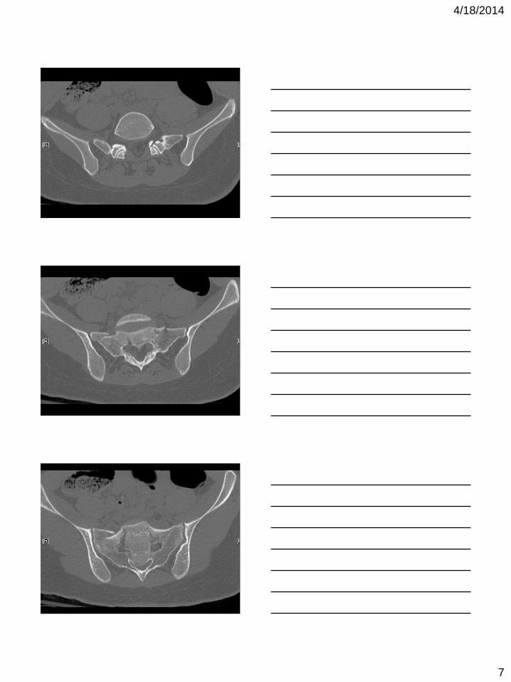
Choice CT Planes to Assess Fracture
Measurement CT Image
AP Displacement Axial
Vertical Translation Coronal
AP Translation Sagittal
Sagittal Angulation Sagittal
Canal Occlusion Axial
≥ 1 cm of displacement (either static
or on loading) generally marker of
pelvic instability
Conceptual Approach to Sacral Fracutres
• Presence of active bleeding
• Presence of open fracture
• Neurological injury
• Pattern and stability of the skeletal injury
• Systemic injury load
Classification Schemes
• Denis classification
• Denis Zone-I fractures
• Denis Zone-II fractures
• Denis Zone-III fractures
• Roy-Camille subclassification
• Denis Zone-III fractures
• Complex sacral fractures
• Denis Zone-III fractures
• Tile classification
• Global pelvic stability
• Isler classification
• Involvement of lumbosacral
articulation

5
Denis Classification – Overview
• Retrospective review (1988): 11 years, 236 patients
• Medial fracture excursion was closely associated with
mechanism of injury and neurological deficits
• Confirmed by Pohlemann (1992) with series of 377 patients, but
neurological deficit more correlated with Tile classification of
pelvic stability
Shortcoming – does not inform about mechanical stability
Denis Classification – Type I
• Location – fracture lateral to the sacral foramina
• Frequency – most common, 50%
• Neurological injury – infrequent, 6%, typically involves L5 nerve root
• Entrapment of L5 nerve root between the upwardly migrated fracture
fragment and the L5 transverse process
• Reduction of the sacral ala may promote L5 recovery
Denis Classification – Type II
• Location – fracture through the sacral foramina
• Frequency – second most common, 34%
• Neurological injury – common, 28%, frequently involves L5, S1, or S2
nerve roots
• Bladder dysfunction is rare

6
Denis Classification – Type III
• Location – fracture medial to the sacral foramina
• Frequency – least common, 16%
• Neurological injury – frequent, 57%, bowel, bladder, sexual function
impairment in 76% of those with neurological injury
• Vertical – almost always associated with pelvic ring fracture
• Horizontal – significant displacement produces severe deficit
Roy Camille Subclassification – Denis Type III
• Injury severity, likelihood of neurological injury, and therapeutic
implications directly related to increasingly severe types
• Type 1 – flexion deformity of the sacrum
• Type 2 – partially translated and hyperkyphotic
• Type 3 – completely translated and no fracture overlap
• Type 4 – segmental S1 comminution
Complex Sacral Fractures – Denis Type III
• H-type
• U-type
• Lambda-type
• T-type

7
Tile Classification of Pelvic Stability
Pohlemann (1992)
•Tile A – rare neurological deficit
•Tile B – maximum rate 10%
•Tile C – Denis I (33%), Denis II (43%), Denis III (64%)
Isler Classification – Lumbosacral Articulation
• Isler 1 - fracture occurs lateral to the L5/S1 facet
• Isler 2 - fractures line involves the L5/S1 facet
• Isler 3 - fracture line extends medially to the L5/S1 facet
Question 1
• 37F with history of major depression
• History
• Suicide attempt, jump from 3rd floor
• Physical
• No open lesions
• Lower extremity power intact
• Absent rectal tone
• Perianal anesthesia
• Urinary retention
• Classification
• Pattern?
• Neurology?
• Stable or unstable?
4/21/2014
1
Pelvic Ring Injuries
Paul A Anderson MD
University of Wisconsin
Purpose
• Mechanism of Injury
– Pelvic
– Sacrum
• Classification
• Treatment
Mechanism of Injury
• High energy trauma
• Multiple injuries
• Significant soft tissue injury

4/21/2014
2
Bone Anatomy
• Paired ilia
• Sacrum
• No inherent stability
Biomechanical Function Sacrum
• Keystone
Resist outward movement Rest of pelvic ring
Biomechanical Function Sacrum
• Inlet view
– Reverse keystone
– Sacrum displaced internally

4/21/2014
3
Biomechanical Function Sacrum
• Keystone
• Inlet view
– Reverse keystone Sacrum displaced internally
• Outlet view
– Keystone
Important Stabilizing Ligaments
• Illiolumbar (L5)
• Sacroiliac ligaments
– Posterior
– Anterior
Tile JAAOS 1996
Important Stabilizing Ligaments
• Sacrotuberous
• Sacrospinous
• Very strong
• Essential to pelvic stability
Tile JAAOS 1996

4/21/2014
4
Important Stabilizing Ligaments
• Symphyseal
• Fibrocartilagenous joint
– Disc
– Reinforcing capsule
Internal Anatomy
• Lots of stuff!!!
• Don’t go there
Pelvic Ring Fracture
90% sacral fractures Pelvic ring injuries

4/21/2014
5
Mechanism of Injury
• Force direct to pelvis
– Ilium
– Pubis
– Ischium
• Indirect forces
– Hip / femur
External rotation
Direct lateral compression
Tile JAAOS 1996
Classification Pelvic Ring Injuries
• Morphology – Letournel
• Mechanism/ stability
– Young-Burgess
– Tile
• AO/ OTA
– Very complicated
Young - Burgess
• Anteroposterior compression (APC)
• Lateral compression
• Shear

4/21/2014
6
Anteroposterior Compression
• APC 1
– Symphysis disruption
– Posterior intact
APC 1
Tile JAAOS 1996
Anteroposterior Compression
• APC 1
– Posterior intact
– Symphysis disruption
• AP 2
– Partial lig disruption
APC 2 Tile JAAOS 1996
Anteroposterior Compression
• APC 1
– Posterior intact
– Symphysis disruption
• APC 2
– Partial lig disruption
• APC 3
– Complete sacroiliac disruption
APC 3
Tile JAAOS 1996

4/21/2014
7
Lateral Compression
• LC 1
– Sacral compression
– Pubis overlapping
LC 1
Tile JAAOS 1996
Lateral Compression
• LC 1
– Sacral compression
• LC 2
– Iliac wing fracture
LC 2
Tile JAAOS 1996
Lateral Compression
• LC 1
– Sacral compression
• LC 2
– Iliac wing fracture
• LC 3
– Windswept pelvis
LC 3
Tile JAAOS 1996

4/21/2014
8
Vertical Shear
• Highly unstable
• Complete sacroiliac disruption
• High degree nerve injury
• L5-S1 disruption
– Facets
– Spondylolisthesis
• Multiple TP fx
Tile JAAOS 1996
Treatment of Pelvic Ring Fractures Goals
• Reduce pelvis volume
• Correct hip malalignment
– Leg length
– Center of head displacement
• Stability
– Load transfer
– Sitting
– Standing/ walking
Indications
• Large topic
• Poor agreement
• Asc sacral fracture – Assess pelvic ring
– Unstable
– Consider stabilization • Pubis
• Acetabulum (if fractured)
• Posterior SI joint and sacrum

4/21/2014
9
Temporary Stabilization
• Hemodynamic instability
• Reduce pelvic volume
• Correct pelvic displacement
• Stabilize to allow clotting
Anterior Techniques
• External fixation
– Unfavorable
– Infections
– Poorly controls posterior
• Infix
– Pedicle screws systems
– Percutaneous
Anterior Techniques
• External fixation
– Unfavorable
– Infections
– Poorly controls posterior
• Infix
– Pedicle screws systems
– Percutaneous Langford JAAOS 2013

4/21/2014
10
Anterior Techniques
• Internal Fixation
– Pubic symphysis plating
– Screws
– Pelvic brim plating
Langford JAAOS 2013
Posterior Fixation
• Old techniques
– Plate across posterior ilium
– Trans-iliac rods
– Anterior plate SI joint
• Iliac ORIF
Langford JAAOS 2013
Posterior Fixation
• Sacroiliac screws
• Iliolumbar fixation

4/21/2014
11
Posterior Fixation
• Sacroiliac screws
• Iliolumbar fixation
Conclusion
• Pelvis ring injuries
• Component of sacral fractures
• Team approach

4/17/2014
1
RD Orr
Center for Spine Health
Cleveland Clinic
Very little in the literature
No Level 1 or 2 studies
There are no clear guidelines
Treatment usually done on case by case basis
Pattern/Mechanism
Energy
Associated injuries
Bone Quality
Lever arms

4/17/2014
2
Stress fracture
Low energy fall
High energy
As energy increases so does damage to soft tissue, displacement, neurologic injury and deformity
As energy increases likelihood of surgery increases
What was the dominant force exerted in the injury?
The human spine does not tolerate shear well If the fracture occurred as a result of shear forces or
if unstable in shear then risk of progression
Is this a Pelvic fracture or a distal spinal fracture? Look for associated fractures in the pelvic ring
U or H-type fractures with >1 cm displacement
> 20(?) degrees angulation
Will surgical treatment of the sacral fracture assist the recovery of the associated injuries?
Will treatment of the associated injuries affect the healing of the sacral injury? (i.e. protected weight bearing)

4/17/2014
3
Osteoporotic sacral fractures often low energy and have less associated soft tissue injury
Risk of fixation failure higher
Tends towards non-operative treatment
Sacral fractures often occur below lumbar fusions
The longer the adjacent level the higher the risk of further displacement
Sacral kyphosis leads to loss of sagittal balance which increases deforming forces
Energy of injury
High
Displaced >1 cm or>20 degrees Surgery
Not displaced
Associated injuries improved by
treatment Surgery
Associated injures unaffected/harmed
by treatment
Non operative treatment
Low
Isolated injury
Yes Non operative
treatment
No Treatment based on
associated injury
Associated long lever arm
Yes Surgery
No Non-operative
treatment

4/18/2014
1
SACRAL
FRACTURES
Timothy Moore, MD Depts of Orthopaedic Surgery and
Neurosciences
MetroHealth Medical Center
Associate Professor, Case Western Reserve School of Medicine
Case #1
• 54yo male
• Hi speed MVC
• Mult extremity injuries
• Head bleed
• Pelvic ring with sacrum
• ? Neuro
MetroHealth Department of Neurosciences

4/18/2014
2

4/18/2014
3

4/18/2014
4

4/18/2014
5

4/18/2014
6
Case #2
• 19yo female
• Car vs pedestrian
• Isolated injury
• Searing R le pain
• 0/5 plantar flexion R
MetroHealth Department of Neurosciences
4/18/2014
7

4/18/2014
8

4/18/2014
9

4/18/2014
10
Case #2
• 42yo male
• Hi speed MVC
• Multiple injuries
• Neuro intact
MetroHealth Department of Neurosciences

4/18/2014
11

4/18/2014
12

4/18/2014
13

4/18/2014
14

4/18/2014
15

4/18/2014
16
What is the most important
anatomic landmark to see when
inserting iliac bolts?
• Superior end plate of S1
• Superimposed sciatic notches
• Femoral heads
• PSIS starting point
• Teardrop
MetroHealth Department of Neurosciences