Diagnosis and classification of periodontal disease
16
Australian Dental Journal 2009; 54:(1 Suppl): S11–S26 doi: 10.1111/j.1834-7819.2009.01140.x Diagnosis and classification of periodontal disease J Highfield* *Discipline of Periodontics, Faculty of Dentistry, The University of Sydney, New South Wales. ABSTRACT Periodontal diseases have been recognized and treated for at least 5000 years. Clinicians have recognized for many years that there are apparent differences in the presentation of periodontal diseases and have attempted to classify these diseases. Systems of classifications of disease have arisen allowing clinicians to develop structures which can be used to identify diseases in relation to aetiology, pathogenesis and treatment. It allows us to organize effective treatment of our patients’ diseases. Once a disease has been diagnosed and classified, the aetiology of the condition and appropriate evidence-based treatment is suggested to the clinician. Common systems of classification also allow effective communication between health care professionals using a common language. Early attempts at classification were made on the basis of the clinical characteristics of the diseases or on theories of their aetiology. These attempts were unsupported by any evidence base. As scientific knowledge expanded, conventional pathology formed the basis of classification. More recently, this has been followed by systems of classification based upon our knowledge of the various periodontal infections and the host response to them. Classification of periodontal diseases has, however, proved problematic. Over much of the last century clinicians and researchers have grappled with the problem and have assembled periodically to review or develop the classification of the various forms of periodontal disease as research has expanded our knowledge of these diseases. This has resulted in frequent revisions and changes. A classification, however, should not be regarded as a permanent structure. It must be adaptable to change and evolve with the development of new knowledge. It is expected that systems of classification will change over time. This review examines the past and present classifications of the periodontal diseases. Keywords: Diagnosis, classification, periodontal diseases, gingivitis, periodontitis. Abbreviations and acronyms: AST = aspartate aminotransferase; CAL = clinical loss of attachment; GCF = gingival crevicular fluid; LJP = localized juvenile periodontitis; NUG = necrotizing ulcerative gingivitis; NUP = necrotizing ulcerative peridontitis. INTRODUCTION Periodontal disease is a disease, or more likely a number of diseases of the periodontal tissues that results in attachment loss and destruction of alveolar bone. The natural history of periodontal disease, in some but not all patients, results in tooth loss. 1 Periodontal disease, however, encompasses a wider spectrum of diseases than just periodontitis and the recognition of these diseases requires a diagnosis be made. Diagnosis is the recognition of the presence of a disease. 2 Clinical diagnosis of periodontal disease is made by the recognition of various signs and symptoms in the periodontal tissues which herald a departure from health. The diagnosis of periodontal disease demands a firm knowledge of what constitutes peri- odontal health. The healthy periodontium, 3 of which only the gingival tissues may be directly observed, is described as being stippled, pale pink or coral pink, in the Caucasian (Fig 1), with various degrees of pigmen- tation in other races. It is tightly adapted to the underlying tissues, with a knife edge margin where it abuts the tooth, The gingival margin is located, in the absence of pathology, at the cemento-enamel junction. It displays a scalloped edge configuration highest interdentally, where it constitutes the interdental papilla and lowest buccally and lingually. There is a gingival crevice where it abuts the tooth which in health is 1–3 mm deep. There is an absence of bleeding from the crevice on gentle probing. The crevice in health will show a small amount of interstitial fluid, gingival crevicular fluid. 4 The lateral wall of the crevice constitutes the free gingival margin. From the most apical extent of the free gingival to the mucogingival junction is the attached gingival which varies in width from 1 to 9 mm and has a stippled surface. It is an immobile tissue tightly bound down to the bone as a mucoperiostium and is a keratinized mucosa well suited to resist injury. Apical from the mucogingival junction ª 2009 Australian Dental Association S11
-
Upload
truongdieu -
Category
Documents
-
view
232 -
download
0
Transcript of Diagnosis and classification of periodontal disease
Australian Dental Journal 2009; 54:(1 Suppl): S11–S26
doi: 10.1111/j.1834-7819.2009.01140.x
Diagnosis and classification of periodontal disease
J Highfield*
*Discipline of Periodontics, Faculty of Dentistry, The University of Sydney, New South Wales.
ABSTRACT
Periodontal diseases have been recognized and treated for at least 5000 years. Clinicians have recognized for many yearsthat there are apparent differences in the presentation of periodontal diseases and have attempted to classify these diseases.Systems of classifications of disease have arisen allowing clinicians to develop structures which can be used to identifydiseases in relation to aetiology, pathogenesis and treatment. It allows us to organize effective treatment of our patients’diseases. Once a disease has been diagnosed and classified, the aetiology of the condition and appropriate evidence-basedtreatment is suggested to the clinician. Common systems of classification also allow effective communication between healthcare professionals using a common language. Early attempts at classification were made on the basis of the clinicalcharacteristics of the diseases or on theories of their aetiology. These attempts were unsupported by any evidence base. Asscientific knowledge expanded, conventional pathology formed the basis of classification. More recently, this has beenfollowed by systems of classification based upon our knowledge of the various periodontal infections and the host responseto them. Classification of periodontal diseases has, however, proved problematic. Over much of the last century cliniciansand researchers have grappled with the problem and have assembled periodically to review or develop the classification ofthe various forms of periodontal disease as research has expanded our knowledge of these diseases. This has resulted infrequent revisions and changes. A classification, however, should not be regarded as a permanent structure. It must beadaptable to change and evolve with the development of new knowledge. It is expected that systems of classification willchange over time. This review examines the past and present classifications of the periodontal diseases.
Keywords: Diagnosis, classification, periodontal diseases, gingivitis, periodontitis.
Abbreviations and acronyms: AST = aspartate aminotransferase; CAL = clinical loss of attachment; GCF = gingival crevicular fluid; LJP =localized juvenile periodontitis; NUG = necrotizing ulcerative gingivitis; NUP = necrotizing ulcerative peridontitis.
INTRODUCTION
Periodontal disease is a disease, or more likely anumber of diseases of the periodontal tissues thatresults in attachment loss and destruction of alveolarbone. The natural history of periodontal disease, insome but not all patients, results in tooth loss.1
Periodontal disease, however, encompasses a widerspectrum of diseases than just periodontitis and therecognition of these diseases requires a diagnosis bemade.
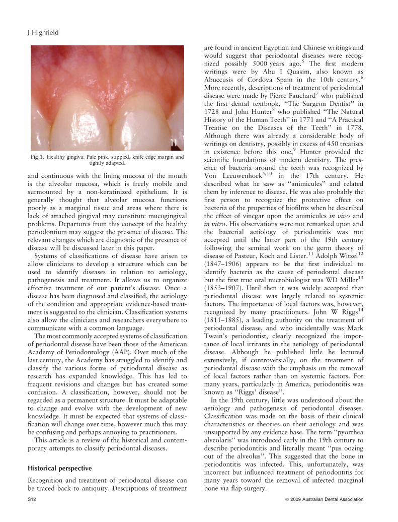
Diagnosis is the recognition of the presence of adisease.2 Clinical diagnosis of periodontal disease ismade by the recognition of various signs and symptomsin the periodontal tissues which herald a departurefrom health. The diagnosis of periodontal diseasedemands a firm knowledge of what constitutes peri-odontal health. The healthy periodontium,3 of whichonly the gingival tissues may be directly observed, isdescribed as being stippled, pale pink or coral pink, in
the Caucasian (Fig 1), with various degrees of pigmen-tation in other races. It is tightly adapted to theunderlying tissues, with a knife edge margin where itabuts the tooth, The gingival margin is located, in theabsence of pathology, at the cemento-enamel junction.It displays a scalloped edge configuration highestinterdentally, where it constitutes the interdentalpapilla and lowest buccally and lingually. There is agingival crevice where it abuts the tooth which in healthis 1–3 mm deep. There is an absence of bleeding fromthe crevice on gentle probing. The crevice in health willshow a small amount of interstitial fluid, gingivalcrevicular fluid.4 The lateral wall of the creviceconstitutes the free gingival margin. From the mostapical extent of the free gingival to the mucogingivaljunction is the attached gingival which varies in widthfrom 1 to 9 mm and has a stippled surface. It is animmobile tissue tightly bound down to the bone as amucoperiostium and is a keratinized mucosa well suitedto resist injury. Apical from the mucogingival junction
ª 2009 Australian Dental Association S11
and continuous with the lining mucosa of the mouthis the alveolar mucosa, which is freely mobile andsurmounted by a non-keratinized epithelium. It isgenerally thought that alveolar mucosa functionspoorly as a marginal tissue and areas where there islack of attached gingival may constitute mucogingivalproblems. Departures from this concept of the healthyperiodontium may suggest the presence of disease. Therelevant changes which are diagnostic of the presence ofdisease will be discussed later in this paper.
Systems of classifications of disease have arisen toallow clinicians to develop a structure which can beused to identify diseases in relation to aetiology,pathogenesis and treatment. It allows us to organizeeffective treatment of our patient’s disease. Once adisease has been diagnosed and classified, the aetiologyof the condition and appropriate evidence-based treat-ment is suggested to the clinician. Classification systemsalso allow the clinicians and researchers everywhere tocommunicate with a common language.
The most commonly accepted systems of classificationof periodontal disease have been those of the AmericanAcademy of Periodontology (AAP). Over much of thelast century, the Academy has struggled to identify andclassify the various forms of periodontal disease asresearch has expanded knowledge. This has led tofrequent revisions and changes but has created someconfusion. A classification, however, should not beregarded as a permanent structure. It must be adaptableto change and evolve with the development of newknowledge. It must be expected that systems of classi-fication will change over time, however much this maybe confusing and perhaps annoying to practitioners.
This article is a review of the historical and contem-porary attempts to classify periodontal diseases.
Historical perspective
Recognition and treatment of periodontal disease canbe traced back to antiquity. Descriptions of treatment
are found in ancient Egyptian and Chinese writings andwould suggest that periodontal diseases were recog-nized possibly 5000 years ago.5 The first modernwritings were by Abu I Quasim, also known asAbuccusis of Cordova Spain in the 10th century.6
More recently, descriptions of treatment of periodontaldisease were made by Pierre Fauchard7 who publishedthe first dental textbook, ‘‘The Surgeon Dentist’’ in1728 and John Hunter8 who published ‘‘The NaturalHistory of the Human Teeth’’ in 1771 and ‘‘A PracticalTreatise on the Diseases of the Teeth’’ in 1778.Although there was already a considerable body ofwritings on dentistry, possibly in excess of 450 treatisesin existence before this one,9 Hunter provided thescientific foundations of modern dentistry. The pres-ence of bacteria around the teeth was recognized byVon Leeuwenhoek5,10 in the 17th century. Hedescribed what he saw as ‘‘animicules’’ and relatedthem by inference to disease. He was also probably thefirst person to recognize the protective effect onbacteria of the properties of biofilms when he describedthe effect of vinegar upon the animicules in vivo andin vitro. His observations were not remarked upon andthe bacterial aetiology of periodontitis was notaccepted until the latter part of the 19th centuryfollowing the seminal work on the germ theory ofdisease of Pasteur, Koch and Lister.11 Adolph Witzel12
(1847–1906) appears to be the first individual toidentify bacteria as the cause of periodontal diseasebut the first true oral microbiologist was WD Miller13
(1853–1907). Until then it was widely accepted thatperiodontal disease was largely related to systemicfactors. The importance of local factors was, however,recognized by many practitioners. John W Riggs14
(1811–1885), a leading authority on the treatment ofperiodontal disease, and who incidentally was MarkTwain’s periodontist, clearly recognized the impor-tance of local irritants in the aetiology of periodontaldisease. Although he published little he lecturedextensively, if controversially, on the treatment ofperiodontal disease with the emphasis on the removalof local factors rather than on systemic factors. Formany years, particularly in America, periodontitis wasknown as ‘‘Riggs’ disease’’.
In the 19th century, little was understood about theaetiology and pathogenesis of periodontal diseases.Classification was made on the basis of their clinicalcharacteristics or theories on their aetiology and wasunsupported by any evidence base. The term ‘‘pyorrheaalveolaris’’ was introduced early in the 19th century todescribe periodontitis and literally meant ‘‘pus oozingout of the alveolus’’. This suggested that the bone inperiodontitis was infected. This, unfortunately, wasincorrect but influenced treatment of periodontitis formany years toward the removal of infected marginalbone via flap surgery.
Fig 1. Healthy gingiva. Pale pink, stippled, knife edge margin andtightly adapted.
S12 ª 2009 Australian Dental Association
J Highfield
Early attempts at classification reflect what Armit-age15 has termed the clinical characteristics paradigmwhich was in vogue from 1870 to 1920. He describesthree major paradigms of understanding which havehad a major influence on our attempts at classifyingperiodontal diseases: the clinical characteristics para-digm, the classic pathology paradigm and the infec-tion ⁄ host response paradigm. From 1920 to 1970 themajor influence was the classic pathology paradigm,and from 1970 to the present day the infection ⁄ hostresponse paradigm.
It had long been recognized clinically that thereappeared to be different types of periodontal diseaseand various attempts were made to define them.Gottlieb in 1921 attempted to split up the broad fieldof pyorrhea alveolaris into schmutz pyorrhea or filthpyorrhea, paradontal pyorrhea where there is deepseated disease within the gingival crevice that hygienemeasures do not alleviate, diffuse atrophy of thealveolar bone and accelerated eruption.16 Fish17
described pyorrhea simplex where there is gradualequal deepening of the sulcus and pyorrhea profundawhere isolated deep pockets existed with little generaldeepening of the sulcus around most teeth. Stillman andMcCall18 argued for the terms, gingivitis, ulatrophia,alveoloclasia, and pericementoclasia for disease pro-cesses attacking primarily the gingival tissues, thepericementum or the alveolar bone. Box19 dividedchronic periodontitis into complex and simplex, andforcefully ascribed a prominent role for occlusal traumain the aetiology of complex and, interestingly, cites insupport of his theory an article published in the DentalScience Journal of Australia in 1926 by R MorseWithycombe, the first periodontist to practise inSydney, Australia.
Almost every periodontist of note seemed to have hisindividual terminology. There was little agreement orcoordination until 1942 when Orban20 proposed aclassification scheme based on the principles of basicpathology. This was accepted by the American Acad-emy of Periodontology (AAP) and gained wide accep-tance. Periodontal disease was classified into broadgroups: inflammatory, dystrophic and traumatic distur-bances. Periodontitis was classified into simplex andcomplex. This was an attempt to classify the differencesin the presentation of periodontitis seen clinically.Simplex was secondary to gingivitis and characterizedby bone loss, pockets, abscess formation and calculusdeposits. Complex was secondary to periodontosis,which he considered a degenerative disease, havingsimilar aetiological factors to periodontitis and little orno calculus.
The terms simplex and complex gained fairly wideacceptance. Definitions, however, varied. MacPhee andCowley21 defined simplex as being characterized bypocket formation of regular depth throughout the
mouth and a pattern of horizontal bone loss. Complexwas characterized by advanced tissue destructionrelative to the age of the patient, pocket formation ofirregular depth around the mouth and irregularvertical bone loss. Complex also inferred that thedisease was not a simple response to local irritants butsuggested a co-factor in the aetiology, such as systemicfactors or local co-factors such as occlusal trauma.Many texts, however, continued to classify inflamma-tory periodontal disease simply as gingivitis andperiodontitis.22,23
The AAP further addressed the issue of classificationin the 1966 World Workshop in Periodontics.24 Theterm chronic marginal periodontitis was accepted butthe workshop failed to produce a definite system ofclassification for periodontitis. No agreement could bereached on the existence of periodontosis as a separatedisease and the suggestion of Loe that periodontosisbe called periodontitis complex was not supported.Emslie suggested further research be undertaken intoperiodontosis. The outcome of the workshop resultedin only one form of periodontitis, chronic marginal,being recognized. In 1977, however, the term juve-nile periodontitis, which had largely replaced theterm periodontosis, was accepted by the AAP. TheAcademy then recognized two distinct forms ofperiodontitis.
From 1970 to the present, Armitage15 stated thatthe infection ⁄ host response paradigm has been dom-inant. This has led to the development of the conceptthat periodontitis comprises a spectrum of relatedbut distinct diseases that differ in aetiology, naturalhistory progression and response to treatment. In1982, Page and Schroeder25 stated they could identifyat least five distinctly different forms of periodontitisin humans. They subclassified marginal periodontitisas adult periodontitis and rapidly progressive peri-odontitis of which they felt there may be at leastseveral types. They designated the forms of periodon-titis as prepubertal, juvenile, rapidly progressive,adult and acute necrotizing ulcerative gingivo-peri-odontitis. In November 1986, the AAP adopted anew classification which embraced these groups asfollows:
I. Juvenile periodontitisA. Prepubertal periodontitisB. Localized juvenile periodontitisC. Generalized juvenile periodontitis
II. Adult periodontitisIII. Necrotizing ulcerative gingivo-periodontitisIV. Refractory periodontitisA further workshop convened by the AAP at Princetonin 198926 amended the classification further. Thisremained the generally accepted classification for thenext 10 years. The main features of the revisedclassification were:
ª 2009 Australian Dental Association S13
Diagnosis and classification of periodontal disease

I. Adult periodontitisII. Early-onset periodontitis
A. Prepubertal periodontitis1. Generalized2. Localized
B. Juvenile periodontitis1. Generalized2. Localized
C. Rapidly progressive periodontitisIII. Periodontitis associated with systemic diseaseIV. Necrotizing ulcerative periodontitisV. Refractory periodontitis
This classification was based upon:1. Presence ⁄ absence of clinically detectable inflamma-
tion.2. Extent and pattern of attachment loss.3. Patient’s age at onset.4. Rate of progression.5. Presence ⁄ absence of miscellaneous signs and symp-
toms, including pain, ulceration and amount ofobservable plaque and calculus.
There were, however, voices of dissent at the workshopregarding this process. Hubert Newman, for example,argued that all periodontal diseases could be classifiedalong the lines of conventional pathology as had beendone previously.
A similar but simplified classification focusing uponadult, early onset and necrotizing ulcerative periodon-titis was produced by the First European Workshop onPeriodontics in 1993.27
Classification – the current situation
The 1989 classification and the simplified Europeanclassification gained widespread acceptance and usethroughout the world. Over time various problems withthe application of the classifications were observed andcriticisms arose. As observed by Armitage,28 thecriticisms largely related to the emphasis on age ofonset and rates of progression in the classificationwhich was felt to be inappropriate. ‘‘Early onset’’implies we have knowledge of when the disease startedand ‘‘rapidly progressive’’ implies knowledge of the rateof progression which in many cases we do not have. Itwas also observed that there was often considerableoverlap of disease categories, difficulty in fitting somepatients into any of the categories and the classificationcriteria were frequently found to be unclear or inade-quate. It was also observed that a gingival componentto the classification was absent. A further WorldWorkshop in Periodontics held by the AAP in 199629
did not produce a new classification. Reasons statedwere ‘‘considerable overlap among disease categories,certain patients do not fit in any category and manymicrobiological and host response features are sharedby multiple disease categories’’.
These concerns were further addressed and theclassification was revised in 1999 when the Interna-tional Workshop for Classification of PeriodontalDiseases and Conditions was convened.30 This resultedin the introduction of a gingival disease category. Adultperiodontitis was replaced by chronic periodontitis andearly onset periodontitis was replaced by aggressiveperiodontitis. Periodontitis associated with systemicdisease was redefined as periodontitis as a manifesta-tion of systemic disease and the new category necrotiz-ing periodontal diseases incorporated both necrotizinggingivitis and necrotizing periodontitis. Separate cate-gories for abscesses of the periodontium, periodontitisassociated with endodontic lesions and development oracquired conditions were added. Refractory periodon-titis was removed as a disease category.
The 1999 AAP Classification28 (Fig 2) is encyclo-paedic. It is very complete, detailed and complex andperhaps does not lend itself for use in its entirety on adaily basis by practitioners. A more convenient andsimplified summary is:
I. Gingival diseasesA. Plaque inducedB. Non-plaque induced
II. Chronic periodontitisA. LocalizedB. Generalized
III. Aggressive periodontitisA. LocalizedB. Generalized
IV. Periodontitis as a manifestation of systemicdisease
V. Necrotizing periodontal diseasesVI. Abscesses of the periodontium
VII. Periodontitis associated with endodontic lesionsVIII. Developmental or acquired deformities and
conditions.
Gingival conditions
This is an important inclusion in the classification.Gingival lesions are classified into two broad categories.Plaque induced and non-plaque induced. Dental plaqueinduced lesions (gingivitis) may be purely plaque relatedwith or without local contributing factors (Fig 3) or maybe modified by systemic factors, medications (Fig 4) orby malnutrition. It should be noted that, although bydefinition gingivitis has been traditionally described asbeing associated with a periodontium where there hasbeen no loss of attachment, it is possible for gingivitis tooccur on a periodontium with a reduced attachmentlevel which is stable and not experiencing progressiveloss of attachment. Non-plaque induced gingival lesionsencompass those caused by specific bacterial, fungal orviral infections, genetic origin, systemic conditions(dermatological conditions, allergic reactions), foreign
S14 ª 2009 Australian Dental Association
J Highfield

(a)
(b)
Fig 2. Classification of periodontal diseases and conditions. The American Academy of Periodontology. (Reproduced with permission from theAmerican Academy of Periodontology). *Can occur on a periodontium with no attachment loss or on a periodontium with attachment loss thatis not progressing. �Can be further classified on the basis of extent and severity. As a general guide, extent can be characterized as localized = £ ofsites involved and generalized = > of sites involved. Severity can be characterized on the basis of the amount of clinical attachment loss (CAL)
as follows: slight=1 or 2 mm CAL; moderate = 3 or 4 mm CAL; and severe = ‡ mm CAL.
ª 2009 Australian Dental Association S15
Diagnosis and classification of periodontal disease
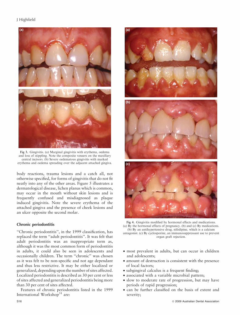
body reactions, trauma lesions and a catch all, nototherwise specified, for forms of gingivitis that do not fitneatly into any of the other areas. Figure 5 illustrates adermatological disease, lichen planus which is common,may occur in the mouth without skin lesions and isfrequently confused and misdiagnosed as plaqueinduced gingivitis. Note the severe erythema of theattached gingiva and the presence of cheek lesions andan ulcer opposite the second molar.
Chronic periodontitis
‘‘Chronic periodontitis’’, in the 1999 classification, hasreplaced the term ‘‘adult periodontitis’’. It was felt thatadult periodontitis was an inappropriate term as,although it was the most common form of periodontitisin adults, it could also be seen in adolescents andoccasionally children. The term ‘‘chronic’’ was chosenas it was felt to be non-specific and not age dependantand thus less restrictive. It may be either localized orgeneralized, depending upon the number of sites affected.Localized periodontitis is described as 30 per cent or lessof sites affected and generalized periodontitis being morethan 30 per cent of sites affected.
Features of chronic periodontitis listed in the 1999International Workshop31 are:
• most prevalent in adults, but can occur in childrenand adolescents;
• amount of destruction is consistent with the presenceof local factors;
• subgingival calculus is a frequent finding;• associated with a variable microbial pattern;• slow to moderate rate of progression, but may have
periods of rapid progression;• can be further classified on the basis of extent and
severity;
(a)
(b)
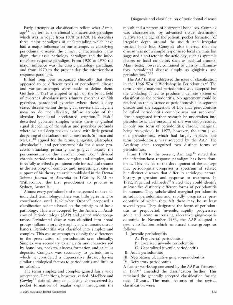
Fig 3. Gingivitis. (a) Marginal gingivitis with erythema, oedemaand loss of stippling. Note the composite veneers on the maxillarycentral incisors. (b) Severe oedematous gingivitis with marked
erythema and oedema spreading over the adjacent attached gingiva.
(a)
(b)
(c)
Fig 4. Gingivitis modified by hormonal effects and medications.(a) By the hormonal effects of pregnancy. (b) and (c) By medications.
(b) By an antihypertensive drug, nifedipine, which is a calciumantagonist. (c) By cyclosporine, an immunosuppressant use to prevent
organ graft rejection.
S16 ª 2009 Australian Dental Association
J Highfield
• can be associated with local predisposing factors (e.g.,tooth-related or iatrogenic factors);
• may be modified by and ⁄ or associated with systemicdiseases (e.g., diabetes mellitus, HIV infection);
• can be modified by factors other than systemic diseasesuch as cigarette smoking and emotional stress.
The workshop also produced the following workingdefinition and features of chronic periodontitis: ‘‘Aninfectious disease resulting in inflammation within thesupporting structures of teeth, progressive attachmentand bone loss. It is characterized by pocket formationand ⁄ or gingival recession. It is recognized as the mostfrequently occurring form of periodontitis. Its onsetmay be at any age but is most commonly detected inadults. The prevalence and severity of the diseaseincreases with age. It may affect a variable number ofteeth and has variable rates of progression.’’
As a guide, severity of the disease has traditionallybeen characterized as being slight or early where boneloss is in the coronal third of the root, moderate wherebone loss is in the middle third of the root andadvanced when in the apical third of the root length.The workshop categorized a general guide for severityon the basis of clinical loss of attachment (CAL) as
follows: slight = 1–2 mm CAL; moderate = 3 to 4 mmCAL; and severe = 5 mm CAL.
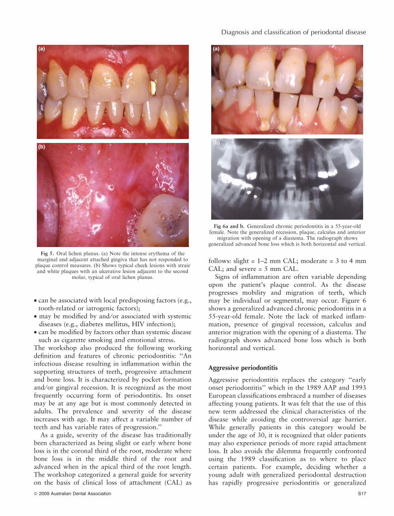
Signs of inflammation are often variable dependingupon the patient’s plaque control. As the diseaseprogresses mobility and migration of teeth, whichmay be individual or segmental, may occur. Figure 6shows a generalized advanced chronic periodontitis in a55-year-old female. Note the lack of marked inflam-mation, presence of gingival recession, calculus andanterior migration with the opening of a diastema. Theradiograph shows advanced bone loss which is bothhorizontal and vertical.
Aggressive periodontitis
Aggressive periodontitis replaces the category ‘‘earlyonset periodontitis’’ which in the 1989 AAP and 1993European classifications embraced a number of diseasesaffecting young patients. It was felt that the use of thisnew term addressed the clinical characteristics of thedisease while avoiding the controversial age barrier.While generally patients in this category would beunder the age of 30, it is recognized that older patientsmay also experience periods of more rapid attachmentloss. It also avoids the dilemma frequently confrontedusing the 1989 classification as to where to placecertain patients. For example, deciding whether ayoung adult with generalized periodontal destructionhas rapidly progressive periodontitis or generalized
(a)
(b)
Fig 6a and b. Generalized chronic periodontitis in a 55-year-oldfemale. Note the generalized recession, plaque, calculus and anterior
migration with opening of a diastema. The radiograph showsgeneralized advanced bone loss which is both horizontal and vertical.
(a)
(b)
Fig 5. Oral lichen planus. (a) Note the intense erythema of themarginal and adjacent attached gingiva that has not responded toplaque control measures. (b) Shows typical cheek lesions with straieand white plaques with an ulcerative lesion adjacent to the second
molar, typical of oral lichen planus.
ª 2009 Australian Dental Association S17
Diagnosis and classification of periodontal disease

juvenile periodontitis under the 1989 classificationhighlights the problem with age dependency and ratesof progression. A diagnosis of generalized juvenileperiodontitis, which has persisted into adulthood,requires knowledge of the time of onset. Alternatively,the condition may be a rapidly progressive periodon-titis. However, making this diagnosis requiresknowledge of rates of progression and time of onsetwhich can only be gained from previous records, whichin many cases may or may not be available (Fig 7).Further, is it appropriate to classify it as a juvenileperiodontitis when the patient is now an adult? Shouldthe classification be changed to something else and, ifso, what? It may not fit into any of our categories. Onepurpose of a classification system is to provide aframework in which to undertake orderly treatmentof a disease. In this instance the classification system is
contributing to confusion and is not suggesting appro-priate treatment. Should we be carrying out treatmentappropriate for a rapidly progressive disease or treat-ment for a generalized juvenile periodontitis which mayno longer be rapidly progressive? Localized juvenileperiodontitis has a circumpubertal onset and progressesvery rapidly for a number of years then frequently goesinto remission,32 becoming more generalized and, asSuzuki33 suggests, clinically similar to adult (chronic)periodontitis. Thus, we may be treating what under the1999 classification is a chronic periodontitis. The newclassification system attempts to avoid these problemsby simplifying the diagnosis. The treatment dilemma,however, may still exist with the new classification whenwe have imperfect knowledge of the disease history.
The workshop decided to discard all classificationsthat were age dependant or where knowledge of rates ofprogression was required. Patients previously classifiedas having rapidly progressive periodontitis were classi-fied as having either generalized aggressive periodontitisor chronic periodontitis, depending upon their clinicalcharacteristics. The term localized juvenile periodontitiswas replaced by localized aggressive periodontitis, andprepubertal periodontitis was removed as a separatedisease category. In most patients given the classificationof generalized prepubertal periodontitis, the periodon-titis was found to be a manifestation of a systemicdisease. Those prepubescent children with periodontitiswithout any modifying systemic conditions would fitunder the chronic or aggressive disease categories.
Common features of localized and generalized formsof aggressive periodontitis listed in the 1999 work-shop34 are:• except for the presence of periodontitis, patients are
otherwise clinically healthy;• rapid attachment loss and bone destruction;• familial aggregation.Secondary features that are generally, but not univer-sally, present are:• amounts of microbial deposits are inconsistent with
the severity of periodontal tissue destruction;• elevated proportions of Actinobacillus actinomyce-
temcomitans (Aggregatibacter actinomycetemcomi-tans) and, in some populations, Porphyromonasgingivalis may be elevated;
• phagocyte abnormalities;• hyper-responsive macrophage phenotype, including
elevated levels of PGE2 and IL-1b;• progression of attachment loss and bone loss may be
self arresting.Further specific features were identified:
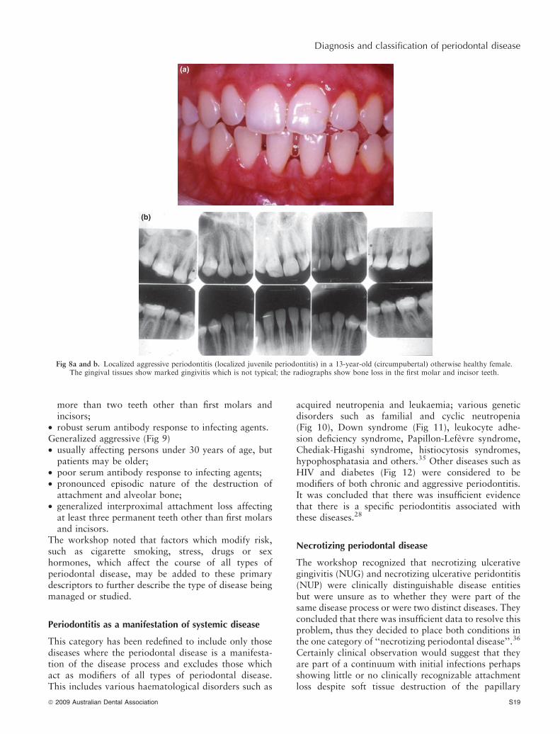
Localized aggressive (Fig 8)• circumpubertal onset;• localized first molar ⁄ incisor presentation with inter-
proximal attachment loss on at least two permanentteeth, one of which is a first molar, and involving no
(a)
(b)
(c)
Fig 7. Rapidly progressive periodontitis (generalized aggressive peri-odontitis). (a) Clinical photo of male aged 21 years showing anteriorgingival recession reflecting attachment loss. Inflammation is not
marked as the patient had received some treatment and the disease wasin remission. (b) Radiograph at age 16 showing normal bone levels.
(c) Radiograph at 21 showing advanced bone destructionillustrating rapid destruction over five years.
S18 ª 2009 Australian Dental Association
J Highfield
more than two teeth other than first molars andincisors;
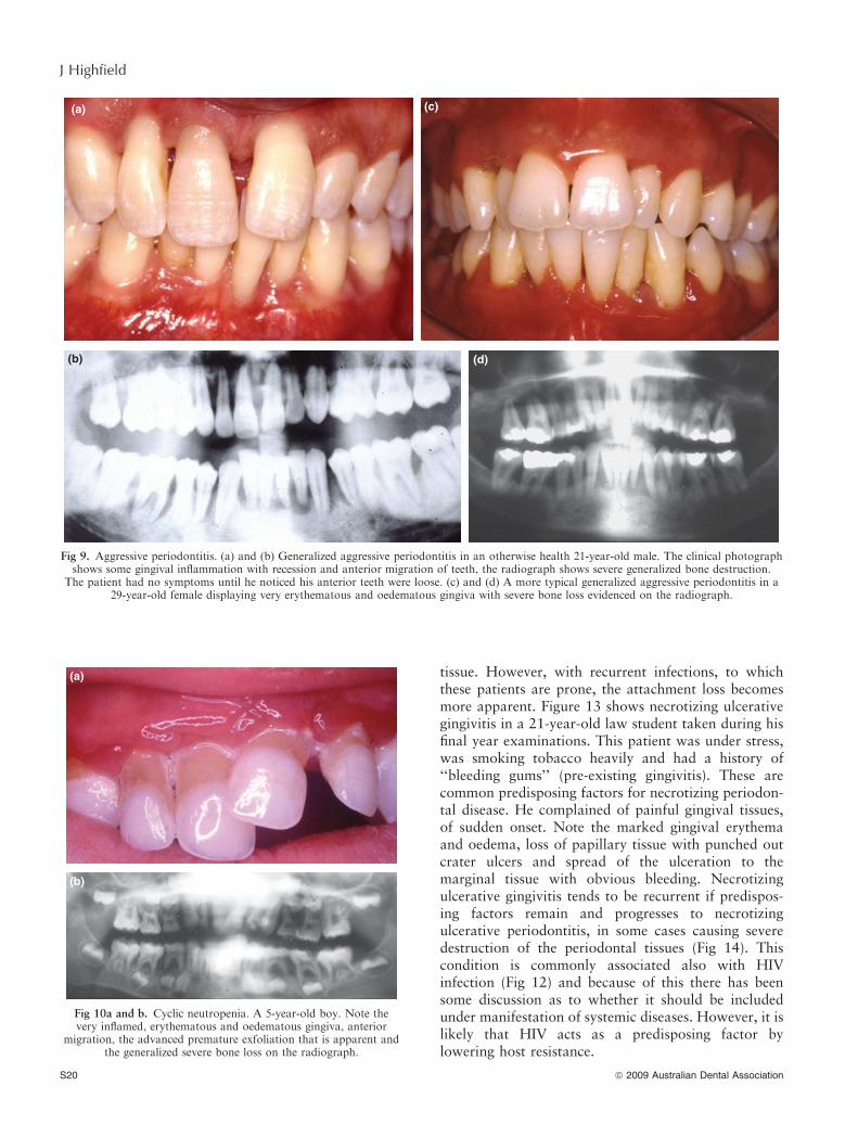
• robust serum antibody response to infecting agents.Generalized aggressive (Fig 9)• usually affecting persons under 30 years of age, but
patients may be older;• poor serum antibody response to infecting agents;• pronounced episodic nature of the destruction of
attachment and alveolar bone;• generalized interproximal attachment loss affecting
at least three permanent teeth other than first molarsand incisors.
The workshop noted that factors which modify risk,such as cigarette smoking, stress, drugs or sexhormones, which affect the course of all types ofperiodontal disease, may be added to these primarydescriptors to further describe the type of disease beingmanaged or studied.
Periodontitis as a manifestation of systemic disease
This category has been redefined to include only thosediseases where the periodontal disease is a manifesta-tion of the disease process and excludes those whichact as modifiers of all types of periodontal disease.This includes various haematological disorders such as
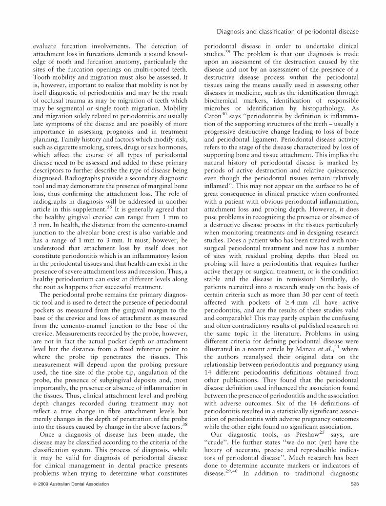
acquired neutropenia and leukaemia; various geneticdisorders such as familial and cyclic neutropenia(Fig 10), Down syndrome (Fig 11), leukocyte adhe-sion deficiency syndrome, Papillon-Lefevre syndrome,Chediak-Higashi syndrome, histiocytosis syndromes,hypophosphatasia and others.35 Other diseases such asHIV and diabetes (Fig 12) were considered to bemodifiers of both chronic and aggressive periodontitis.It was concluded that there was insufficient evidencethat there is a specific periodontitis associated withthese diseases.28
Necrotizing periodontal disease
The workshop recognized that necrotizing ulcerativegingivitis (NUG) and necrotizing ulcerative peridontitis(NUP) were clinically distinguishable disease entitiesbut were unsure as to whether they were part of thesame disease process or were two distinct diseases. Theyconcluded that there was insufficient data to resolve thisproblem, thus they decided to place both conditions inthe one category of ‘‘necrotizing periodontal disease’’.36
Certainly clinical observation would suggest that theyare part of a continuum with initial infections perhapsshowing little or no clinically recognizable attachmentloss despite soft tissue destruction of the papillary
(a)
(b)
Fig 8a and b. Localized aggressive periodontitis (localized juvenile periodontitis) in a 13-year-old (circumpubertal) otherwise healthy female.The gingival tissues show marked gingivitis which is not typical; the radiographs show bone loss in the first molar and incisor teeth.
ª 2009 Australian Dental Association S19
Diagnosis and classification of periodontal disease
tissue. However, with recurrent infections, to whichthese patients are prone, the attachment loss becomesmore apparent. Figure 13 shows necrotizing ulcerativegingivitis in a 21-year-old law student taken during hisfinal year examinations. This patient was under stress,was smoking tobacco heavily and had a history of‘‘bleeding gums’’ (pre-existing gingivitis). These arecommon predisposing factors for necrotizing periodon-tal disease. He complained of painful gingival tissues,of sudden onset. Note the marked gingival erythemaand oedema, loss of papillary tissue with punched outcrater ulcers and spread of the ulceration to themarginal tissue with obvious bleeding. Necrotizingulcerative gingivitis tends to be recurrent if predispos-ing factors remain and progresses to necrotizingulcerative periodontitis, in some cases causing severedestruction of the periodontal tissues (Fig 14). Thiscondition is commonly associated also with HIVinfection (Fig 12) and because of this there has beensome discussion as to whether it should be includedunder manifestation of systemic diseases. However, it islikely that HIV acts as a predisposing factor bylowering host resistance.
(a) (c)
(d)(b)
Fig 9. Aggressive periodontitis. (a) and (b) Generalized aggressive periodontitis in an otherwise health 21-year-old male. The clinical photographshows some gingival inflammation with recession and anterior migration of teeth, the radiograph shows severe generalized bone destruction.
The patient had no symptoms until he noticed his anterior teeth were loose. (c) and (d) A more typical generalized aggressive periodontitis in a29-year-old female displaying very erythematous and oedematous gingiva with severe bone loss evidenced on the radiograph.
(a)
(b)
Fig 10a and b. Cyclic neutropenia. A 5-year-old boy. Note thevery inflamed, erythematous and oedematous gingiva, anterior
migration, the advanced premature exfoliation that is apparent andthe generalized severe bone loss on the radiograph.
S20 ª 2009 Australian Dental Association
J Highfield

Periodontal abscess
Periodontal abscess was added as a separate category.It was felt that, although periodontal abscess forma-tion is a clinical feature of both chronic and aggres-sive periodontitis, it presents as a distinct clinicalentity that requires specific diagnosis and treatmentand thus deserves a separate classification. In mostcases periodontal abscess formation reflects the acuteexacerbation of a pre-existing periodontal pocket(Fig 15). It may be more correctly called an acuteperiodontitis. Abscesses may occur because of othercauses such as foreign body impaction, trauma, includ-ing occlusal trauma causing vertical and horizontal rootfractures or cemental tears. They can present a diag-nostic dilemma, so perhaps a separate classification isjustified.
Periodontal–endodontic lesions
The workshop included a single category, the ‘‘com-bined lesion’’ where an endodontic lesion is drainingthrough a pre-existing periodontal pocket.
Developmental or acquired deformities and conditions
This category appears to have been added for com-pleteness. It includes localized tooth related factors andmucogingival conditions which may modify or predis-pose to disease. Many of these conditions do notconstitute disease entities in their own rights but theymodify and alter susceptibility to disease. Therefore, itis debatable whether they should be included in adisease classification. Occlusal trauma, primary andsecondary is included. Primary occlusal trauma, wherean injury to the attachment or tooth is the result of
(a)
(b)
Fig 11a and b. Down syndrome. Note the gingival inflammation,splayed teeth and macroglossia. The radiograph shows severe
bone loss.
(a)
(b)
(c)
Fig 12. Periodontitis modified by systemic disease. (a) and (b) Severenecrotizing ulcerative periodontitis in a patient with HIV infection
(previously classified as HIV associated periodontitis). Note the severeinterproximal gingival destruction, recession and necrotizing lesions.
(c) A patient with advanced periodontitis modified by poorlycontrolled insulin dependant diabetes.
ª 2009 Australian Dental Association S21
Diagnosis and classification of periodontal disease

excessive occlusal forces, is recognized as a condition.However, secondary occlusal trauma, where normalforces cause a traumatic lesion in a periodontium withreduced support is a debatable and perhaps doubtfulphenomena. Conditions such as recession, excessivegingival display and ridge deformities which mayrequire treatment for reasons other than treatment ofdisease, are also included and this is easier to justify.
Diagnosis
Traditionally, the diagnosis of the presence of peri-odontal diseases is made on the basis of evaluation ofclinical signs and symptoms and may be supported byevidence from radiographs. Gingival changes includingcolour, contour, texture alterations and the presenceof bleeding on probing from the gingival tissues allowthe diagnosis of plaque induced gingival diseases. Non-plaque induced gingival diseases may necessitate otherinvestigations such as histopathology, microbiology or
serology to effect a diagnosis. Periodontitis is diagnosedby the presence of gingival changes as may be evidencedfor gingivitis plus the presence of reduced resistanceof the tissues to periodontal probing with a deepergingival sulcus or ‘‘pocket’’ which reflects loss ofperiodontal attachment.37 It is important to recognizethat ‘‘pockets’’ may have a horizontal as well as avertical dimension, thus the clinician in carrying outtheir probing for attachment loss must be careful to
Fig 14. Necrotizing periodontitis. Note the tissue destruction that hasoccurred following recurrent episodes of infection. The disease isnot self limiting and will continue if untreated with periods of
remission and exacerbation.
Fig 13. Necrotizing periodontal disease. Necrotizing ulcerative gingi-vitis. A 21-year-old male, under stress and a smoker. Complains ofpain of sudden onset, fetor oris and gingival bleeding. Note thepapillary and gingival ulcers, marked bleeding and presence of a
pseudomembrane at the margin, the result of necrosis.
(a)
(b)
Fig 15a and b. Acute periodontal abscess. Patient complains of painof sudden onset and gingival swelling. Note the well circumscribedswelling on the attached gingiva with points of fluctuation. The
radiograph shows pre-existing periodontal marginal bone loss on themesial of 11, indicative of a periodontal pocket and an apical
rarefaction that is unrelated to the periodontal abscess but whichconfuses the differential diagnosis.
S22 ª 2009 Australian Dental Association
J Highfield
evaluate furcation involvements. The detection ofattachment loss in furcations demands a sound knowl-edge of tooth and furcation anatomy, particularly thesites of the furcation openings on multi-rooted teeth.Tooth mobility and migration must also be assessed. Itis, however, important to realize that mobility is not byitself diagnostic of periodontitis and may be the resultof occlusal trauma as may be migration of teeth whichmay be segmental or single tooth migration. Mobilityand migration solely related to periodontitis are usuallylate symptoms of the disease and are possibly of moreimportance in assessing prognosis and in treatmentplanning. Family history and factors which modify risk,such as cigarette smoking, stress, drugs or sex hormones,which affect the course of all types of periodontaldisease need to be assessed and added to these primarydescriptors to further describe the type of disease beingdiagnosed. Radiographs provide a secondary diagnostictool and may demonstrate the presence of marginal boneloss, thus confirming the attachment loss. The role ofradiographs in diagnosis will be addressed in anotherarticle in this supplement.55 It is generally agreed thatthe healthy gingival crevice can range from 1 mm to3 mm. In health, the distance from the cemento-enameljunction to the alveolar bone crest is also variable andhas a range of 1 mm to 3 mm. It must, however, beunderstood that attachment loss by itself does notconstitute periodontitis which is an inflammatory lesionin the periodontal tissues and that health can exist in thepresence of severe attachment loss and recession. Thus, ahealthy periodontium can exist at different levels alongthe root as happens after successful treatment.
The periodontal probe remains the primary diagnos-tic tool and is used to detect the presence of periodontalpockets as measured from the gingival margin to thebase of the crevice and loss of attachment as measuredfrom the cemento-enamel junction to the base of thecrevice. Measurements recorded by the probe, however,are not in fact the actual pocket depth or attachmentlevel but the distance from a fixed reference point towhere the probe tip penetrates the tissues. Thismeasurement will depend upon the probing pressureused, the tine size of the probe tip, angulation of theprobe, the presence of subgingival deposits and, mostimportantly, the presence or absence of inflammation inthe tissues. Thus, clinical attachment level and probingdepth changes recorded during treatment may notreflect a true change in fibre attachment levels butmerely changes in the depth of penetration of the probeinto the tissues caused by change in the above factors.38
Once a diagnosis of disease has been made, thedisease may be classified according to the criteria of theclassification system. This process of diagnosis, whileit may be valid for diagnosis of periodontal diseasefor clinical management in dental practice presentsproblems when trying to determine what constitutes
periodontal disease in order to undertake clinicalstudies.39 The problem is that our diagnosis is madeupon an assessment of the destruction caused by thedisease and not by an assessment of the presence of adestructive disease process within the periodontaltissues using the means usually used in assessing otherdiseases in medicine, such as the identification throughbiochemical markers, identification of responsiblemicrobes or identification by histopathology. AsCaton40 says ‘‘periodontitis by definition is inflamma-tion of the supporting structures of the teeth – usually aprogressive destructive change leading to loss of boneand periodontal ligament. Periodontal disease activityrefers to the stage of the disease characterized by loss ofsupporting bone and tissue attachment. This implies thenatural history of periodontal disease is marked byperiods of active destruction and relative quiescence,even though the periodontal tissues remain relativelyinflamed’’. This may not appear on the surface to be ofgreat consequence in clinical practice when confrontedwith a patient with obvious periodontal inflammation,attachment loss and probing depth. However, it doespose problems in recognizing the presence or absence ofa destructive disease process in the tissues particularlywhen monitoring treatments and in designing researchstudies. Does a patient who has been treated with non-surgical periodontal treatment and now has a numberof sites with residual probing depths that bleed onprobing still have a periodontitis that requires furtheractive therapy or surgical treatment, or is the conditionstable and the disease in remission? Similarly, dopatients recruited into a research study on the basis ofcertain criteria such as more than 30 per cent of teethaffected with pockets of ‡ 4 mm all have activeperiodontitis, and are the results of these studies validand comparable? This may partly explain the confusingand often contradictory results of published research onthe same topic in the literature. Problems in usingdifferent criteria for defining periodontal disease wereillustrated in a recent article by Manau et al.,41 wherethe authors reanalysed their original data on therelationship between periodontitis and pregnancy using14 different periodontitis definitions obtained fromother publications. They found that the periodontaldisease definition used influenced the association foundbetween the presence of periodontitis and the associationwith adverse outcomes. Six of the 14 definitions ofperiodontitis resulted in a statistically significant associ-ation of periodontitis with adverse pregnancy outcomeswhile the other eight found no significant association.
Our diagnostic tools, as Preshaw25 says, are‘‘crude’’. He further states ‘‘we do not (yet) have theluxury of accurate, precise and reproducible indica-tors of periodontal disease’’. Much research has beendone to determine accurate markers or indicators ofdisease.29,40 In addition to traditional diagnostic
ª 2009 Australian Dental Association S23
Diagnosis and classification of periodontal disease
procedures, assessments of inflammation and damageto the periodontal tissues, these indicators includebiochemical mediators as markers, microbiologicaldiagnostic procedures, immunological methods andgenetic methods. Despite extensive research into bio-chemical and microbiological markers, several of whichhave been developed into commercial products, little ofvalue to the clinician has evolved. One product whichwas marketed for a brief time used the ability ofthree putative periodontal pathogens, B. gingivalis,B. forsythus and Trepenoma denticola, to hydrolyzethe synthetic trypsin substrate, N-benzoyl-DL-arginine-2-naphthylamide (BANA) forming a colour reaction todetect the presence of these putative periodontal patho-gens in the subgingival plaque.42 Another detected thepresence of aspartate aminotransferase (AST) in thegingival fluid.43 AST is an enzyme present in all cells andreleased in abundance on cell death. In medicine it isused as a non-specific marker of cell death and routinelyused as a diagnostic indicator of myocardial infarction.Gingival crevicular fluid (GCF) levels of AST have beenshown to increase with inflammation and there isevidence that AST levels in GCF correlate with diseaseactivity as assessed by probing attachment loss and withsevere gingivitis.44 The problem with the test was indistinguishing between severe gingivitis and diseaseactivity. These tests added little to our diagnostic abilityand their place in clinical practice was unclear. Therewas uncertainty about their ability to reliably distin-guish between sites that were progressing and sites thatwere inflamed but not progressing. Periodontal patho-gens are found in sites with and without disease activityand also in population groups resistant to periodonti-tis.45 AST is found at sites with gingivitis, non-progress-ing and progressing lesions thus the ability of the test todiscriminate is questionable.46 Some microbiologicinvestigations such as DNA probes and monoclonalantibody investigations may be of use particularly in thetreatment of refractory forms of periodontitis.
Therefore, despite enormous research there has beenno major breakthrough in diagnosis of periodontaldiseases. Although there have been major advances inour understanding of periodontal diseases, we still mustrely upon our traditional diagnostic procedures, assess-ment of inflammation and assessment of damage to theperiodontal tissues. Signs of inflammation indicate thatthe tissues are no longer healthy but cannot distinguishbetween gingivitis and periodontitis. Signs of inflam-mation must be evaluated in combination with evidenceof attachment loss and probing depth. Inflamed peri-odontal tissues may exhibit some or all of the cardinalsigns of inflammation, rubor (redness), tumor (swell-ing), calor (heat), dolor (pain) and functio laesa (loss offunction), although the two last signs usually occur latein the disease process. In periodontal disease we mayalso diagnose bleeding on probing, suppuration, and
increased gingival fluid flow. Bleeding on probing andsuppuration are generally used by clinicians aspresumptive indicators of disease activity. Bleeding onprobing, while a reliable indicator of gingival inflam-mation,47 correlates poorly with disease activity and isa poor predictor of the progression of periodontitis.48,49
A good diagnostic test should have high sensitivity, theability to detect true negatives (unlikely to be negative ifsomeone has the disease), and high specificity, theability to detect true positives (unlikely to be positive ifsomeone truly does not have the disease). Sensitivity,however, often comes at the expense of specificity andvice versa. Bleeding on probing has low specificityfor predicting disease activity, however sensitivity isapparently high.40 The use of bleeding on probing as aprimary diagnostic tool means that we may be treatingsites which are inactive. Conversely, the absence ofbleeding on probing is used by clinicians as an indicatorof periodontal stability.50 Suppuration has a highspecificity for further disease progression in populationswith a high prevalence of periodontitis.29,51 One wouldsuspect that most practitioners would find the presenceof suppuration disturbing.
Statistically significant increase in probing attach-ment level is the gold standard for the measurement ofperiodontal disease activity at a site.52 This can only bereliably done using histological evidence of periodontalbreakdown. Clinically, it must be done by longitudinalmonitoring of sites with probing measurements. Prac-tically, this is difficult given the errors inherent inperiodontal probing detailed above and the difficulty ofdetecting the cemento-enamel junction or some otherfeature as a fixed reference point. When using manualprobes, changes in measurements of between 2 mm to3 mm must occur before we can be certain that diseaseprogression has occurred and the change is not the errorin the system. Obviously, this is clinically unacceptable.As Caton40 states ‘‘this favours specificity over sensi-tivity and standards may have to be lowered to reducefalse negatives in patient management’’. Most practi-tioners I suspect would treat a site where lesser amountsof attachment loss or increased probing depth aredetected.
CONCLUSIONS
Despite intensive research efforts to develop newtechnologies to improve diagnostic ability, traditionaldiagnostic procedures based upon clinical signs ofinflammation, probing depths and clinical attachmentloss still form the basis upon which periodontaldiagnosis is made. Absence of gingival inflammationand shallow probing depths have a strong negativepredictive values for periodontal stability.48,53 Theclinician should strive to achieve this endpoint in thetreatment of our patients.
S24 ª 2009 Australian Dental Association
J Highfield

Diagnosis of the disease also involves classification.Systems of classification are continually evolving. Clas-sification of periodontal diseases is difficult and allclassification systems produced to date have theirimperfections and their critics. Van der Velden54 arguedthat the 1999 reclassification was not helpful andsuggested a classification based on age, which appearsto be a simplified version of the 1989 AAP classification,and further classified on the basis of extent, severity andclinical characteristics. This classification allows thepossibility of making an accurate clinical diagnosis forany patient with periodontitis. Milward and Chapple,56
while recognizing the 1999 World Workshop attemptedto produce a classification from an evidence-basedperspective, criticized the 1999 classification as beingunnecessarily complex and not suited for routinegeneral dental practice. Lopez and Baelum57 argue froman epidemiological point of view that there is littlejustification for the use of complicated classificationsystems and favour an approach based upon simpleclinical attachment loss measurements such as ‡ 3 mmetc., a minimalist approach that does not try todifferentiate between different types of periodontitis.
Why is classification of periodontal disease sodifficult and so controversial? The answer lies in theheterogenicity of the clinical presentation and our lackof understanding of the true nature of the differencesbetween the different clinical presentations of thedisease. We attempt to classify using evidence basedupon the different infections represented and on thehost response. However, in most cases our knowledge isincomplete or confused. Much of the certitude that wasfelt in the 1980s that we had reached a point where wecould truly distinguish between the different diseasepresentations in a scientific manner, has largely evap-orated. It is of interest that localized juvenile periodon-titis (LJP), one disease which has long been recognizedas being a distinct or separate disease entity on the basisof clinical characteristics, microbiological factors andhost response, the features of which are well docu-mented, has been discarded as a distinct disease entityand has been reclassified as localized aggressive peri-odontitis. This apparently was done to conform to thepredetermined criteria of the classification and inparticular the removal of the emphasis on age. However,I have yet to see an LJP in anyone other than a juvenileand the circumpubertal age of onset is an integral part ofthe disease description and its deletion does notappear to have been a progressive step. We should alsoconsider that perhaps there is just one periodontitiswith different clinical expressions in different hosts.
Armitage,15 in a thoughtful article on classification,stated that ‘‘the classification system proposed by the‘1999 International Workshop for a Classification ofPeriodontal Diseases and Conditions’ has correctedsome of the problems associated with the previous
system that had been in use since 1989. Nevertheless,the new system is far from perfect and will need to bemodified once there are sufficient new data to justifyrevisions’’. He is of the opinion that current diseasedesignations such as ‘‘chronic periodontitis’’ areconstellations of polymicrobial and polygenic infectionswhose clinical expression is altered by importantenvironmental and host modifying conditions.
It would seem that we are trying to classify diseasesof which we do not have sufficient knowledge. Thepresent classification of periodontitis looks surprisinglylike a return to simplex and complex. Until we havegreater understanding of the aetiology, the bacteriaassociated with different periodontal infections and thepathogenesis and genetics of periodontal diseases, it isvery likely that we will see further reclassifications atregular intervals.
REFERENCES
1. Loe H, Anerud A, Boysen H, Morrison E. Natural history ofperiodontal disease in man. Rapid, moderate and no loss ofattachment in Sri Lankan laborers 14 to 46 years of age. J ClinPeriodontol 1986;13:431–445.
2. Fowler HW, Fowler FG, eds. The Consise Oxford Dictionary.5th edn. Oxford: Oxford University Press, 1964.
3. Lindhe J, Karring T, Lang N, eds. Clinical periodontology andimplant dentistry. 4th edn. Munskgaard: Blackwell, 2003:3–48.
4. Brill N, Krasse B. The passage of tissue fluid into the clinicallyhealthy gingival pocket. Acta Odont Scand 1958;16:233–245.
5. Gold SI. Periodontics. The past. Part (1). Early sources. J ClinPeriodontol 1985;12:79–97.
6. Weinberger BW. An introduction to the history of dentistry.Volume I. St Louis: Mosby, 1948:203–208.
7. Fauchard P. Le chirurgien dentiste, au traite des dens. Paris:Pierre-Jean Maruiette, 1728. Reprinted in facsimile, Paris: Prelat,1961. English translation by Lillian Lindsay, London: Butter-worth, 1946.
8. Hunter J. The natural history of the human teeth. 3rd edn.London: Johnson, 1803.
9. Weinberger BW. Dental literature, its origins and development.J Dent Res 1926;6:309–313.
10. Dobell C. Anthony von Leeuwenhoeck and his ‘‘little animals’’.New York: Russell, 1958:19–35, 237–255.
11. Gold SI. Periodontics. The past Part III. Microbiology. J ClinPeriodontol 1985;12:79–269.
12. Witzel A. The treatment of of pyorrhea alveolaris or infectiousalveolitis. Br J Dent Sci 1882;153:209–263.
13. Miller WD. The micro-organisms of the human mouth. Phila-delphia: The S. S. White Dental Mfg. Co., 1890:321–336.
14. Riggs JW. Suppurative inflammation of the gums and absorptionof the gums and alveolar process. Pa J Dent Sci 1876;3:99–104.Reprinted in Arch Clin Oral Pathol 1938;2:423.
15. Armitage G. Classifying periodontal diseases–a longstandingdilemma. Periodontol 2000 2002;30:9–23.
16. Gottleib B, Orban B. Biology and pathology of the tooth and itssupporting mechanism. New York: Macmillan, 1938:1–3.
17. Fish EW. Parodontal Disease. 2nd edn. London: Eyre & Spotti-swoode, 1952:55.
18. Stillman PR, McCall. A textbook of clinical periodontia. NewYork: Macmillan, 1922:151–153.
ª 2009 Australian Dental Association S25
Diagnosis and classification of periodontal disease

19. Box HK. Treatment of the periodontal pocket. Toronto: Uni-versity of Toronto Press, 1928:11–28.
20. Orban B. Classification and nomenclature of periodontal diseases.J Periodontol 1942;13:88–91.
21. Macphee T, Cowley G. Essentials of periodontology and peri-odontics. 3rd edn. Oxford: Blackwell, 1981:132–133.
22. Grant DA, Stern IB, Everett FG. Orban’s periodontics. 3rd edn.St Louis: Mosby, 1968:119–120.
23. Pritchard JF. Advanced periodontal disease: surgical and pros-thetic management. Philadelphia: Saunders, 1965:79–81.
24. The American Academy of Periodontology. Proceedings of theWorld Workshop in Periodontics. Ann Arbor, MI: University ofMichigan, 1966:69–126.
25. Page RC, Schroeder HE. Periodontitis in man and other animals.A comparative review. Karger, 1982:222–239.
26. American Academy of Periodontology. Proceedings of the WorldWorkshop in Clinical Periodontics. Chicago: American Academyof Periodontology, 1989:1 ⁄ 23–1 ⁄ 24.
27. Attstrom R, van der Velden U. Consensus report (epidemiology).In: Lang NP, Karring T, eds. Proceedings of the First EuropeanWorkshop on Periodontics, 1993. London: Quintessence,1994:120–126.
28. Armitage GC. Development of a classification system for peri-odontal diseases and conditions. Ann Periodontol 1999;4:1–6.
29. Armitage GC. Periodontal diseases: diagnosis. 1996 WorldWorkshop in Periodontics. Ann Periodontol 1996;1:37–215.
30. International Workshop for a Classification of Periodontal Dis-eases and Conditions. Papers. Oak Brook, Illinois, 30 October–2 November 1999. Ann Periodontol 1999;4:1–112.
31. Lindhe J, Ranney R, Lamster I, et al. Consensus report: Chronicperiodontitis. Ann Periodontol 1999;4:38.
32. Baer PN, Benjamin S. Resorptive lesions of the alveolar bone.Periodontal diseases in children and adolescents. Philadelphia,PA: Lippincourt, 1974:139–181.
33. Suzuki JB. Diagnosis and classification of the periodontaldiseases. Dent Clin North Am 1988;32:195.
34. Lang N, Bartold PM, Cullinan M, et al. Consensus report:aggressive periodontitis. Ann Periodontol 1999;4:53.
35. Lindhe J, Ranney R, Lamster I, et al. Consensus report: peri-odontitis as a manifestation of systemic diseases. Ann Periodontol1999;4:64.
36. Lang N, Soskolne WA, Greenstein G, et al. Consensus report:necrotizing periodontal diseases. Ann Periodontol 1999;4:78.
37. Listgarten MA. Pathogenesis of periodontitis. J Clin Periodontol1986;13:418–430.
38. Fowler C, Garrett S, Crigger M, Egelberg J. Histologic probeposition in treated and untreated human periodontal tissues.J Clin Periodontol 1982;9:373–385.
39. Preshaw PM. Definitions of periodontal disease in research. J ClinPeriodontol 2009;36:1–2.
40. Caton J. Periodontal diagnosis and diagnostic aids. AmericanAcademy of Periodontology. Proceedings of the World Workshopin Clinical Periodontics. Chicago: American Academy of Peri-odontology, 1989:1 ⁄ 5–1 ⁄ 22.
41. Manau C, Echeverria A, Agueda A, Guerrero A, Echeverria JJ.Periodontal disease definition may determine the associationbetween periodontitis and pregnancy outcomes. J Clin Period-ontol 2008;35:385–397.
42. Loesche WJ, Bretz WA, Lopain D, et al. Multi-center clinicalevaluation of a chairside method for detecting certain periodon-
topathic bacteria in periodontal disease. J Periodontol 1990;61:189–196.
43. Chambers DA, Crawford JM, Mukerjee S, Cohen RL. Aspar-tate aminotransferase in crevicular fluid during experimentalgingivitis in beagle dogs. J Periodontol 1984;55:525–530.
44. Persson GR, DeRouen TA, Page RC. Relationship betweengingival crevicular fluid levels of aspartate aminotransferase andactive tissue destruction in treated chronic periodontitis patients.J Periodont Res 1990;25:81–87.
45. Africa CW, Parker JR, Reddy J. Bacteriological studies of sub-gingival plaque in a periodontitis-resistant population. I. Dark-field microscopic studies. J Perio Res 1985;20:1–7.
46. Chambers DA, Imrey PB, Cohen RL, Crawford JM, Alves MEAF,McSwiggin TA. A longitudinal study of aspartate aminotrans-ferase in human gingival crevicular fluid. J Periodont Res1991;26:65–74.
47. Greenstein G. The role of bleeding upon probing in the diagnosisof periodontal disease. A literature review. J Periodontol1984;55:684–688.
48. Haffajee AD, Socransky SS, Goodson JM. Clinical parametersas predictors of destructive periodontal disease activity. J ClinPeriodontol 1983;10:257–265.
49. Claffey N, Egelberg J. Clinical indicators of probing attach-ment loss following initial periodontal treatment inadvanced periodontitis patients. J Clin Periodontol 1995;22:690–696.
50. Lang NP, Adler R, Joss A, Nyman S. Absence of bleeding onprobing. An indicator of periodontal stability. J Clin Periodontol1990;17:714–721.
51. Magnusson I, Marks RG, Clark WB, Walker CB, Low SB,McArthur WP. Clinical, microbiological and immunologicalcharacteristics of subjects with ‘refractory’ periodontal disease.J Clin Periodontol 1991;18:291–299.
52. Goodson JM. Clinical measurement of periodontitis. J ClinPeriodontol 1986;13:446–455.
53. Okamato H, Yoneyama T, Lindhe J, Haffajee A, Socransky S.Methods of evaluating periodontal disease data in epidemiologicresearch. J Clin Periodontol 1988;15:430–439.
54. Van der Velden U. Diagnosis of periodontitis. J Clin Periodontol2000;27:960–961.
55. Corbet EF. Radiographs in periodontal disease diagnosis andmanagement. Aust Dent J 2009;54(1 Suppl):S27–S43.
56. Milward MR, Chapple ILC. Classification of periodontal dis-eases: Where were we? Where are we? Where are we going?Dental Update 2003;30:37–44.
57. Lopez R, Baelum V. Classifying periodontitis among adolescents:implications for epidemiological research. Community Dent OralEpidemiol 2003;3:136–143.
Address for correspondence:Adjunct Associate Professor John Highfield
Discipline of PeriodonticsFaculty of Dentistry
The University of Sydney2 Chalmers Street
Sydney NSW 2010Email: [email protected]
S26 ª 2009 Australian Dental Association
J Highfield