Development/Plasticity/Repair ... · large-scale pruning A number of larval sensory neurons in...
12
Development/Plasticity/Repair An Interaction Screen Identifies headcase as a Regulator of Large-Scale Pruning Nicolas Loncle and Darren W. Williams Medical Research Council Centre for Developmental Neurobiology, King’s College London, London SE1 1UL, United Kingdom Large-scale pruning, the removal of long neuronal processes, is deployed widely within the developing nervous system and is essential for proper circuit formation. In Drosophila the dendrites of the class IV dendritic arborization sensory neuron ddaC undergo large-scale pruning by local degeneration controlled by the steroid hormone ecdysone. The molecular mechanisms that control such events are largely unknown. To identify new molecules that orchestrate this developmental degeneration, we performed a genetic interaction screen. Our approach combines the strength of Drosophila forward genetics with detailed in vivo imaging of ddaC neurons. This screen allowed us to identify headcase (hdc) as a new gene involved in dendrite pruning. hdc is evolutionarily conserved, but the protein’s function is unknown. Here we show that hdc is expressed just before metamorphosis in sensory neurons that undergo remodeling. hdc is required in a cell- autonomous manner to control dendrite severing, the first phase of pruning. Our epistasis experiments with known regulators of dendrite pruning reveal hdc as a founding member of a new pathway downstream of ecdysone signaling. Introduction In recent years our understanding of neural development has increased dramatically (Mason, 2009), but we still know relatively little about the cellular and molecular mechanisms that control neuron pruning (Luo and O’Leary, 2005). Large-scale pruning, where long neuronal processes are removed, is deployed widely within the developing nervous system and is essential for the proper construction of neural circuits. The best studied example occurs during the development of cortical layer 5 projection neu- rons whose subcortical axons undergo a branch-specific pruning, dependent on their region of origin (Stanfield et al., 1982). Data from retinal ganglion cells (Nakamura and O’Leary, 1989) and thalamocortical neurons (Portera-Cailliau et al., 2005) reveal that pruned axons are removed by local degeneration, not by distal to proximal branch retraction. In insects that undergo a complete metamorphosis, a number of larval neurons are remodeled during the pupal–adult transi- tion to become incorporated into adult circuits (Truman and Reiss, 1976; Truman, 1990). Pioneering studies of Drosophila mushroom body -neurons (-MB) reveal that both axons and dendrites prune by local degeneration (Watts et al., 2003). De- tailed observations of these events show that branch thinning is coincident with changes in the microtubule cytoskeleton, fol- lowed by severing and fragmentation. These features are also evident in the remodeling of sensory neuron dendrites (Kuo et al., 2005; Williams and Truman, 2005). During metamorphosis, pruning is gated by a nuclear hor- mone receptor complex containing the ecdysone receptor (EcR) and ultraspiricle (usp), the fly RXR homolog (Schubiger et al., 1998). This nuclear receptor complex detects changes in the titer of the steroid hormone 20-hydroxy ecdysone (referred to hereaf- ter as ecdysone). The loss of either partner results in a complete block of the pruning (Schubiger et al., 1998; Lee et al., 2000). The transcription factor Sox14, a downstream target of EcR, has re- cently been identified as a regulator of branch severing in a path- way that includes the large multidomain cytosolic protein Mical (Kirilly et al., 2009). Alongside this, a small number of other molecules have been identified as players in the pruning pathway, including components of the ubiquitin proteasome system (UPS) (Watts et al., 2003; Kuo et al., 2006), caspases (Kuo et al., 2006; Williams et al., 2006), ik2, and Kat60L (Lee et al., 2009). Nevertheless, the relationship between these molecules remains unclear. To identify new molecules that orchestrate large-scale prun- ing and find links between known players, we developed a genome-wide interaction screen. This combines the strength of Drosophila forward genetics with detailed in vivo imaging of class IV dendritic arborizing (da) sensory neurons. Using this ap- proach, we identified a novel regulator of pruning encoded by the gene called headcase (hdc). By developing new tools to test the requirement of pruning regulators in class IV da neurons, we have shown that hdc acts in a cell-autonomous manner to control the severing of primary dendrites in sensory neurons. Epistatic experiments reveal that hdc belongs to a new pathway down- stream of the ecdysone receptor, independent of the transcrip- tion factor Sox14. Received March 20, 2012; revised Sept. 17, 2012; accepted Sept. 24, 2012. Author contributions: N.L. and D.W. designed research; N.L. performed research; N.L. and D.W. analyzed data; N.L. and D.W. wrote the paper. This work was funded by the Medical Research Council and the Wellcome Trust. We thank Shigeo Hayashi, Gine ´s Morata, Christos Samakovlis, Rob White, Fengwei Yu, the NIG-FLY (Fly Stocks of National Institute of Genetics, Japan), the Vienna Drosophila RNAi Center, and the Bloomington Stock Center for generously providing fly stocks and other reagents. We also thank David Brierley, Muriel Boube, Matthias Landgraf, and Margrit Schubiger for helpful comments and suggestions on the manuscript. Correspondence should be addressed to Dr. Nicolas Loncle at the above address. E-mail: [email protected]. DOI:10.1523/JNEUROSCI.1391-12.2012 Copyright © 2012 the authors 0270-6474/12/3217086-11$15.00/0 17086 • The Journal of Neuroscience, November 28, 2012 • 32(48):17086 –17096
Transcript of Development/Plasticity/Repair ... · large-scale pruning A number of larval sensory neurons in...

Development/Plasticity/Repair
An Interaction Screen Identifies headcase as a Regulator ofLarge-Scale Pruning
Nicolas Loncle and Darren W. WilliamsMedical Research Council Centre for Developmental Neurobiology, King’s College London, London SE1 1UL, United Kingdom
Large-scale pruning, the removal of long neuronal processes, is deployed widely within the developing nervous system and is essential forproper circuit formation. In Drosophila the dendrites of the class IV dendritic arborization sensory neuron ddaC undergo large-scalepruning by local degeneration controlled by the steroid hormone ecdysone. The molecular mechanisms that control such events arelargely unknown.
To identify new molecules that orchestrate this developmental degeneration, we performed a genetic interaction screen. Our approachcombines the strength of Drosophila forward genetics with detailed in vivo imaging of ddaC neurons. This screen allowed us to identifyheadcase (hdc) as a new gene involved in dendrite pruning. hdc is evolutionarily conserved, but the protein’s function is unknown. Herewe show that hdc is expressed just before metamorphosis in sensory neurons that undergo remodeling. hdc is required in a cell-autonomous manner to control dendrite severing, the first phase of pruning. Our epistasis experiments with known regulators ofdendrite pruning reveal hdc as a founding member of a new pathway downstream of ecdysone signaling.
IntroductionIn recent years our understanding of neural development hasincreased dramatically (Mason, 2009), but we still know relativelylittle about the cellular and molecular mechanisms that controlneuron pruning (Luo and O’Leary, 2005). Large-scale pruning,where long neuronal processes are removed, is deployed widelywithin the developing nervous system and is essential for theproper construction of neural circuits. The best studied exampleoccurs during the development of cortical layer 5 projection neu-rons whose subcortical axons undergo a branch-specific pruning,dependent on their region of origin (Stanfield et al., 1982). Datafrom retinal ganglion cells (Nakamura and O’Leary, 1989) andthalamocortical neurons (Portera-Cailliau et al., 2005) reveal thatpruned axons are removed by local degeneration, not by distal toproximal branch retraction.
In insects that undergo a complete metamorphosis, a numberof larval neurons are remodeled during the pupal–adult transi-tion to become incorporated into adult circuits (Truman andReiss, 1976; Truman, 1990). Pioneering studies of Drosophilamushroom body �-neurons (�-MB) reveal that both axons anddendrites prune by local degeneration (Watts et al., 2003). De-tailed observations of these events show that branch thinning iscoincident with changes in the microtubule cytoskeleton, fol-
lowed by severing and fragmentation. These features are alsoevident in the remodeling of sensory neuron dendrites (Kuo etal., 2005; Williams and Truman, 2005).
During metamorphosis, pruning is gated by a nuclear hor-mone receptor complex containing the ecdysone receptor (EcR)and ultraspiricle (usp), the fly RXR homolog (Schubiger et al.,1998). This nuclear receptor complex detects changes in the titerof the steroid hormone 20-hydroxy ecdysone (referred to hereaf-ter as ecdysone). The loss of either partner results in a completeblock of the pruning (Schubiger et al., 1998; Lee et al., 2000). Thetranscription factor Sox14, a downstream target of EcR, has re-cently been identified as a regulator of branch severing in a path-way that includes the large multidomain cytosolic protein Mical(Kirilly et al., 2009). Alongside this, a small number of othermolecules have been identified as players in the pruning pathway,including components of the ubiquitin proteasome system(UPS) (Watts et al., 2003; Kuo et al., 2006), caspases (Kuo et al.,2006; Williams et al., 2006), ik2, and Kat60L (Lee et al., 2009).Nevertheless, the relationship between these molecules remainsunclear.
To identify new molecules that orchestrate large-scale prun-ing and find links between known players, we developed agenome-wide interaction screen. This combines the strength ofDrosophila forward genetics with detailed in vivo imaging of classIV dendritic arborizing (da) sensory neurons. Using this ap-proach, we identified a novel regulator of pruning encoded by thegene called headcase (hdc). By developing new tools to test therequirement of pruning regulators in class IV da neurons, wehave shown that hdc acts in a cell-autonomous manner to controlthe severing of primary dendrites in sensory neurons. Epistaticexperiments reveal that hdc belongs to a new pathway down-stream of the ecdysone receptor, independent of the transcrip-tion factor Sox14.
Received March 20, 2012; revised Sept. 17, 2012; accepted Sept. 24, 2012.Author contributions: N.L. and D.W. designed research; N.L. performed research; N.L. and D.W. analyzed data;
N.L. and D.W. wrote the paper.This work was funded by the Medical Research Council and the Wellcome Trust. We thank Shigeo Hayashi, Gines
Morata, Christos Samakovlis, Rob White, Fengwei Yu, the NIG-FLY (Fly Stocks of National Institute of Genetics,Japan), the Vienna Drosophila RNAi Center, and the Bloomington Stock Center for generously providing fly stocksand other reagents. We also thank David Brierley, Muriel Boube, Matthias Landgraf, and Margrit Schubiger forhelpful comments and suggestions on the manuscript.
Correspondence should be addressed to Dr. Nicolas Loncle at the above address. E-mail: [email protected]:10.1523/JNEUROSCI.1391-12.2012
Copyright © 2012 the authors 0270-6474/12/3217086-11$15.00/0
17086 • The Journal of Neuroscience, November 28, 2012 • 32(48):17086 –17096

Materials and MethodsFly stocks. In this study Drosophila of either sex were used.
The following GAL4 driver strains were used: C161-GAL4 (Shepherdand Smith, 1996), expressed in five dorsal da neurons; ppk1.9-GAL4driver (Grueber et al., 2003), expressed in ddaC and occasionally in iso-lated epidermal cells; 201Y-GAL4 (Yang et al., 1995), expressed in �-MBneurons; and elav c155-GAL4, a general neuronal driver (Lin et al., 1994).
For this study we used the following flies: hdc43 and hdc50 (Weaver andWhite, 1995); UAS-hdc�2 and UAS-hdc (Steneberg et al., 1998); UAS-EcR-RNAi CA104 (Schubiger et al., 2005); UAS-Sox14, Mical15256 (Kirillyet al., 2009); UAS-Sp1-RNAi and UAS-btd-RNAi (Estella et al., 2003);EsgG66B FRT40A (Hayashi, 1996); UAS-Brm DN (K804R) (Elfring et al.,1998); and CBP-�Q(Kumar et al., 2004). UAS-hdc-RNAi-15532R2 wasobtained from the National Institute of Genetics (Japan). UAS-Sox14-RNAi-10856GD and UAS-Mical-RNAi-105837KK were obtained fromthe Vienna Drosophila RNAi Centre. The RNAi lines used for the screenwere obtained from the National Institute of Genetics (Japan) and theVienna Drosophila RNAi Centre (Dietzl et al., 2007). Deficiencies usedfor the screen were obtained from Bloomington Stock Center (http://flystocks.bio.indiana.edu), as well as the following flies, UAS-Dicer2, hs-FLP 122, and Ecrm554fs.
Mosaic analysis with a repressible cell marker. For mosaic analysis witha repressible cell marker (MARCM) (Lee and Luo, 1999) of da sensoryneurons, clones were induced in the embryo by a double heat shockmethod (Grueber et al., 2002). The following flies were generated: fe-male: w, elav C155-GAL4, UAS-RedStinger, hs-FLP 122; /�; FRT82B, tub-GAL80/TM6B Tb cross with male: w; ppk-eGFP; FRT82B hdc43/TM6BTb and female: w, elav C155-GAL4, UAS-redstinger, hs-FLP 122; FRT40Atub-GAL80/CyO to cross with male: w/w; esgG66B FRT40A/CyO;ppk-eGFP.
ddaC neurons were identified at 18 h after puparium formation (APF)by the ppk-eGFP, with MARCM clones expressing a nuclear RedStingerreporter protein.
For the MARCM analysis of the gamma MB neurons, the cloneswere induced in first instar larvae by applying a 1 h heat shock at 37°Cas described previously (Lee et al., 1999; Lee and Luo, 1999) withfemale: y, w, hs-FLP; 201Y-GAL4, mCD8-GFP/CyO; FRT82B, tub-GAL80/TM6B Tb.
Staging of animals. Individual animals were collected at pupariationand maintained at 25°C in a Petri dish with moist filter paper. Staging wasdenoted as hours after puparium formation or APF.
Immunocytochemistry. Immunocytochemistry was performed as de-scribed by Truman et al. (2004)). Primary antibodies used were rabbitanti-GFP diluted 1/500 (Invitrogen), mouse-anti HDC mAb U33.7diluted 1:5 and kindly provided by Robert White (University of Cam-bridge, Cambridge, UK) (Weaver and White, 1995), and mouse anti-Sox14 diluted 1:200 kindly provided by Fengwei Yu (National Universityof Singapore, Singapore) (Kirilly et al., 2009).
Secondary antibodies used were FITC donkey anti-rabbit IgG diluted1:500 from Jackson ImmunoResearch Laboratories and Cy3-conjugateddonkey anti-rat or anti-mouse IgG diluted 1:500 Stratech Scientific.
Imaging, image analysis, and quantification. Confocal images weretaken at 1 �m intervals using a Zeiss LSM 510 system.
Stacks were assembled in NIH ImageJ (http://rsb.info.nih.gov/ij/). Im-ages were adjusted only for brightness and contrast using Adobe Photo-shop (Adobe Systems).
Third-instar larvae, white pre-pupae, and pupae until 12 h APF weredirectly imaged using confocal microscopy. After 12 h APF, pupae werepeeled out of the pupal case. Images of neurons were taken as maximumprojections of 15– 40 optical sections at 1 �m intervals. Quantification ofall live images was carried out by counting the number of primary andsecondary dendrites in a 230 � 230 �m region of the dendritic field of theddaC neurons, originating from the second to fifth abdominal segments.
Statistical analyses. Mann–Whitney and Kruskal–Wallis statistical testswere performed in Graphpad Prism 5 with values of p � 0.05 deemed tobe significant.
ResultsA genetic interaction screen to uncover molecules that controllarge-scale pruningA number of larval sensory neurons in Drosophila undergo re-modeling during metamorphosis to become components of theadult nervous system (Williams and Shepherd, 1999). This re-modeling is achieved by the removal of larval arborizations fol-lowed by the outgrowth of adult-specific processes (Williams andTruman, 2004). Dorsal dendritic arborizing sensory neuron C(ddaC) is a class IV dendritic arborizing neuron located in thedorsal body wall of each larval hemisegment. ddaC pruning takesplace in two distinct phases: severing and clearance. The firstphase begins at the onset of metamorphosis at 0 h after pupariumformation, APF. At this time, the proximal dendrites of ddaCbegin to thin and form bead-like structures (Fig. 1A). By 10 hAPF (Fig. 1B), 80% of the primary dendrites are severed (n � 50).In the second phase of pruning, these severed dendrites are re-moved. By 18 h APF, the dendrites of ddaC have been completelyremoved in 85% of the dorsal body wall territories examined (n �50) (Fig. 1C). The remaining 15% of dorsal fields contain only afew small pieces of the severed dendrites that are soon removed(Fig. 1C). The timing of these events relative to puparium forma-tion is highly stereotyped.
Relatively few molecules involved in large-scale pruning havebeen identified (Fig. 1D). To isolate novel regulators, we de-signed a genetic interaction screen. As the ecdysone receptor is atthe apex of this pathway (Fig. 1E), we used it to generate a sensi-tized background. We recombined the null allele EcRM554fs withppk-GAL4�CD8::GFP to visualize ddaC neurons directly in liveanimal. Flies heterozygous for EcR show normal pruning. Defi-ciencies were brought into this background, and the appropriateF1 pupae were imaged at 18 h APF to determine whether therewere any changes in pruning (Fig. 1F). Using this approach, wescreened 135 deficiencies that cover �80% of the third chromo-some. On average, the deficiencies used removed �40 genes. Ourtrans-heterozygous F1s fell into three distinct categories. Onegroup showed wild-type pruning at 18 h APF (57 deficiencies of135). Importantly, in this group there were F1 genotypes in whicha large number of genes, i.e., �140, were removed and pruningwas found to be normal (Fig. 1G), suggesting that the loss of onecopy of many different genes is not enough to generate pruningphenotypes. In the second category of deficiencies (34 deficien-cies of 135), we observed clearance defects. In such trans-heterozygotes, severing appears to take place, and yet there is anincreased frequency of hemisegments containing dendritic frag-ments (Fig. 1H). In the third category, we identified a number ofdeficiencies that resulted in severing defects (44 deficiencies of135). In this group, dendrites were still found attached to the cellbody at 18 h APF (Fig. 1 I). Within the third group we identifiedthe deficiency, Df(3R)ED5196, which removed kat-60L1, a genethat has been shown to be required for branch severing (Lee et al.,2009), suggesting that the screen is specific. We chose to focus onthe third category, as severing is one of the first steps in pruning,and little is known about the molecular machinery that orches-trates it. Within this group we decided to further investigateDf(3R)ED6332 (Fig. 1 J), as it removed only a small number ofgenes.
To identify the gene(s) responsible for the phenotype ob-served with Df(3R)ED6332, we expressed all of the availableRNAi lines against the three genes covered by this deficiency andimaged the morphology of ddaC at 18 h APF. We found that twoRNAi lines against the gene headcase (hdc), CG15532, were able
Loncle and Williams • Role of Drosophila Gene headcase in Large-Scale Pruning J. Neurosci., November 28, 2012 • 32(48):17086 –17096 • 17087

to generate the severing defect (Fig. 1K). To confirm these results,we performed a genetic interaction test between EcRM554fs andtwo null alleles of headcase, hdc43 (Fig. 1L) and hdc50 (data notshown). Both alleles, when trans-heterozygous with EcRm554fs,recapitulate the severing defects (9%, n � 22 for hdc43 and 37%,n � 8 for hdc50) observed with either the deficiency (16.7%, n �6) or RNAi (80%, n � 10 for 15532R2). Taken together, these
data strongly suggest that the gene headcase is the causative factorwithin Df(3R)ED6332.
Spatial and temporal dynamics of hdcTo explore where and when hdc is expressed, we performed im-munostaining with an antibody against HDC. headcase starts tobe detectable in da sensory neurons only in late wandering third
Figure 1. A screen to uncover new players of ddaC pruning. Live imaging of ddaC neurons labeled by ppk-GAL4 expressing UAS-CD8-GFP. A–C, A timeline of ddaC pruning. Z-projections of dorsalabdominal body wall at 0 (A), 10 (B), and 18 h APF (C), respectively. At 10 h APF, almost all the branches are severed close to the soma. By 18 h APF, the severed dendrites are cleared, only the somaand axon remain. D, Scheme showing the genes known to be involved in ddaC pruning; arrows represent direct interactions. E, The expression of EcR-RNAi CA104 in ddaC robustly blocks the earlystages of ddaC pruning. Dendritic branches are not severed at 18 h APF. F, Interaction screen: flies carrying ppk-GAL4, UAS-CD8-GFP, and a null mutation for EcR are crossed with balanceddeficiencies. Pupal dominant markers on balancer chromosomes allow the selection of the appropriate genotypes. The F1 pupae trans-heterozygous for EcR and a deficiency are imaged at 18 h APFand assessed for defects in pruning. G–I, ddaC neurons from pupae trans-heterozygous for EcR and three different deficiencies at 18 h APF. G, In category i of F1 pupae, pruning is normal, meaningthat there is no genetic interaction. H and I show F1 pupae in which genetic interactions are evident. H, In category ii, clearance defects occur and pieces of dendrite are still in the field at 18 h APF;severing does not appear to be affected. I, In category iii, severing and clearance defects are evident with some dendrites still attached to the soma. J–L, Identification of the gene responsible forpruning defects within the deficiency Df(3R)ED6332. J, Trans-heterozygous neuron for Df(3R)ED6332 and EcR at 18 h APF. K, RNAi-15532R2, which targets headcase (hdc), one of the genes removedby Df(3R)ED6332, at 18 h APF. L, Genetic interaction between EcR and hdc43, a null allele of hdc, at 18 h APF.
17088 • J. Neurosci., November 28, 2012 • 32(48):17086 –17096 Loncle and Williams • Role of Drosophila Gene headcase in Large-Scale Pruning

instar larvae (wL3) (Fig. 2B). Its expression becomes strongerfollowing puparium formation (0 h APF) (Fig. 2C) and is main-tained until at least 5 h APF (Fig. 2D).
The staining reveals a cytoplasmic localization of HDC in daneurons, with an enrichment close to the nucleus. Although thisperinuclear staining is intense, HDC is also present at lower levelsin the dendrites and the axon. Between 0 and 5 h APF there is noobvious change in the localization of HDC within ddaC. Stainingwas found to be completely absent in homozygous null mutants(data not shown). We also observed staining in three of the neigh-boring sensory neurons (Fig. 2C, arrows). We tested HDC anti-body on flies expressing an RNAi against hdc specifically in ddaCneurons. In these individuals, we saw a loss of HDC staining inthese neurons, whereas HDC was still detectable in the neighbor-ing da neurons (Fig. 2E). As a further control, we overexpressedHDC in ddaC neurons in flies which were homozygous mutantsfor hdc. Under these conditions, we observed a strong HDC stain-ing only in ddaC neurons (Fig. 2F). In summary, hdc is expressedat a time and in a location that is compatible with a role in sensoryneuron pruning.
hdc is necessary for dendrite severingThe localization and onset of hdc expression in ddaC neuronsand the targeted expression of RNAi tools designed to knockdown hdc suggest that it may act in a cell-autonomous man-ner. To confirm this, we performed mosaic analysis with theMARCM system, generating ddaC neurons homozygous for anull allele of hdc.
Although we, along with others, have routinely used theMARCM system to investigate the cell-autonomous requirementof gene products during pruning, we felt the need to advance thisapproach. We combined a ppk-eGFP reporter construct with anuclear localized RedStinger reporter under the control of theenhancer trap elav C155-GAL4. ppk-eGFP labels all class IV daneurons whether clonal or nonclonal, whereas the homozygousmutant clones are marked by elav C155-GAL4, which expressesstrongly in the nervous system and also at low level in othertissues, including the epidermis. We have thus optimized theMARCM system to unequivocally identify our clones as ddaCneurons, visualize both the location and frequency of clones inneighboring tissues, and, importantly, compare our homozygousmutant ddaC clones with adjacent heterozygous control ddaCneurons in the same animal (Fig. 3C). To test this system, we usedflies heterozygous for the null allele esgG66B, where ddaC pruningis wild type (data not shown). However, in pupae with MARCMclones, we either found heterozygous esg control (nonclonal)neurons that prune normally (Fig. 3A) or heterozygous neuronswhere pruning was strongly delayed (Fig. 3B). With our en-hanced system we can now visualize and remove pupae withglobal developmental defects from our future analysis.
Using our modified MARCM technique, we performed clonalanalysis with hdc 43. We generated 16 pupae containing a singleclonal (mutant) ddaC neuron and imaged the two adjacent non-clonal heterozygous ddaC neurons as controls (Fig. 3C). We ob-served severing defects in 50% of ddaC neurons homozygousmutant for hdc (Fig. 3E) where the neighboring control neuronswere presenting no phenotype (Fig. 3D,F). Figure 3E is represen-tative of how these severing phenotypes appear, with predomi-nantly one or two dendrites remaining attached to the soma at18 h APF. From these experiments, we conclude that hdc is re-quired for the severing of dendrites in ddaC neurons and acts in acell-autonomous manner.
hdc is required but not sufficient for ddaC branch severingAs the expression of hdc coincides with the onset of dendriticpruning and is required cell autonomously for this process, wewanted to establish whether the precocious expression of hdc issufficient to initiate branch severing. The expression of wild-typeheadcase from early larval life using ppk-GAL4 did not disrupt thedevelopment of the proximal dendrites of ddaC, and there was noevidence of degeneration or cell death (Fig. 4A,C). We thenlooked at the same animals following pupariation, when nativehdc is expressed, to determine whether overexpression can causeprecocious severing. We quantified the number of I° (brancheslocated between the soma and the first branch point within thearborization) and II° branches (those from the first to the secondbranch point) still attached to the soma at 6 h APF. This proximalregion was chosen because it is where severing normally takesplace. At 0 h APF, the average number of I° and II° dendrites weresimilar between ppk-GAL4/w 1118 flies and ppk-GAL4�UAS-hdcflies with 10 (n � 21) and 9 (n � 20) dendrites, respectively. At 6 hAPF, we found 6.2 and 5.2 dendrites in ppk-GAL4 (n � 38) andppk�UAS-hdc (n � 19) flies, respectively. There was no statisti-cally significant difference between the two groups (p � 0.3057).We conclude that hdc overexpression during either larval life orsoon after puparium formation is not sufficient to induce oraccelerate dendrite severing.
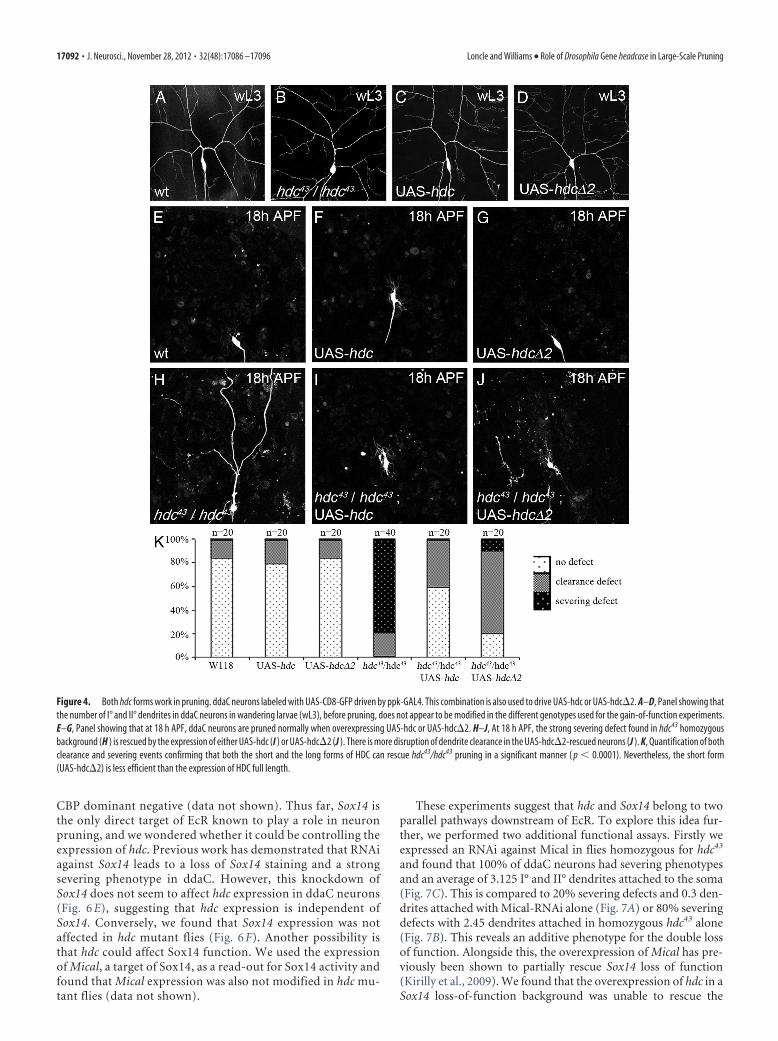
The short form of hdc is able to rescue hdc loss of functionduring pruningA curious feature of hdc is the presence of a stop codon in themiddle of the gene, allowing the generation of two distinct pro-teins from the same transcript. headcase generates a short formtermed hdcS (70 kDa) and a full-length form (120 kDa) referredto as hdcFL (Steneberg et al., 1998). To determine which formwas important during pruning, we performed rescue experi-ments that took advantage of the fact that homozygous mutanthdc animals can survive until late pupal stages, after pruning iscompleted. We used two transgenes, UAS-hdc�2, which allowsthe generation of a protein equivalent to hdcS, and UAS-hdc,which can produce both the full-length and the short form in flies(Steneberg et al., 1998). In homozygous hdc43 animals we ob-served �80% of ddaC neurons with severing defects at 18 h APF(Fig. 4H,K). The specific expression of either the UAS-hdc orUAS-hdc�2 transgene in ddaC neurons was able to rescue thesedefects at 18 h APF, with only 0 –10% of the neurons showing afailure in severing (Fig. 4 I–K). Although the short form gener-ated by UAS-hdc�2 was sufficient to rescue hdc function in thesevering of ddaC neurons, qualitative analyses showed that inaround 80% of neurons the rescue was incomplete, with severedpieces of dendrites in the field for UAS-hdc�2 pupae against only40% for UAS-HDC pupae (Fig. 4 H–J ). This data suggests thathdc�2 is sufficient to rescue ddaC pruning but is not as efficientas hdcFL.
headcase is also required for pruning in otherneuronal classesDuring metamorphosis, larval neurons undergo remodeling orprogrammed cell death (Truman et al., 1990). Within the dorsalgroup of sensory neurons there are 6 da neurons, dda A–F, whichrepresent the four different classes of da neurons (Grueber et al.,2002). At the onset of metamorphosis, the class I da neurons,ddaD and ddaE, and class IV ddaC are remodeled, whereas theclass II neuron ddaB and class III neurons ddaA and ddaF un-dergo apoptosis (Williams and Truman, 2005). To determinewhich of these neurons express hdc, we performed immunostain-
Loncle and Williams • Role of Drosophila Gene headcase in Large-Scale Pruning J. Neurosci., November 28, 2012 • 32(48):17086 –17096 • 17089

Figure 2. Spatial localization and temporal dynamics of hdc expression. A–D, Sensory neurons revealed by ppk-GAL4 driving UAS-CD8-GFP. These larval fillets are stained against HDC,and the GFP is observed without antibody. The arrowhead shows the position of ddaC neurons. A, hdc expression is not detectable in ddaC at early third instar larva (eL3). B–D, hdcexpression starts during the wandering stage of L3 (wL3) (B), reaches a maximum at 0 h APF (C), and is still evident at 5 h APF (D). hdc is also expressed in some neighboring neurons (C,arrows). E and F are control experiments for HDC antibody. E, The specific expression of hdc-RNAi in ddaC neurons results in an absence of HDC staining (arrowhead), whereas this stainingis still present in the neighboring da neurons (arrow). F, In hdc43 homozygous mutant background, HDC staining is absent in da neurons (arrows) except in ddaC (arrowheads), where weare overexpressing HDC.
17090 • J. Neurosci., November 28, 2012 • 32(48):17086 –17096 Loncle and Williams • Role of Drosophila Gene headcase in Large-Scale Pruning

ing on all the multidendritic sensory neurons in the dorsal grouplabeled by the enhancer trap line C161-GAL4. We found that theclass I, II, and III da neurons were HDC positive, alongside thedorsal bipolar dendrite neuron dbd (Fig. 5A–C).
To determine whether hdc is deployed in the remodeling ofthese neurons, we used the enhancer trap C161-GAL4 to look atthe effect of hdc loss of function in class I, II, and III ddaA B, D, E,and F neurons. At 0 h APF we saw no obvious differences betweenthese neurons in wild-type and hdc homozygous mutant flies,suggesting that their development does not depend on hdc (Fig.5D and data not shown). By 24 h APF in wild-type flies, the larvaldendrites of ddaD, ddaE, and dbd neurons were completelypruned back, whereas in hdc43/hdc43 flies there were still a num-ber of branches attached to the soma in 85% of the dorsal dagroups (n � 20) (Fig. 5E,F). At 24 h APF, ddaA, B, and F wereabsent in both wild-type and hdc mutant flies, revealing that thehdc function was not required for the programmed cell death ofthese neurons. Moreover, we found that the increased expressionof hdc in ddaA, B, or F did not prevent their death (data notshown).
Mushroom body �-MB neurons remodel their axons anddendrites during metamorphosis before elaborating their adult-specific projections (Watts et al., 2003). To determine whetherhdc is required during �-neuron pruning, we used the driver201Y-GAL4 to express hdc-RNAi. Alongside this, we generatedhomozygous hdc43 MB neuroblast clones and visualized the mor-phology of mutant (hdc/hdc) at 24 h APF, when axonal and den-dritic pruning is largely complete (Fig. 5G). We found noevidence for a disruption in the development of �-MB neuronsmutant for hdc (Fig. 5H, n � 8), whereas expression of EcR RNAiwith 201Y-GAL4 leads to a robust block of pruning (Fig. 5I).
These data are consistent with a lack of HDC staining in �-MBneurons (data not shown).
We conclude that hdc is involved in the pruning of differentclasses of sensory neurons but is not required in �-MB neuronpruning, whereas EcR is required for pruning in both. We alsofind that removing hdc does not disrupt the programmed celldeath of class II and III da neurons.
hdc is regulated by EcR but not by Sox14Previous studies have shown that headcase can be regulated inimaginal tissues and the tracheal system by the transcription fac-tors escargot (esg), buttonhead (btd), and Sp1 (Steneberg et al.,1998; Estella et al., 2003). To determine whether any of these playa role in pruning, we generated esg mutant MARCM clones n �10 (Fig. 6A) and expressed RNAi against either btd or Sp1 usingthe ppk driver (Fig. 6B, C). None of these regulators disruptedpruning in ddaC, suggesting that hdc is controlled differently inremodeling neurons.
As hdc expression closely follows the prepupal pulse of ecdy-sone, we asked whether hdc is regulated by ecdysone signaling.EcR was downregulated in ddaC by expressing EcR-RNAi CA104
with ppk-GAL4 (Fig. 1E). This cell-autonomous knockdown ofEcR resulted in a lack of hdc expression at 0 h APF, when it isnormally at its strongest (Fig. 6D). These data show that hdc isdownstream of EcR and that it could be either a direct or indirecttarget. Recently, Brahma (Brm) and CREB binding protein(CBP) have been shown to be epigenetic factors that facilitateactivation of downstream ecdysone response genes during prun-ing (Kirilly et al., 2011). The specific expression of a Brm or CBPdominant negative in ddaC results in a diminution of hdc ex-pression at 0 h APF; this reduction is more pronounced with
Figure 3. Cell-autonomous requirement of hdc in ddaC pruning. This figure shows live imaging of ddaC neurons labeled with ppk-eGFP; the MARCM clone is labeled with UAS-RedStinger, whichprovides a strong nuclear RFP signal, driven by elav C155-GAL4. A and B show two neurons from different 18 h APF pupae in which heat shocks were used to produce esgG66B MARCM clones. Neitherof these neurons are MARCM clones, so they are heterozygous (esg G66B/�) and thus should not affect ddaC pruning. In A, the neuron undergoes wild-type pruning. In B, the neuron shows a strongpruning defect, not due to a cell autonomous effect but to a global developmental defect, as suggested by the high number of clonal epidermal cells expressing RedStinger (esgG66B homozygous;magenta nucleus). C, Modified MARCM strategy: selection of a MARCM clone ddaC neuron by its nuclear expression of UAS-RedStinger (red dot) and the two nonclonal neighboring neurons, whichwere used as controls. D–F show three adjacent ddaC neurons at 18 h APF belonging to the same pupa. D and F are not clonal, as they show no RedStinger expression (insets). The pruning of theseneurons is wild type, and no pieces of dendrite are left in the field. The neuron in E is homozygous mutant for hdc, as revealed by RedStinger expression (inset). This neuron exhibits a severing defect,as a robust primary dendritic branch is still attached to the soma.
Loncle and Williams • Role of Drosophila Gene headcase in Large-Scale Pruning J. Neurosci., November 28, 2012 • 32(48):17086 –17096 • 17091
CBP dominant negative (data not shown). Thus far, Sox14 isthe only direct target of EcR known to play a role in neuronpruning, and we wondered whether it could be controlling theexpression of hdc. Previous work has demonstrated that RNAiagainst Sox14 leads to a loss of Sox14 staining and a strongsevering phenotype in ddaC. However, this knockdown ofSox14 does not seem to affect hdc expression in ddaC neurons(Fig. 6 E), suggesting that hdc expression is independent ofSox14. Conversely, we found that Sox14 expression was notaffected in hdc mutant flies (Fig. 6 F). Another possibility isthat hdc could affect Sox14 function. We used the expressionof Mical, a target of Sox14, as a read-out for Sox14 activity andfound that Mical expression was also not modified in hdc mu-tant flies (data not shown).
These experiments suggest that hdc and Sox14 belong to twoparallel pathways downstream of EcR. To explore this idea fur-ther, we performed two additional functional assays. Firstly weexpressed an RNAi against Mical in flies homozygous for hdc43
and found that 100% of ddaC neurons had severing phenotypesand an average of 3.125 I° and II° dendrites attached to the soma(Fig. 7C). This is compared to 20% severing defects and 0.3 den-drites attached with Mical-RNAi alone (Fig. 7A) or 80% severingdefects with 2.45 dendrites attached in homozygous hdc43 alone(Fig. 7B). This reveals an additive phenotype for the double lossof function. Alongside this, the overexpression of Mical has pre-viously been shown to partially rescue Sox14 loss of function(Kirilly et al., 2009). We found that the overexpression of hdc in aSox14 loss-of-function background was unable to rescue the
Figure 4. Both hdc forms work in pruning. ddaC neurons labeled with UAS-CD8-GFP driven by ppk-GAL4. This combination is also used to drive UAS-hdc or UAS-hdc�2. A–D, Panel showing thatthe number of I° and II° dendrites in ddaC neurons in wandering larvae (wL3), before pruning, does not appear to be modified in the different genotypes used for the gain-of-function experiments.E–G, Panel showing that at 18 h APF, ddaC neurons are pruned normally when overexpressing UAS-hdc or UAS-hdc�2. H–J, At 18 h APF, the strong severing defect found in hdc43 homozygousbackground (H ) is rescued by the expression of either UAS-hdc (I ) or UAS-hdc�2 (J ). There is more disruption of dendrite clearance in the UAS-hdc�2-rescued neurons (J ). K, Quantification of bothclearance and severing events confirming that both the short and the long forms of HDC can rescue hdc43/hdc43 pruning in a significant manner ( p � 0.0001). Nevertheless, the short form(UAS-hdc�2) is less efficient than the expression of HDC full length.
17092 • J. Neurosci., November 28, 2012 • 32(48):17086 –17096 Loncle and Williams • Role of Drosophila Gene headcase in Large-Scale Pruning

Sox14 phenotype (Fig. 7D–G,J). Interestingly, we found that hdcoverexpression limits the severing defects induced by EcR-RNAi CA104 in ddaC at 18 h APF, even though they remain verystrong (Fig. 7H–J). In a reverse experiment, we removed one copyof EcR in flies homozygous for hdc43 and found that 94% of ddaCneurons had severing phenotypes with an average of 3.5 I°and II°dendrites remaining attached to the soma (n � 16), compared to80% with severing defects and 2.45 dendrites remaining in flieshomozygous for hdc43 (n � 40). Taken together, these datastrongly suggest that hdc and Sox14 belong to different parallelpathways downstream of EcR in pruning neurons.
DiscussionLarge-scale pruning is deployed widely within the developingnervous system and yet little is known about the cellular andmolecular mechanisms controlling it. This active removal of longneuronal processes is dynamic, with individual branches under-going remodeling, severing, and clearance. Like programmed celldeath, local branch-specific auto-destruction programs must betightly regulated but also robust in their execution. We have ex-ploited the metamorphic remodeling of the Drosophila sensory
system to develop an interaction screen that allowed us to identifyheadcase, a molecule that controls branch severing.
A genetic interaction screen reveals a role for hdc indendrite severingMost of the genes involved in neuronal pruning have been iden-tified using candidate-based approaches. With the exception ofSox14, these players are required for EcR expression (Lee et al.,1999; Schuldiner et al., 2008; Boulanger et al., 2011) or are rela-tively far downstream, i.e., direct regulators of the cytoskeleton(Lee et al., 2009). Our goal is to bridge this gap by identifyingmolecules that link EcR with these downstream targets. We es-tablished a genetic interaction screen that is both open ended andspecific due to the detailed in vivo imaging of class IV dendriticarborizing sensory neurons. Interaction screens by their naturereveal targets that are in close genetic proximity. Here we used thenull allele EcRM554fs as a sensitized background, as the ecdysonereceptor sits at the apex of the pruning pathway. By focusing ondisruptions to branch severing events, we have also biased thescreen toward the discovery of molecules controlling the earlieststeps of the pruning pathway. Using defined chromosomal defi-
Figure 5. Role of hdc in other neurons that undergo remodeling. A–C, We used C161-GAL4 to express UAS-CD8::GFP in 5 other da neurons and stain the wL3 fillets with HDC antibody. hdc isexpressed in the other class of da neurons, dbd neuron (purple arrows), and ddaC (yellow arrowhead). D–F, We used C161-GAL4 expressing UAS-CD8::GFP to visualize dorsal sensory neurons (D).In wild-type pupae at 24 h APF (E), only two da neurons and dbd neuron remain in the field (arrows), and they have undergone pruning. The three other da neurons labeled in C161-GAL4 haveundergo apoptosis by this time. F, In hdc43/hdc43 at 24 h APF, ddaA, B and F are absent, demonstrating that hdc is not required for the apoptosis of da neurons. The remaining neurons, ddaD, ddaE,and dbd, show consistent severing defects (arrows). G–I, Twenty-four hour APF �-MB neurons labeled with 201Y-Gal4�UAS-mCD8-GFP. G, �-MB neurons have almost completely pruned boththeir axons (arrows) and dendrites. H, In MARCM clones for hdc43, the pruning does not appear to be modified, and only a few axons remain (arrows). I, Pruning is suppressed by downregulating EcRwith EcR-RNAi CA104, resulting in the persistence of axons at 24 h APF (arrows).
Loncle and Williams • Role of Drosophila Gene headcase in Large-Scale Pruning J. Neurosci., November 28, 2012 • 32(48):17086 –17096 • 17093

ciencies, we removed large numbers ofgenes at a time and could then locate thegene of interest with relative ease. Thisstrategy allowed us to identify headcase(hdc), CG15532, as the gene responsiblefor the severing phenotype. Previous workin Drosophila revealed that a loss of hdcdisrupts the differentiation of imaginalprimordia during pupal development andthat its expression closely prefigures there-entry of imaginal cells into the cell cycle(Weaver and White, 1995). Headcase is anevolutionarily conserved protein, andgrowing evidence suggests that the humanhomolog HECA plays a key role in carci-nogenesis (Makino et al., 2001; Chien etal., 2006; Dowejko et al., 2009; Dowejko etal., 2012). It is a highly basic protein (pI,9.6) and could therefore be involved inprotein–protein or protein–RNA interac-tions. Currently, there are no obvious do-mains that give an indication of how HDCacts within the cell. This shows one of thebenefits of such an open-ended screen, aswe would clearly not have selected hdc us-ing candidate-based approaches. We de-cided to explore its biological functionand establish where it fits within the prun-ing pathway.
hdc acts cell autonomously duringdendrite pruningWe have improved MARCM-based ap-proaches for studying pruning in the re-modeling of sensory neurons by using aGAL4-independent tool (ppk-eGFP) tovisualize all class IV da sensory neurons incombination with a nuclear localized Red-Stinger protein to reveal MARCM clones(GAL80 minus). This approach has a num-ber of advantages. Firstly, it allows the easyselection of clones and unequivocal identifi-cation of ddaC neurons. Secondly, it pro-vides the opportunity to compare mutant ddaC clones with adjacentheterozygous ddaC “control neurons” within the same pupa. This isa robust internal control, which is particularly important as the longembryonic heat shocks used to generate mitotic clones within thesensory system also induce clones in many other tissues. As elavC155-GAL4 also expresses GAL4 in epidermal cells, it gives us insight intothe global frequency of mitotic clones. Such “invisible” non-neuralclones can disrupt the overall timing of puparium development,leading to non-cell autonomous effects on neuron pruning. Withthis modified version of the MARCM, we were able to account forthese issues and confirm a cell-autonomous role for HDC in branchsevering.
hdc overexpression did not result in precocious or acceleratedsevering, indicating that it is not a limiting factor. These data andthe lack of known catalytic domain points toward HDC acting inconcert with other factors in a complex.
hdc expression is regulated by EcR but not by Sox14Our expression data show that hdc is expressed in da sensoryneurons from the end of larval life. This immunostaining shows
that HDC is localized close to the nucleus, but not in it. It is alsopresent at lower levels in proximal axons and dendrites. Afterbranch severing begins, the majority of HDC remains perinu-clear, suggesting that the specificity of HDC action is not likely tocome from gross changes in its subcellular localization. The factthat headcase is not ubiquitously expressed but has a clear tissue-specific and temporal expression profile suggests that it is not ageneral factor but a notable component for gating the severingprocess. Normally, two forms of headcase are generated from asingle transcript by a readthrough mechanism. Using transgenes,we find that both isoforms of HDC can rescue the pruning phe-notype and that the long form is more effective. This mirrors therequirement for HDC function in the developing tracheal system(Steneberg et al., 1998; Steneberg and Samakovlis, 2001).
Previous studies have shown that hdc is regulated by button-head and Sp1 in imaginal discs or by escargot in the trachea. Wewondered if one of these transcription factors could also regulatehdc expression in ddaC neurons. The loss of function of thesegenes does not lead to any pruning defects, demonstrating thatthey are not required for hdc regulation within the peripheral
Figure 6. Regulation of hdc expression. A, MARCM clones for esg G66B show no defects in pruning at 18 h APF, (RedStingerexpression in nucleus of homozygous mutant clone), B, C, ppk-GAL4�btd-RNAi (B) and ppk-GAL4�Sp1-RNAi (C) at 18 h APF; thepruning of ddaC neurons is not disrupted in either background. D–F, Fillets of 0 h APF pupa where ddaC neurons are labeled withUAS-CD8-GFP driven by ppk-GAL4, which is also used to drive EcR-RNAiCA104 or Sox14-RNAi when necessary. D, The expression ofEcR-RNAiCA104 in ddaC neurons (arrowhead) suppresses hdc expression in a cell autonomous manner. HDC can still be seen in theneighboring da neurons (arrows). E, The expression of Sox14-RNAi in ddaC neurons does not affect hdc expression. F, In a hdc43/hdc43 background, Sox14 staining is still present in the nucleus of ddaC neurons (arrowhead)s.
17094 • J. Neurosci., November 28, 2012 • 32(48):17086 –17096 Loncle and Williams • Role of Drosophila Gene headcase in Large-Scale Pruning

nervous system. Thus, depending on the tissue, hdc seems to beregulated by different transcription factors. Importantly, the re-moval of EcR abolishes hdc expression in ddaC neurons, con-firming that it is under the control of ecdysone signaling,downstream of EcR in the pruning pathway. It is unlikely that hdcis a direct target of EcR since it was never found in previousmicroarray analyses looking for EcR targets (Lee et al., 2003; Liand White, 2003; Beckstead et al., 2005). As Sox14 is the onlydownstream transcription factor known to be involved in neuronpruning, we wanted to determine whether it could regulate hdcexpression. Both immunostaining and rescue experiments leadus to the conclusion that there is no cross-regulation betweenSox14 and hdc in ddaC. Altogether, this data strongly supports theproposal that hdc and Sox14 belong to two parallel pathwaysdownstream of EcR. This observation of two and probably moreindependent pathways is consistent with the need for the precise
and active control of this auto-destructivemachinery in different neuron types anddifferent compartments within the cell.
hdc function is conserved in otherclasses of sensory neuronsTo explore the role of hdc in other celltypes, we looked at different neurons thatremodel during metamorphosis. Our im-munostaining revealed that hdc is ex-pressed in all of the multidendritic (md)neurons we looked at, apart from dmd1.We found that loss of hdc function leads toa severing defect in class I and IV da sen-sory neurons and the dorsal bipolar den-drite neuron, dbd. Moreover, hdc loss orgain of function in class II and III da neu-rons does not change their fate, as they stillundergo apoptosis (data not shown). Wealso found that headcase does not play arole in the �-MB neuron pruning. Thisobservation is interesting as we are look-ing at three distinct types of remodelingdecisions within the fly nervous system:the loss of dendrites in ddaC pruning, theremoval of both axons and dendrites in�-MB, and cell death for class II and III daneurons. None of these appear to be a de-fault state, because suppression of thepruning does not trigger apoptosis as seenin this study, nor does the blockage of celldeath initiate branch severing (Williamsand Truman, 2005). All three outcomesare triggered by the same ecdysone signalvia EcR and one of its targets, Sox14. Here,we discovered a downstream target of theecdysone signaling specific to dendritepruning in a subset of remodeling neu-rons. This target is the founding memberof a new pathway parallel to Sox14. Wehave exploited the metamorphic develop-ment of neurons and ecdysone signaling,which is of course specific to the phylumEcdyzoa, but our expectation is that thebatteries of downstream genes deployedduring pruning are likely to be evolution-arily conserved and, thus, a useful entry
point for investigating general principles of neurite pruning.
ConclusionWe have revealed for the first time that hdc is required in a cell-autonomous manner at the severing step during md neuronpruning. Importantly, our data also reveals that hdc acts indepen-dently of Sox14, despite both being under the control of EcR.These observations highlight that pruning is an association ofmultiple parallel pathways downstream of EcR.
Furthermore, we show through the identification of hdcthat we have developed a genetic interaction screen that is anefficient and elegant way to populate the pruning pathway.This is a fundamental step toward understanding the complexregulation of this active and tightly regulated auto-destructionprocess.
Figure 7. Epistasis experiments between hdc, Sox14, and EcR. ddaC neurons labeled with UAS-CD8-GFP driven by ppk-GAL4 at18 h APF were also used to drive Mical-RNAi, UAS-hdc, Sox14-RNAi, UAS-Sox14, Mical-RNAi, and EcR-RNAi CA104 alone or incombination. A, The expression of Mical-RNAi can lead to severing defect in 20% of ddaC neurons. B, Severing defect observed in80% of flies homozygous for hdc43. C, The expression of Mical-RNAi in ddaC neurons of homozygous hdc43 flies has an additiveeffect with 100% of the ddaC neurons presenting severing defect. D, hdc gain-of-function does not affect pruning of ddaC neurons.E, F, The strong severing defect observed with the expression of Sox14-RNAi (E) is completely rescued by the over expression ofUAS-Sox14 (F ). G, The expression of UAS-hdc is not sufficient to rescue the severing defect generated by Sox14-RNAi. H, I, Thealmost complete suppression of pruning in ddaC neuron expressing EcR-RNAi CA104 (H ) is partially rescued by the overexpression ofhdc (I ). J, Quantification of I° and II° dendrites remaining attached to the soma. There is no significant difference betweenSox14-RNAi and the combination of Sox14-RNAi and UAS-hdc. Although not total, the rescue of EcR-RNAi CA104 phenotype byUAS-hdc is significant ( p � 0.0001).
Loncle and Williams • Role of Drosophila Gene headcase in Large-Scale Pruning J. Neurosci., November 28, 2012 • 32(48):17086 –17096 • 17095

ReferencesBeckstead RB, Lam G, Thummel CS (2005) The genomic response to 20-
hydroxyecdysone at the onset of Drosophila metamorphosis. GenomeBiol 6:R99. CrossRef Medline
Boulanger A, Clouet-Redt C, Farge M, Flandre A, Guignard T, Fernando C,Juge F, Dura JM (2011) ftz-f1 and Hr39 opposing roles on EcR expres-sion during Drosophila mushroom body neuron remodeling. Nat Neuro-sci 14:37– 44. CrossRef Medline
Chien CC, Chang CC, Yang SH, Chen SH, Huang CJ (2006) A homologueof the Drosophila headcase protein is a novel tumor marker for early-stagecolorectal cancer. Oncol Rep 15:919 –926. Medline
Dietzl G, Chen D, Schnorrer F, Su KC, Barinova Y, Fellner M, Gasser B,Kinsey K, Oppel S, Scheiblauer S, Couto A, Marra V, Keleman K, DicksonBJ (2007) Nature 448:151–156. CrossRef Medline
Dowejko A, Bauer RJ, Muller-Richter UD, Reichert TE (2009) The humanhomolog of the Drosophila headcase protein slows down cell division ofhead and neck cancer cells. Carcinogenesis 30:1678 –1685. CrossRefMedline
Dowejko A, Bauer R, Bauer K, Muller-Richter UD, Reichert TE (2012) Thehuman HECA interacts with cyclins and CDKs to antagonize Wnt-mediated proliferation and chemoresistance of head and neck cancercells. Exp Cell Res 318:489 – 499. CrossRef Medline
Elfring LK, Daniel C, Papoulas O, Deuring R, Sarte M, Moseley S, Beek SJ,Waldrip WR, Daubresse G, DePace A, Kennison JA, Tamkun JW (1998)Genetic analysis of brahma: the Drosophila homolog of the yeast chroma-tin remodeling factor SWI2/SNF2. Genetics 148:251–265. Medline
Estella C, Rieckhof G, Calleja M, Morata G (2003) The role of buttonheadand Sp1 in the development of the ventral imaginal discs of Drosophila.Development 130:5929 –5941. CrossRef Medline
Grueber WB, Jan LY, Jan YN (2002) Tiling of the Drosophila epidermis bymultidendritic sensory neurons. Development 129:2867–2878. Medline
Grueber WB, Jan LY, Jan YN (2003) Different levels of the homeodomainprotein cut regulate distinct dendrite branching patterns of Drosophilamultidendritic neurons. Cell 112:805– 818. CrossRef Medline
Hayashi S (1996) A Cdc2 dependent checkpoint maintains diploidy in Dro-sophila. Development 122:1051–1058. Medline
Kirilly D, Gu Y, Huang Y, Wu Z, Bashirullah A, Low BC, Kolodkin AL, WangH, Yu F (2009) A genetic pathway composed of Sox14 and Mical gov-erns severing of dendrites during pruning. Nat Neurosci 12:1497–1505.CrossRef Medline
Kirilly D, Wong JJ, Lim EK, Wang Y, Zhang H, Wang C, Liao Q, Wang H,Liou YC, Yu F (2011) Intrinsic epigenetic factors cooperate with thesteroid hormone ecdysone to govern dendrite pruning in Drosophila.Neuron 72:86 –100. CrossRef Medline
Kumar JP, Jamal T, Doetsch A, Turner FR, Duffy JB (2004) CREB bindingprotein functions during successive stages of eye development in Dro-sophila. Genetics 168:877– 893. CrossRef Medline
KuoCT,JanLY,JanYN (2005) Dendrite-specificremodelingofDrosophilasensoryneurons requires matrix metalloproteases, ubiquitin-proteasome, and ecdysonesignaling. Proc Natl Acad Sci U S A 102:15230–15235. CrossRef Medline
Kuo CT, Zhu S, Younger S, Jan LY, Jan YN (2006) Identification of E2/E3ubiquitinating enzymes and caspase activity regulating Drosophila sen-sory neuron dendrite pruning. Neuron 51:283–290. CrossRef Medline
Lee CY, Clough EA, Yellon P, Teslovich TM, Stephan DA, Baehrecke EH(2003) Genome-wide analyses of steroid- and radiation-triggered pro-grammed cell death in Drosophila. Curr Biol 13:350 –357. CrossRefMedline
Lee HH, Jan LY, Jan YN (2009) Drosophila IKK-related kinase Ik2 and Ka-tanin p60-like 1 regulate dendrite pruning of sensory neuron duringmetamorphosis. Proc Natl Acad Sci U S A 106:6363– 6368. CrossRefMedline
Lee T, Luo L (1999) Mosaic analysis with a repressible cell marker for studiesof gene function in neuronal morphogenesis. Neuron 22:451– 461.CrossRef Medline
Lee T, Lee A, Luo L (1999) Development of the Drosophila mushroom bod-ies: sequential generation of three distinct types of neurons from a neu-roblast. Development 126:4065– 4076. Medline
Lee T, Marticke S, Sung C, Robinow S, Luo L (2000) Cell-autonomous re-quirement of the USP/EcR-B ecdysone receptor for mushroom body neu-ronal remodeling in Drosophila. Neuron 28:807– 818. CrossRef Medline
Li TR, White KP (2003) Tissue-specific gene expression and ecdysone-
regulated genomic networks in Drosophila. Dev Cell 5:59 –72. CrossRefMedline
Lin DM, Fetter RD, Kopczynski C, Grenningloh G, Goodman CS (1994)Genetic analysis of Fasciclin II in Drosophila: defasciculation, refascicula-tion, and altered fasciculation. Neuron 13:1055–1069. CrossRef Medline
Luo L, O’Leary DD (2005) Axon retraction and degeneration in develop-ment and disease. Annu Rev Neurosci 28:127–156. CrossRef Medline
Makino N, Yamato T, Inoue H, Furukawa T, Abe T, Yokoyama T, Yatsuoka T,Fukushige S, Orikasa S, Takahashi T, Horii A (2001) Isolation and char-acterization of the human gene homologous to the Drosophila headcase(hdc) gene in chromosome bands 6q23– q24, a region of common dele-tion in human pancreatic cancer. DNA Seq 11:547–553. Medline
Mason C (2009) The development of developmental neuroscience. J Neu-rosci 29:12735–12747. CrossRef Medline
Nakamura H, O’Leary DD (1989) Inaccuracies in initial growth and ar-borization of chick retinotectal axons followed by course corrections andaxon remodeling to develop topographic order. J Neurosci 9:3776 –3795.Medline
Portera-Cailliau C, Weimer RM, De Paola V, Caroni P, Svoboda K (2005)Diverse modes of axon elaboration in the developing neocortex. PLoSBiol 3:e272. CrossRef Medline
Schubiger M, Wade AA, Carney GE, Truman JW, Bender M (1998) Dro-sophila EcR-B ecdysone receptor isoforms are required for larval moltingand for neuron remodeling during metamorphosis. Development 125:2053–2062. Medline
Schubiger M, Carre C, Antoniewski C, Truman JW (2005) Ligand-dependent de-repression via EcR/USP acts as a gate to coordinate thedifferentiation of sensory neurons in the Drosophila wing. Development132:5239 –5248. CrossRef Medline
Schuldiner O, Berdnik D, Levy JM, Wu JS, Luginbuhl D, Gontang AC, Luo L(2008) piggyBac-based mosaic screen identifies a postmitotic functionfor cohesin in regulating developmental axon pruning. Dev Cell 14:227–238. CrossRef Medline
Shepherd D, Smith SA (1996) Central projections of persistent larval sen-sory neurons prefigure adult sensory pathways in the CNS of Drosophila.Development 122:2375–2384. Medline
Stanfield BB, O’Leary DD, Fricks C (1982) Selective collateral elimination inearly postnatal development restricts cortical distribution of rat pyrami-dal tract neurones. Nature 298:371–373. CrossRef Medline
Steneberg P, Samakovlis C (2001) A novel stop codon readthrough mecha-nism produces functional Headcase protein in Drosophila trachea. EMBORep 2:593–597. CrossRef Medline
Steneberg P, Englund C, Kronhamn J, Weaver TA, Samakovlis C (1998)Translational readthrough in the hdc mRNA generates a novel branchinginhibitor in the Drosophila trachea. Genes Dev 12:956 –967. CrossRefMedline
Truman JW (1990) Metamorphosis of the central nervous system of Dro-sophila. J Neurobiol 21:1072–1084. CrossRef Medline
Truman JW, Reiss SE (1976) Dendritic reorganization of an identified mo-toneuron during metamorphosis of the tobacco hornworm moth. Science192:477– 479. CrossRef Medline
Truman JW, Fahrbach SE, Kimura K (1990) Hormones and programmedcell death: insights from invertebrate studies. Prog Brain Res 86:25–35.CrossRef Medline
Truman JW, Schuppe H, Shepherd D, Williams DW (2004) Developmentalarchitecture of adult-specific lineages in the ventral CNS of Drosophila.Development 131:5167–5184. CrossRef Medline
Watts RJ, Hoopfer ED, Luo L (2003) Axon pruning during Drosophila meta-morphosis: evidence for local degeneration and requirement of theubiquitin-proteasome system. Neuron 38:871– 885. CrossRef Medline
Weaver TA, White RA (1995) headcase, an imaginal specific gene requiredfor adult morphogenesis in Drosophila melanogaster. Development 121:4149 – 4160. Medline
Williams DW, Shepherd D (1999) Persistent larval sensory neurons in adultDrosophila melanogaster. J Neurobiol 39:275–286. CrossRef Medline
Williams DW, Truman JW (2004) Mechanisms of dendritic elaboration ofsensory neurons in Drosophila: insights from in vivo time lapse. J Neurosci24:1541–1550. CrossRef Medline
Williams DW, Truman JW (2005) Cellular mechanisms of dendrite prun-ing in Drosophila: insights from in vivo time-lapse of remodeling den-dritic arborizing sensory neurons. Development 132:3631–3642.CrossRef
17096 • J. Neurosci., November 28, 2012 • 32(48):17086 –17096 Loncle and Williams • Role of Drosophila Gene headcase in Large-Scale Pruning

Williams DW, Kondo S, Krzyzanowska A, Hiromi Y, Truman JW (2006)Local caspase activity directs engulfment of dendrites during pruning. NatNeurosci 9:1234 –1236. CrossRef Medline
Yang MY, Armstrong JD, Vilinsky I, Strausfeld NJ, Kaiser K (1995) Subdi-vision of the Drosophila mushroom bodies by enhancer-trap expressionpatterns. Neuron 15:45–54. CrossRef Medline
Loncle and Williams • Role of Drosophila Gene headcase in Large-Scale Pruning J. Neurosci., November 28, 2012 • 32(48):17086 –17096 • 17096a