Development/Plasticity/Repair ... · fromgeneticremovalofTGF...
10
Development/Plasticity/Repair Defective Retinal Vascular Endothelial Cell Development As a Consequence of Impaired Integrin V8-Mediated Activation of Transforming Growth Factor- Thomas D. Arnold, 1 Gina M. Ferrero, 2 Haiyan Qiu, 3,6 Isabella T. Phan, 4 Rosemary J. Akhurst, 5,7 Eric J. Huang, 3,6 and Louis F. Reichardt 2,8 Departments of 1 Pediatrics, 2 Physiology, 3 Pathology, 4 Ophthalmology, and 5 Anatomy, 6 Pathology Service 113B, VA Medical Center, 7 Helen Diller Family Comprehensive Cancer Center, and 8 Program in Neuroscience, University of California, San Francisco, San Francisco, California 95158 Deletions of the genes encoding the integrin V8(Itgav, Itgb8) have been shown to result in abnormal vascular development in the CNS, including prenatal and perinatal hemorrhage. Other work has indicated that a major function of this integrin in vivo is to promote TGF activation. In this paper, we show that Itgb8 mRNA is strongly expressed in murine Mu ¨ller glia and retinal ganglion cells, but not astrocytes. We further show that Itgb8 deletion in the entire retina severely perturbs development of the murine retinal vasculature, elevating vascular branch point density and vascular coverage in the superficial vascular plexus, while severely impairing formation of the deep vascular plexus. The stability of the mutant vasculature is also impaired as assessed by the presence of hemorrhage and vascular basal lamina sleeves lacking endothelial cells. Specific deletion of Itgb8 in Mu ¨ller glia and neurons, but not deletion in astrocytes, recapitulates the phenotype observed following Itgb8 in the entire retina. Consistent with V8’s role in TGF1 activation, we show that retinal deletion of Tgfb1 results in very similar retinal vascular abnormalities. The vascular deficits appear to reflect impaired TGF signaling in vascular endothelial cells because retinal deletion of Itgb8 reduces phospho-SMAD3 in endothelial cells and endothelial cell-specific deletion of the TGFRII gene recapitulates the major deficits observed in the Itgb8 and TGF1 mutants. Of special interest, the retinal vascular phenotypes observed in each mutant are remarkably similar to those of others following inhibition of neuropilin-1, a receptor previously implicated in TGF activation and signaling. Introduction Vascular development within the brain and retina depends upon interactions of vascular cells with neighboring cells and extracel- lular matrix (ECM). These cells secrete growth factors, such as vascular endothelial growth factor (VEGF) and TGF, which are essential for vascular growth and stability. Integrins are cell- surface proteins implicated in numerous aspects of vascular development. Of particular interest are the V-containing integ- rins, several of which promote activation of TGF (Glinka et al., 2011; Shi et al., 2011), which in turn regulates differentiation of endothelial cells and pericytes (Carmeliet and Jain, 2011). In the nervous and immune systems, the integrin V8 is an important regulator of TGF (Yang et al., 2007; Mu et al., 2008; Aluwihare et al., 2009). Binding of V8 to an RGD site in the latent complex of TGF results in release of the active protein (Mu et al., 2002; Cambier et al., 2005). In contrast to other integ- rins, V8 is likely to be constitutively active (Helsten et al., 2008). Thus, regulation of V8-dependent TGF activation likely depends on control of integrin expression levels as well as the presence other TGF latent complex-binding proteins, such as neuropilins, or proteases that cooperate with this integrin (Glinka et al., 2011; Shi et al., 2011). In the absence of V8, vascular development in the brain is highly abnormal with elevated proliferation, impaired organiza- tion of endothelial cells and extensive hemorrhage, phenotypes mimicked by combined TGF1 and 3 mutation (McCarty et al., 2002; Zhu et al., 2002; Aluwihare et al., 2009). During brain vas- cular development, V8 appears to be required on neuroepi- thelial cells, but not pericytes or endothelial cells (McCarty et al., 2005; Proctor et al., 2005). It is thought that neuroepithelial- derived V8 activates ECM-sequestered TGF, which then has direct effects on surrounding vessels. Prior work showed that TGF signaling is required to main- tain retinal vascular stability and retinal function in adult mice (Walshe et al., 2009), and TGF signaling has been implicated in several retinal diseases (Carmeliet and Jain, 2011). It is therefore important to understand how TGF signaling in the retina is regulated. In this paper, we characterize the role of V8-TGF signal- ing in murine retinal vascular development. Our results show that deletion of the Itgb8 gene from retinal ganglia cells and Mu ¨l- ler glia, but not astrocytes, results in highly abnormal vascular patterning and instability. An almost identical phenotype results Received Nov. 9, 2011; accepted Dec. 5, 2011. Author contributions: T.D.A., E.J.H., and L.F.R. designed research; T.D.A., G.M.F., H.Q., and I.T.P. performed research; R.J.A. contributed unpublished reagents/analytic tools; T.D.A. analyzed data; L.F.R. wrote the paper. This work has been supported by a Leducq Foundation Transatlantic Network of Excellence, NIH Grant NS19090, and the Pediatric Scientist Development Program (NICHD). Correspondence should be addressed to Louis F. Reichardt, Rock Hall 284A, UCSF Mission Bay, 1550 Fourth Street, San Francisco, CA 94158. E-mail: [email protected]. DOI:10.1523/JNEUROSCI.5648-11.2012 Copyright © 2012 the authors 0270-6474/12/321197-10$15.00/0 The Journal of Neuroscience, January 25, 2012 • 32(4):1197–1206 • 1197
-
Upload
truongliem -
Category
Documents
-
view
216 -
download
0
Transcript of Development/Plasticity/Repair ... · fromgeneticremovalofTGF...

Development/Plasticity/Repair
Defective Retinal Vascular Endothelial Cell DevelopmentAs a Consequence of Impaired Integrin �V�8-MediatedActivation of Transforming Growth Factor-�
Thomas D. Arnold,1 Gina M. Ferrero,2 Haiyan Qiu,3,6 Isabella T. Phan,4 Rosemary J. Akhurst,5,7 Eric J. Huang,3,6
and Louis F. Reichardt2,8
Departments of 1Pediatrics, 2Physiology, 3Pathology, 4Ophthalmology, and 5Anatomy, 6Pathology Service 113B, VA Medical Center, 7Helen Diller FamilyComprehensive Cancer Center, and 8Program in Neuroscience, University of California, San Francisco, San Francisco, California 95158
Deletions of the genes encoding the integrin �V�8 (Itgav, Itgb8) have been shown to result in abnormal vascular development in the CNS,including prenatal and perinatal hemorrhage. Other work has indicated that a major function of this integrin in vivo is to promote TGF�activation. In this paper, we show that Itgb8 mRNA is strongly expressed in murine Muller glia and retinal ganglion cells, but notastrocytes. We further show that Itgb8 deletion in the entire retina severely perturbs development of the murine retinal vasculature,elevating vascular branch point density and vascular coverage in the superficial vascular plexus, while severely impairing formation of thedeep vascular plexus. The stability of the mutant vasculature is also impaired as assessed by the presence of hemorrhage and vascularbasal lamina sleeves lacking endothelial cells. Specific deletion of Itgb8 in Muller glia and neurons, but not deletion in astrocytes,recapitulates the phenotype observed following Itgb8 in the entire retina. Consistent with �V�8’s role in TGF�1 activation, we show thatretinal deletion of Tgfb1 results in very similar retinal vascular abnormalities. The vascular deficits appear to reflect impaired TGF�signaling in vascular endothelial cells because retinal deletion of Itgb8 reduces phospho-SMAD3 in endothelial cells and endothelialcell-specific deletion of the TGF�RII gene recapitulates the major deficits observed in the Itgb8 and TGF�1 mutants. Of special interest,the retinal vascular phenotypes observed in each mutant are remarkably similar to those of others following inhibition of neuropilin-1, areceptor previously implicated in TGF� activation and signaling.
IntroductionVascular development within the brain and retina depends uponinteractions of vascular cells with neighboring cells and extracel-lular matrix (ECM). These cells secrete growth factors, such asvascular endothelial growth factor (VEGF) and TGF�, which areessential for vascular growth and stability. Integrins are cell-surface proteins implicated in numerous aspects of vasculardevelopment. Of particular interest are the �V-containing integ-rins, several of which promote activation of TGF� (Glinka et al.,2011; Shi et al., 2011), which in turn regulates differentiation ofendothelial cells and pericytes (Carmeliet and Jain, 2011).
In the nervous and immune systems, the integrin �V�8 is animportant regulator of TGF� (Yang et al., 2007; Mu et al., 2008;Aluwihare et al., 2009). Binding of �V�8 to an RGD site in thelatent complex of TGF� results in release of the active protein(Mu et al., 2002; Cambier et al., 2005). In contrast to other integ-rins, �V�8 is likely to be constitutively active (Helsten et al.,
2008). Thus, regulation of �V�8-dependent TGF� activationlikely depends on control of integrin expression levels as well asthe presence other TGF� latent complex-binding proteins, suchas neuropilins, or proteases that cooperate with this integrin(Glinka et al., 2011; Shi et al., 2011).
In the absence of �V�8, vascular development in the brain ishighly abnormal with elevated proliferation, impaired organiza-tion of endothelial cells and extensive hemorrhage, phenotypesmimicked by combined TGF�1 and 3 mutation (McCarty et al.,2002; Zhu et al., 2002; Aluwihare et al., 2009). During brain vas-cular development, �V�8 appears to be required on neuroepi-thelial cells, but not pericytes or endothelial cells (McCarty et al.,2005; Proctor et al., 2005). It is thought that neuroepithelial-derived �V�8 activates ECM-sequestered TGF�, which then hasdirect effects on surrounding vessels.
Prior work showed that TGF� signaling is required to main-tain retinal vascular stability and retinal function in adult mice(Walshe et al., 2009), and TGF� signaling has been implicated inseveral retinal diseases (Carmeliet and Jain, 2011). It is thereforeimportant to understand how TGF� signaling in the retina isregulated.
In this paper, we characterize the role of �V�8-TGF� signal-ing in murine retinal vascular development. Our results showthat deletion of the Itgb8 gene from retinal ganglia cells and Mul-ler glia, but not astrocytes, results in highly abnormal vascularpatterning and instability. An almost identical phenotype results
Received Nov. 9, 2011; accepted Dec. 5, 2011.Author contributions: T.D.A., E.J.H., and L.F.R. designed research; T.D.A., G.M.F., H.Q., and I.T.P. performed
research; R.J.A. contributed unpublished reagents/analytic tools; T.D.A. analyzed data; L.F.R. wrote the paper.This work has been supported by a Leducq Foundation Transatlantic Network of Excellence, NIH Grant NS19090,
and the Pediatric Scientist Development Program (NICHD).Correspondence should be addressed to Louis F. Reichardt, Rock Hall 284A, UCSF Mission Bay, 1550 Fourth Street,
San Francisco, CA 94158. E-mail: [email protected]:10.1523/JNEUROSCI.5648-11.2012
Copyright © 2012 the authors 0270-6474/12/321197-10$15.00/0
The Journal of Neuroscience, January 25, 2012 • 32(4):1197–1206 • 1197
from genetic removal of TGF�1 from the retina. Our results showthat deletion of Itgb8 results in decreased phosphorylation ofSMAD3 in retinal vascular endothelial cells, and specific removalof the TGF�RII receptor from these cells recapitulates the majordeficits observed in Itgb8 and Tgfb1 mutants. In addition, retinaldeletion of Itgb8, Tgfb1, or Tgfbr2 also dramatically impairs for-mation of the retina’s outer deep vascular plexus, as was previ-ously seen following inhibition of neuropilin-1, a receptorpreviously implicated in TGF� activation and signaling (Cao etal., 2010; Glinka et al., 2011).
Materials and MethodsMice. To generate heterozygous Itgb8icr mice, Itgb8�/� mice (Zhu et al.,2002) bred on a C57BL/6129S4 genetic background, were backcrossedfor two generations with the ICR/CD-1 outbred strain. Itgb8icr �/�
heterozygotes were then bred to generate mutant Itgb8icr�/� (�8icr�/�),mouse pups and Itgb8icr �/� littermate controls, which were used forexperiments. To specifically delete Itgb8 from retinal neuroepitheliumincluding Muller glia, we used the nesCre mouse line (also known asNesCre8 (Petersen et al., 2002). Itgb8 flox/flox females (Proctor et al., 2005)were crossed with Itgb8 flox/�;NesCre tg/� males to obtain Itgb8 flox/flox;NesCre tg/�mutant pups (�8;nesCre) and Itgb8 flox/�;NesCre tg/� litter-mate controls. To target retinal astrocytes for recombination, we used themGFAP-Cre line 73.12 (Garcia et al., 2004).
Tgfb1 heterozygotes (Tgfb1�/�) were bred on an NIH/Ola back-ground, and then were intercrossed to generate mutant homozygous-null Tgfb1 �/� mice. To conditionally delete Transforming GrowthFactor � Receptor II (Tgfbr2) from retinal vascular endothelial cells, wecrossed Tgfbr2 flox/flox female mice (Leveen et al., 2002) with Platelet-Derived Growth Factor-B (Pdgfb)-inducible Cre tg/�-iCreER TM2 mice(Claxton et al., 2008). Resultant Tgfbr2flox/�;Pdgfb-iCreERTM2 mice werecrossed with Tgfbr2flox/flox mice to generate Tgfbr2 flox/flox;Pdgfb-iCreERTM2 and Tgfbr2flox/flox;Pdgfb-iCreERTM2 pups for experiments.
To evaluate for efficiency and specificity of Cre-mediated recombina-tion we used Rosa26EYFP (Srinivas et al., 2001), or ROSA26tm14(CAG-tdTomato) (Ai14) (Madisen et al., 2010) reporter mice. Other thanItgb8icr and Tgfb1 mice, all mice were maintained on mixed geneticbackgrounds. Mice were genotyped by PCR. Procedures were performedaccording to University of California, San Francisco Institutional AnimalCare and Use Committee (UCSF IACUC)-approved guidelines.
Inducible genetic experiments. For EC-specific loss-of-function ex-periments, Tgfbr2flox/flox;Pdgfb-iCreERTM2 and Tgfbr2flox/�;Pdgfb-iCreERTM2 littermate controls were used. Tgfbr2 inactivation wasinduced by intragastric injection of 50 �l of tamoxifen solution (Sigma,T5648 –1G, 1 mg/ml, generated by diluting a 10 mg/ml tamoxifen stocksolution in 1:4 ethanol/corn oil with corn oil) once daily on postnatal day1 (P1), P2, and P3 (for analysis at P6), or P5 and P6 (for analysis at P9). Ahypodermic 27 G needle was used to administer tamoxifen solution.“TGF�RII;i�EC” refers to Tgfbr2flox/flox;Pdgfb-iCreERTM2 mutants afteradministration of tamoxifen. Inducible genetic experiments were per-formed under UCSF IACUC-approved guidelines.
Immunohistochemistry. Pups of the described crosses were killed anddissected at P6, P9, or P14 for investigation of retinal primary vascularplexus sprouting angiogenesis (P6) and deeper plexus vasculogenesis(P9, P14) (Pitulescu et al., 2010). For retina whole mounts, dissected eyesunderwent weak fixation (for ease of hyaloid vasculature removal) in �2ml of 4% paraformaldehyde (PFA) in PBS for 1 h on ice. Dissected retinaswere further fixed at 4°C overnight in 4% PFA in PBS, and then stored at4°C in PBS. Whole retinas were blocked in 5% BSA, 0.3% Triton X-100 inPBS (block buffer) overnight at 4°C before isolectin incubation and an-tibody administration. For isolectin labeling, retinas were transferred toPblec buffer (PBS, pH 6.8, containing, in mM: 1 CaCl2, 1 MgCl2, 0.1MnCl2, 1% Triton X-100) and then stained with isolectin B4 (IB4) over-night as previously described (Pitulescu et al., 2010). The following day,primary antibody labeling was performed in block buffer overnight at4°C. After primary antibody incubation, retinas were labeled with SA-647 or SA-488 and fluorophore-conjugated secondary antibodies inblock buffer for 2 h at room temperature. Stained whole retinas were
flat-mounted with Prolong Gold (Invitrogen). For thin sectioning, wholeeyes were fixed in 4% PFA overnight, then transferred to 30% sucrose inPBS overnight. Eyes were embedded in OCT then cryosectioned at 15�m. Cryosections were permeabilized and blocked with 0.3% TritonX-100 and 3% BSA in PBS. Sections were incubated with primary anti-bodies overnight at 4°C, then with fluorophore-conjugated secondaryantibodies (Invitrogen) and ToPro3 (Invitrogen) nuclear stain and thenmounted with Prolong Gold (Invitrogen).
Antibodies. Primary antibodies used were: monoclonal rat anti-CD31clone MEC13.3 (BD PharMingen, 550274, 1:100), polyclonal rabbit anti-mouse Collagen IV (AbD Serotec, 2150-1470, 1:100), monoclonal mouseanti-� Smooth Muscle Actin Clone 1A4 (Sigma, A 2547, 1:100), mono-clonal rat anti-mouse TER-119 Erythroid Antigen Antibody (R&D Sys-tems, MAB1125, 1:100), polyclonal rabbit anti-phospho Histone H3(Ser10) (Millipore, 06 –570, 1:100), polyclonal rabbit anti-Glial FibrillaryAcidic Protein (Dako Cytomation, Z 0334, 1:100), monoclonal mouseanti-rat nestin clone rat-401 (4d4) (AbD Serotec, 21263, 1:100), poly-clonal rabbit anti-glutamine synthetase (Thermo Scientific PA1-46165,1:250), monoclonal mouse anti-Cellular Retinaldehyde-Binding Protein(Novus Biologicals, (B2) NB100-74392, 1:250), monoclonal rabbit anti-phospho-SMAD3 (Epitomics, 1:100) biotinylated GSL I-isolectin B4(Vector Laboratories, B-1205, 1:25), mouse. Conjugated rabbit anti-GFP-Alexa Fluor 488 (1:100) was used to amplify endogenous eYFPsignal from Rosa26 EYFP Cre reporter mice. Secondary antibodies usedwere: Alexa Fluor 488- or 647-conjugated goat anti-rat (Invitrogen,A21311 or A31852, 1:100), Alexa Fluor 555-conjugated goat anti-rabbit(Invitrogen, A31851, 1:100) and Alexa Fluor 488-, 555-, or 647-conjugated goat anti-mouse (Invitrogen, A21311 or A31851 or A31852,1:100). Alexa Fluor 488- or 647-conjugated streptavidin (Invitrogen,S32354 or S32357, 1:100) was used to recognize Biotinylated GSLI-isolectin B4.
In situ hybridization. In situ hybridization was performed on frozensections as described previously (Wilkinson, 1999). The digoxigeninRNA labeling and detection kit from Roche Biochemicals was used togenerate RNA probes using a 200 bp integrin �8 cDNA fragment fromexon 4 as template. Tissue sections were hybridized overnight withdigoxigenin-labeled RNA probes (0.4 mg/ml) at 60oC, washed in 2� SSCat 65oC, incubated with RNase (1 mg/ml in 2� SSC), washed in 0.2�SSC at 65°C, blocked in TBS with 10% sera, and incubated in alkalinephosphatase labeled anti-DIG antibody (Roche) (1:2000, 10% sera)overnight. Sections were incubated with NBT (nitroblue tetrazolium)and BCIP (5-bromo-4-chloro-3-indolyl-phosphate) (Roche) for the col-orimetric detection of alkaline phosphatase activity. Staining wasstopped after visual inspection. Sections were washed, fixed in 4% para-formaldehyde, and coverslipped.
Imaging. Confocal imaging was performed on a Zeiss LSM5 Pascalmicroscope.
Measurements and statistics. All quantifications were done using high-resolution confocal images representing a thin optical section of the sam-ple. Images were analyzed using ImageJ software using previouslyreported guidelines (Pitulescu et al., 2010). Each group contained at leastthree mutants and three littermate controls; four images from each retina(one image in each quadrant) was used to analyze angiogenic parameters.The number of branchpoints and the percentage area covered by CD31-positive endothelial cells were calculated from fields sized 460 � 230 �musing images from the central vascular plexus in the area between anartery and vein, without the angiogenic front. The number of CollagenIV-positive stained, isolectin B4-negative stained sleeves were counted infields sized 230 � 230 �m from the central vascular plexus in an areabetween an artery and vein, without the angiogenic front. Phospho-Histone 3 labeled CD31-positive endothelial cells were counted in fieldssized 460 � 460 �m from the angiogenic front. Quantification ofphospho-SMAD3 immunofluorescent staining was determined usingstained cryosections from three mutants and three controls at P9. Threerandomly chosen confocal images from each sample were taken using thesame confocal settings. ImageJ software was used to quantify the numberof endothelial cell nuclei (ToPro3-positive, CD31-positive) per totalCD31-positive vessel length per image, and the intensity of phospho-SMAD3 staining in each endothelial cell nuclei. p-values were defined
1198 • J. Neurosci., January 25, 2012 • 32(4):1197–1206 Arnold et al. • �V�8 Regulation of Retinal Vasculature

using Student’s t test for paired comparisons and ANOVA for group-wisecomparisons with Tukey’s post hoc test analysis to compare individualgroups. Control groups from experiments with �8icr, �8nesCre, and�8mGFAPcre mice (Fig. 1; see Fig. 3), and TGF�1 and TGF�RII mice(see Fig. 4) were not significantly different in any parameter measured,and were therefore grouped together and used as a collective “control.”
ResultsTo examine the role of the Itgb8 gene in retinal vascular develop-ment, we first characterized the phenotype resulting from Itgb8gene deletion throughout the animal, including all cells of theretina. For this, we used our original Itgb8 knock-out, outbredonto the ICR background (�8icr�/�) to permit survival intoadulthood (Zhu et al., 2002; Lakhe-Reddy et al., 2006). Results,presented in Figure 1, show that complete Itgb8 deletion results inhemorrhage within the retina at P6 (Fig. 1A). Vascular organiza-tion in the superficial (primary) plexus at the level of the retinalganglion cell layer is highly abnormal with excessive branchingand increased vessel coverage of the retinal surface (Fig. 1B,F).The stability of the developing vasculature appears to be signifi-cantly reduced, as assessed by the presence of collagen IV seg-ments lacking endothelial cells (Fig. 1C,F). Formation of thesuperficial vascular plexus appears to be transiently delayed in
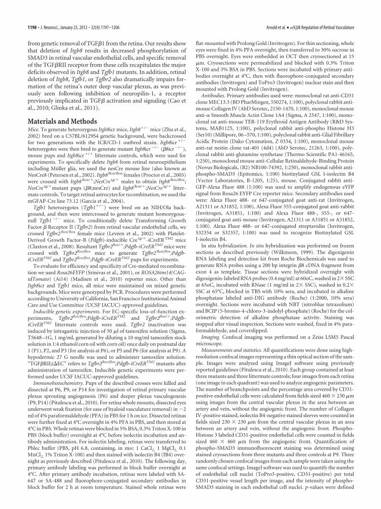
Itgb8 mutants (data not shown), but the densities and morphol-ogies of vascular tip cells appear comparatively normal (Fig. 1D).Consistent with our prior observations within the CNS (Zhu etal., 2002), mutant retinal endothelial cells have an abnormallyhigh rate of proliferation (Fig. 1E,F) and a very low level ofapoptosis, not different from that in controls (data not shown).In addition, complete loss of Itgb8 resulted in no apparent alter-ation in pericyte coverage as assessed by colocalization of severalpericyte-specific markers—�-smooth muscle actin (�SMA),NG2 and desmin (Fig. 2A–C). Normal expression of the arterialand arteriolar marker ephrinB2-eGFP in conditional Itgb8;ne-sCre mutants (see below) suggests that absence of this integrindoes not prevent normal arterial-venous differentiation (Fig.2D) (Wang et al., 1998; Adams and Klein, 2000). Thus, develop-ment of the superficial vascular plexus is not prevented by theabsence of the integrin �V�8, but the plexus appears unstableand exhibits other abnormalities that are likely to be the cause ofhemorrhage.
Following initial formation of a superficial vascular plexus atthe level of the retinal ganglion cell layer, vessels sprout into thedepth of the retina, growing along Muller glial cell processes, andestablish two deep vascular plexuses (see diagrams, Fig. 3A,B).
Figure 1. Absence of integrin �V�8 perturbs development of the retinal vasculature. A–D, Flatmounts of P6 retinas from control, Itgb8 complete knock-out (�8icr�/�), nesCre-specific Itgb8(�8;nesCre), and mGFAPCre-specific (�8;mGFAPCre) mutant retinas. A, Visualization of vasculature (collagen IV) and red blood cells (Ter119) reveals extensive hemorrhage in retinas of �8icr �/�
mutants and substantial, but reduced hemorrhage in �8;nesCre mutants. �8;mGFAPCre retinas are comparatively normal. B, Higher-magnification images of vascular plexuses illustrate perturbedvascular branching and increased vascular density in �8icr �/� and �8;nesCre mutants compared with �8;mGFAPCre mutants and controls. C, Vascular endothelia (IB4) and collagen IV stainingdemonstrate elevated numbers of collagen IV sleeves lacking endothelia (arrows) in �8icr �/� and �8;nesCre mutants compared with �8;mGFAPCre mutants and controls. D, Images of angiogenicfront in the control and three mutants reveals no overt differences in tip cell or filopodial morphology. E, Panels from P6 control, Itgb8 complete knock-out (�8icr�/�) and nesCre-specific Itgb8(�8;nesCre) mutant retinas colabeled with anti-CD31 (endothelia) and anti-phospho-histone H3 (PH3, proliferating cells). Endothelial proliferation was not analyzed in �8;mGFAPCre mutants. F,Quantifications of vascular branch point density (number per 0.106 mm 2 field), vessel coverage (percentage of area covered by CD31-expressing endothelial cells), empty collagen IV sleeves asillustrated in C (number per 0.0529 mm 2 field), and endothelial cell proliferation (number of proliferating (PH3�), CD31� endothelial cells per field as illustrated in E) demonstrate significantdifferences between control and �8icr �/� or �8;nesCre mutants, but no significant difference between control and �8;mGFAPCre mutants [ANOVA p-values�0.0005; Tukey’s subgroup analysis:*p � 0.05, **p � 0.005, ***p � 0.0005, NS � not significant. N � 16 (combined controls), N � 4 (�8icr �/�, �8;nesCre), N � 3 (�8;mGFAPCre)]. Error bars represent SEM.
Arnold et al. • �V�8 Regulation of Retinal Vasculature J. Neurosci., January 25, 2012 • 32(4):1197–1206 • 1199
The outer deep vascular plexus first formsat the outer boundary of the inner nuclearlayer by P9, followed by the inner deepvascular plexus which forms at the innerboundary of the inner nuclear layer byP14. When we compared development ofthe outer deep vascular plexus at P9 andP14 in control and Itgb8 mutant retinas,we observed that its formation is dramat-ically impaired in the mutant (Fig. 3A,B).Thus, integrin �V�8 appears to be re-quired for development of this plexus. Incontrast, Itgb8 deletion does not preventdevelopment of the inner deep vascularplexus. The vessels in the inner deep vas-cular plexus, however, are clearly abnor-mal, most obviously being thicker than inthe control.
In prior work to identify the cells thatmust express integrin �V�8 to preventhemorrhage within the developing dorsalforebrain, we demonstrated through useof cell-specific Itgb8 knock-outs that ex-pression of this integrin is required in theneuroepithelium and astroglia, but notwithin neurons or endothelial cells (Proc-tor et al., 2005). Analysis of cell-specificitgav knock-outs resulted in the same con-clusion (McCarty et al., 2005). We thereforehypothesized that loss of Itgb8 expressionfrom retinal glial populations (astrocytesand/or Muller glia) would also disrupt thedeveloping retinal vasculature, similar toour observations in the complete Itgb8knock-out. To explore this, we used two dif-ferent cre transgenes: the nesCre transgene,which promotes Cre-mediated recombina-tion in CNS neuroglial precursors starting atE8.5 (Petersen et al., 2002), and themGFAP-driven Cre 73.12 transgene (Garciaet al., 2004), which efficiently recombinesbrain astrocytes. As shown in Figure 1A,nesCre-mediated deletion of Itgb8 virtuallyphenocopied mutants with complete loss ofItgb8 (�8icr�/�), displaying highly disorga-nized and unstable vasculature with excessive branch points, in-creased vessel coverage, increased empty collagen IV sleeves, andhemorrhage. Endothelial cells of the superficial vascular plexus hadabnormally high levels of proliferation (Fig. 1E,F). Formation of theouter deep vascular plexus in �8nesCre (Itgb8;nesCre) mutants wasseverely disrupted (data not shown). As with �8icr mutants, condi-tional �8nesCre mutants had comparatively normal tip cells (Fig.1D) as well as normal arteriovenous differentiation, and pericyterecruitment (Fig. 2). Surprisingly, when Itgb8 was deleted using themGFAPCre transgene, the retinal vasculature appeared to be verysimilar to that in control retinas, with no hemorrhage or abnormal-ities in vessel coverage or branch point density (Fig. 1). While thedata suggest that there may be an elevated number of empty collagenIV segments in this mutant, the difference was not statisticallysignificant.
To better understand the difference in phenotypes between�8nesCre and �8mGFAPCre mutants, we examined expressionof itgb8 mRNA within the P6 retina by in situ analysis (Fig. 4A).
For this purpose, we examined expression of Itgb8 exon 4, theexon flanked by loxP sites in the conditional itgb8 mutant (Proc-tor et al., 2005). In situ hybridization for Itgb8 mRNA demon-strates strong expression of Itgb8 in the inner nuclear layer,ganglion cell layer, and nerve fiber layer, consistent with expres-sion in Muller glia cell bodies, descending processes, and endfeet,respectively, as well as retinal ganglion cells. Notably, expressionof Itgb8 exon 4 is completely abolished by nesCre-mediated re-combination, arguing that expression of �V�8 is limited tocells—neurons or glia—targeted by nesCre.
To further examine the cellular specificity of the nesCre andmGFAPCre lines, we used two different reporters—Rosa26;tdTo-mato;Ai14 (Madisen et al., 2010) and R26R-EYFP (Srinivas et al.,2001). Results using each reporter, presented in Figure 4, B and C,show that nesCre mediates recombination in a large majority ofneural and glial cells within the retina, including but not re-stricted to nestin-, glutamine synthetase-, and CRALBP-expressing Muller Glia (Close et al., 2005). Interestingly, data
Figure 2. Normal mural cell coverage and arteriovenous differentiation in absence of integrin �V�8 in P6 retinas. A, Panelsillustrate colocalization of endothelial cells (CD31) and pericytes (�-smooth muscle actin, �SMA) in control, complete Itgb8knock-out (�8icr�/�) and nesCre-specific Itgb8 (�8;nesCre) mutant retinas. In these images, two veins (v) are depicted on the leftand right with an artery (a) present in the middle of each panel. B, Panels present sample images colabeled with CD31 (endothelialcells) and NG2 (pericytes) to visualize pericyte coverage in the control and mutants. C, Panels present representative images ofretinas colabeled with CD31(endothelial cells) and desmin (pericytes). Note normal localization of the pericyte markers �SMA, NG2and desmin in each mutant. D, Panels show representative images of arteries visualized with ephrinB2::eGFP that demonstratenormal arterial differentiation in control and representative �8;nesCre mutant. Scale bars, 100 �m.
1200 • J. Neurosci., January 25, 2012 • 32(4):1197–1206 Arnold et al. • �V�8 Regulation of Retinal Vasculature

using the EYFP reporter show that nesCre does not promotedetectable recombination in GFAP-expressing retinal astrocytes(Fig. 4C), even though these cells migrate into the eye from theoptic nerve (Watanabe and Raff, 1988). In contrast, our datashow that the mGFAP-driven Cre 73.12 transgene promotes ef-ficient recombination in GFAP-expressing astrocytes, but notwithin nestin-expressing Muller glia (Fig. 3C,D). Together withthe in situ data that indicated that nesCre-mediated recombina-tion virtually eliminated itgb8 expression within the retina (Fig.4 A), the results argue that retinal astrocytes do not play asignificant role in �V�8-mediated vascular development. Thecomplete loss of itgb8 mRNA expression observed follow-ing nesCre-mediated recombination indicates that GFAP-expressing astrocytes do not express significant levels of �V�8.Together with prior observations within the CNS that excluded aneuronal role in �V�8-mediated signaling (Proctor et al., 2005),our results suggest that �V�8 expression on Muller glia is re-quired for formation of a normal and stable vascular plexus andto prevent hemorrhage.
As summarized in the introduction, there is compelling evi-dence that a major role of integrin �V�8 in vivo is to facilitaterelease of active TGF� from large and small latent TGF�-containing inactive complexes (Mu et al., 2002; Aluwihare et al.,2009). To obtain direct evidence that the presence of integrin�V�8 promotes TGF� signaling within the developing retina, wequantified phospho-SMAD3 levels in vessel-associated nuclei incontrol and Itgb8 mutant retinas. Results (Fig. 5A,B) show thatelevated numbers of endothelial cells, identified through colabel-ing with the endothelial cell marker CD31, are present in themutant retina. This is consistent with data above demonstratingelevated endothelial cell proliferation in the Itgb8 mutant. Whenphospho-SMAD3 levels within each endothelial cell nucleus werequantified, however, significantly less phospho-SMAD3 was ob-served in each of the mutant nuclei (Fig. 5A,B). On balance, theseobservations indicate that absence of the integrin �V�8 results inreduced TGF� signaling within endothelial cells.
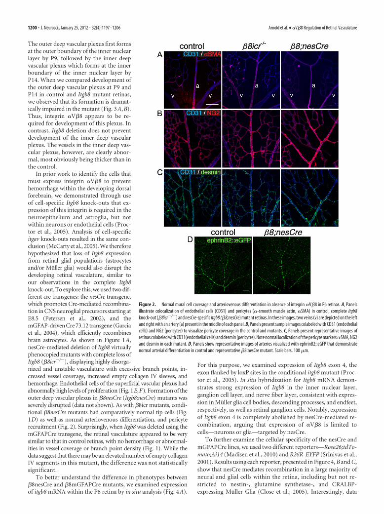
To strengthen this conclusion, we examined the phenotypeswith in the retina of mice lacking TGF�1 ligand (Tgfb1�/� mu-tant). In results presented in Figure 6, we observed the presence of
hemorrhage and a highly abnormal vasculature in this mutant.The superficial vascular plexus in this mutant exhibited an ele-vated density of branch points, elevated coverage of the retinalsurface and increased proliferation of the endothelial cells. Thevasculature also appeared to be unstable as documented by in-creased numbers of collagen IV sleeves lacking endothelial cells.These observations indicate that mice lacking TGF�1 ligand ex-hibit retinal vascular phenotypes very similar to those observed inthe �8icr�/� and �8nesCre conditional mutants (Fig. 1).
To provide direct evidence that TGF� signaling within endo-thelial cells is required for normal vascular development withinthe retina, we examined retinas in mice with conditional deletionfrom endothelial cells of the gene encoding the essential TGF�receptor, TGF�RII. This receptor subunit is required for bothAlk1 and Alk5 (type I TGF� receptors)-mediated TGF� signaltransduction (Schmierer and Hill, 2007). To examine the likelyinvolvement of endothelial cells, we deleted Tgfbr2 by crossingTgfbr2flox mice with inducible platelet-derived growth factorB-Cre-ER TM2 mice (Leveen et al., 2002; Claxton et al., 2008). Atamoxifen-dependent Cre was used for these experiments be-cause the constitutive absence of TGF�RII in endothelial cellsresults in early embryonic lethality due to cardiovascular defi-cits(Jiao et al., 2006; Carvalho et al., 2007; Robson et al., 2010). Topromote postnatal Tgfbr2 deletion, newborn pups were injectedwith tamoxifen at P1, P2, and P3. Conditional recombinationwas followed in mutant and control animals with the Rosa26;tdTomato;Ai14 Cre reporter (Madisen et al., 2010). When thesuperficial vascular plexus was examined at P6, the mutantshowed a very similar phenotype to that observed in the completeabsence of retinal �V�8 or TGF�1 including prominent retinalhemorrhage (Fig. 6A). In addition, the mutant retinas formedvascular plexuses with an abnormally high density of branchesand elevated coverage of the retinal surface by the vasculature(Fig. 6A,B). Proliferation of endothelial cells was abnormallyhigh in the mutant (Fig. 6B). The increased density of collagen IVsleeves lacking endothelial cells indicated that vascular stabilitywas reduced (Fig. 6A,B). As in the Itgb8 and Tgfb1 mutants, theapparently normal association of �-smooth muscle actin andNG2-expressing cells suggests that pericyte coverage of the vas-
Figure 3. Integrin �V�8 is required for the development of the outer deep vascular plexus. A, B, Diagrams illustrate schematically the development of the deep retinal vascular plexus. By P9,vessels have sprouted from the superficial vascular plexus (SVP; green) and establish the outer deep vascular plexus (oDVP; red). Subsequently, the oDVP remodels, and the inner deep vascularpleuxus (iDVP) is established (B) by P14. Below diagrams: A, Flat-mounted retinas at P9 were stained for red blood cells (anti-Ter119) and vessels [anti-collagen IV (col IV), or anti-CD31]. Left panelsdemonstrate vascular instability and retinal hemorrhage in P9 �8icr �/� mutants. Middle and right panels are representative confocal optical slices taken from the superficial vascular plexus (SVP;green), and the outer deep vascular plexus (oDVP; red) within flat-mounted P9 retinas stained with the endothelial cell marker, anti-CD31. Middle panels are overlays of the SVP and DVP. Rightpanels in A are higher-magnification insets of outer DVP from boxed areas in middle panels. In contrast to control, �8icr�/� mutants do not establish a deep vascular plexus. B, Optical slices fromflat mounted P14 retinas stained with IB4 illustrate perturbed remodeling of superficial vascular plexus (SVP; green), abnormally thickened vessels in the inner deep vascular plexus (iDVP; blue), andcontinued lack of an outer deep vascular plexus (oDVP; red) in �8icr �/� mutants compared with control. Scale bars, 100 �m.
Arnold et al. • �V�8 Regulation of Retinal Vasculature J. Neurosci., January 25, 2012 • 32(4):1197–1206 • 1201

culature was comparatively normal (Fig. 6A) and the density andmorphology of endothelial tip cells at the angiogenic front ap-peared similar in both the control and mutant retinas (Fig. 6A).Thus, endothelial cells appear to be an essential target for integrin�V�8-dependent TGF� signaling that facilitates formation of anormal superficial vascular plexus.
To examine the potential role of endothelial cell TGF� signal-ing in promoting vessel stability after formation, and in the for-mation of the deep vascular plexus, we examined Tgfb1 andconditional endothelial cell-specific Tgfrb2flox mutants, the latterinitiated by tamoxifen injections into pups at P5 and P6. Whenthe retinas were examined at P9, the preestablished superficialvascular plexus appeared to be disrupted with tortuous vesselsegments and associated hemorrhage (Fig. 6C,D). In addition,the outer deep vascular plexus in the outer portion of the innernuclear layer was virtually absent (Fig. 6C,D) in both mutants. As
with the �8icr�/� and �8nesCre mutants, the inner deep vascu-lar plexus of Tgfb1 mutants developed by P14, but the outer deepplexus did not form. Even though the inner deep vascular plexusdoes form, it was clearly abnormal with vessels much thicker thanthose present in the control. (Endothelial cell-specific Tgfbr2 mu-tants were significantly smaller than controls at P14, and weretherefore not studied at this time point.) Thus, these results, to-gether with the requirement for integrin �V�8, indicate that in-tegrin �V�8 promotes release of active TGF�1, which thensignals through endothelial cells to promote primary angiogene-sis and vascular stabilization in the retina.
DiscussionResults presented in this paper show that absence in the develop-ing retina of integrin �V�8 as a result of Itgb8 gene deletionresults in major abnormalities in retinal vessel formation, includ-
Figure 4. Itgb8 expression in Muller glia and retinal ganglion cells, but not retinal astrocytes. A, In situ hybridization for Itgb8 mRNA in P9 wild-type retina (top), and �8;nesCre mutant retinas(bottom). Itgb8-expressing Muller glia span the full thickness of the retina with heavy expression in the inner nuclear layer (INL), where Muller glia cell bodies reside, and in the nerve fiber layer (NFL)where Muller glial endfeet and astrocytes normally reside. There is staining for Itgb8 mRNA in the ganglion cell layer (GCL), indicating that retinal ganglion cells, in addition to Muller glia, expressItgb8. Note that all staining for Itgb8 mRNA is lost in the �8;nesCre mutant. The dark brown dots in the �8;nesCre mutant are red blood cells from retinal hemorrhage in this mutant. B–D, NesCrerecombines Muller glia and retinal ganglion cells, but not retinal astrocytes, while mGFAPCre specifically recombines retinal astrocytes. B, Images depict nesCre-directed Rosa26RtdTomato;AI14reporter expression (red), colabeled with Muller glia markers, nestin (green), Cellular retinaldehyde-binding protein (Cralp; green), and glutamine synthetase (GS; blue). NesCre directs recombina-tion of Muller glia, retinal ganglion cells, and other neural cells of the retina. Arrowheads point to heavy expression of endogenous nestin on recombined Muller glia next to vessels penetrating intothe deep retina. C, D, RosaR26-eYFP reporter expression (green) with endogenous nestin or GFAP (red), optical sections from flat mount retinas taken at the level of the NFL (C) or INL (D). C, Panelsdepict overlap of endogenous nestin with that of the nesCre-eYFP reporter. Arrowheads point to recombined Muller glial endfeet wrapping around blood vessels (unstained). Note absence of overlapof endogenous GFAP present in retinal astrocytes with the eYFP reporter. Arrow points to an exemplary astrocyte cell body labeled by GFAP, but lacking eYFP recombination marker. Note thatbecause nesCre promotes recombination in progenitors of many retinal cells in addition to Muller Glia, there is extensive expression of the eYFP reporter in cells that do not express nestin. Right panelsdepict overlap of endogenous GFAP (red) with mGFAPCre (mGfap-Cre73.12)-mediated recombination as visualized using the RosaR26-eYFP reporter (green). Arrow points to an astrocyte cell bodylabeled by GFAP with strong overlapping eYFP recombination marker. D, Fibers of Muller glia extend into the deeper retina and are recombined by nesCre, whereas there are no GFAP-stainingastrocyte fibers in this layer, and no recombination from the mGFAPcre transgene in this layer. Scale bars, 50 �m.
1202 • J. Neurosci., January 25, 2012 • 32(4):1197–1206 Arnold et al. • �V�8 Regulation of Retinal Vasculature

ing increased endothelial proliferation, elevated vascular branch-ing, reduced vasculature segment stability and hemorrhage. Inaddition, absence of this integrin severely compromised forma-tion of the deep vascular plexus. In contrast, endothelial tip cellsin the primary plexus and pericyte coverage of the nascent vesselsin this plexus appeared comparatively normal. Thus, absence of�V�8 appears to affect primarily reorganization and maturationof endothelial cells within the primary plexus, although it clearlyhas a more dramatic effect on the formation of the outer deepvascular plexus.
The phenotype of the Itgb8 mutant is mimicked byneuroepithelial-specific deletion of Itgb8 using the nesCre trans-gene, indicating that TGF� is mostly likely activated by �V�8 onsurfaces of neuroepithelial cells and Muller glia. Indeed, we seeintense staining for Itgb8 mRNA in a distribution similar to thatof Muller glial cell bodies and processes, which is entirely lost inthe conditional �8nesCre mutants. Interestingly, retinal astro-cytes do not appear to have an essential role in controlling TGF�signaling because hemorrhage and vascular abnormalities are notobserved following specific deletion of Itgb8 gene deletion withinthese cells, using mGFAPCre. These cells seem unlikely to expresssignificant levels of �V�8 because expression of Itgb8 mRNAappears to be almost absent from the retina in the conditional�8nesCre mutant, despite the presence of a normal Itgb8 genewithin GFAP-expressing astrocytes,. Thus, it is not surprisingthat the presence of a normal Itgb8 gene within retinal astrocytesdoes not prevent the vascular abnormalities observed in the �8;nesCre mutant.
Our observations are consistent with the observed colocaliza-tion of integrin �8 with markers of radial glia in a perivasculardistribution in the developing CNS (Cambier et al., 2005). Withtheir close apposition to developing blood vessels of both thesuperficial vascular plexus and the penetrating vessels whichform the deep vascular plexus, retinal Muller glia are well posi-tioned to provide important paracrine signaling effectors such asVEGF or TGF�. Indeed, Muller glia are known to secrete variousgrowth factors involved in both physiologic and pathologic reti-nal angiogenesis (Harada et al., 2005, 2011; Ye et al., 2009, 2011).Our observations on the apparently minor importance of astro-cyte �V�8 are similar to those indicating that retinal astrocyte-derived VEGF is also relatively dispensable during development(Scott et al., 2010).
Another major finding of the present study is that levels ofphospho-SMAD3 within endothelial cell nuclei are reduced sig-nificantly in the Itgb8 mutant compared with control endothelialcells, providing direct evidence that absence of this integrin im-pairs TGF� signaling within the developing vasculature. This re-sult is entirely consistent with recent evidence showing that in cellculture, astroglial-derived �V�8 binds to and activates latentTGF�1 and 3, which then transactivates brain endothelial cells(Mu et al., 2002; Cambier et al., 2005); with the immunohisto-chemical localization of TGF�1 and 3 in perivascular basementmembranes of the retina (Lutty et al., 1993); and with work thatdemonstrates an essential role for TGF� signaling in maintainingdevelopmental and adult retinal microvascular homeostasis andbarrier function (Walshe et al., 2009; Mahmoud et al., 2010).Moreover, we show that the vascular phenotypes found in Itgb8mutants are also observed following abolition of TGF� signalingin retinal endothelial cells through tamoxifen-induced, Cre-mediated deletion of the essential TGF� receptor subunit,TGF�RII. These previous studies, and this current work there-fore establish that �V�8-mediated activation of TGF� signalingin endothelial cells in vivo has an essential role in enhancingvascular maturation and stability within the developing retina.This work also suggests that �V�8-TGF� signaling could play animportant role in vascular patterning and barrier formation invarious developmental and pathological contexts outside of theretina.
Overall these results are consistent with, but extend our priorstudies on the role of this integrin in CNS vascular development.In prior work, we showed that loss of Itgb8 leads to vasculardysplasia and hemorrhage in the brain (Zhu et al., 2002). Theseeffects were specific to integrin �V�8 expressed in neuroepithe-lial cells and radial glia, as neuroepithelial deletion of integrin�V�8 completely recapitulates the Itgb8 KO brain phenotype. Incontrast, Itgb8 deletion from brain astrocytes, microglia, peri-cytes, postmitotic neurons, or endothelial cells, did not result inan obvious vascular deficit (Proctor et al., 2005; our unpublishedobservations). Our present studies extend this work by providinga direct demonstration that canonical TGF� signaling is reducedin the mutant vasculature as well as showing the importance ofthis integrin within the developing retina.
The retinal vascular phenotypes resulting from loss of �V�8-TGF�1 signaling, and in particular the loss of outer deep vascular
Figure 5. Phospho-SMAD3 signaling is reduced, in endothelial cells of integrin �8 mutants. A, Top, Representative thin sections from P9 retinas illustrating phospho-SMAD3 (pSMAD3, red)colocalization with endothelial cell-specific-CD31 (green) in control and itgb8 complete knock-out retinas. Bottom are rainbow spectrum intensity maps of phospho-SMAD3 staining delimited byCD31 staining from boxed regions in panels above (red, most intense staining; blue, least intense staining). Whereas most endothelial cells in the superficial and deep vascular plexuses of controlmice are strongly positive (arrows) and few weakly positive (arrowheads) for pSMAD3 staining, relatively few endothelial cells are strongly positive and most are weakly positive for pSMAD3 inmutants. Note the thickened superficial vascular plexus (SVP) and lack of a deep vascular plexus (DVP) in the mutant versus control sections. B, Quantification of vascular nuclei per 100 �m lengthof the vasculature documents a significant increase in the density of vasculature-associated endothelial cells in the mutant. The intensity of phospho-SMAD3 labeling of each endothelial nucleus wasquantified in B in arbitrary units. The intensity of labeling in individual nuclei as well as the mean leveling densities presented in this graph documents a significant reduction in endothelialcell-specific phospho-SMAD3 levels in mutants compared with controls. p-values from Student’s t test: *p � 0.019, ***p � 0.0001; N � 3 (controls), N � 3 (�8icr�/� mutants). Error bars in allgraphs represent SEM. Scale bars: (A), 100 �m; (C), 50 �m.
Arnold et al. • �V�8 Regulation of Retinal Vasculature J. Neurosci., January 25, 2012 • 32(4):1197–1206 • 1203
Figure 6. Absence of Tgfb1, and inhibition of TGF� signaling in retinal endothelial cells recapitulate the vascular abnormalities observed in the Itgb8 mutants. A, Representative images of retinaflat-mounts of control, Tgfb1 �/� and endothelial-specific Tgfbr2 mutant (TGF�RII;i�EC) P6 retinas. TGF�RII;i�EC mutant retinas were generated using animals homozygous for the Tgfbr2-floxallele bearing also a Pdgfb promoter-regulated Cre-ER-T2 transgene. Cre recombination was induced by intragastric tamoxifen injections (50 �g) at P1, P2, and P3. Loss of TGF�1 or TGF�RII resultsin hemorrhage at P6 as illustrated by extravascular red blood cells (Ter119) in the mutant retinas (left panels). Endothelial cells, visualized by endothelial cell-specific expression of tdTomato(Pdgfb-CreER TM2-mediated expression of recombination marker tdTomato from Rosa26RtdTomato;AI14 allele) and/or CD31 labeling illustrate enhanced vascular branching density in P6 Tgfb1 �/�
and TGF�RII;i�EC mutant retinas. Localization of vascular endothelia (IB4) and collagen IV illustrate increased presence of collagen IV sleeve segments lacking endothelial cells (IB4-negative;arrows) in Tgfb1 �/� and TGF�RII;i�EC mutants. The relative distribution of �-smooth muscle actin (SMA) and NG2-expressing pericytes compared with CD31-expressing endothelial cells incontrol compared with mutants suggests that pericyte coverage is normal in the mutants. The same panels document reasonably normal arterial (a) and venous (v) differentiation in mutants.Colabeling with anti-CD31 and the tdTomato recombination reporter (to colabel endothelial cells) and phosphohistone3 (PH3) illustrate an increased density of proliferating endothelial cells in P6Tgfb1 �/� and TGF�RII;i�EC mutant retinas. Images of the angiogenic front in control and mutant P6 retinas indicate that the morphologies and numbers of endothelial tip cells and their filopodiaare not obviously perturbed in the Tgfb1 �/� or endothelial-specific Tgfbr2 (TGF�RII;i�EC) mutants. B, Panels present quantification of changes in retinal vascular development at P6 as a result ofTgfb1 �/� deletion or endothelial cell-specific deletion of Tgfbr2. Results demonstrate that as a result of loss of TGF�1 or impaired TGF�RII function, there are increases in vascular branch pointdensity, the area of retina covered by vascular endothelial cells, the number of collagen IV sleeve segments lacking endothelial cell coverage, and the number of proliferating endothelial cells. ANOVAp-values: branch points per field, p � 0.0056; vessel coverage (%), Col IV sleeves per field, and PH3 �/CD31 � cells per field, p � 0.0001. Tukey’s subgroup analysis: (Figure legend continues.)
1204 • J. Neurosci., January 25, 2012 • 32(4):1197–1206 Arnold et al. • �V�8 Regulation of Retinal Vasculature

plexus formation, are similar to those seen in several other mousemutants. Of note, Ang2-defient mice (Hackett et al., 2000, 2002);mice with defective Norrin/Frizzled-4/Lrp5 signaling (Ye etal., 2009); mice with defective Jagged1/Dll4/Notch signaling(Benedito et al., 2009); and mice with defective neuropilin-1 sig-naling (Pan et al., 2007) all lack formation of the outer deepvascular plexus.
Neuropilin-1 is of particularly relevance to this study becauseits ligands include active and latent TGF� (Pan et al., 2007; Glinkaand Prud’homme, 2008; Cao et al., 2010; Glinka et al., 2011).Neuropilin-1 inhibition has been shown to increase vascular den-sity within the primary vascular plexus and to severely impairformation of the deep vascular plexus with little effect on VEGF-RII signaling (Pan et al., 2007). Within the CNS, the abnormalendothelial proliferation and organization seen in the neuropilin-1mutant is strikingly similar to that observed in the Itgb8 mutant (Zhuet al., 2002; Gerhardt et al., 2004). As recent data indicate that theneuropilins bind both latent and active TGF�, facilitate TGF� acti-vation, and control the balance of phosphorylation of differentSMAD effectors of TGF� signaling (Glinka and Prud’homme, 2008;Cao et al., 2010; Glinka et al., 2011), it is not clear precisely how�V�8 and neuropilin signaling intersect in the retina. As �V�8binding alone does not appear sufficient to activate latent TGF�complexes (Mu et al., 2002), it is possible that neuropilin is requiredfor latent complex activation, perhaps through generation of tension(Shi et al., 2011).
Together, our findings suggest a novel paradigm for couplingneuroglia cells to blood vessels during development through�V�8-TGF� signaling. Considering the already well character-ized role for TGF� in angiogenic pathologies, it is tempting tospeculate that �V�8 may play parallel roles in human gliovascu-lar diseases. Angiogenesis and associated vessel instability arehallmark features of retinopathy of prematurity and proliferativediabetic retinopathy. Intriguingly, TGF� is dysregulated in bothhuman and experimental retinopathy of prematurity (Shih et al.,2003; Drenser, 2009). Similarly, genetic polymorphisms inTGF�1 are associated with increased risk for proliferative dia-betic retinopathy (Beranek et al., 2002). TGF� levels are altered inboth human and experimental proliferative diabetic retinopathy(Pfeiffer et al., 1997; Gerhardinger et al., 2009), and systemicTGF� blockade in adult mice disrupts retinal vessel architectureand barrier function, reminiscent of pathological changes associ-ated with proliferative diabetic retinopathy (Walshe et al., 2009).Finally, some studies show that selective inhibition of integrin�V, �8’s only known dimerization partner, significantly attenu-
ates neo-angiogenesis in experimental proliferative retinopathies(Chavakis et al., 2002). Selective inhibition of integrin �8 in thesesettings might similarly affect pathological retinal angiogenesis.
Note added in proof. A related paper was published recently afterthe submission of our revised manuscript (Hirota et al., 2011).Both papers used nestin-driven Cre expression to delete Itgb8within the retinal precursor cells and observed similar retinalhemorrhagic phenotypes. Harada et al. (2011) report a deficit intip cell filopodia density not observed in our study. The authorsdid not include a description of the abnormalities in vascularbranch point density, coverage, or stability that were observed inour study. Both papers provide data that strongly supports a roleof this integrin in development of the secondary (deep) vascularplexus. Hirota et al. (2011) did not examine the role in the outerversus inner deep vascular plexus. Retinal anti-TGF injection byHirota et al. (2011) provided data complementary to ours on therole of TGF as a downstream effector of this integrin. The mostimportant disagreement between our papers concerns the role ofV8 in retinal astrocytes versus Muller glia. Hirota et al. (2011)concluded that retinal astrocyte-expressed V8 is required for nor-mal retinal angiogenesis because they observed expression of thisintegrin in cultured retinal astrocytes. Our data indicates thatnestin-Cre promotes efficient gene deletion in Muller glia, butnot retinal astrocytes in vivo. We also observed expression of the8 subunit in Muller glia, but not retinal astrocytes in vivo. Follow-ing astrocyte-specific, GFAP-Cre-mediated Itgb8 deletion in vivo,we did not detect retinal hemorrhage or significant abnormalitiesin vascular branching, coverage, or stability. It seems likely thatthe cultures examined by Hirota et al. (2011) contained Mullerglia that mediated TGF activation.
ReferencesAdams RH, Klein R (2000) Eph receptors and ephrin ligands. essential me-
diators of vascular development. Trends Cardiovasc Med 10:183–188.Aluwihare P, Mu Z, Zhao Z, Yu D, Weinreb PH, Horan GS, Violette SM,
Munger JS (2009) Mice that lack activity of alphavbeta6- andalphavbeta8-integrins reproduce the abnormalities of Tgfb1- and Tgfb3-null mice. J Cell Sci 122:227–232.
Benedito R, Roca C, Sorensen I, Adams S, Gossler A, Fruttiger M, Adams RH(2009) The notch ligands Dll4 and Jagged1 have opposing effects onangiogenesis. Cell 137:1124 –1135.
Beranek M, Kankova K, Benes P, Izakovicova-Holla L, Znojil V, Hajek D, VlkovaE, Vacha J (2002) Polymorphism R25P in the gene encoding transforminggrowth factor-beta (TGF-beta1) is a newly identified risk factor for prolifer-ative diabetic retinopathy. Am J Med Genet 109:278–283.
Cambier S, Gline S, Mu D, Collins R, Araya J, Dolganov G, Einheber S,Boudreau N, Nishimura SL (2005) Integrin alpha(v)beta8-mediated ac-tivation of transforming growth factor-beta by perivascular astrocytes: anangiogenic control switch. Am J Pathol 166:1883–1894.
Cao Y, Szabolcs A, Dutta SK, Yaqoob U, Jagavelu K, Wang L, Leof EB, UrrutiaRA, Shah VH, Mukhopadhyay D (2010) Neuropilin-1 mediates diver-gent R-Smad signaling and the myofibroblast phenotype. J Biol Chem285:31840 –31848.
Carmeliet P, Jain RK (2011) Molecular mechanisms and clinical applica-tions of angiogenesis. Nature 473:298 –307.
Carvalho RL, Itoh F, Goumans MJ, Lebrin F, Kato M, Takahashi S, Ema M,Itoh S, van Rooijen M, Bertolino P, Ten Dijke P, Mummery CL (2007)Compensatory signalling induced in the yolk sac vasculature by deletionof TGFbeta receptors in mice. J Cell Sci 120:4269 – 4277.
Chavakis E, Riecke B, Lin J, Linn T, Bretzel RG, Preissner KT, Brownlee M,Hammes HP (2002) Kinetics of integrin expression in the mouse model ofproliferative retinopathy and success of secondary intervention with cy-clic RGD peptides. Diabetologia 45:262–267.
Claxton S, Kostourou V, Jadeja S, Chambon P, Hodivala-Dilke K, Fruttiger M(2008) Efficient, inducible Cre-recombinase activation in vascular endo-thelium. Genesis 46:74 – 80.
Close JL, Gumuscu B, Reh TA (2005) Retinal neurons regulate proliferation
4
(Figure legend continued.) *p � 0.05, **p � 0.005, ***p � 0.0005, NS � not significant;N � 9 (combined controls), N � 3 (Tgfb1�/�, TGF�RII;i�EC). Error bars represent SEM. C,Representative images of flat-mounts of control, Tgfb1 �/� and endothelial-specific Tgfbr2(TGF�RII;i�EC) mutant P9 and P14 retinas. Endothelial-specific Tgfbr2 mutant retinas wereanalyzed following tamoxifen administration (50 �g) on P5 and P6, retinal vascular pheno-types were analyzed on P9 by immunostaining vessels (IB4; green) and red blood cells (Ter119).Confocal optical slices were taken from the superficial vascular plexus (SVP, green), the innerdeep vascular plexus (iDVP; blue), and the outer deep vascular plexus (oDVP, red) as illustratedin diagram in Figure 1. At P9, absence of TGF�1 or acute loss of TGF�RII from endothelial cellsleads to disruption of the SVP with corresponding leakage of red blood cells. At P9 formation ofthe outer DVP is dramatically inhibited in the absence of TGF�1 or TGF� receptor 2 in retinalendothelial cells. Right panels in P9 retina image sets are higher-magnification insets of outerDVP from boxed areas in middle panels. At P14 absence of TGF� results in continued failure offormation of the outer deep vascular plexus. The inner deep vascular plexus does form in themutant with abnormally thickened vessels, similar to the phenotypes in the Itgb8 mutant. AtP14, TGF�RII;i�EC mutants were sickly and obviously smaller than littermate controls, and sowere not analyzed. Scale bars, 100 �m.
Arnold et al. • �V�8 Regulation of Retinal Vasculature J. Neurosci., January 25, 2012 • 32(4):1197–1206 • 1205

of postnatal progenitors and Muller glia in the rat retina via TGF betasignaling. Development 132:3015–3026.
Drenser KA (2009) Anti-angiogenic therapy in the management of retinopa-thy of prematurity. Dev Ophthalmol 44:89 –97.
Garcia AD, Doan NB, Imura T, Bush TG, Sofroniew MV (2004) GFAP-expressing progenitors are the principal source of constitutive neurogen-esis in adult mouse forebrain. Nat Neurosci 7:1233–1241.
Gerhardinger C, Dagher Z, Sebastiani P, Park YS, Lorenzi M (2009) Thetransforming growth factor-beta pathway is a common target of drugsthat prevent experimental diabetic retinopathy. Diabetes 58:1659 –1667.
Gerhardt H, Ruhrberg C, Abramsson A, Fujisawa H, Shima D, Betsholtz C(2004) Neuropilin-1 is required for endothelial tip cell guidance in thedeveloping central nervous system. Dev Dyn 231:503–509.
Glinka Y, Prud’homme GJ (2008) Neuropilin-1 is a receptor for transform-ing growth factor beta-1, activates its latent form, and promotes regula-tory T cell activity. J Leukoc Biol 84:302–310.
Glinka Y, Stoilova S, Mohammed N, Prud’homme GJ (2011) Neuropilin-1exerts co-receptor function for TGF-beta-1 on the membrane of cancercells and enhances responses to both latent and active TGF-beta. Carci-nogenesis 32:613– 621.
Hackett SF, Ozaki H, Strauss RW, Wahlin K, Suri C, Maisonpierre P, Yanco-poulos G, Campochiaro PA (2000) Angiopoietin 2 expression in theretina: upregulation during physiologic and pathologic neovasculariza-tion. J Cell Physiol 184:275–284.
Hackett SF, Wiegand S, Yancopoulos G, Campochiaro PA (2002)Angiopoietin-2 plays an important role in retinal angiogenesis. J CellPhysiol 192:182–187.
Hammes HP, Brownlee M, Jonczyk A, Sutter A, Preissner KT (1996) Subcu-taneous injection of a cyclic peptide antagonist of vitronectin receptor-type integrins inhibits retinal neovascularization. Nat Med 2:529 –533.
Harada C, Harada T, Quah HM, Namekata K, Yoshida K, Ohno S, Tanaka K,Parada LF (2005) Role of neurotrophin-4/5 in neural cell death duringretinal development and ischemic retinal injury in vivo. Invest Ophthal-mol Vis Sci 46:669 – 673.
Harada C, Guo X, Namekata K, Kimura A, Nakamura K, Tanaka K, ParadaLF, Harada T (2011) Glia- and neuron-specific functions of TrkB sig-nalling during retinal degeneration and regeneration. Nat Commun2:189.
Helsten TL, Bunch TA, Kato H, Yamanouchi J, Choi SH, Jannuzi AL, FeralCC, Ginsberg MH, Brower DL, Shattil SJ (2008) Differences in regula-tion of Drosophila and vertebrate integrin affinity by talin. Mol Biol Cell19:3589 –3598.
Hirota S, Liu Q, Lee HS, Hossain MG, Lacy-Hulbert A, McCarty JH (2011)The astrocyte-expressed integrin V8 governs blood vessel sprouting in thedeveloping retina. Development 138:5157– 66.
Jiao K, Langworthy M, Batts L, Brown CB, Moses HL, Baldwin HS (2006) Tgf-beta signaling is required for atrioventricular cushion mesenchyme remod-eling during in vivo cardiac development. Development 133:4585–4593.
Lakhe-Reddy S, Khan S, Konieczkowski M, Jarad G, Wu KL, Reichardt LF,Takai Y, Bruggeman LA, Wang B, Sedor JR, Schelling JR (2006) Beta8integrin binds Rho GDP dissociation inhibitor-1 and activates Rac1 toinhibit mesangial cell myofibroblast differentiation. J Biol Chem281:19688 –19699.
Leveen P, Larsson J, Ehinger M, Cilio CM, Sundler M, Sjostrand LJ, HolmdahlR, Karlsson S (2002) Induced disruption of the transforming growthfactor beta type II receptor gene in mice causes a lethal inflammatorydisorder that is transplantable. Blood 100:560 –568.
Lutty GA, Merges C, Threlkeld AB, Crone S, McLeod DS (1993) Heteroge-neity in localization of isoforms of TGF-beta in human retina, vitreous,and choroid. Invest Ophthalmol Vis Sci 34:477– 487.
Madisen L, Zwingman TA, Sunkin SM, Oh SW, Zariwala HA, Gu H, Ng LL,Palmiter RD, Hawrylycz MJ, Jones AR, Lein ES, Zeng H (2010) A robustand high-throughput Cre reporting and characterization system for thewhole mouse brain. Nat Neurosci 13:133–140.
Mahmoud M, Allinson KR, Zhai Z, Oakenfull R, Ghandi P, Adams RH,Fruttiger M, Arthur HM (2010) Pathogenesis of arteriovenous malfor-mations in the absence of endoglin. Circ Res 106:1425–1433.
McCarty JH, Monahan-Earley RA, Brown LF, Keller M, Gerhardt H, Rubin K,Shani M, Dvorak HF, Wolburg H, Bader BL, Dvorak AM, Hynes RO(2002) Defective associations between blood vessels and brain paren-
chyma lead to cerebral hemorrhage in mice lacking alphav integrins. MolCell Biol 22:7667–7677.
McCarty JH, Lacy-Hulbert A, Charest A, Bronson RT, Crowley D, HousmanD, Savill J, Roes J, Hynes RO (2005) Selective ablation of alphav integ-rins in the central nervous system leads to cerebral hemorrhage, seizures,axonal degeneration and premature death. Development 132:165–176.
Mu D, Cambier S, Fjellbirkeland L, Baron JL, Munger JS, Kawakatsu H,Sheppard D, Broaddus VC, Nishimura SL (2002) The integrin alpha(v-)beta8 mediates epithelial homeostasis through MT1-MMP-dependentactivation of TGF-beta1. J Cell Biol 157:493–507.
Mu Z, Yang Z, Yu D, Zhao Z, Munger JS (2008) TGFbeta1 and TGFbeta3 arepartially redundant effectors in brain vascular morphogenesis. Mech Dev125:508 –516.
Pan Q, Chanthery Y, Liang WC, Stawicki S, Mak J, Rathore N, Tong RK,Kowalski J, Yee SF, Pacheco G, Ross S, Cheng Z, Le Couter J, Plowman G,Peale F, Koch AW, Wu Y, Bagri A, Tessier-Lavigne M, Watts RJ (2007)Blocking neuropilin-1 function has an additive effect with anti-VEGF toinhibit tumor growth. Cancer Cell 11:53– 67.
Petersen PH, Zou K, Hwang JK, Jan YN, Zhong W (2002) Progenitor cellmaintenance requires numb and numblike during mouse neurogenesis.Nature 419:929 –934.
Pfeiffer A, Spranger J, Meyer-Schwickerath R, Schatz H (1997) Growth factoralterations in advanced diabetic retinopathy: a possible role of blood ret-ina barrier breakdown. Diabetes 46 [Suppl 2]:S26 –S30.
Pitulescu ME, Schmidt I, Benedito R, Adams RH (2010) Inducible genetargeting in the neonatal vasculature and analysis of retinal angiogenesisin mice. Nat Protoc 5:1518 –1534.
Proctor JM, Zang K, Wang D, Wang R, Reichardt LF (2005) Vascular devel-opment of the brain requires beta8 integrin expression in the neuroepi-thelium. J Neurosci 25:9940 –9948.
Robson A, Allinson KR, Anderson RH, Henderson DJ, Arthur HM (2010)The TGFbeta type II receptor plays a critical role in the endothelial cellsduring cardiac development. Dev Dyn 239:2435–2442.
Schmierer B, Hill CS (2007) TGFbeta-SMAD signal transduction: molecu-lar specificity and functional flexibility. Nat Rev Mol Cell Biol 8:970 –982.
Scott A, Powner MB, Gandhi P, Clarkin C, Gutmann DH, Johnson RS, Ferrara N,Fruttiger M (2010) Astrocyte-derived vascular endothelial growth factorstabilizes vessels in the developing retinal vasculature. PLoS One 5:e11863.
Shih SC, Ju M, Liu N, Mo JR, Ney JJ, Smith LE (2003) Transforming growthfactor beta1 induction of vascular endothelial growth factor receptor 1:mechanism of pericyte-induced vascular survival in vivo. Proc Natl AcadSci U S A 100: 15859 –15864.
Shi M, Zhu J, Wang R, Chen X, Mi L, Walz T, Springer TA (2011) LatentTGF-beta structure and activation. Nature 474:343–349.
Srinivas S, Watanabe T, Lin CS, William CM, Tanabe Y, Jessell TM, Costan-tini F (2001) Cre reporter strains produced by targeted insertion ofEYFP and ECFP into the ROSA26 locus. BMC Dev Biol 1:4.
Walshe TE, Saint-Geniez M, Maharaj AS, Sekiyama E, Maldonado AE,D’Amore PA (2009) TGF-beta is required for vascular barrier function,endothelial survival and homeostasis of the adult microvasculature. PLoSOne 4:e5149.
Wang HU, Chen ZF, Anderson DJ (1998) Molecular distinction and angio-genic interaction between embryonic arteries and veins revealed byephrin-B2 and its receptor Eph-B4. Cell 93:741–753.
Watanabe T, Raff MC (1988) Retinal astrocytes are immigrants from theoptic nerve. Nature 332:834 – 837.
Wilkinson DG (1999) In situ hybridization: a practical approach. NewYork: Oxford UP.
Yang Z, Mu Z, Dabovic B, Jurukovski V, Yu D, Sung J, Xiong X, Munger JS(2007) Absence of integrin-mediated TGFbeta1 activation in vivo reca-pitulates the phenotype of TGFbeta1-null mice. J Cell Biol 176:787–793.
Ye X, Wang Y, Cahill H, Yu M, Badea TC, Smallwood PM, Peachey NS, NathansJ (2009) Norrin, frizzled-4, and Lrp5 signaling in endothelial cells controls agenetic program for retinal vascularization. Cell 139:285–298.
Ye X, Smallwood P, Nathans J (2011) Expression of the Norrie disease gene(Ndp) in developing and adult mouse eye, ear, and brain. Gene ExprPatterns 11:151–155.
Zhu J, Motejlek K, Wang D, Zang K, Schmidt A, Reichardt LF (2002) beta8integrins are required for vascular morphogenesis in mouse embryos.Development 129:2891–2903.
1206 • J. Neurosci., January 25, 2012 • 32(4):1197–1206 Arnold et al. • �V�8 Regulation of Retinal Vasculature