Development/Plasticity/Repair ... · Development/Plasticity/Repair...
13
Development/Plasticity/Repair Unexpected Survival of Neurons of Origin of the Pyramidal Tract after Spinal Cord Injury Jessica L. Nielson, 1,2 Ilse Sears-Kraxberger, 1 Melissa K. Strong, 1,2 Jamie K. Wong, 1,3 Rafer Willenberg, 1,2 and Oswald Steward 1,2,3,4 1 Reeve–Irvine Research Center, Departments of 2 Anatomy and Neurobiology, 3 Neurobiology and Behavior, and 4 Neurosurgery, University of California, Irvine, Irvine, California 92697 There is continuing controversy about whether the cells of origin of the corticospinal tract (CST) undergo retrograde cell death after spinal cord injury (SCI). All previous attempts to assess this have used imaging and/or histological techniques to assess upper motoneu- rons in the cerebral cortex. Here, we address the question in a novel way by assessing Wallerian degeneration and axon numbers in the medullary pyramid of Sprague Dawley rats after both acute SCI, either at cervical level 5 (C5) or thoracic level 9 (T9), and chronic SCI at T9. Our findings demonstrate that only a fraction of a percentage of the total axons in the medullary pyramid exhibit any sign of degeneration at any time after SCI—no more so than in uninjured control rats. Moreover, design-based counts of myelinated axons revealed no decrease in axon number in the medullary pyramid after SCI, regardless of injury level, severity, or time after injury. Spinal cord-injured rats had fewer myelinated axons in the medullary pyramid at 1 year after injury than aged matched controls, suggesting that injury may affect ongoing myelination of axons during aging. We conclude that SCI does not cause death of the CST cell bodies in the cortex; therefore, therapeutic strategies aimed at promoting axon regeneration of the CST in the spinal cord do not require a separate intervention to prevent retrograde degeneration of upper motoneurons in the cortex. Introduction Before the advent of retrograde tracing, cells of origin for fiber tracts were discovered by cutting axons and looking for cells that displayed signs of retrograde degeneration, specifically chroma- tolysis (Nissl, 1892). The cells of origin of the corticospinal tract (CST) were first identified in this way (Holmes and May, 1909), and since then, there has been continuing debate about whether corticospinal neurons die as a result of axotomy. Within the con- text of developing therapies to promote axon regeneration and recovery after spinal cord injury (SCI), this is a crucial question that needs a definitive answer. Many therapies currently being explored aim to enhance axon regeneration at the lesion site (Thuret et al., 2006). Such therapies require, however, that the cells of origin of CST axons in the motor cortex survive, because when a cell body dies, the axon dies as well (Carlson et al., 2000, 2001), which would make therapeutic interventions to induce axon regeneration futile. Numerous studies have addressed this question by assessing changes in the motor cortex after injury, with conclusions ranging from no cell death to extensive retro- grade degeneration. Assessments have included counts of neu- rons in the cortex (Feringa and Vahlsing, 1985; Giehl and Tetzlaff, 1996; Hammond et al., 1999; Bonatz et al., 2000; Hains et al., 2003; Klapka et al., 2005), changes in gene expression in the cell bodies (Kost-Mikucki and Oblinger, 1991; Mikucki and Oblinger, 1991; Kost and Oblinger, 1993; Mason et al., 2003), changes in forelimb representation (Schmidlin et al., 2004, 2005), assessment of motor responses after stimulation in the cortex (Piecharka et al., 2005), shrinkage of the cell bodies (Holmes and May, 1909; Kalil and Schneider, 1975; Ganchrow and Bernstein, 1985; Merline and Kalil, 1990; Wannier et al., 2005), chromatol- ysis (Holmes and May, 1909; Lassek, 1942; Bodian, 1946), and activation of cell death markers (Hains et al., 2003; Lee et al., 2004). There have also been reports of no response to injury (Mason et al., 2003; Crawley et al., 2004). One of the difficulties in all of these analyses is that the assessments were performed over widespread areas of the cerebral cortex and did not involve quan- titative assessment techniques that take into account changes in neuron size (i.e., unbiased sampling and counting). Based on these considerations, the present study takes a different ap- proach—analyzing the axons of the CST in the one site that they are found in a single, definable tract, the medullary pyramid. Essentially all CST axons travel through the medullary pyramid en route to the spinal cord. If CST cell bodies die after SCI, their axons must die too, which should be detectable as Wallerian degeneration of the axons [called indirect Wallerian degenera- tion (van Gehuchten, 1903)] and time-dependent decreases in axon number in the medullary pyramid. Materials and Methods Animal use and care All procedures were approved by the Institutional Animal Care and Use Committee at the University of California, Irvine, in compliance with the Received March 16, 2010; revised July 2, 2010; accepted July 8, 2010. This work was supported by National Institutes of Health Grant NS047718 (O.S.) and Grants RR07-227 and RR08-263 from the Roman Reed Spinal Cord Injury Research Fund of California. The colony of rats with chronic spinal cord injury was supported by private donations to the Reeve–Irvine Research Center from “Research for Cure.” Thanks to Kelli Sharp for technical assistance. Correspondence should be addressed to Dr. Oswald Steward, 1105 GNRF, 837 Health Sciences Drive, University of California, Irvine, Irvine, CA 92697. E-mail: [email protected]. DOI:10.1523/JNEUROSCI.1433-10.2010 Copyright © 2010 the authors 0270-6474/10/3011516-13$15.00/0 11516 • The Journal of Neuroscience, August 25, 2010 • 30(34):11516 –11528
Transcript of Development/Plasticity/Repair ... · Development/Plasticity/Repair...

Development/Plasticity/Repair
Unexpected Survival of Neurons of Origin of the PyramidalTract after Spinal Cord Injury
Jessica L. Nielson,1,2 Ilse Sears-Kraxberger,1 Melissa K. Strong,1,2 Jamie K. Wong,1,3 Rafer Willenberg,1,2
and Oswald Steward1,2,3,4
1Reeve–Irvine Research Center, Departments of 2Anatomy and Neurobiology, 3Neurobiology and Behavior, and 4Neurosurgery, University of California,Irvine, Irvine, California 92697
There is continuing controversy about whether the cells of origin of the corticospinal tract (CST) undergo retrograde cell death afterspinal cord injury (SCI). All previous attempts to assess this have used imaging and/or histological techniques to assess upper motoneu-rons in the cerebral cortex. Here, we address the question in a novel way by assessing Wallerian degeneration and axon numbers in themedullary pyramid of Sprague Dawley rats after both acute SCI, either at cervical level 5 (C5) or thoracic level 9 (T9), and chronic SCI atT9. Our findings demonstrate that only a fraction of a percentage of the total axons in the medullary pyramid exhibit any sign ofdegeneration at any time after SCI—no more so than in uninjured control rats. Moreover, design-based counts of myelinated axonsrevealed no decrease in axon number in the medullary pyramid after SCI, regardless of injury level, severity, or time after injury. Spinalcord-injured rats had fewer myelinated axons in the medullary pyramid at 1 year after injury than aged matched controls, suggesting thatinjury may affect ongoing myelination of axons during aging. We conclude that SCI does not cause death of the CST cell bodies in thecortex; therefore, therapeutic strategies aimed at promoting axon regeneration of the CST in the spinal cord do not require a separateintervention to prevent retrograde degeneration of upper motoneurons in the cortex.
IntroductionBefore the advent of retrograde tracing, cells of origin for fibertracts were discovered by cutting axons and looking for cells thatdisplayed signs of retrograde degeneration, specifically chroma-tolysis (Nissl, 1892). The cells of origin of the corticospinal tract(CST) were first identified in this way (Holmes and May, 1909),and since then, there has been continuing debate about whethercorticospinal neurons die as a result of axotomy. Within the con-text of developing therapies to promote axon regeneration andrecovery after spinal cord injury (SCI), this is a crucial questionthat needs a definitive answer. Many therapies currently beingexplored aim to enhance axon regeneration at the lesion site(Thuret et al., 2006). Such therapies require, however, that thecells of origin of CST axons in the motor cortex survive, becausewhen a cell body dies, the axon dies as well (Carlson et al., 2000,2001), which would make therapeutic interventions to induceaxon regeneration futile. Numerous studies have addressed thisquestion by assessing changes in the motor cortex after injury,with conclusions ranging from no cell death to extensive retro-grade degeneration. Assessments have included counts of neu-rons in the cortex (Feringa and Vahlsing, 1985; Giehl and
Tetzlaff, 1996; Hammond et al., 1999; Bonatz et al., 2000; Hains etal., 2003; Klapka et al., 2005), changes in gene expression in thecell bodies (Kost-Mikucki and Oblinger, 1991; Mikucki andOblinger, 1991; Kost and Oblinger, 1993; Mason et al., 2003),changes in forelimb representation (Schmidlin et al., 2004, 2005),assessment of motor responses after stimulation in the cortex(Piecharka et al., 2005), shrinkage of the cell bodies (Holmes andMay, 1909; Kalil and Schneider, 1975; Ganchrow and Bernstein,1985; Merline and Kalil, 1990; Wannier et al., 2005), chromatol-ysis (Holmes and May, 1909; Lassek, 1942; Bodian, 1946), andactivation of cell death markers (Hains et al., 2003; Lee et al.,2004). There have also been reports of no response to injury(Mason et al., 2003; Crawley et al., 2004). One of the difficulties inall of these analyses is that the assessments were performed overwidespread areas of the cerebral cortex and did not involve quan-titative assessment techniques that take into account changes inneuron size (i.e., unbiased sampling and counting). Based onthese considerations, the present study takes a different ap-proach—analyzing the axons of the CST in the one site that theyare found in a single, definable tract, the medullary pyramid.Essentially all CST axons travel through the medullary pyramiden route to the spinal cord. If CST cell bodies die after SCI, theiraxons must die too, which should be detectable as Walleriandegeneration of the axons [called indirect Wallerian degenera-tion (van Gehuchten, 1903)] and time-dependent decreases inaxon number in the medullary pyramid.
Materials and MethodsAnimal use and careAll procedures were approved by the Institutional Animal Care and UseCommittee at the University of California, Irvine, in compliance with the
Received March 16, 2010; revised July 2, 2010; accepted July 8, 2010.This work was supported by National Institutes of Health Grant NS047718 (O.S.) and Grants RR07-227 and
RR08-263 from the Roman Reed Spinal Cord Injury Research Fund of California. The colony of rats with chronic spinalcord injury was supported by private donations to the Reeve–Irvine Research Center from “Research for Cure.”Thanks to Kelli Sharp for technical assistance.
Correspondence should be addressed to Dr. Oswald Steward, 1105 GNRF, 837 Health Sciences Drive, University ofCalifornia, Irvine, Irvine, CA 92697. E-mail: [email protected].
DOI:10.1523/JNEUROSCI.1433-10.2010Copyright © 2010 the authors 0270-6474/10/3011516-13$15.00/0
11516 • The Journal of Neuroscience, August 25, 2010 • 30(34):11516 –11528
National Institutes of Health guidelines. All animals used for this studywere Sprague Dawley outbred rats (Harlan Laboratories), maintained ona 12 h light/dark cycle at 25°C. For surgery, animals were anesthetizedwith an intraperitoneal injection of ketamine and xylazine (100 and 10mg/kg, respectively; Western Medical Supply). Postoperatively, animalsreceived subcutaneous injections of 10 ml of 0.9% saline, 0.5 mg/kgBaytril, and 0.01 mg/kg buprenorphine, and were kept warm on an iso-thermic pad until mobile and alert. After injury, animals received salineand Baytril for 10 consecutive days or until animals were killed for anal-ysis, and buprenorphine was given for 7 consecutive days. For SCI ani-mals, bladders were manually expressed twice per day for the duration ofthe experiments.
Injury paradigmsThoracic vertebral level 9 dorsal funiculus lesion. To assess CST axon de-generation after axotomy at a site relatively distal to the cells of origin inthe cortex, young male rats (150 –175 g at time of surgery) received adorsal funiculus (DF) lesion at thoracic vertebral level 9 (T9), usingophthalmic microscissors (World Precision Instruments; 7 cm long,curved 3 mm blades, 0.1 mm tips). This injury paradigm bilaterallytransects the dorsal CST and has been described previously (Neumannand Woolf, 1999; Hains et al., 2003). The completeness of the injury wasverified using an operating microscope; the muscles were sutured inlayers, and the skin was closed with wound clips. This injury causesparalysis of the hindlimbs, and only transient bladder impairment duringthe first day after surgery, and did not affect normal eating, drinking, orgrooming. Animals were allowed to survive for either 1 week (n � 16) or4 weeks (n � 7) after injury.
Cervical vertebral level 5 lateral hemisection lesion. To assess axon de-generation after axotomy at a site relatively proximal to the cells of originin the cortex, adult female rats (225–250 g at time of surgery) received aright lateral hemisection lesion (Hx) at cervical vertebral level 5 (C5)using a Moria microknife (FST catalog #7040A; Fine Science Tools). Thisinjury paradigm unilaterally transects the crossed dorsal and dorsolateraland uncrossed ventral components of the CST and has been describedpreviously (Anderson et al., 2005). The completeness of the injury wasverified using an operating microscope; the muscles were sutured inlayers, and the skin was closed with wound clips. This injury causesparalysis of the forearm and paw ipsilateral to the injury, with partialipsilateral hindlimb paralysis, and transient bladder impairment for thefirst day after surgery. Eating and drinking were not impaired; however,grooming was predominantly with the left forelimb. Animals were al-lowed to survive for either 1 week (n � 8) or 3 weeks (n � 8) after injury.
Retrograde tracing. Immediately after both the T9 DF and C5 Hx injuryparadigms, the majority of animals received a 0.5 � 0.5 � 0.5 mm pieceof Gelfoam (Pfizer) impregnated with 5 �l of 4% (w/v in 0.9% saline)Fluorogold (FG) (Fluorochrome) placed into the injury site (FG animals:T9 DF 1 week, n � 16; 4 weeks, n � 7; C5 Hx 1 week, n � 8; 3 weeks, n �1; no FG animals: C5 Hx 3 weeks, n � 5).
T9 contusions. To assess CST axon degeneration in a more severe, andclinically relevant injury paradigm (Bresnahan et al., 1991), we took ad-vantage of a colony of rats that had received contusion injuries �1 yearpreviously. These animals were part of a colony of spinal cord-injuredrats that had been maintained specifically to allow experiments on ratswith chronic spinal cord injuries. The adult female rats (225–250 g attime of surgery) received moderate contusive injuries at T9, using theInfinite Horizon (IH) Impactor equipped with a 2.5-mm-diameter im-pactor probe (Precision Systems and Instrumentation). The vertebralcolumn was stabilized by clamping the vertebrae immediately rostral andcaudal to the exposed spinal cord with stabilizing forceps. The force ofthe impact to the spinal cord was set at either 200 or 250 kdyn. Thecompleteness of the injury was verified using an operating microscope;the muscles were sutured in layers, and the skin closed with wound clips.Animals were assessed for locomotor recovery after injury using theBasso, Beattie, and Breshnahan (BBB) scale for locomotor recovery(Basso et al., 1995), a 21-point scale that rates locomotor function, with 0being complete paralysis, and 21, normal locomotion. Nine rats wereselected from the chronically injured colony that received moderate con-tusions (200 –250 kDa) and exhibited BBB scores ranging from 10 to 11
at 1 year. This severity of injury ranges from occasional weight supportedplantar steps without coordination of the forelimbs and hindlimbs (FLs–HLs), to frequent weight supported plantar steps without coordinationof the FLs–HLs (Basso et al., 1995). Another group of young female rats(3 months of age; n � 5) received moderate contusion injuries and werekilled 3 weeks after injury.
Axon degeneration controls. To assess the nature and extent of axonaldegeneration in the medullary pyramid after destruction of cortical mo-toneurons, tissue from animals from a previous study (Strong et al.,2009) was used. Briefly, the right sensorimotor cortex was removed byaspiration in adult female rats (225–250 g). After injury, the scalp wassutured. This injury impairs motor function of the contralateral forepaw,described previously (Strong et al., 2009). Animals were allowed to sur-vive for 4 (n � 6) or 11 weeks (n � 5) after injury. CST axons projectexclusively within the ipsilateral medullary pyramid, so the contralateralpyramid from the 4 week time point that contains axons from the unin-jured side of the cortex served as an intraanimal control for axon counts(n � 6).
CST tract tracing. To determine the distribution of CST axons in themedullary pyramid to accurately define the area of the pyramidal tract foraxon counts, adult female rats (225–250 g at time of surgery) receivedunilateral cortical injections of the anterograde tracer biotin-conjugateddextran amine (BDA), 10,000 molecular weight (mini-ruby-BDA; In-vitrogen) using a 10 �l Hamilton microsyringe fitted with a pulled glassmicropipette. BDA was injected into six sites in the sensorimotor cortexof one hemisphere at coordinates of 2.5 and 1.5 mm lateral from themidline, either at bregma or 1 mm rostral and caudal. The detailed pro-cedure has been described previously (Anderson et al., 2005; Strong et al.,2009). Because BDA is conjugated to mini-ruby, the tracer can be visu-alized by fluorescence microscopy in sections without any additionalstaining (see Fig. 1 A). Rats were killed humanely either 5 d after injec-tions (n � 2) for assessment of labeled myelinated and unmyelinatedaxons in the pyramid, or 21 d after injection (n � 5). These cases werealso used to assess Wallerian degeneration in the medullary pyramidafter partial injury to the cortex caused by multiple penetrations ofthe injection micropipette. A separate set of animals from a previousstudy (Strong et al., 2009) that had received unilateral cortical ablationinjuries also received BDA injections into the contralateral cortex 21 dbefore the end of the experiment (n � 5). These animals were used toquantify axon degeneration associated with minimal cortical damagefrom these injections.
Uninjured controls. Adult female rats were used as uninjured, unoper-ated controls for comparison of axon counts. Animals were killed hu-manely as age-matched controls both for acute (8 –12 weeks; n � 5) andchronic (1 year; n � 5) time points.
PerfusionsAt the end of each experiment, animals received an intraperitoneal injec-tion of Euthasol (195 mg/ml pentobarbital sodium and 25 mg/ml phe-nytoin sodium; Delmarva Laboratories) and were transcardially perfusedwith either 4% paraformaldehyde in 0.1 M PBS, pH 7.4, for light micro-scope analysis, or 2% glutaraldehyde/2% paraformaldehyde (0.1 M caco-dylate buffer, 4 mM magnesium sulfate, 2 mM calcium chloride) forelectron microscope (EM) analysis.
HistologyBrains and spinal cords were dissected and postfixed in 4% paraformal-dehyde in 0.1 M PBS, pH 7.4. Brainstems were prepared for embedding inplastic as described below. Forebrains and spinal cords were cryopro-tected in 30 and 27% sucrose, respectively, frozen in TissueTek OCT(VWR International), and stored at �80°C until they were sectioned.Pyramids from animals that received BDA and were perfused for EManalysis were sectioned coronally into 100 �m vibratome sections andstained for BDA labeling using an avidin– biotin system combined with aperoxidase/DAB (diaminobenzidine)-nickel reaction (Vector Laborato-ries) before resin embedding.
Resin embedding. The brainstem was blocked by hand into 1 mm coro-nal blocks, and the block containing the main part of the pyramid (seeFig. 1C,D, site 3) was embedded in resin. Tissue was rinsed in 0.1 M
Nielson et al. • Corticospinal Motoneurons Survive SCI J. Neurosci., August 25, 2010 • 30(34):11516 –11528 • 11517
cacodylate buffer (Electron Microscopy Sciences) overnight and againfor 15 min the next day. The blocks were postfixed with 1% osmiumtetroxide (Electron Microscopy Sciences) in 0.1 M cacodylate buffer for1 h, rinsed in double distilled H2O (ddH2O) and en bloc stained with0.1% uranyl acetate (Electron Microscopy Sciences) in ddH2O. Blockswere then rinsed in ddH2O two times for 10 min each time, dehydrated inincreasing concentrations of ethanol (70, 85, 95, 100, 100%) for 10 mineach, and then in propylene oxide (intermediate solvent; Electron Mi-croscopy Sciences) two times for 10 min each time. Tissue blocks werethen incubated in propylene oxide/Spurr’s resin (1:1 mix) for 1 h, andthen in Spurr’s resin (Electron Microscopy Sciences) overnight. Theblocks were put in a fresh change of resin in BEEM capsules (ElectronMicroscopy Sciences) the next day and polymerized overnight at 60°C.
Sectioning. Frozen brains and spinal cords were sectioned into 20 �msections on a MICROM HM505 NP Series cryostat set at �20°C. Brainswere sectioned coronally, maintaining serial order, and sections werestored in PBS with 0.02% sodium azide in sterile 48-well plates at 4°Cuntil processing. Spinal cords were sectioned into 20 �m sagittal andhorizontal sections for T9 dorsal funiculus lesions and C5 hemisections,respectively, and directly mounted onto Fisherbrand Superfrost Plusglass slides (Thermo Fisher Scientific) and allowed to dry overnight atroom temperature, and either stained immediately, or stored at �80°Cuntil stained for lesion verification. Medullary pyramids embedded inresin were sectioned into 1 �m coronal sections using a Leica UltracutUCT ultramicrotome (Leica Microsystems) and stained with toluidineblue O (Sigma-Aldrich; catalog #198161) (1% toluidine blue and 2%sodium borate in ddH2O) at 60°C for 3 min, and mounted onto Fisher-brand Superfrost Plus slides and coverslipped.
Samples from rats with unilateral cortical lesions, as well as uninjuredcontrols, were also prepared for electron microscopy. Pyramids wereembedded in resin, and ultrathin sections of 60 nm thickness were cutusing a Leica Ultracut UCT ultramicrotome, mounted on copper grids,stained with uranyl acetate and lead citrate, and viewed on a JEOL 1400transmission electron microscope. Images were captured using a GatanDigital camera.
Lesion verification. Sections through lesion sites were mounted in serialorder, and one section every 100 �m was stained for hemotoxylin andeosin (H&E) for assessing lesion size. Slides were washed in PBS threetimes for 5 min each time, and then dehydrated in increasing concentra-tions of ethanol for 2 min each (70, 95, 100, 100%), defatted in xylene for5 min, and rehydrated in decreasing concentrations of ethanol for 2 mineach (100, 100, 95, 70%). Slides were then rinsed in tap water twice,stained with hemotoxylin for 5 min, rinsed in tap water, dipped in acidalcohol, rinsed in tap water, dipped several times in ammonia water, andrinsed in tap water. Slides were then rinsed in 95% ethanol for 2 min, andstained with eosin for 15 s, dehydrated in 95 and 100% ethanol for 2 mineach, defatted in xylenes three times for 3 min each, and then cover-slipped with DPX mounting media (Sigma-Aldrich) and viewed under abright-field microscope.
Microscopy and axon quantificationImages were captured on an Olympus AX-80 microscope (Olympus)using MagnaFire SP2.1B software (Optronics Software). To define thelocation of the CST axons in the pyramid, BDA-mini ruby was visualizedusing the fluorescence of the conjugated mini-ruby (see Fig. 1 A).
Total myelinated axon counts. Toluidine blue-stained plastic sectionswere used to count the total number of myelinated axons in the medul-lary pyramid (see Fig. 1 B). This was estimated by systematic randomsampling (SRS) and the fractionator counting technique using the StereoInvestigator system (MicroBrightField; version 7.003 software; MBF Bio-science). Axon counts were performed blind with respect to injury par-adigms and controls. The cross-sectional area of the medullary pyramidwas determined by tracing its outlines at 20� magnification. Myelinatedaxons were then counted at high magnification (100� under oil immer-sion) in 5 � 5 �m fractionator grids (25 �m 2) at a total of 25 samplingsites throughout the pyramid on each side. Because CST axons projectlongitudinally through the pyramid, the same sampling strategy can beused as for a nerve bundle; any single cross section will contain all theaxons that project longitudinally through the medullary pyramid (see
Fig. 1C,D). The following equation was used to estimate the total numberof myelinated axons per side in the pyramid: Total axons � [(NS �AS) � AP], where NS is the average number of axons per sampling site,AS is the area of each sampling site (25 �m 2), and AP is the area of thepyramid.
Degenerating axon counts. Toluidine blue-stained plastic sections wereused to count degenerating axons; counts were performed blind withrespect to injury paradigms and controls. The entire cross section of themedullary pyramid was scanned at high magnification (120�, under oilimmersion) to count the total number of axons displaying degenerativechanges. The main criterion for classifying an axon as degenerating was adark dense cytoplasm (Griffin et al., 1995; Rosenbluth, 1995); supple-mental indicators included myelin breakdown and demyelination(Rosenbluth, 1995). For cases with cortical lesions where the number ofaxons displaying degenerative changes was too high to count using thismethod, the SRS method with fractionator counting (described above)was used, with uninjured controls counted in the same fashion to correctfor between group comparisons using the two different counting meth-ods (direct counting vs SRS counting). It is important to note here thatdirect counting through the microscope allowed focusing up and down,which allowed a better assessment of the morphology of the axons. Incontrast, with the SRS method, counts are made from the computerscreen, which decreases the resolution of the image.
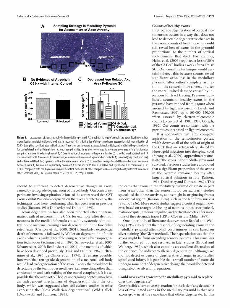
Assessment of axonal atrophy. To assess possible axon atrophy, we mea-sured the cross-sectional area of axons in the pyramid using the sametoluidine blue-stained plastic sections described above, a technique thathas been shown previously to be a measure of axonal atrophy after injury(Gold et al., 1992). High-magnification images (120�, under oil immer-sion) of pyramids were obtained and assessed using NIH ImageJ (imageprocessing and analysis). Representative samples from C5 hemisection (3weeks; n � 2), T9 contusions (3 weeks, n � 1; 1 year, n � 2), anduninjured controls (8 –12 weeks, n � 2; 1 year, n � 1) were assessed foraxonal atrophy. A total of three images per side (lateral, middle, medial)was taken for each pyramid (see Fig. 8 A), with three sampling sites as-sessed per image, for a total of nine sites. Axons at each site were assessedin 5 � 5 �m fractionator grids (25 �m 2), and only axons within theaccepted boundaries of the grid were analyzed for area. Using ImageJ, theperimeters of axons within the sampling area were traced, includingthe myelin sheath.
Quantification of BDA-labeled axons. Electron microscope images ofaxons in the pyramid of animals 5 d after cortical BDA injections (n � 2)were quantified to determine whether unmyelinated axons originate inthe sensorimotor cortex. Images were taken from the center of the pyra-mid and quantified based on electron density indicative of positive BDAlabeling. BDA-labeled myelinated and unmyelinated axons were countedin each sample site.
Statistical analysisData were analyzed for statistical significance between time points, injurytype, and controls using a one-way ANOVA with Bonferroni’s multiple-comparison test for post hoc analysis using Prism 4 for Macintosh.Graphs are plotted as the average from each group tested, with the errorbars representing the SD.
ResultsQuantitative assessment of myelinated axons in themedullary pyramidCST axons that project from the sensorimotor cortex to the spinalcord pass through the ventral medulla in the pyramid (Kuypers,1981). Thus, for purposes of axon counting, the pyramid can beconsidered as a nerve bundle with defined boundaries. The ven-tral and lateral boundaries of the medullary pyramid are obvious,but the dorsal boundary with the ventral medulla is less so. Todefine the boundaries of the CST in the medullary pyramid, CSTaxons were labeled by injecting mini-ruby-BDA into the sensori-motor cortex of one hemisphere (Fig. 1A). The region occupiedby BDA-labeled axons was used as a guide to define the region
11518 • J. Neurosci., August 25, 2010 • 30(34):11516 –11528 Nielson et al. • Corticospinal Motoneurons Survive SCI
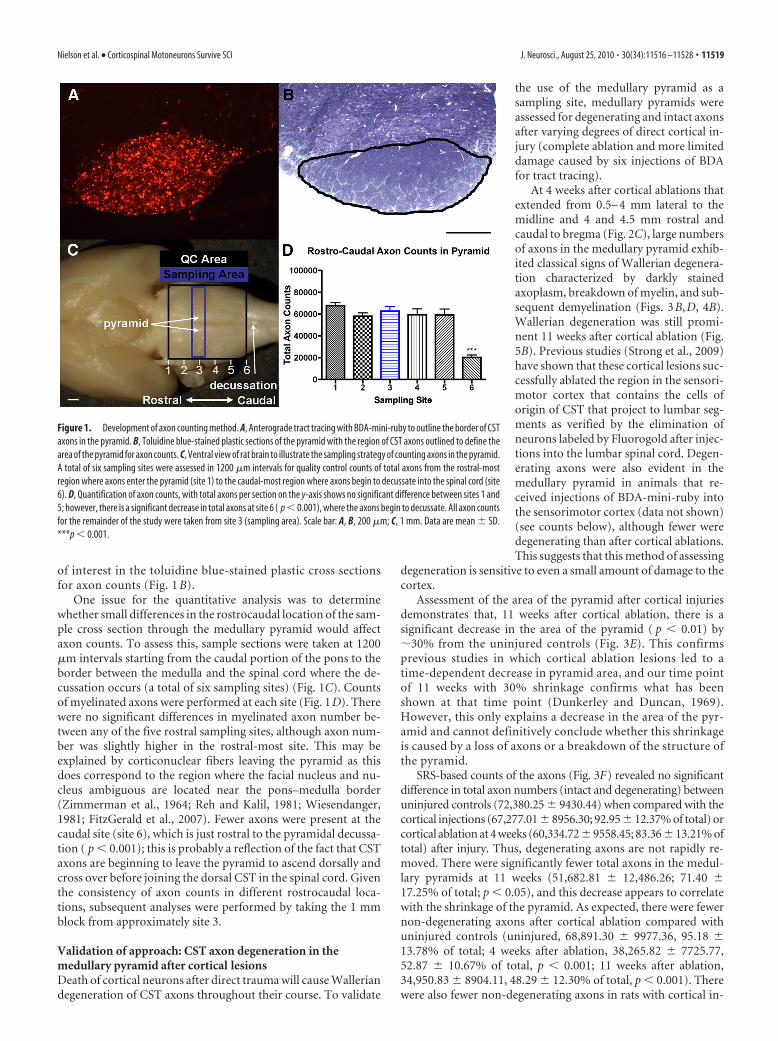
of interest in the toluidine blue-stained plastic cross sectionsfor axon counts (Fig. 1 B).
One issue for the quantitative analysis was to determinewhether small differences in the rostrocaudal location of the sam-ple cross section through the medullary pyramid would affectaxon counts. To assess this, sample sections were taken at 1200�m intervals starting from the caudal portion of the pons to theborder between the medulla and the spinal cord where the de-cussation occurs (a total of six sampling sites) (Fig. 1C). Countsof myelinated axons were performed at each site (Fig. 1D). Therewere no significant differences in myelinated axon number be-tween any of the five rostral sampling sites, although axon num-ber was slightly higher in the rostral-most site. This may beexplained by corticonuclear fibers leaving the pyramid as thisdoes correspond to the region where the facial nucleus and nu-cleus ambiguous are located near the pons–medulla border(Zimmerman et al., 1964; Reh and Kalil, 1981; Wiesendanger,1981; FitzGerald et al., 2007). Fewer axons were present at thecaudal site (site 6), which is just rostral to the pyramidal decussa-tion ( p � 0.001); this is probably a reflection of the fact that CSTaxons are beginning to leave the pyramid to ascend dorsally andcross over before joining the dorsal CST in the spinal cord. Giventhe consistency of axon counts in different rostrocaudal loca-tions, subsequent analyses were performed by taking the 1 mmblock from approximately site 3.
Validation of approach: CST axon degeneration in themedullary pyramid after cortical lesionsDeath of cortical neurons after direct trauma will cause Walleriandegeneration of CST axons throughout their course. To validate
the use of the medullary pyramid as asampling site, medullary pyramids wereassessed for degenerating and intact axonsafter varying degrees of direct cortical in-jury (complete ablation and more limiteddamage caused by six injections of BDAfor tract tracing).
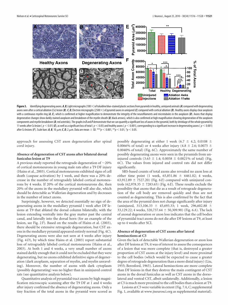
At 4 weeks after cortical ablations thatextended from 0.5– 4 mm lateral to themidline and 4 and 4.5 mm rostral andcaudal to bregma (Fig. 2C), large numbersof axons in the medullary pyramid exhib-ited classical signs of Wallerian degenera-tion characterized by darkly stainedaxoplasm, breakdown of myelin, and sub-sequent demyelination (Figs. 3B,D, 4B).Wallerian degeneration was still promi-nent 11 weeks after cortical ablation (Fig.5B). Previous studies (Strong et al., 2009)have shown that these cortical lesions suc-cessfully ablated the region in the sensori-motor cortex that contains the cells oforigin of CST that project to lumbar seg-ments as verified by the elimination ofneurons labeled by Fluorogold after injec-tions into the lumbar spinal cord. Degen-erating axons were also evident in themedullary pyramid in animals that re-ceived injections of BDA-mini-ruby intothe sensorimotor cortex (data not shown)(see counts below), although fewer weredegenerating than after cortical ablations.This suggests that this method of assessing
degeneration is sensitive to even a small amount of damage to thecortex.
Assessment of the area of the pyramid after cortical injuriesdemonstrates that, 11 weeks after cortical ablation, there is asignificant decrease in the area of the pyramid ( p � 0.01) by�30% from the uninjured controls (Fig. 3E). This confirmsprevious studies in which cortical ablation lesions led to atime-dependent decrease in pyramid area, and our time pointof 11 weeks with 30% shrinkage confirms what has beenshown at that time point (Dunkerley and Duncan, 1969).However, this only explains a decrease in the area of the pyr-amid and cannot definitively conclude whether this shrinkageis caused by a loss of axons or a breakdown of the structure ofthe pyramid.
SRS-based counts of the axons (Fig. 3F) revealed no significantdifference in total axon numbers (intact and degenerating) betweenuninjured controls (72,380.25 � 9430.44) when compared with thecortical injections (67,277.01 � 8956.30; 92.95 � 12.37% of total) orcortical ablation at 4 weeks (60,334.72 � 9558.45; 83.36 � 13.21% oftotal) after injury. Thus, degenerating axons are not rapidly re-moved. There were significantly fewer total axons in the medul-lary pyramids at 11 weeks (51,682.81 � 12,486.26; 71.40 �17.25% of total; p � 0.05), and this decrease appears to correlatewith the shrinkage of the pyramid. As expected, there were fewernon-degenerating axons after cortical ablation compared withuninjured controls (uninjured, 68,891.30 � 9977.36, 95.18 �13.78% of total; 4 weeks after ablation, 38,265.82 � 7725.77,52.87 � 10.67% of total, p � 0.001; 11 weeks after ablation,34,950.83 � 8904.11, 48.29 � 12.30% of total, p � 0.001). Therewere also fewer non-degenerating axons in rats with cortical in-
Figure 1. Development of axon counting method. A, Anterograde tract tracing with BDA-mini-ruby to outline the border of CSTaxons in the pyramid. B, Toluidine blue-stained plastic sections of the pyramid with the region of CST axons outlined to define thearea of the pyramid for axon counts. C, Ventral view of rat brain to illustrate the sampling strategy of counting axons in the pyramid.A total of six sampling sites were assessed in 1200 �m intervals for quality control counts of total axons from the rostral-mostregion where axons enter the pyramid (site 1) to the caudal-most region where axons begin to decussate into the spinal cord (site6). D, Quantification of axon counts, with total axons per section on the y-axis shows no significant difference between sites 1 and5; however, there is a significant decrease in total axons at site 6 ( p � 0.001), where the axons begin to decussate. All axon countsfor the remainder of the study were taken from site 3 (sampling area). Scale bar: A, B, 200 �m; C, 1 mm. Data are mean � SD.***p � 0.001.
Nielson et al. • Corticospinal Motoneurons Survive SCI J. Neurosci., August 25, 2010 • 30(34):11516 –11528 • 11519
jections (55,932.38 � 12,326.47; 77.28 � 17.03% of total in con-trol rats), but this difference was not statistically significant.
It is noteworthy that complete ablation of the sensorimotorcortex led to the loss of only �50% of the axons in the medullarypyramid by 11 weeks, suggesting that �50% of the myelinatedaxons in the pyramid come from areas other than the sensorimo-tor cortex. This is consistent with previous studies (Miller, 1987)(see Discussion).
Counts revealed abundant degenerating axons 4 and 11 weeksafter ablation (11,456.77 � 3368.15, 15.83 � 4.65% of total;10,838.02 � 1661.20, 14.97 � 2.30% of total); however, these twotime points were not significantly different from each other. Thenumber of degenerating axons after cortical ablation was signif-icantly higher than what was seen after BDA injections ( p � 0.01and p � 0.001, respectively; 4085.13 � 3144.52; 5.64 � 4.34% oftotal).
SRS-based assessments of the medullary pyramids from unin-jured controls did reveal a small and variable number of possibly
degenerating axons (550.77 � 507.04; 0.76 � 0.70% of total).However, counts of the same samples using direct microscopicevaluation yielded values for degenerating axons that were ap-proximately two orders of magnitude lower; we feel the lowercounts are more accurate given the increased resolution ofmorphology when the tissue is examined at high powerthrough the microscope. Importantly, even the higher numberof possibly degenerating axons from SRS-based analyses rep-resents only a fraction of a percentage of the total. A caveat isthat there may also be false positives in the counts of fromanimals with cortical lesions that would not be scored as de-generating if the counts were done by direct microscopic vi-sualization. However, the degree of degeneration that is seenwith SRS-based counting is proportional to the amount ofdamage in the cortex.
Thus, even with the differences between SRS-based and directcounts, it is clear that degenerating axons are easy to detect inthe pyramid when present after a cortical lesion, validating the
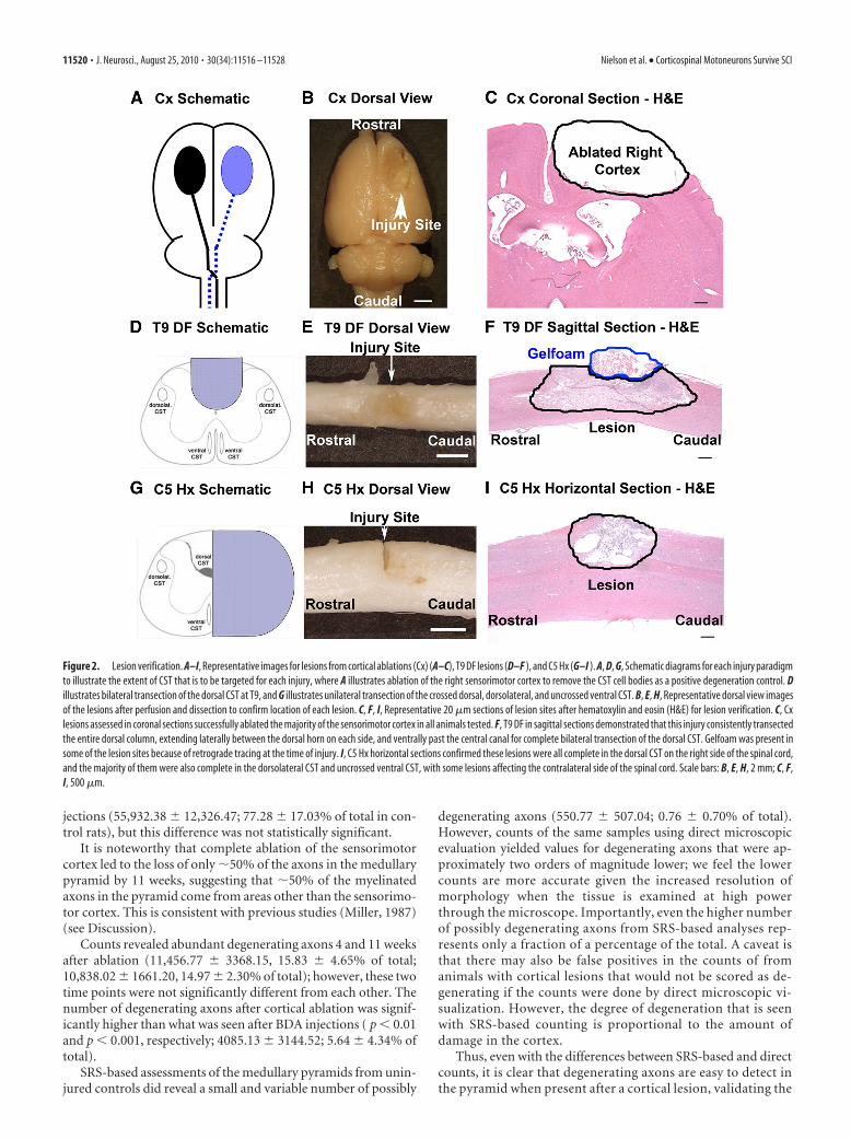
Figure 2. Lesion verification. A–I, Representative images for lesions from cortical ablations (Cx) (A–C), T9 DF lesions (D–F ), and C5 Hx (G–I ). A, D, G, Schematic diagrams for each injury paradigmto illustrate the extent of CST that is to be targeted for each injury, where A illustrates ablation of the right sensorimotor cortex to remove the CST cell bodies as a positive degeneration control. Dillustrates bilateral transection of the dorsal CST at T9, and G illustrates unilateral transection of the crossed dorsal, dorsolateral, and uncrossed ventral CST. B, E, H, Representative dorsal view imagesof the lesions after perfusion and dissection to confirm location of each lesion. C, F, I, Representative 20 �m sections of lesion sites after hematoxylin and eosin (H&E) for lesion verification. C, Cxlesions assessed in coronal sections successfully ablated the majority of the sensorimotor cortex in all animals tested. F, T9 DF in sagittal sections demonstrated that this injury consistently transectedthe entire dorsal column, extending laterally between the dorsal horn on each side, and ventrally past the central canal for complete bilateral transection of the dorsal CST. Gelfoam was present insome of the lesion sites because of retrograde tracing at the time of injury. I, C5 Hx horizontal sections confirmed these lesions were all complete in the dorsal CST on the right side of the spinal cord,and the majority of them were also complete in the dorsolateral CST and uncrossed ventral CST, with some lesions affecting the contralateral side of the spinal cord. Scale bars: B, E, H, 2 mm; C, F,I, 500 �m.
11520 • J. Neurosci., August 25, 2010 • 30(34):11516 –11528 Nielson et al. • Corticospinal Motoneurons Survive SCI
approach for assessing CST axon degeneration after spinalcord injury.
Absence of degeneration of CST axons after bilateral dorsalfuniculus lesion at T9A previous study reported the retrograde degeneration of �20%of cortical motoneurons in young male rats after a T9 DF injury(Hains et al., 2003). Cortical motoneurons exhibited signs of celldeath (caspase activation) by 1 week, and there was a 20% de-crease in the number of retrogradely labeled cortical motoneu-rons by 4 weeks. If 20% of the cortical motoneurons die, then20% of the axons in the medullary pyramid will also die, whichshould be detectable as Wallerian degeneration and by decreasesin the number of intact axons.
Surprisingly, however, we detected essentially no sign of de-generating axons in the medullary pyramid 1 week after DF le-sions at T9 that ablated the dorsal column bilaterally, with thelesion extending ventrally into the gray matter past the centralcanal, and laterally into the dorsal horn (for an example of thelesion, see Fig. 2F). Based on the results of Hains et al. (2003),there should be extensive retrograde degeneration, but CST ax-ons in the medullary pyramid appeared entirely normal (Fig. 4C).Degenerating axons were also not seen at 4 weeks after injury(Fig. 4D), by which time Hains et al. (2003) report substantialloss of retrogradely labeled cortical motoneurons (Hains et al.,2003). At both 1 and 4 weeks, a very small number of axonsstained darkly enough that it could not be excluded that they weredegenerating, but no axons exhibited definitive signs of degener-ation (dark axoplasm, separation of myelin, and myelin unravel-ing). Moreover, the number of axons with dark cytoplasm(possibly degenerating) was no higher than in uninjured controlrats (see quantitative analysis below).
Quantitative analysis of pyramidal tract axons by high magni-fication microscopic scanning after the T9 DF at 1 and 4 weeksafter injury confirmed the absence of degenerating axons. Only atiny fraction of the total axons in the pyramid were scored as
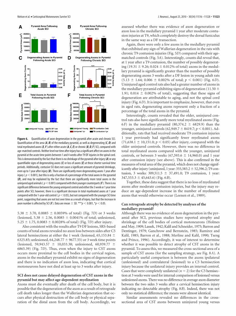
possibly degenerating at either 1 week (6.7 � 4.2; 0.0108 �0.0066% of total) or 4 weeks after injury (4.8 � 2.6; 0.0075 �0.0040% of total) (Fig. 6C). Approximately the same number ofpossibly degenerating axons were seen in the pyramids from un-injured controls (3.63 � 1.4; 0.0058 � 0.0021% of total) (Fig.6C). The values from injured and control rats did not differsignificantly.
SRS-based counts of total axons also revealed no axon loss ateither time point (1 week, 65,851.86 � 6461.02; 4 weeks,63,912.89 � 7527.20) (Fig. 6E) compared with uninjured con-trols (62,978.35 � 7283.65) (Fig. 6E). These results exclude thepossibility that axons that die as a result of retrograde degenera-tion of the cell body are removed quickly and thus are notcounted as degenerating. This is also confirmed by the fact thatthe area of the pyramid does not change significantly after injury(uninjured, 313,106.33 � 45,693.33; 1 week, 296,602.88 �33,129.22; 4 weeks, 320,737.64 � 50,309.90) (Fig. 6A). The lackof axonal degeneration or axon loss indicates that the cell bodiesof pyramidal tract axons do not die after DF lesions at T9, at leastup to 4 weeks after SCI.
Absence of degeneration of CST axons after lateralhemisections at C5Given the lack of detectable Wallerian degeneration or axon lossafter DF lesions at T9, it was of interest to assess the consequencesof a lesion that was more complete (that is, destroyed a greaterproportion of CST axons at the injury level) and more proximalto the cell bodies (which would be expected to cause a greaterdegree of retrograde degeneration than a more distal injury) (Liu,1955; Beresford, 1965). Lateral hemisections are more completethan DF lesions in that they destroy the main contingent of CSTaxons in the dorsal funiculus as well as CST axons in the dorso-lateral and ventral CST, albeit unilaterally. Additionally, a lesionat C5 is much more proximal to the cell bodies than a lesion at T9.
Lesions at C5 were variable in extent (Fig. 7A,C; supplementalFig. 1, available at www.jneurosci.org as supplemental material).
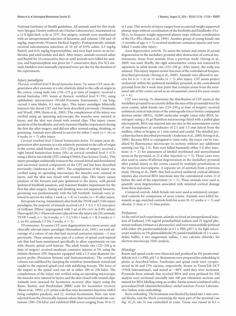
Figure 3. Identifying degenerating axons. A, B, Light micrographs (100�) of toluidine blue-stained plastic sections from pyramids in healthy, uninjured animals (A) compared with degeneratingaxons seen after a cortical ablation (Cx) lesion (B). C, D, Electron micrographs (2500�) of pyramid axons in uninjured (C) compared with cortical ablation (D). Healthy axons display clear axoplasmwith a continuous myelin ring (A, C), which is confirmed at higher magnification to demonstrate the integrity of the neurofilaments and microtubules in the axoplasm (D). Axons that displaydegenerative changes show darkly stained axoplasm and breakdown of the myelin sheath (B, black arrows), which is also confirmed at high magnification showing degeneration of the axoplasmcomponents and myelin breakdown (D, red asterisks). The graphs in E and F demonstrate that we can quantify a significant loss of axons in the pyramid, both by shrinkage of the whole pyramid by11 weeks after Cx lesion ( p � 0.01) (E), as well as a significant loss of total ( p � 0.05) and healthy axons ( p � 0.001), corresponding to a significant increase in degenerating axons ( p � 0.001)after Cx lesions (F ). Scale bars: A, B, 10 �m; C, D, 2 �m. Data are mean � SD. ***p � 0.001, **p � 0.01, *p � 0.05.
Nielson et al. • Corticospinal Motoneurons Survive SCI J. Neurosci., August 25, 2010 • 30(34):11516 –11528 • 11521

Lesions were reconstructed by assessing horizontal sectionsthrough the lesion site at 100 �m intervals and representing theseon schematic cross sections. In the reconstructed images, lesionsin different animals are shown in gray superimposing imagesfrom different animals. In this way, the darkest shade of grayindicates the areas damaged in all animals; the lightest shade ofgray illustrates the area damaged in one animal only (supplemen-tal Fig. 1, gray area, available at www.jneurosci.org as supplemen-tal material). The quantitative analyses were limited to cases inwhich lesions extended to the midline so as to completely destroythe ipsilateral dorsal CST (Fig. 2 I) (1 week, n � 8; 3 weeks, n � 6)and also included cases in which lesions extended over to thecontralateral side. For axon counts after C5 hemisections, data
are organized into three categories: lesioned side refers to thepyramids contralateral to the hemisection containing damagedaxons from the crossed dorsal and dorsolateral CST; unlesionedside refers to the pyramids ipsilateral to the hemisection thatinclude mainly undamaged axons, but also includes axons of theuncrossed ventral CST and axons affected by over-hemisections;uninjured refers to control animals with no lesions.
Even though lesions were predominantly unilateral, the pyra-mid on the side contralateral to the lesion that would contain themajority of the injured axons (lesioned side) was indistinguish-able from the side where there are mostly uninjured axons thatserved as an internal control (unlesioned), both qualitatively andquantitatively. Importantly, however, the sides were distinguish-able based on the presence of degenerating axons lateral to thepyramid indicating Wallerian degeneration of spinothalamic andspinoreticular axons (Fig. 4H); degenerating axons were presentonly ipsilateral to the lesion (compare with Fig. 4G). This internalcontrol again confirms that Wallerian degeneration can be de-tected in these sections when present.
Histologically, CST axons in the medullary pyramid appearedentirely normal, at both 1 week (Fig. 4E) and 3 weeks after C5hemisections (Fig. 4F). This was true whether the C5 hemisec-tion injury was limited to one side, or extended over the mid-line to affect the contralateral spinal cord (for analysis of therelationship between lesion size and axon counts, see supple-mental Fig. 2, available at www.jneurosci.org as supplementalmaterial). There was no significant difference in the area of thepyramid at 1 week (lesioned, 316,825.40 � 33,210.00 �m 2; un-lesioned, 307,191.10 � 36,433.09 �m 2) or 3 weeks (lesioned,294,301.13 � 37,521.80 �m2; unlesioned, 292,127.25 � 41,377.14�m2) after injury compared with uninjured controls (Fig. 7B), andcounts by high magnification microscopic scanning again re-vealed only a fraction of a percentage of the total that exhibitedpossible signs of degeneration in either pyramid at 1 week (le-sioned, 4.25 � 2.76, 0.0067 � 0.0044% of total; unlesioned,
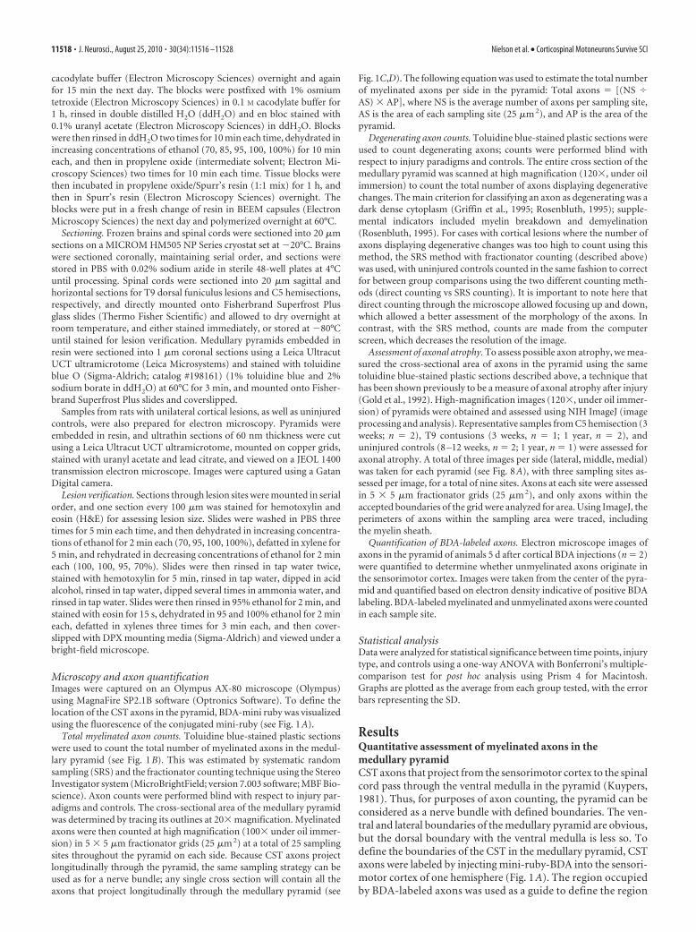
Figure 4. Histological assessment of myelinated axons in the pyramid after acute transec-tion SCI. A, B, Representative sections of toluidine blue-stained plastic sections (100�) toassess axon health and morphology after injury. Uninjured (A), healthy axons have clear axo-plasm and a continuous myelin ring, whereas degenerating axons, seen after cortical ablationlesion (B) causes significant amounts of detectable degeneration, have darkly stained axoplasmand myelin degradation (black arrow). G, H, Light micrographs (120�) of toluidine blue-stained plastic sections from the brainstem to show integrity of the ascending spinothalamictract (STT) lateral to the pyramid in uninjured (G) and degenerating (H ) axons after Hx SCI,which induces Wallerian degeneration (black arrowheads). The axons in the pyramid after a T9DF lesion, 1 week (C) and 4 weeks (D), as well as after a C5 Hx injury, 1 week (E) and 3 weeks (F ),did not result in significant degeneration such as the Cx lesion. There are a few axons thatexhibit degenerative changes (C, red arrow) and are quantified in Figures 6 and 7, respec-tively. Scale bars, 10 �m (for all panels); (in F ) A–F, images taken at 100�; (in H ) G, H,images taken at 120�.
Figure 5. Histological assessment of myelinated axons in the pyramid after contusive andchronic SCI. Representative sections of toluidine blue-stained plastic sections to assess axonhealth and morphology after a more severe type of injury at chronic time points. Uninjuredanimals aged 1 year (A) have healthy axons with clear axoplasm and a continuous myelin ring,compared with cortical lesion positive controls displaying widespread axon degeneration (B,black arrows). The axons in the pyramid after a T9 contusion both at 3 weeks (C) and even up to1 year (D) after injury do not show any detectable signs of degeneration to suggest their cellbodies had died as a result of the injury. Scale bar, 10 �m.
11522 • J. Neurosci., August 25, 2010 • 30(34):11516 –11528 Nielson et al. • Corticospinal Motoneurons Survive SCI
5.38 � 3.70, 0.0085 � 0.0059% of total) (Fig. 7D) or 3 weeks(lesioned, 5.38 � 2.56, 0.0085 � 0.0041% of total; unlesioned,5.25 � 1.75, 0.0083 � 0.0028% of total) (Fig. 7D) after injury.
Also consistent with the results after T9 DF lesions, SRS-basedcounts of total axons revealed no axon loss between sides after C5lateral hemisections at either the 1 week (lesioned, 65,153.84 �6325.85; unlesioned, 64,248.77 � 9677.55) or 3 week time points(lesioned, 59,943.57 � 10,033.38; unlesioned, 60,939.77 �6865.39) (Fig. 7D). Thus, even when the injury to CST axonsoccurs more proximal to the cell bodies in the cervical region,axons in the medullary pyramid exhibit no signs of degenerationand there is no indication of axon loss, indicating that corticalmotoneurons have not died at least up to 3 weeks after injury.
SCI does not cause delayed degeneration of CST axons in thepyramid but may affect age-dependent myelinationAxons must die eventually after death of the cell body, but it ispossible that the degeneration of the axon as a result of retrogradecell death takes longer than the Wallerian degeneration that oc-curs after physical destruction of the cell body or physical sepa-ration of the distal axon from the cell body. Accordingly, we
assessed whether there was evidence of axon degeneration oraxon loss in the medullary pyramid 1 year after moderate contu-sion injuries at T9, which completely destroy the dorsal funiculusin the same way as a DF transection.
Again, there were only a few axons in the medullary pyramidthat exhibited any sign of Wallerian degeneration in the rats withchronic T9 contusion injuries (Fig. 5D) compared with their age-matched controls (Fig. 5A). Interestingly, counts did reveal that,at 1 year after a T9 contusion, the number of possibly degenerat-ing (19.31 � 9.26; 0.024 � 0.012% of total) axons in the medul-lary pyramid is significantly greater than the number of possiblydegenerating axons 3 weeks after a DF lesion in young adult rats(5.13 � 1.64; 0.006 � 0.002% of total; p � 0.001) (Fig. 6D).Uninjured aged control rats also had a greater number of axons inthe medullary pyramid exhibiting signs of degeneration (11.50 �1.91; 0.014 � 0.002% of total), suggesting that these signs ofdegeneration are attributable to aging, and not the spinal cordinjury (Fig. 6D). It is important to emphasize, however, that evenin aged rats, degenerating axons represent only a fraction of apercentage of the total axons in the pyramid.
Interestingly, counts revealed that the older, uninjured con-trol rats also have significantly more total myelinated axons (Fig.6F) in the medullary pyramid (80,374.2 � 6829.9) than theyounger, uninjured controls (62,949.7 � 8419.7; p � 0.001). Ad-ditionally, rats that had received moderate T9 contusion injuries1 year previously had significantly fewer myelinated axons(71,638.1 � 10,151.0; p � 0.05) after injury, compared with theolder uninjured controls. However, there was no difference intotal myelinated axons compared with the younger, uninjuredcontrols, or between 3 weeks (67,593.6 � 14,960.0) and 1 yearafter contusion injury (see above). This is also confirmed in themeasures of total area of the pyramid, which does not change signif-icantly after injury (uninjured, 1 year: 355,583.2 � 52,596.2; T9 con-tusion, 3 weeks: 309,511.5 � 37,491.0; T9 contusion, 1 year:347,553.3 � 45,643.6) (Fig. 6B).
Together, these data suggest that there is no loss of myelinatedaxons after moderate contusion injuries, but the injury may re-duce an age-dependent increase in the number of myelinatedaxons that would otherwise occur (Leenen et al., 1989).
Can retrograde atrophy be detected by analyses of themedullary pyramid?Although there was no evidence of axon degeneration in the pyr-amid after SCI, previous studies have reported atrophy andshrinkage of the cell bodies of cortical motoneurons (Holmesand May, 1909; Lassek, 1942; Kalil and Schneider, 1975; Barron andDentinger, 1979; Ganchrow and Bernstein, 1985; Ramirez andKalil, 1985; Barron et al., 1988; Merline and Kalil, 1990; Tsengand Prince, 1996). Accordingly, it was of interest to determinewhether it was possible to detect atrophy of CST axons in thepyramid. To assess this, we measured the cross-sectional area of asample of CST axons (for the sampling strategy, see Fig. 8A). Aparticularly useful comparison is between the axons ipsilateral(unlesioned) and contralateral (lesioned) to a C5 hemisectioninjury because the unilateral injury provides an internal control.Cases that were completely unilateral (n � 2) for the C5 hemisec-tion at 3 weeks were used for internal comparison of lesioned versusunlesioned axons. There was no difference in average axon diameterbetween the two sides 3 weeks after a cervical hemisection injuryindicating no detectable atrophy (Fig. 8B). Indeed, there was notonly no statistical difference; the means were almost identical.
Similar assessments revealed no differences in the cross-sectional area of CST axons between uninjured young versus
Figure 6. Quantification of axon degeneration in the pyramid after acute and chronic SCI.Quantification of the area (A, B) of the medullary pyramid, as well as degenerating (C, D) andtotal myelinated axons (E, F ) after either acute (A, C, E) or chronic (B, D, F ) SCI, compared withage-matched controls. Neither level nor time after injury has a significant affect on axons in thepyramid at the acute time points between 1 and 4 weeks after T9 DF injuries in the spinal cord.This is demonstrated by the fact that there is no shrinkage of the pyramid after injury (A) or anyquantifiable signs of degenerating axons (C) or loss of axons (E) at these shorter survival timeperiods. Additionally, contusive SCI does not cause a significant amount of pyramid shrinkage,even up to 1 year after injury (B). There are significantly more degenerating axons 1 year afterinjury ( p � 0.001), but this is only a fraction of a percentage of the total axons in the pyramid(D), and may be explained by the fact that there are significantly more total axons in theuninjured aged animals ( p�0.001) compared with their younger counterparts (F ). There is nosignificant difference between the young uninjured control and either the 3 week or 1 year timepoints after SCI; however, there is a significant decrease in total myelinated axons at 1 yearcompared with the 1-year-old control ( p � 0.05), but not compared with the younger SCI timepoint, suggesting that axons are not lost over time as a result of injury, but that the increase inaxon number is affected by SCI (F ). Data are mean � SD. ***p � 0.001, *p � 0.05.
Nielson et al. • Corticospinal Motoneurons Survive SCI J. Neurosci., August 25, 2010 • 30(34):11516 –11528 • 11523

age-matched rats with T9 contusion inju-ries 3 weeks after contusion (Fig. 8C).Interestingly, however, axonal cross-sectional area was less in rats that receivedT9 contusions 1 year previously comparedwith age-matched controls (Fig. 8C). Thevalues from 1-year-old uninjured controlrats were also significantly higher than thevalues from animals 3 weeks after hemisec-tion. None of the other comparisons werestatistically significant, however (Fig. 8C),including the trend for increased axon areain the old versus young uninjured rats—aphenomenon that has previously been re-ported for axons in dorsal roots (Rao andKrinke, 1983).
Together, these results reveal that thereare no detectable changes in axon sizeduring the time that cortical motoneu-rons exhibit retrograde atrophy after SCI,but SCI may affect age-related changes inaxon size that otherwise occur.
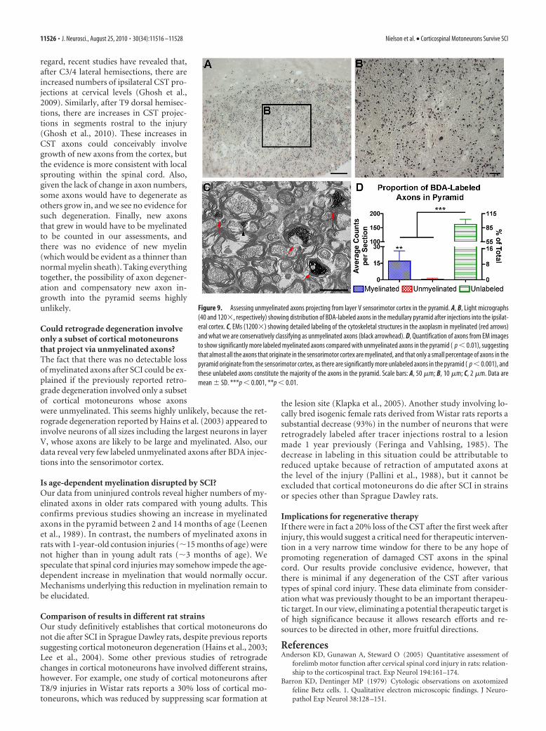
Could spinal cord lesions lead to aselective death of unmyelinated axonsand sparing of myelinated axons?One possible explanation for the absenceof degeneration of myelinated axons isthat cortical motoneurons that die afterspinal cord injury project via unmyeli-nated axons. This is highly unlikely inprinciple because cells that have beenreported to exhibit caspase staining arelarge neurons in layer V. Nevertheless, torule out this possibility, we assessed whethera significant number of CST axons (identi-fied by BDA tracing) were unmyelinated.EM analyses after BDA tract-tracing injec-tions into the sensorimotor cortexrevealed that the vast majority of BDA-labeled axons in the pyramid are myelinated (Fig. 9C). Therewere very few electron-dense processes that we conservativelyclassified as labeled unmyelinated axons because they appearedto contain neurofilaments and microtubules, but definitiveidentification of these as unmyelinated axons was difficultbecause the BDA labeling obscured the cytoplasm. Even in-cluding these questionable elements, the number of labeled unmy-elinated axons was low (16.9 � 9.3 labeled myelinated axons/275�m2 vs 0.7 � 1.1 labeled unmyelinated axons/275 �m2).
DiscussionGiven the long-standing controversy over whether there is retro-grade cell death of the CST after SCI, the present study aimed toresolve this matter definitively in a novel way, by assessing CSTaxons in the medullary pyramid. Our results reveal that there isno degeneration of CST axons or axon loss in the medullarypyramid, even in situations that have previously been reported tocause extensive retrograde degeneration of cortical motoneurons(Hains et al., 2003). Thus, cortical motoneurons and their axonsin the medullary pyramid survive the injury to the distal axon aftervarious types of spinal cord injury. The important implication is thatinterventions to promote regeneration of the CST do not requireseparate manipulations to preserve the cells of origin in the cortex.
It should be noted that we undertook this study with the ex-pectation that retrograde degeneration did occur and that itwould be possible to easily detect and rigorously quantify CSTaxon degeneration in the medullary pyramid. Thus, our resultswere as surprising to us as they are likely to be to the field.
Assessment of CST axon degenerationOur assessment of CST axon degeneration in the medullary pyr-amid is based on the assumption that axons of neurons that die asa result of retrograde degeneration would exhibit degenerativechanges similar to what are seen during Wallerian degeneration(condensation of the axonal cytoplasm, dark staining in toluidineblue-stained plastic sections, and eventually myelin fragmenta-tion). There has been speculation about this issue after SCI inprimates (Davison, 1937; Lassek, 1942), but no definitive evi-dence has been reported to our knowledge. Wallerian degenera-tion of an axon occurs after disconnection of the axon from itsrespective cell body (Waller, 1850) and has been reported exten-sively in the distal segment of the axon after injury (Ramon yCajal, 1928; Raff et al., 2002). Based on the rapid onset of degen-erative changes in axons after damage to the cell body (Ranson,1914; Dunkerley and Duncan, 1969; Beirowski et al., 2004;Coleman, 2005), time points of 1, 3, and 4 weeks after injury
Figure 7. Effect of lesion extent on axon degeneration in the medullary pyramid. A, C, Lesions at C5 were reconstructed byassessing horizontal sections through the lesion epicenter at 100 �m intervals and representing these on schematic cross sections.Shown are 1 week (A) and 3 week (C) survival. B, D, Quantification of area, degenerating axons, and total axons in the medullarypyramid after unilateral lesions to the spinal cord demonstrating that the extent of the lesion does not have a significant effect ineither the area of the pyramid (B) or axon degeneration as measured by both total axons as well as axons displaying degenerativechanges (D). There is no significant effect of lesion size on axons either in the pyramid affected by the lesions (black bars, lesioned),or the side unaffected by the lesion (gray bars, unlesioned), compared with each other and with uninjured controls (green bars,uninjured). The average lesion size was determined by multiplying the reconstructed images in Adobe Photoshop to show theareas of the spinal cord that were mostly affected across all the animals. The gray scale under each reconstruction depicts thevariability in the lesion sizes, withdarkerregionsrepresentingco-incidenceof lesionarea,showninA and C.Thedarkestgrayindicatesthearea damaged in all animals; the lightest indicates the area damaged in only one animal. Scale bar, 500 �m. Data are mean � SD.
11524 • J. Neurosci., August 25, 2010 • 30(34):11516 –11528 Nielson et al. • Corticospinal Motoneurons Survive SCI
should be sufficient to detect degenerative changes in axonscaused by retrograde degeneration of the cell body. Our control ex-periments involving aspiration lesions of the cortex reveal that CSTaxons exhibit Wallerian degeneration that is easily detectable by thetechniques used here, confirming what has been seen in previousstudies (Ranson, 1914; Dunkerley and Duncan, 1969).
Axon degeneration has also been reported after nontrau-matic death of neurons in the CNS, for example, after death ofneurons in the medial habenula caused by systemic adminis-tration of nicotine, causing axon degeneration in the fasciculusretroflexus (Carlson et al., 2000, 2001). Similarly, excitotoxicdeath of neurons is followed by Wallerian degeneration of theiraxons, which is easily detectable using selective silver impregna-tion techniques (Schmued et al., 1995; Schauwecker et al., 2000;Schauwecker, 2002; Benkovic et al., 2004), the methods of whichhave been described previously (Fink and Heimer, 1967; Beltra-mino et al., 1993; de Olmos et al., 1994). It remains possible,however, that retrograde degeneration of a neuronal cell bodywould lead to degeneration of the axon in a way that would not bedetectable by the techniques used here (i.e., something other thancondensation and dark staining of the axonal cytoplasm). It is alsopossible that the axons of cell bodies undergoing apoptosis may havean independent mechanism of degeneration than the cellbody, which was suggested after cell culture studies in miceexpressing the “slow Wallerian degeneration” (Wld s) allele(Deckwerth and Johnson, 1994).
Counts of healthy axonsIf retrograde degeneration of cortical mo-toneurons occurs in a way that does notlead to detectable degenerative changes inthe axons, counts of healthy axons wouldstill reveal loss of axons in the pyramidproportional to the number of corticalmotoneurons that died. For example,Hains et al. (2003) reported a loss of 20%of the CST cell bodies 1 week after a T9 DFSCI. Our counting technique would cer-tainly detect this because counts revealsignificant axon loss in the medullarypyramid after either complete aspira-tion of the sensorimotor cortex, or afterthe more limited damage caused by in-jections for tract tracing. Previous pub-lished counts of healthy axons in thepyramid have ranged from 73,000 whenassessed by light microscopy (Lassek andRasmussen, 1940), up to 103,000–150,000when assessed by electron-microscopiccounts (Leenen et al., 1985, 1989; Gorgels,1990). Our counts are consistent with theprevious counts based on light microscopy.
It is noteworthy that, after completeaspiration of the sensorimotor cortex,which destroys all of the cells of origin ofthe CST that are retrogradely labeled byFluorogold injections into the spinal cord(Strong et al., 2009), approximately one-half of the axons in the medullary pyramidsurvived. Previous studies have also notedthat a significant proportion of the axonsin the pyramid remained healthy afterlarge cortical ablations in rats (Ranson,1914; Dunkerley and Duncan, 1969). This
indicates that axons in the medullary pyramid originate in partfrom areas other than the sensorimotor cortex. Early studiesspeculated that these surviving axons might be originating from asubcortical region (Ranson, 1914) such as the lentiform nucleus(Swank, 1936). More recent studies suggest a cortical origin, how-ever, based on retrograde labeling of neurons in posterior parietal,rostral occipital, anterior cingulate, and prefrontal cortex after injec-tions of the retrograde tracer HRP at C5/6 in rats (Miller, 1987).
One other body of literature deserves note: Brodal and Wal-berg (1952) do report the presence of degenerating axons in themedullary pyramid after spinal cord injuries in cats based onsilver staining (the Glees method). Their speculation was that theaxons might be from ascending sensory systems. This issue wasfurther explored, but not resolved in later studies (Brodal andWalberg, 1982), which also contains an excellent discussion ofthe evidence for indirect Wallerian degeneration. Although wedid not detect evidence of degenerative changes in axons afterspinal cord injury, it is possible that a small number of axons doundergo some sort of degenerative change that would be detectedusing selective silver impregnation.
Could new axons grow into the medullary pyramid to replaceones that degenerate?One possible alternative explanation for the lack of any detectableloss of myelinated axons in the medullary pyramid is that newaxons grow in at the same time that others degenerate. In this
Figure 8. Assessment of axonal atrophy in the medullary pyramid. A, Sampling strategy of axons in the pyramid, shown at lowmagnification in toluidine blue-stained plastic sections (10�). Both sides of the pyramid were assessed at high magnification of120� (sampling size illustrated in black boxes). Three sites per side were assessed, lateral, middle, and medial in the pyramid bothfor contralateral and ipsilateral sides. At each sampling site, three sites were used to measure axon area using fractionatorsampling, and quantified using ImageJ. B, C, Quantification of axon area in the pyramids after C5 Hx with 3 week survival, and T9contusion with both 3 week and 1 year survival, compared with uninjured age-matched controls. B, Lesioned (gray checkered bar)and unlesioned (black bar) pyramids within the same animal after a C5 Hx results in no significant difference between axon areabetween sides. C, Axon area is significantly decreased 3 weeks after a C5 Hx ( p � 0.05), and 1 year after a T9 contusion ( p �0.001), compared with the 1-year-old uninjured control; however, all other comparisons are not significantly different from eachother. Scale bar, 200 �m. Data are mean � SD. *p � 0.05, ***p � 0.001.
Nielson et al. • Corticospinal Motoneurons Survive SCI J. Neurosci., August 25, 2010 • 30(34):11516 –11528 • 11525
regard, recent studies have revealed that,after C3/4 lateral hemisections, there areincreased numbers of ipsilateral CST pro-jections at cervical levels (Ghosh et al.,2009). Similarly, after T9 dorsal hemisec-tions, there are increases in CST projec-tions in segments rostral to the injury(Ghosh et al., 2010). These increases inCST axons could conceivably involvegrowth of new axons from the cortex, butthe evidence is more consistent with localsprouting within the spinal cord. Also,given the lack of change in axon numbers,some axons would have to degenerate asothers grow in, and we see no evidence forsuch degeneration. Finally, new axonsthat grew in would have to be myelinatedto be counted in our assessments, andthere was no evidence of new myelin(which would be evident as a thinner thannormal myelin sheath). Taking everythingtogether, the possibility of axon degener-ation and compensatory new axon in-growth into the pyramid seems highlyunlikely.
Could retrograde degeneration involveonly a subset of cortical motoneuronsthat project via unmyelinated axons?The fact that there was no detectable lossof myelinated axons after SCI could be ex-plained if the previously reported retro-grade degeneration involved only a subsetof cortical motoneurons whose axonswere unmyelinated. This seems highly unlikely, because the ret-rograde degeneration reported by Hains et al. (2003) appeared toinvolve neurons of all sizes including the largest neurons in layerV, whose axons are likely to be large and myelinated. Also, ourdata reveal very few labeled unmyelinated axons after BDA injec-tions into the sensorimotor cortex.
Is age-dependent myelination disrupted by SCI?Our data from uninjured controls reveal higher numbers of my-elinated axons in older rats compared with young adults. Thisconfirms previous studies showing an increase in myelinatedaxons in the pyramid between 2 and 14 months of age (Leenenet al., 1989). In contrast, the numbers of myelinated axons inrats with 1-year-old contusion injuries (�15 months of age) werenot higher than in young adult rats (�3 months of age). Wespeculate that spinal cord injuries may somehow impede the age-dependent increase in myelination that would normally occur.Mechanisms underlying this reduction in myelination remain tobe elucidated.
Comparison of results in different rat strainsOur study definitively establishes that cortical motoneurons donot die after SCI in Sprague Dawley rats, despite previous reportssuggesting cortical motoneuron degeneration (Hains et al., 2003;Lee et al., 2004). Some other previous studies of retrogradechanges in cortical motoneurons have involved different strains,however. For example, one study of cortical motoneurons afterT8/9 injuries in Wistar rats reports a 30% loss of cortical mo-toneurons, which was reduced by suppressing scar formation at
the lesion site (Klapka et al., 2005). Another study involving lo-cally bred isogenic female rats derived from Wistar rats reports asubstantial decrease (93%) in the number of neurons that wereretrogradely labeled after tracer injections rostral to a lesionmade 1 year previously (Feringa and Vahlsing, 1985). Thedecrease in labeling in this situation could be attributable toreduced uptake because of retraction of amputated axons atthe level of the injury (Pallini et al., 1988), but it cannot beexcluded that cortical motoneurons do die after SCI in strainsor species other than Sprague Dawley rats.
Implications for regenerative therapyIf there were in fact a 20% loss of the CST after the first week afterinjury, this would suggest a critical need for therapeutic interven-tion in a very narrow time window for there to be any hope ofpromoting regeneration of damaged CST axons in the spinalcord. Our results provide conclusive evidence, however, thatthere is minimal if any degeneration of the CST after varioustypes of spinal cord injury. These data eliminate from consider-ation what was previously thought to be an important therapeu-tic target. In our view, eliminating a potential therapeutic target isof high significance because it allows research efforts and re-sources to be directed in other, more fruitful directions.
ReferencesAnderson KD, Gunawan A, Steward O (2005) Quantitative assessment of
forelimb motor function after cervical spinal cord injury in rats: relation-ship to the corticospinal tract. Exp Neurol 194:161–174.
Barron KD, Dentinger MP (1979) Cytologic observations on axotomizedfeline Betz cells. 1. Qualitative electron microscopic findings. J Neuro-pathol Exp Neurol 38:128 –151.
Figure 9. Assessing unmyelinated axons projecting from layer V sensorimotor cortex in the pyramid. A, B, Light micrographs(40 and 120�, respectively) showing distribution of BDA-labeled axons in the medullary pyramid after injections into the ipsilat-eral cortex. C, EMs (1200�) showing detailed labeling of the cytoskeletal structures in the axoplasm in myelinated (red arrows)and what we are conservatively classifying as unmyelinated axons (black arrowhead). D, Quantification of axons from EM imagesto show significantly more labeled myelinated axons compared with unmyelinated axons in the pyramid ( p � 0.01), suggestingthat almost all the axons that originate in the sensorimotor cortex are myelinated, and that only a small percentage of axons in thepyramid originate from the sensorimotor cortex, as there are significantly more unlabeled axons in the pyramid ( p � 0.001), andthese unlabeled axons constitute the majority of the axons in the pyramid. Scale bars: A, 50 �m; B, 10 �m; C, 2 �m. Data aremean � SD. ***p � 0.001, **p � 0.01.
11526 • J. Neurosci., August 25, 2010 • 30(34):11516 –11528 Nielson et al. • Corticospinal Motoneurons Survive SCI
Barron KD, Dentinger MP, Popp AJ, Mankes R (1988) Neurons of layer Vbof rat sensorimotor cortex atrophy but do not die after thoracic cordtransection. J Neuropathol Exp Neurol 47:62–74.
Basso DM, Beattie MS, Bresnahan JC (1995) A sensitive and reliable loco-motor rating scale for open field testing in rats. J Neurotrauma 12:1–21.
Beirowski B, Berek L, Adalbert R, Wagner D, Grumme DS, Addicks K,Ribchester RR, Coleman MP (2004) Quantitative and qualitative analy-sis of Wallerian degeneration using restricted axonal labelling in YFP-Hmice. J Neurosci Methods 134:23–35.
Beltramino CA, de Olmos JS, Gallyas F, Heimer L, Zaborszky L (1993) Silverstaining as a tool for neurotoxic assessment. NIDA Res Monogr 136:101–126; discussion 126 –132.
Benkovic SA, O’Callaghan JP, Miller DB (2004) Sensitive indicators of in-jury reveal hippocampal damage in C57BL/6J mice treated with kainicacid in the absence of tonic-clonic seizures. Brain Res 1024:59 –76.
Beresford WA (1965) A discussion on retrograde changes in nerve fibres.Prog Brain Res 14:33–56.
Bodian D (1946) Spinal projections of brain-stem in rhesus monkey, de-duced from retrograde chromatolysis. Anat Rec 94:512–513.
Bonatz H, Rohrig S, Mestres P, Meyer M, Giehl KM (2000) An axotomymodel for the induction of death of rat and mouse corticospinal neuronsin vivo. J Neurosci Methods 100:105–115.
Bresnahan JC, Beattie MS, Stokes BT, Conway KM (1991) Three-dimensional computer-assisted analysis of graded contusion lesions inthe spinal cord of the rat. J Neurotrauma 8:91–101.
Brodal A, Walberg F (1952) Ascending fibers in pyramidal tract of cat. AMAArch Neurol Psychiatry 68:755–775.
Brodal A, Walberg F (1982) A re-evaluation of the question of ascendingfibers in the pyramidal tract. Brain Res 232:271–281.
Carlson J, Armstrong B, Switzer RC 3rd, Ellison G (2000) Selective neuro-toxic effects of nicotine on axons in fasciculus retroflexus further supportevidence that this a weak link in brain across multiple drugs of abuse.Neuropharmacology 39:2792–2798.
Carlson J, Noguchi K, Ellison G (2001) Nicotine produces selective degen-eration in the medial habenula and fasciculus retroflexus. Brain Res906:127–134.
Coleman M (2005) Axon degeneration mechanisms: commonality amid di-versity. Nat Rev Neurosci 6:889 – 898.
Crawley AP, Jurkiewicz MT, Yim A, Heyn S, Verrier MC, Fehlings MG, MikulisDJ (2004) Absence of localized grey matter volume changes in the motorcortex following spinal cord injury. Brain Res 1028:19–25.
Davison C (1937) Syndrome of the anterior spinal artery of the medulla.AMA Arch Neurol Psychiatry 37:91–107.
Deckwerth TL, Johnson EM Jr (1994) Neurites can remain viable after de-struction of the neuronal soma by programmed cell death (apoptosis).Dev Biol 165:63–72.
de Olmos JS, Beltramino CA, de Olmos de Lorenzo S (1994) Use of anamino-cupric-silver technique for the detection of early and semiacuteneuronal degeneration caused by neurotoxicants, hypoxia, and physicaltrauma. Neurotoxicol Teratol 16:545–561.
Dunkerley GB, Duncan D (1969) A light and electron microscopic study ofthe normal and the degenerating corticospinal tract in the rat. J CompNeurol 137:155–183.
Feringa ER, Vahlsing HL (1985) Labeled corticospinal neurons one yearafter spinal cord transection. Neurosci Lett 58:283–286.
Fink RP, Heimer L (1967) Two methods for selective silver impregnation ofdegenerating axons and their synaptic endings in the central nervoussystem. Brain Res 4:369 –374.
FitzGerald MJT, Gruener G, Mtui E (2007) Clinical neuroanatomy andneuroscience, Ed 5. Philadelphia: Elsevier Saunders.
Ganchrow D, Bernstein JJ (1985) Thoracic dorsal funicular lesions affect thebouton patterns on, and diameters of, layer VB pyramidal cell somata inrat hindlimb cortex. J Neurosci Res 14:71– 81.
Ghosh A, Sydekum E, Haiss F, Peduzzi S, Zorner B, Schneider R, Baltes C,Rudin M, Weber B, Schwab ME (2009) Functional and anatomical re-organization of the sensory-motor cortex after incomplete spinal cordinjury in adult rats. J Neurosci 29:12210 –12219.
Ghosh A, Haiss F, Sydekum E, Schneider R, Gullo M, Wyss MT, Mueggler T,Baltes C, Rudin M, Weber B, Schwab ME (2010) Rewiring of hindlimbcorticospinal neurons after spinal cord injury. Nat Neurosci 13:97–104.
Giehl KM, Tetzlaff W (1996) BDNF and NT-3, but not NGF, prevent
axotomy-induced death of rat corticospinal neurons in vivo. Eur J Neu-rosci 8:1167–1175.
Gold BG, Griffin JW, Price DL (1992) Somatofugal axonal atrophy precedesdevelopment of axonal degeneration in acrylamide neuropathy. ArchToxicol 66:57– 66.
Gorgels TG (1990) A quantitative analysis of axon outgrowth, axon loss,and myelination in the rat pyramidal tract. Brain Res Dev Brain Res54:51– 61.
Griffin JW, George EB, Hsieh ST, Glass JD (1995) Axonal degeneration anddisorders of the axonal cytoskeleton. New York, Oxford: Oxford UP.
Hains BC, Black JA, Waxman SG (2003) Primary cortical motor neuronsundergo apoptosis after axotomizing spinal cord injury. J Comp Neurol462:328 –341.
Hammond EN, Tetzlaff W, Mestres P, Giehl KM (1999) BDNF, but notNT-3, promotes long-term survival of axotomized adult rat corticospinalneurons in vivo. Neuroreport 10:2671–2675.
Holmes G, May WP (1909) On the exact origin of the pyramidal tract inman and other mammals. Brain 32:1– 43.
Kalil K, Schneider GE (1975) Retrograde cortical and axonal changes fol-lowing lesions of the pyramidal tract. Brain Res 89:15–27.
Klapka N, Hermanns S, Straten G, Masanneck C, Duis S, Hamers FP, MullerD, Zuschratter W, Muller HW (2005) Suppression of fibrous scarring inspinal cord injury of rat promotes long-distance regeneration of cortico-spinal tract axons, rescue of primary motoneurons in somatosensory cor-tex and significant functional recovery. Eur J Neurosci 22:3047–3058.
Kost SA, Oblinger MM (1993) Immature corticospinal neurons respond toaxotomy with changes in tubulin gene expression. Brain Res Bull30:469 – 475.
Kost-Mikucki SA, Oblinger MM (1991) Changes in glial fibrillary acidicprotein mRNA expression after corticospinal axotomy in the adult ham-ster. J Neurosci Res 28:182–191.
Kuypers HGJM (1981) Anatomy of the descending pathways. Baltimore:Williams and Wilkins.
Lassek AM (1942) The pyramidal tract: a study of retrograde degenerationin the monkey. Arch Neurol Psychiatr (Chicago) 48:561–567.
Lassek AM, Rasmussen GL (1940) A comparative fiber and numerical anal-ysis of the pyramidal tract. J Comp Neurol 72:417– 428.
Lee BH, Lee KH, Kim UJ, Yoon DH, Sohn JH, Choi SS, Yi IG, Park YG (2004)Injury in the spinal cord may produce cell death in the brain. Brain Res1020:37– 44.
Leenen LP, Meek J, Posthuma PR, Nieuwenhuys R (1985) A detailedmorphometrical analysis of the pyramidal tract of the rat. Brain Res359:65– 80.
Leenen LP, Meek J, Posthuma PR, Nieuwenuys R (1989) Differences in thefiber composition of the pyramidal tract in two- and 14-month-old rats.Neuroscience 28:635– 643.
Liu CN (1955) Time pattern in retrograde degeneration after trauma ofcentral nervous system of mammals. In: Regeneration in the central ner-vous system (Windle WF, ed), pp 84 –93. Springfield, IL: Thomas.
Mason MR, Lieberman AR, Anderson PN (2003) Corticospinal neuronsup-regulate a range of growth-associated genes following intracortical,but not spinal, axotomy. Eur J Neurosci 18:789 – 802.
Merline M, Kalil K (1990) Cell death of corticospinal neurons is induced byaxotomy before but not after innervation of spinal targets. J Comp Neurol296:506 –516.
Mikucki SA, Oblinger MM (1991) Corticospinal neurons exhibit a novelpattern of cytoskeletal gene expression after injury. J Neurosci Res30:213–225.
Miller MW (1987) The origin of corticospinal projection neurons in rat.Exp Brain Res 67:339 –351.
Neumann S, Woolf CJ (1999) Regeneration of dorsal column fibers intoand beyond the lesion site following adult spinal cord injury. Neuron23:83–91.
Nissl F (1892) Ueber die Veranderungen der Ganglienzellen amFacialiskern des Kaninchens nach Ausreissung der Nerven. Allgen ZPsychiatr 48:197–198.
Pallini R, Fernandez E, Sbriccoli A (1988) Retrograde degeneration of cor-ticospinal axons following transection of the spinal cord in rats. A quan-titative study with anterogradely transported horseradish peroxidase.J Neurosurg 68:124 –128.
Piecharka DM, Kleim JA, Whishaw IQ (2005) Limits on recovery in thecorticospinal tract of the rat: partial lesions impair skilled reaching and
Nielson et al. • Corticospinal Motoneurons Survive SCI J. Neurosci., August 25, 2010 • 30(34):11516 –11528 • 11527
the topographic representation of the forelimb in motor cortex. Brain ResBull 66:203–211.
Raff MC, Whitmore AV, Finn JT (2002) Axonal self-destruction and neu-rodegeneration. Science 296:868 – 871.
Ramirez LF, Kalil K (1985) Critical stages for growth in the development ofcortical neurons. J Comp Neurol 237:506 –518.
Ramon y Cajal S (1928) Cajal’s degeneration and regeneration of the ner-vous system. New York, Oxford: Oxford UP.
Ranson SW (1914) A note on the degeneration of the fasciculus cerebro-spinalis in the albino rat. J Comp Neurol 24:503–507.
Rao RS, Krinke G (1983) Changes with age in the number and size of my-elinated axons in the rat L4 dorsal spinal root. Acta Anat (Basel) 117:187–192.
Reh T, Kalil K (1981) Development of the pyramidal tract in the hamster. I.A light microscopic study. J Comp Neurol 200:55– 67.
Rosenbluth (1995) Pathology of demylinated and dysmyelinated axons.New York, Oxford: Oxford UP.
Schauwecker PE (2002) Modulation of cell death by mouse genotype: dif-ferential vulnerability to excitatory amino acid-induced lesions. Exp Neu-rol 178:219 –235.
Schauwecker PE, Ramirez JJ, Steward O (2000) Genetic dissection of thesignals that induce synaptic reorganization. Exp Neurol 161:139 –152.
Schmidlin E, Wannier T, Bloch J, Rouiller EM (2004) Progressive plasticchanges in the hand representation of the primary motor cortex parallelincomplete recovery from a unilateral section of the corticospinal tract atcervical level in monkeys. Brain Res 1017:172–183.
Schmidlin E, Wannier T, Bloch J, Belhaj-Saif A, Wyss AF, Rouiller EM (2005)Reduction of the hand representation in the ipsilateral primary motor
cortex following unilateral section of the corticospinal tract at cervicallevel in monkeys. BMC Neurosci 6:56.
Schmued LC, Scallet AC, Slikker W Jr (1995) Domoic acid-induced neuro-nal degeneration in the primate forebrain revealed by degeneration spe-cific histochemistry. Brain Res 695:64 –70.
Strong MK, Blanco JE, Anderson KD, Lewandowski G, Steward O (2009)An investigation of the cortical control of forepaw gripping after cervicalhemisection injuries in rats. Exp Neurol 217:96 –107.
Swank RL (1936) The pyramidal tract. An experimental study of the corti-cospinal tract and other components in the rabbit. AMA Arch NeurolPsychiatry 36:530 –541.
Thuret S, Moon LD, Gage FH (2006) Therapeutic interventions after spinalcord injury. Nat Rev Neurosci 7:628 – 643.
Tseng GF, Prince DA (1996) Structural and functional alterations in ratcorticospinal neurons after axotomy. J Neurophysiol 75:248 –267.
Van Gehuchten A (1903) La degenerescence dite retrograde ou degeneres-cence wallerienne indirecte. Nevraxe 5:1–108.
Waller A (1850) Experiments on the section of the glossopharyngeal andhypoglossal nerves of the frog, and observations of the alterations pro-duced thereby in the structure of their primitive fibres. Philos Trans R SocLondon 140:423– 429.
Wannier T, Schmidlin E, Bloch J, Rouiller EM (2005) A unilateral section ofthe corticospinal tract at cervical level in primate does not lead to mea-surable cell loss in motor cortex. J Neurotrauma 22:703–717.
Wiesendanger M (1981) The pyramidal tract. Its structure and function.New York: Plenum.
Zimmerman EA, Chambers WW, Liu CN (1964) An experimental study ofthe anatomical organization of the cortico-bulbar system in the albino rat.J Comp Neurol 123:301–323.
11528 • J. Neurosci., August 25, 2010 • 30(34):11516 –11528 Nielson et al. • Corticospinal Motoneurons Survive SCI