Development/Plasticity/Repair ComprehensiveCorticospinalLabelingwith mu crystallin ... ·...
16
Development/Plasticity/Repair Comprehensive Corticospinal Labeling with mu-crystallin Transgene Reveals Axon Regeneration after Spinal Cord Trauma in ngr1 / Mice X Kathren L. Fink, 1,2 X Stephen M. Strittmatter, 1,2 * and X William B.J. Cafferty 1 * 1 Department of Neurology and 2 Program in Cellular Neuroscience, Neurodegeneration and Repair, Yale University School of Medicine, New Haven, Connecticut 06520 Spinal cord injury interrupts descending motor tracts and creates persistent functional deficits due to the absence of spontaneous axon regeneration. Of descending pathways, the corticospinal tract (CST) is thought to be the most critical for voluntary function in primates. Even with multiple tracer injections and genetic tools, the CST is visualized to only a minor degree in experimental studies. Here, we identify and validate the mu-crystallin (crym) gene as a high-fidelity marker of the CST. In transgenic mice expressing green fluorescent protein (GFP) under crym regulatory elements (crym-GFP), comprehensive and near complete CST labeling is achieved throughout the spinal cord. Bilateral pyramidotomy eliminated the 17,000 GFP-positive CST axons that were reproducibly labeled in brainstem from the spinal cord. We show that CST tracing with crym-GFP is 10-fold more efficient than tracing with biotinylated dextran amine (BDA). Using crym-GFP, we reevaluated the CST in mice lacking nogo receptor 1 (NgR1), a protein implicated in limiting neural repair. The number and trajectory of CST axons in ngr1 / mice without injury was indistinguishable from ngr1 / mice. After dorsal hemisection in the midthoracic cord, CST axons did not significantly regenerate in ngr1 / mice, but an average of 162 of the 6000 labeled thoracic CST axons (2.68%) regenerated 100 m past the lesion site in crym-GFP ngr1 / mice. Although traditional BDA tracing cannot reliably visualize regenerating ngr1 / CST axons, their regenerative course is clear with crym-GFP. Therefore the crym-GFP transgenic mouse is a useful tool for studies of CST anatomy in experimental studies of motor pathways. Key words: corticospinal tract; nogo receptor; regeneration; spinal cord injury; transgene Introduction Spinal cord injury (SCI) results in permanent disability due to the limited growth capacity of adult CNS axons. In part, cell-intrinsic programs limit axonal regeneration (Liu et al., 2011). In addition, the adult CNS is an inhibitory environment due to the expression of chondroitin sulfate proteoglycans (Yiu and He, 2006) and myelin-associated inhibitors (NogoA, myelin-associated glyco- protein, oligodendrocyte myelin glycoprotein; Akbik et al., 2012). These molecules restrict axon growth by signaling through neuron-specific Nogo Receptors 1 and 3 (NgR1, (Fournier et al., 2001; Dickendesher et al., 2012), PirB (Atwal et al., 2008), PTP- sigma (Shen et al., 2009; Fry et al., 2010), leukocyte common Received Aug. 23, 2015; revised Sept. 15, 2015; accepted Sept. 24, 2015. Author contributions: K.L.F., S.M.S., and W.B.J.C. designed research; K.L.F. and W.B.J.C. performed research; K.L.F. and W.B.J.C. analyzed data; K.L.F., S.M.S., and W.B.J.C. wrote the paper. This work was supported by the National Institutes of Health (Grant R01NS080388 to S.M.S.) and the Falk Medical Research Trust (S.M.S.). S.M.S. is a cofounder of Axerion Therapeutics, seeking to develop NgR- and PrP-based therapeutics. The remain- ing authors declare no competing financial interests. *S.M.S. and W.B.J.C. contributed equally to this work. Correspondence should be addressed to either Stephen M. Strittmatter or William B.J.Cafferty, Depart- ment of Neurology and Program in Cellular Neuroscience, Neurodegeneration and Repair, Yale University School of Medicine, 8300F 300 George Street, New Haven, CT 06520, E-mail: [email protected] or [email protected]. DOI:10.1523/JNEUROSCI.3165-15.2015 Copyright © 2015 the authors 0270-6474/15/3515403-16$15.00/0 Significance Statement Axon regeneration fails in the adult CNS, resulting in permanent functional deficits. Traditionally, inefficient extrinsic tracers such a biotinylated dextran amine (BDA) are used to label regenerating fibers after therapeutic intervention. We introduce crym-green fluorescent protein (GFP) transgenic mice as a comprehensive and specific tool with which to study the primary descending motor tract, the corticospinal tract (CST). CST labeling with crym-GFP is 10 times more efficient compared with BDA. The enhanced sensitivity afforded by crym-GFP revealed significant CST regeneration in NgR1 knock-out mice. Therefore, crym- GFP can be used as a standardized tool for future CST spinal cord injury studies. The Journal of Neuroscience, November 18, 2015 • 35(46):15403–15418 • 15403
Transcript of Development/Plasticity/Repair ComprehensiveCorticospinalLabelingwith mu crystallin ... ·...

Development/Plasticity/Repair
Comprehensive Corticospinal Labeling with mu-crystallinTransgene Reveals Axon Regeneration after Spinal CordTrauma in ngr1�/� Mice
X Kathren L. Fink,1,2 X Stephen M. Strittmatter,1,2* and X William B.J. Cafferty1*1Department of Neurology and 2Program in Cellular Neuroscience, Neurodegeneration and Repair, Yale University School of Medicine, New Haven,Connecticut 06520
Spinal cord injury interrupts descending motor tracts and creates persistent functional deficits due to the absence of spontaneous axonregeneration. Of descending pathways, the corticospinal tract (CST) is thought to be the most critical for voluntary function in primates.Even with multiple tracer injections and genetic tools, the CST is visualized to only a minor degree in experimental studies. Here, weidentify and validate the mu-crystallin (crym) gene as a high-fidelity marker of the CST. In transgenic mice expressing green fluorescentprotein (GFP) under crym regulatory elements (crym-GFP), comprehensive and near complete CST labeling is achieved throughout thespinal cord. Bilateral pyramidotomy eliminated the 17,000 GFP-positive CST axons that were reproducibly labeled in brainstem from thespinal cord. We show that CST tracing with crym-GFP is 10-fold more efficient than tracing with biotinylated dextran amine (BDA). Usingcrym-GFP, we reevaluated the CST in mice lacking nogo receptor 1 (NgR1), a protein implicated in limiting neural repair. The number andtrajectory of CST axons in ngr1 � / � mice without injury was indistinguishable from ngr1 �/� mice. After dorsal hemisection in themidthoracic cord, CST axons did not significantly regenerate in ngr1 �/� mice, but an average of 162 of the 6000 labeled thoracic CSTaxons (2.68%) regenerated �100 �m past the lesion site in crym-GFP ngr1 � / � mice. Although traditional BDA tracing cannot reliablyvisualize regenerating ngr1 � / � CST axons, their regenerative course is clear with crym-GFP. Therefore the crym-GFP transgenic mouseis a useful tool for studies of CST anatomy in experimental studies of motor pathways.
Key words: corticospinal tract; nogo receptor; regeneration; spinal cord injury; transgene
IntroductionSpinal cord injury (SCI) results in permanent disability due to thelimited growth capacity of adult CNS axons. In part, cell-intrinsicprograms limit axonal regeneration (Liu et al., 2011). In addition,the adult CNS is an inhibitory environment due to the expression
of chondroitin sulfate proteoglycans (Yiu and He, 2006) andmyelin-associated inhibitors (NogoA, myelin-associated glyco-protein, oligodendrocyte myelin glycoprotein; Akbik et al.,2012). These molecules restrict axon growth by signaling throughneuron-specific Nogo Receptors 1 and 3 (NgR1, (Fournier et al.,2001; Dickendesher et al., 2012), PirB (Atwal et al., 2008), PTP-sigma (Shen et al., 2009; Fry et al., 2010), leukocyte common
Received Aug. 23, 2015; revised Sept. 15, 2015; accepted Sept. 24, 2015.Author contributions: K.L.F., S.M.S., and W.B.J.C. designed research; K.L.F. and W.B.J.C. performed research;
K.L.F. and W.B.J.C. analyzed data; K.L.F., S.M.S., and W.B.J.C. wrote the paper.This work was supported by the National Institutes of Health (Grant R01NS080388 to S.M.S.) and the Falk Medical
Research Trust (S.M.S.).S.M.S. is a cofounder of Axerion Therapeutics, seeking to develop NgR- and PrP-based therapeutics. The remain-
ing authors declare no competing financial interests.*S.M.S. and W.B.J.C. contributed equally to this work.
Correspondence should be addressed to either Stephen M. Strittmatter or William B.J.Cafferty, Depart-ment of Neurology and Program in Cellular Neuroscience, Neurodegeneration and Repair, Yale UniversitySchool of Medicine, 8300F 300 George Street, New Haven, CT 06520, E-mail: [email protected] [email protected].
DOI:10.1523/JNEUROSCI.3165-15.2015Copyright © 2015 the authors 0270-6474/15/3515403-16$15.00/0
Significance Statement
Axon regeneration fails in the adult CNS, resulting in permanent functional deficits. Traditionally, inefficient extrinsic tracerssuch a biotinylated dextran amine (BDA) are used to label regenerating fibers after therapeutic intervention. We introducecrym-green fluorescent protein (GFP) transgenic mice as a comprehensive and specific tool with which to study the primarydescending motor tract, the corticospinal tract (CST). CST labeling with crym-GFP is 10 times more efficient compared with BDA.The enhanced sensitivity afforded by crym-GFP revealed significant CST regeneration in NgR1 knock-out mice. Therefore, crym-GFP can be used as a standardized tool for future CST spinal cord injury studies.
The Journal of Neuroscience, November 18, 2015 • 35(46):15403–15418 • 15403

antigen-related phosphatase (Fisher et al., 2011), and the sphin-golipid receptor S1PR2 (Kempf et al., 2014). Receptor antago-nism via either genetic perturbation (Kim et al., 2003; Simonen etal., 2003; Kim et al., 2004; Dimou et al., 2006; Cafferty et al., 2010;Dickendesher et al., 2012; Bartus et al., 2014) or pharmacologicaltreatment (Schnell and Schwab, 1990; Bradbury et al., 2002;GrandPre et al., 2002; Liebscher et al., 2005; Wang et al., 2006;Wang et al., 2011; Lang et al., 2015) results in enhanced (yetincomplete) recovery of motor function after experimental SCI(Basso et al., 1995; Basso et al., 2006). In general, the anatomicalsubstrate supporting functional recovery remains unclear andcorrelative. In contrast to universally adopted SCI behavioral as-sessments such as BMS and BBB, methods to label motor tractssuch as the corticospinal tract (CST) vary widely (Steward et al.,2008b; Lee et al., 2010; Geoffroy et al., 2015). Therefore, impor-tant anatomical data can be lost, overinterpreted, and difficult toreproduce (Kim et al., 2003; Simonen et al., 2003; Woolf, 2003;
Zheng et al., 2005; Cafferty et al., 2007a; Steward et al., 2007;Steward et al., 2008a; Steward et al., 2012).
Traditionally, CST axon regeneration and plasticity is assessedafter injection of anterograde tracers such as biotinylated dextranamine (BDA) into motor cortex (Tuszynski and Steward, 2012).Cortical BDA injection is convenient because only layer V corti-cospinal motor neurons (CSMNs) project to spinal cord. Multi-ple standardized stereotaxic injection sites targeting the fore andhindlimb motor cortex are used routinely to maximize CSMNlabeling. Despite these measures, CST axon labeling varies greatlybetween laboratories and as much as 70-fold within groups(Steward et al., 2008b).
In addition to inconsistent partial labeling with BDA, ex-trinsic tracer injections further complicate SCI experimentsthrough additional surgeries with unintended damage. Tolimit these issues, genetic approaches have begun to emerge tomore comprehensively and exclusively label CSMNs. One re-
Figure 1. BAC transgenic Tg(Crym-EGFP)GF82Gsat expresses soluble GFP in adult brain and spinal cord. Schematic (A) shows a linear representation of the BAC clone RP24-62I5, which containsTmem159 (cyan), Zp2 (yellow), Anks4b (orange), Crym (red), and Abca14 (purple). Enlarged sequence shows inferred schematic of GENSAT clone BX1076, which contains EGFP (green) with its ownPolyA (blue) 5� to the start codon of Crym (red solid boxes are exons, open boxes are introns). BX1076 was injected into fertilized ova to create Tg(crym-EGFP)GF82 (crym-GFP) mice. Schematic (B)shows location of primary and secondary motor (cyan), sensory (yellow), auditory (orange), and visual cortex (magenta). Stippled lines labeled C, D, and I in schematic B indicate location ofphotomicrographs through the transverse plane of the brain (C–E) and cervical spinal cord (I–K ) of an adult wild-type crym-GFP mouse. Intense GFP labeling is observed in layer V of motor (C,�0.86mm anterior to bregma), sensory (D, �0.86 posterior to bregma), auditory and visual cortex (E, �3.5 mm posterior to bregma). GFP is additionally localized in a diffuse pattern in the striatum (C)and in the CA1 and CA2 regions of the hippocampus (E). High-power photomicrographs (F–H ) show crym-GFP expression in pyramidal neurons in layer V of primary motor cortex. Antibodies to Crymlabel all crym-GFP � pyramidal neurons (G, H ). Low-power photomicrographs (I–K ) of a transverse section of C7 spinal cord show robust crym-GFP expression in the dorsal, lateral, and ventralfuniculi (I ) and throughout dorsal and intermediate gray matter. Low levels of Crym protein are observed in the ventral dorsal column (J ), which overlaps 100% with crym-GFP (K ). Scale bars: C, 1mm; F, 100 �m; I, 500 �m.
15404 • J. Neurosci., November 18, 2015 • 35(46):15403–15418 Fink et al. • Complete CST Labeling with crym-GFP

port describes a double transgenic thy1-STOP-YFP � Emx-Cre mouse in which yellow fluorescent protein (YFP) is drivenvia cre-recombinase-mediated excision of a transcriptionalstop site under the control of thy1 elements (Bareyre et al.,
2005). In this line, 11,000 YFP � CSTaxons are labeled in the cervical cord.However, the utility of this approach islimited by the stochastic nature of thy1promoter elements (Feng et al., 2000;Willenberg and Steward, 2015) and theneed for multiple transgenes. A mousein which a single transgene drives aCST-specific reporter would facilitatebreeding with multiple genetically mod-ified models for SCI studies. Here, weintroduce the mu-crystallin green fluo-rescent protein (crym-GFP) transgenicmouse to study the intact and lesionedadult CST. Crym-GFP mice express solu-ble GFP under the mu-crystallin pro-moter and demonstrate comprehensiveand near specific CST labeling. Further-more, we demonstrate the utility of thecrym-GFP line by crossing it with NgR1knock-out mice and reassessing CSTregeneration after dorsal hemisection(DhX). The increased sensitivity affordedby the crym-GFP line reveals significantCST regeneration in ngr1� / � comparedwith ngr1�/� mice, an anatomical findingnot observed in previous studies usingBDA (Kim et al., 2004). We propose thatthe crym-GFP mice will streamline andstandardize SCI experiments that assessCST regeneration as a primary outcome.
Materials and MethodsMice. BAC transgenic Tg(crym-EGFP)GF82Gsatmice (hereafter referred to as crym-GFP mice) wereprocuredfromtheGENSATproject[stocknumber012003-UCD, The Gene Expression Nervous Sys-tem Atlas (GENSAT) Project, National Institutesof Health–National Institute of NeurologicalDisorders and Stroke Contracts N01NS02331and HHSN271200723701C to The RockefellerUniversity, New York) and back crossed withC57BL/6 mice for 9 generations. Crym-GFPmice were then crossed with ngr1 � / � mice asdescribed previously (Kim et al., 2004), subse-quent crym-GFP ngr1 �/ � mice were crossedwith nontransgenic ngr1 �/ � mice to create ei-ther crym-GFP ngr1 �/� or crym ngr1 � / �
mouse lines. To minimize the number ofanimals used while maintaining enough rigorto achieve our scientific objectives (Festing andAltman, 2002), we used freely available poweranalysis (Hedwig.mgh.harvard.edu/sample_size/js/js_parallel_quant.html) to estimatesample sizes. The experiments are poweredat 90% based on the number of animals ineach group, the SD as determined by our pre-vious BMS data (Cafferty et al., 2010), and asignificance level of 0.05. Before behavioralassessment and surgery, mice were random-ized and split into cages of three. Theseblocks were maintained unless evidence of
fighting was apparent, upon which aggressors were singly housed.Surgery. All procedures and postoperative care were performed in ac-
cordance with the guidelines of the Institutional Animal Use and CareCommittee at Yale University.
Figure 2. Intact crym-GFP transgenic mice illustrate comprehensive labeling of the CST. Low-power photomicrographs oftransverse sections of cervical (C6, A) and lumbar (L4, B) spinal cord from an intact (color schematic in A shows a bilaterally intactCST in green) adult wild-type crym-GFP mouse show intrinsic GFP expression in the dorsal (dCST), dorsolateral (dlCST), and ventralcorticospinal (vCST) tracts and throughout spinal gray matter (GM). Inset schematics (A) reflect intact terminals. Spinal gray andwhite matter are delineated with a white stippled line (A, B). Sagittal sections through the midline (C, cyan line depicts relativelocation of section, compass reflects orientation of section: D, dorsal; V, ventral; R, rostral; C, caudal) and mediolateral (D) thoraciccord show intense GFP labeling in the main dorsal CST and GFP � terminals entering spinal gray matter. Horizontal thoracicsections through the upper (E, cyan line depicts relative location of section, compass reflects orientation of section: M, medial; L,lateral; R, rostral; C, caudal) and lower dorsal horn (F ) show GFP � CST terminals densely localized throughout gray matter. Scalebars: B, 500 �m; D, 1 mm.
Fink et al. • Complete CST Labeling with crym-GFP J. Neurosci., November 18, 2015 • 35(46):15403–15418 • 15405

Retrograde fast blue and anterograde BDAtracing. To complete retrograde fast blue(FB) and anterograde BDA labeling ofCSMNs, adult (7–9 weeks of age) malecrym-GFP ngr1 �/� (n � 6) and crym-GFPngr1 � / �(n � 6) mice were anesthetized withketamine (100 mg/kg) and xylazine (15 mg/kg) and placed in a stereotaxic frame (Stoelt-ing). An incision was made over the cervicalenlargement and the C5–C8 vertebrae wererevealed by blunt dissection of overlyingmuscle. A hemi-laminectomy was performedto expose the underlying C5–C8 spinal cordand a small incision was made in the duramater. The tip of a pulled glass capillary tubeattached to a Micro4 infusion device (WorldPrecision Instruments) was slowly insertedstereotaxically to a depth of 500 �m into theC5 level of the spinal cord and �500 �mlateral from the midline. Thirty seconds afterintroduction of the capillary tube 75 nl of a0.1% solution of FB (Polysciences) was in-fused into the spinal cord over 2 min. The tipwas left in situ for an additional 30 s beforeremoval. This procedure was completedthree additional times at C6, C7, and C8, re-sulting in a total infusion of 300 nl of FB.Anterograde CSMN labeling with BDA wascompleted as described previously (Kim etal., 2003; Kim et al., 2004; Cafferty et al.,2007b; Cafferty et al., 2010). Briefly, burrholes were made over the sensorimotor cor-tex and 5 microinfusions of 75 nl of a 10%solution of BDA were made to a depth of 0.7mm (coordinates, �1 mm to �1 mm poste-rior to bregma and 0.5–1.5 mm lateral tobregma) using a pulled glass capillary tubeattached to a Micro4 infusion device to de-liver a total volume of 375 nl of BDA. Musclewas sutured with Vicryl and skin with mono-filament suture. Two weeks after externaltracer injections, mice were perfused with4% paraformaldehyde and the tissue waspostfixed overnight at 4°C and embedded in10% gelatin for immunohistochemical pro-cessing. An investigator blinded to genotypecompleted all surgical procedures.
DhX and anterograde CSMN tracing. Adult(7–9 weeks of age) female crym-GFP ngr1 �/�
(n � 11) and crym-GFP ngr1 � / � (n � 15)mice were anesthetized with ketamine (100mg/kg) and xylazine (15 mg/kg) and an inci-sion made over their thoracic spinal cord. Alaminectomy was performed to expose thedorsal portion of spinal cord corresponding tothe T6 and T7 levels. The dura mater waspierced and the spinal cord exposed and apledget of gelfoam soaked in 1% lidocaine wasplaced on the exposed cord for 1 min beforelesion. A DhX lesion was performed at T6 witha 30 gauge needle and a pair of microscissors toa depth of 1.0 mm to completely sever the dor-sal and dorsolateral CSTs. The overlying mus-cle was sutured with Vicryl and skin layer withmonofilament suture. Four weeks after SCI,mice received unilateral cortical micro infusion (Micro4; World Preci-sion Instruments) with BDA (10,000 mol/wt; Life Technologies) to an-terogradely label the CST as described above. Six weeks after DhX, micewere perfused with 4% paraformaldehyde and the tissue was postfixed
overnight at 4°C and embedded in 10% gelatin for immunohistochemi-cal processing. An investigator blinded to genotype completed all surgi-cal procedures.
Bilateral pyramidotomy. To complete bilateral pyramidotomy (bPyX),adult wild-type crym-GFP mice (n � 6 ) were anesthetized with ketamine
Figure 3. bPyX results in complete loss of spinal GFP labeling in crym-GFP mice. Low-power photomicrographs of transversesections of cervical (C6, A) and lumbar (L4, B) spinal cord after bPyX [color schematic in A shows bilateral axotomy (red crosses) ofthe CST (green) in the medullary pyramids] in an adult wild-type crym-GFP mouse show a complete loss of GFP in the dCST, dlCST,vCST, and throughout spinal gray matter. Inset schematic (A) reflects lesioned terminals. Midline (C, cyan line depicts relativelocation of section, compass reflects orientation of section: D, dorsal; V, ventral; R, rostral; C, caudal) and mediolateral (D) sagittalsections show a complete loss of GFP labeling in white and gray matter after bPyX. Horizontal sections through the upper (E, cyanline depicts relative location of section, compass reflects orientation of section: M, medial; L, lateral; R, rostral; C, caudal) and lowerdorsal horn also show a complete loss of GFP � CST axons and terminals after bPyX. Few sporadic GFP � cells can be seen scatteredin gray matter in transverse (A, B), sagittal (C, D) and horizontal (E, F ) sections. Scale bars: B, 500 �m; D, 1 mm.
15406 • J. Neurosci., November 18, 2015 • 35(46):15403–15418 Fink et al. • Complete CST Labeling with crym-GFP

(100 mg/kg) and xylazine (15 mg/kg) and placed in a supine position, anincision was made to the left of the trachea, and blunt dissection exposedthe occipital bone at the base of the skull. The occipital bone was removedon either side of the basilar artery with blunt Dumont #2 forceps toexpose the medullary pyramids. The dura mater was pierced with a 30gauge needle and resected. The pyramids were transected bilaterally withfine Dumont #5 forceps to a depth of 0.25 mm or just exposed for shamlesion. No internal sutures were made and skin was closed with mono-filament suture. Four weeks after bPyX, mice were perfused with 4%paraformaldehyde. Tissue was postfixed overnight at 4°C and embeddedin 10% gelatin for immunohistochemical processing.
Behavioral analysis. Mice that underwent DhX lesions were assessedusing the Basso Mouse Score (BMS) (Basso et al., 2006). Data are pre-sented as average BMS SEM. Data were analyzed via repea-ted-measures ANOVA with Bonferonni correction for multiple
comparisons. Post hoc analysis was completedfor statistically significant differences compar-ing BMS score at each time point between ge-notypes with ANOVA. Two investigatorsblinded to genotype completed BMS scoring.
Histology. Mice were killed with an overdoseof ketamine (100 mg/kg) and xylazine (15 mg/kg) and were transcardially perfused with 0.9%NaCl (normal saline) followed by 4% parafor-maldehyde in PBS. Brains and spinal cordswere dissected, postfixed in 4% paraformalde-hyde overnight at 4°C, and subsequently em-bedded in 10% gelatin (Sigma-Aldrich)dissolved in water for vibratome sectioning.Transverse sections (35– 40 �m) of cervicaland lumbar spinal cord (C6 –C7), sagittal andhorizontal sections of thoracic spinal cord(T4 –T8), and coronal sections of brain andbrainstem were processed for BDA withstreptavidin-conjugated secondary antibodies(Life Technologies) and tyramide signal ampli-fication (PerkinElmer). Immunofluorescenceused antibodies directed against green fluores-cent protein (GFP, 1:5000; Life Technologies),Crym (1:500; Abcam), and GFAP (1:10,000;DAKO) with Alexa Fluor 488 and Alexa Fluor594 (1:500; Life Technologies). Direct applica-tion of Nissl-568 (Life Technologies)-labeledneuronal somata in cortex. An investigatorblinded to genotype completed all immunohis-tochemical procedures.
Quantification: percentage of cortical layer Vneuronal somata crym-GFP � and crym-GFP �FB �. Mice that underwent unilateral in-traspinal FB infusion were prepared forimmunohistochemical analysis and thenumber of crym-GFP � and crym-GFP �FB �
CSMNs was determined. Then, 35 �m sectionsthrough forelimb primary motor cortex fromngr1 �/� (n � 6) and ngr1 � / � (n � 6) micewere processed for GFP immunohistochemis-try and counterstained with Nissl-568. FBemits fluorescence at 420 nm under 365 nmillumination and therefore does not requireadditional methods for detection. Layer V ofcortex was identified via depth from pial sur-face and density of large pyramidal shapedsomata. The number of Nissl �, crym-GFP �Nissl �, and crym-GFP �FB � neuronswere counted in a 250 � 250 �m box superim-posed upon five randomly selected sectionsfrom each mouse and photomicrographs takenunder epifluorescent illumination at 20� mag-nification (Leica Microsystems). Data are pre-sented as average percentage of the number of
Nissl � somata that were crym-GFP � and the average number of FB �
somata that were crym-GFP � SEM and analyzed by Student’s t testwith Bonferonni correction.
Number of CST axons in spinal white matter. Mice that underwentsham lesion in the DhX cohort that received intracortical BDA infusionwere prepared for immunohistochemical analysis to determine the ab-solute number of CST axons that were crym-GFP � and BDA � in thebrainstem and dorsal, dorsolateral, and ventral CST in the cervical, tho-racic, and lumbar spinal cord. Then, 35 �m transverse sections frombrainstem and spinal cord from ngr1 �/� (n � 6) and ngr1 � / � (n � 6)mice were processed for GFP immunohistochemistry and BDA detec-tion. Photomicrographs were taken at 63� under oil illumination (ZeissImager Z1) from five randomly selected sections from each location fromeach animal. The absolute number crym-GFP � and BDA � axons were
Figure 4. Crym-GFP is expressed in cortical layer V projection neurons. Photomicrographs (A, B) show robust GFP expression inlayer V (cortical layers shown in K ) cortical neuron somata, axons and primary dendrites (B, D, F are high-power insets from boxshown in E). GFP � somata are exclusively neuronal as 100% of GFP � cells are Nissl � (C–F ). Of all Nissl � cells in layer V (LV),34.44 2.4 (average number of Nissl �GFP � somata SEM) were GFP � in ngr1 �/� and 34.93 2.4 were GFP � inngr1 � / � mice (M ). There was no significant difference between genotypes (Student’s t test). Unilateral injection of the retro-grade tracer FB into the cervical spinal cord resulted in dense labeling of layer V pyramidal neuron somata (G–L) in contralateralcortex 2 weeks after injection (H, J, L are high-power insets from box shown in K ). Of all FB � neurons, 94.69 0.6 were GFP � inngr1 �/� and 94.23 0.4 were GFP � in ngr1 � / � mice (N ). There was no significant difference between genotypes (Student’st test). Scale bars: A, 100 �m; B, 50 �m.
Fink et al. • Complete CST Labeling with crym-GFP J. Neurosci., November 18, 2015 • 35(46):15403–15418 • 15407

counted for the dorsolateral and ventral CSTs.For brainstem and dorsal CST, low-powerphotomicrographs were taken at 10� to deter-mine the area occupied by the pyramidal tractand the CST in the ventral dorsal columns, re-spectively. A 25 � 25 �m grid was then over-laid on the 63� photomicrographs and thecrym-GFP � or BDA � axons in five randomlyselected boxes from within this grid werecounted. The average number of axons fromthese boxes was then scaled up to determine thetotal number of axons labeled. Data are pre-sented as the average number of crym-GFP � orBDA � axonal profiles SEM and analyzed byANOVA with Bonferonni correction.
Density of crym-GFP � and BDA � axons inspinal gray matter. Densitometric analysis ofcrym-GFP � and BDA � axons terminating inthe cervical spinal cord was completed in Im-ageJ version 1.45s as described previously (Caf-ferty and Strittmatter, 2006). Briefly, labeledaxons were selected by thresholding and theaverage fiber length was measured using theskeletonize function in five sections per ani-mal. Data are presented as the average length ofcrym-GFP � and BDA � axons in mm 2 SEMand analyzed by ANOVA with Bonferonnicorrection.
Number of regenerating crym-GFP � CST ax-ons. To quantify the number of regeneratingcrym-GFP � CST axons after DhX, we cut 35�m sagittal sections of thoracic spinal cordthrough the lesion site and completed GFP im-munohistochemistry. All sections with evi-dence of the fasciculated dorsal CST rostral tothe lesion site were included in our analysis(5– 6 sections/animal). The crym-GFP � axonsat the lesion epicenter and at 50, 100, 250, 500, 750, 1000, 1250, 1500,1750, 2000, 2250, and 2500 �m caudal to the lesion site were counted ineach sagittal section. The sum of crym-GFP � CST axons in five sagittalsections was divided by the average number of crym-GFP � CST axons inthe dorsal CST at the mid-thoracic spinal level in intact wild-type crym-GFP mice to derive an axon index value (Lee et al., 2010; Blackmore et al.,2012; Geoffroy et al., 2015; Wang et al., 2015). Data are shown as theaverage number of regenerating crym-GFP � axons after T6 DhX peranimal indexed to the total number of crym-GFP axons in intact crym-GFP mice SEM/genotype and analyzed with two-way ANOVA withrepeated measures with Bonferonni correction for multiple comparisonswith post hoc t test. An investigator blinded to genotype completed allimmunohistochemical analyses.
ResultsCrym-GFP transgenic mice reveal detailed wiring of the CSTThe GENSAT project provides a compendium of mouse lines inwhich bacterial artificial chromosome (BAC) transgenes havebeen introduced to express fluorescent reporters under the con-trol of the promoter and regulatory elements of specific genes ofinterest. These lines facilitate the detailed study of the temporaland spatial expression of specific genes of interest. We browsedtheir transgenic catalog (Gong et al., 2003; Heintz, 2004) insearch of a mouse line that would label CSMNs, their axons, andtheir terminals with high efficiency. We found that the GENSATclone BX1076 fulfilled these criteria. GENSAT used the BACclone RP24-62I5 containing 216,104 base pairs of mouse chro-mosome 7 (Fig. 1A) to make GENSAT BAC clone BX1076 (Fig.1A); that is, �15 kilobases containing the crym promoter, theeight exons, and seven introns of the crym gene. The sequence for
green fluorescent protein (GFP) with its own start codon fol-lowed by a polyadenylation signal was inserted upstream of theATG of the first exon of the crym gene (Fig. 1A) allowing for theexpression of soluble GFP under the crym promoter. We ob-tained the BAC-BX1076 transgenic mouse from GENSAT (Gonget al., 2003) herein referred to as the crym-GFP mouse. Crym is aNADP-regulated thyroid hormone binding protein (Vie et al., 1997;Hallen et al., 2011) and lens protein (Kim et al., 1992) that is alsoenriched in layer V neurons that project to the spinal cord (Arlotta etal., 2005). Other than its use as a marker of CSMNs in cortex (Yas-voina et al., 2013), its functional role remains unknown.Examination of crym knock-out mice revealed no observable ana-tomical or functional differences (data not shown). Gross examina-tion of the GFP expression in the CNS of crym-GFP mice(localization schematized in Fig. 1B) confirmed that crym-GFP isexpressed in neuronal somata in cortical layer V projection neuronsin primary and secondary motor cortex (Fig. 1C,F–H), sensory cor-tex (Fig. 1D), and auditory and visual cortex (Fig. 1E). In addition,crym-GFP can be seen in the septal nuclei (Fig. 1C,D), striatum (Fig.1C), and hippocampus (Fig. 1E). In the spinal cord, the CST is ro-bustly labeled with crym-GFP, revealing the dense innervation pat-tern of the CST into the gray matter from axons exiting the dorsal,dorsolateral, and ventral white matter (Fig. 1I). Next, we investi-gated whether GFP transgenic labeling under the control of the crympromoter was faithful to endogenous Crym expression. Immuno-staining of brain and spinal cord sections using an antibody to Crymrevealed complete overlap between endogenous Crym and trans-genic GFP in layer V neurons of cortex (Fig. 1F–H) and in the spinalcord (Fig. 1I–K). Endogenous Crym expression is weak and espe-
Figure 5. Intrinsic GFP labeling of the CST in crym-GFP transgenic mice is superior to extrinsic labeling with anterogradelytransported BDA. Low-power photomicrographs of brainstem (A–C), cervical (C8, D–F ), thoracic (T8, G–I ), and lumbar (L4, J–L)spinal cord show robust intrinsic GFP � CST axons in the medullary pyramid (A) dorsal, dorsolateral and ventral spinal white andgray matter (D, G, J ). In contrast, fewer CST axons and terminals are BDA � in the pyramids (B) and spinal cords (E, H, K ) comparedwith GFP �-labeled axons and terminals in crym-GFP mice (see overlays C, F, I, L; GFP, green; BDA, red; DAPI, blue). Scale bars in Aand D, 500 �m.
15408 • J. Neurosci., November 18, 2015 • 35(46):15403–15418 Fink et al. • Complete CST Labeling with crym-GFP
cially difficult to detect in the finer CST axon terminals in spinalcord, suggesting that Crym functions within the neuronal somata(Fig. 1J); however, crym-GFP transgenic mice express soluble GFPthat is not restricted to any cellular compartment and therefore fillssomata, axons, and terminals, thereby underscoring its utility as acomprehensive intrinsic CST marker (Fig. 1I–K).
Crym-GFP comprehensively labels the CST throughout thespinal axisWe sought to examine the utility of crym-GFP mice to intrin-sically label the CST for spinal cord injury studies. To this end,we completed bPyX in crym-GFP mice to singularly axotomizethe CST bilaterally in its entirety. Intact crym-GFP mice show
Figure 6. Intrinsic GFP labeling of the CST in crym-GFP transgenic mice is superior to extrinsic labeling with anterogradely transported BDA. Single-channel high-power photomicro-graphs show crym-GFP expression (A1) and BDA tracing (A2) of CST axons (overlay A3; crym-GFP, green; BDA, red) in the medullary pyramid of a wild-type crym-GFP mouse. Sections(A1–A3) were assessed and quantification completed on CST axons in the brainstem rostral to the pyramidal decussation, shown in schematic B. Subsequent evaluation of the numberof CST axons entering the cervical, thoracic, and lumbar spinal cord were parsed into three groups according to the their white matter location (schematic C) in the dCST, dlCST, or vCST.Significantly more CST axons were labeled with GFP compared with BDA in both ngr1 �/� and ngr1 � / � mice (D, *F(3, 99) � 966.652, p 0.0005, one-way ANOVA with Bonferonni posthoc comparisons). There was no significant difference in the number of crym-GFP � axons or BDA � axons between genotypes. Data are shown as average number of labeled axons SEM. Single-channel high-power photomicrographs show crym-GFP expression (E1, F1, G1) and BDA tracing (E2, F2, G2) of CST axons in the dCST in transverse sections of cervical(E1–E3), thoracic (F1–F3), and lumbar (G1–G3) spinal cord of a wild-type crym-GFP mouse. Significantly more CST axons were labeled with GFP compared with BDA (E3, F3, G3,overlays; crym-GFP, green; BDA, red) at all spinal levels in both ngr1 �/� and ngr1 � / � mice (H, *F(11, 273) � 381.683, p 0.0005, one-way ANOVA with Bonferonni post hoccomparisons, data are shown as average number of labeled axons SEM). There was no significant difference in the number of crym-GFP labeled axons or BDA � axons betweengenotypes at any spinal level. Data are shown as average number of labeled axons SEM. Single channel high-power photomicrographs show crym-GFP expression (I1, J1, K1) and BDAtracing (I2, J2, K2) of CST axons in the dlCST in transverse sections of cervical (I1–I3), thoracic (J1–J3), and lumbar (K1–K3) spinal cord of a wild-type crym-GFP mouse. Significantlymore CST axons were labeled with GFP compared with BDA (I3, J3, K3, overlays; crym-GFP, green; BDA, red) at all spinal levels in both ngr1 �/� and ngr1 � / � mice (L, *F(11, 318) �292.458, p 0.0005, one-way ANOVA with Bonferonni post hoc comparisons, data are shown as average number of labeled axons SEM). There was no significant difference in thenumber of crym-GFP labeled axons or BDA � axons between genotypes at any spinal level. Single-channel high-power photomicrographs show crym-GFP expression (M1, N1, O1) andBDA tracing (M2, N2, O2) of CST axons in the vCST in transverse sections of cervical (M1–M3), thoracic (N1–N3), and lumbar (O1–O3) spinal cord of a wild-type crym-GFP mouse.Significantly more CST axons were labeled with GFP compared with BDA (M3, N3, O3, overlays; crym-GFP, green; BDA, red) at all spinal levels in both ngr1 �/� and ngr1 � / � mice (P,*F(11, 313) � 282.994, p 0.0005, one-way ANOVA with Bonferonni post hoc comparisons, data are shown as average number of labeled axons SEM). There was no significantdifference in the number of crym-GFP-labeled axons or BDA � axons between genotypes at any spinal level. Scale bar in A3, 10 �m.
Fink et al. • Complete CST Labeling with crym-GFP J. Neurosci., November 18, 2015 • 35(46):15403–15418 • 15409
robust labeling of CST axons in the dorsal CST (dCST) thedorsolateral CST (dlCST), and ventral CST (vCST) in the cer-vical (Fig. 2A) and lumbar (Fig. 2B) spinal cord. In addition,GFP labeled CST terminals can be seen densely innervatingspinal gray matter in transverse sections of cervical (Fig. 2A)and lumbar (Fig. 2B) cord, and in sagittal (Fig. 2C,D) andhorizontal (Fig. 2 E, F ) sections of thoracic cord. After bPyX,crym-GFP is almost entirely eliminated from cervical (Fig.3A), thoracic (Fig. 3C–F ), and lumbar spinal cord (Fig. 3B). Afew spinal interneurons can be seen along the midline; how-ever, they are easily distinguishable from the axon tracts andcomplex CST innervation pattern (Fig. 2). These data clearlydemonstrate that crym-GFP is a comprehensive and robustlabel of the CST and also reveal the full extent of its complexand dense terminal arborization in both dorsal and ventralspinal gray matter.
Crym-GFP � CSMNs exclusively terminate in spinal cord inngr1 �/� and ngr1 � / � miceThe sensitivity of CST labeling afforded by crym-GFP trans-genic mice allows for comprehensive localization of fine struc-tures that are typical of newly sprouted or regenerating axons(Steward et al., 2003; Tuszynski and Steward, 2012). Previousstudies have shown that mice null mutant for NgR1 exhibitplasticity of uninjured CSMNs after unilateral pyramidotomy(uPyX) (Cafferty and Strittmatter, 2006) and with experience-dependent plasticity in visual cortex (McGee et al., 2005) andsomatosensory cortex (Akbik et al., 2013). Strikingly, how-ever, no CST axon regeneration was observed after DhX inngr1 � / � mice (Kim et al., 2004). Together, these data suggestthat either developmental differences in the wiring of ngr1 � / �
mice account for the anatomical changes observed after uPyXand during activity-dependent plasticity or the methodologyused to label regenerating CST axons after DhX in ngr1 � / �
mice was insufficient to detect regenerating CST axons. Toexplore these possibilities, we completed a comprehensiveevaluation of the CST in ngr1 �/� crym-GFP and ngr1 � / �
crym-GFP mice (see Figs. 4, 5, 6, 7) and assessed CST regener-ation in both lines of mice after DhX SCI (see Figs. 8, 9, 10,11, 12).
We examined crym-GFP labeling in transverse sections of motorcortex (Fig. 4A, B) in intact ngr1�/� and ngr1� /� mice. Costainingfor Nissl (Fig. 4C–F) revealed that crym-GFP labeled 34.4 2.4% oflayer V pyramidal neurons in ngr1�/� mice and 34.9 2.4% of layerV neurons in ngr1� /� mice (Fig. 4M). Because only a subset of layerV neurons were crym-GFP�, we sought to determine whether thissubset was projecting to either the spinal cord, intracortically acrossthe corpus callosum, or to brainstem nuclei. To this end, we injectedthe retrograde tracer FB into the cervical enlargement to labelCSMNs that project to the spinal cord. FB can be seen accumulating
Figure 7. Intrinsic GFP labeling of gray matter CST terminals in crym-GFP transgenic mice issuperior to extrinsic labeling with anterogradely transported BDA. Low-power photomicro-graphs of transverse sections of intact C8 spinal cord from adult ngr1 �/� (A) and ngr1 � / � (B)mice show dense GFP � CST fibers terminating throughout gray matter (A, B, green). BDA �
4
fibers can be seen exiting the dorsal columns and innervating a more sparse area of gray matter(A, B, red). High-power photomicrographs of the dorsal (C–H) and ventral (I–N) horns showsignificantly denser GFP � CST axon terminals (C, D, I, J) compared with BDA � CST axon ter-minals (E, F, K, L). Multichannel overlays (G, H, M, N; GFP, green; BDA, red) illustrate thesuperior efficiency of crym-GFP compared with anterograde BDA tracing. Densitometric analysisshowed that significantly more GFP � CST fibers terminated in intact cervical spinal gray mattercompared with BDA � CST fibers (O, *F(3, 96) � 231.293, p 0.0005, one-way ANOVA withBonferonni post hoc comparisons, data are shown as the average density of CST occupation persquare millimeter for each label SEM). There was no significant difference in the density ofcrym-GFP-labeled terminals or BDA � terminals between genotypes. Scale bars: A, 500 �m; C,10 �m.
15410 • J. Neurosci., November 18, 2015 • 35(46):15403–15418 Fink et al. • Complete CST Labeling with crym-GFP

in a subset of layer V neurons in forelimb motor cortex 2 weeks afterintraspinal injection (Fig. 4G–L). Colabeling sections with GFP re-vealed that 94.7 0.6% and 94.2 0.4% of FB� cells were alsoGFP� in crym-GFP ngr1�/� and crym-GFP ngr1� /� mice, respec-
tively (Fig. 4N). These data confirm that crym-GFP is specificallylabeling spinally projecting layer V neurons in motor cortex and thatthere is no significant difference in the number or location of spinallyprojecting CSMNs in ngr1� /� compared with ngr1�/� mice.
Figure 8. Comprehensive CST labeling in crym-GFP mice reveal axon regeneration of axotomized CST neurons after DhX SCI in ngr1 �/� and ngr1 � / � mice. Low-power photomicrographs ofmid-sagittal sections from ngr1 �/� (A) and ngr1 � / � (B–D) show intense GFP � labeling of the CST rostral to the lesion site (inset compass in A shows orientation; D, dorsal; V, ventral; R, rostral;C, caudal). The epicenter of the lesion is marked with an asterisk. In both ngr1 �/� (A) and ngr1 � / � (B–D) mice GFP � CST axons can be seen caudal to the lesion site. High-power photomicro-graphs of inset boxes labeled i–iii clearly show GFP � CST axons regenerating into the lesion site in ngr1 �/� mice (Ai–Aiii). Significantly more GFP � CST axons can be seen crossing the lesion siteand entering the distal spinal cord in ngr1 � / � mice (Bi–Biii, Ci–Ciii, Di–Diii). Scale bars: A, 500 �m; Ai, 150 �m.
Fink et al. • Complete CST Labeling with crym-GFP J. Neurosci., November 18, 2015 • 35(46):15403–15418 • 15411

CST fascicule labeling is superior in crym-GFP micecompared with anterograde BDA transportWe next sought to examine CST axon number and spinal termi-nal density in crym-GFP ngr1�/� mice and determine whetherthe CST is miswired in crym-GFP ngr1� / � mice. Traditionally,
the CST has been visualized in the spinal cord after injection ofBDA into multiple sites in motor cortex (Tuszynski and Steward,2012). Using this method, normal CST wiring was observed inngr1� / � mice previously (Kim et al., 2004; Cafferty and Stritt-matter, 2006). We sought to determine whether crym-GFP label-
Figure 9. Additional examples of CST regeneration in ngr1 �/� and ngr1 � / � after DhX. The epicenter of the lesion is marked with an asterisk. In both ngr1 �/� (A) and ngr1 � / � (B–D) mice,GFP � CST axons can be seen caudal to the lesion site. High-power photomicrographs of inset boxes labeled i–iii clearly show GFP � CST axons regenerating into the lesion site in ngr1 �/� mice(Ai–Aiii). Significantly more GFP � CST axons can be seen crossing the lesion site and entering the distal spinal cord in ngr1 � / � mice (Bi–Biii, Ci–Ciii, Di–Diii). Scale bars: A, 500 �m; Ai, 150 �m.
15412 • J. Neurosci., November 18, 2015 • 35(46):15403–15418 Fink et al. • Complete CST Labeling with crym-GFP
ing of the CST was more efficient than BDA tracing and thus ableto distinguish any potential differences in CST anatomy betweenngr1�/� and ngr1� / � mice. Transverse sections through thebrainstem (Fig. 5A–C), cervical (Fig. 5D–F), thoracic (Fig. 5G–I),and lumbar (Fig. 5J–L) spinal cord 2 weeks after intracorticalBDA delivery revealed that, qualitatively, BDA labeled a fractionof the CST compared with crym-GFP at all levels of the neuroaxis.
Close inspection of BDA� and crym-GFP� CST axon profilesin the pyramidal tract rostral to the decussation in the brainstem(Fig. 6A,B) showed that, on average, 17205 593 and 17107 1078 CST axons were GFP� in crym-GFP ngr1�/� and crym-GFPngr1� / � mice, respectively (Fig. 6D). However, intracorticalBDA injection (Fig. 6A) labeled only 1634 167 and 1787 204CST axons in crym-GFP ngr1�/� and crym-GFP ngr1� / � mice,respectively, significantly less than the number of GFP� axons(Fig. 6D; *p 0.0001, ANOVA). These data show that crym-GFPis labeling 10 times the number of CST axons that are commonlylabeled with intracortical BDA injection. To determine whetherthis trend was consistent throughout the neuroaxis, we examinedthe number of GFP� CST axon profiles and BDA� profiles in thedCST (Fig. 6C,E–H), dlCST (Fig. 6C,I–L), and vCST (Fig.6C,M–P) in the cervical (Fig. 6E, I,M), thoracic (Fig. 6F, J,N),and lumbar (Fig. 6G,K,O) spinal cord in intact crym-GFPngr1�/� and crym-GFP ngr1� / � mice 2 weeks after intracorticalBDA injection. As expected, the average number of crym-GFP�
and BDA� CST profiles decreased caudally as axons terminatedin spinal gray matter (Fig. 6H,L,P). However, significantly moreGFP� CST axon profiles were observed in all white matter loca-tions at all spinal levels compared with the number of BDA�
axonal profiles (Fig. 6H,L,P; *p 0.001, ANOVA), maintainingat least a 10:1 CST labeling superiority of crym-GFP over BDA.Despite the difference in the efficiency between crym-GFP andBDA in labeling the CST, there was no significant difference inthe number of GFP� CST axon profiles or BDA� profiles be-tween genotypes, confirming normal CST fasciculation inngr1� / � mice.
Crym-GFP reveals extensive CST terminal innervation inspinal gray matter and normal CST wiring in ngr1 � / � miceBecause the number of CST axons labeled in crym-GFP was 10-fold higher than commonly observed with BDA tracing, wesought to explore the CST termination pattern in spinal graymatter (Fig. 7A,B) and also to determine whether spinal CSTinnervation was aberrant in ngr1� / � mice. Densitometric anal-ysis (Fig. 7O) was completed on high-power photomicrographsof transverse sections of crym-GFP (Fig. 7C,D,G– J,M,N) andBDA (Fig. 7E–H, K–N)-labeled axons in the cervical spinal dorsal(Fig. 7C–H) and ventral horns (Fig. 7I–N). Consistent with thewhite matter quantification, crym-GFP labels 10 times more ax-ons in spinal gray matter than BDA (Fig. 7O). No difference ingray matter innervation was observed between crym-GFPngr1�/� and crym-GFP and ngr1� / � mice (Fig. 7O), indicatingthat there are no developmental wiring defects in the CST ofngr1� / � mice.
Comprehensive CST labeling reveals CST regeneration inngr1 � / � miceCSMNs express NgR1 (McGee and Strittmatter, 2003; Zheng etal., 2005), so CST axons are restricted from spontaneous plastic-ity and regeneration by myelin-associated inhibitors expressed inthe intact and degenerating CNS (Schwab and Strittmatter,2014). Previously, we have shown that mice null for NgR1 displayenhanced plasticity of the processes of CSMNs in the adult cortex
(Lee et al., 2004) and spinal cord (Cafferty and Strittmatter,2006). Furthermore, pharmacological inhibition of NgR1 activa-tion resulted in increased CST regeneration after experimentalSCI (Li et al., 2004a; Wang et al., 2006; Wang et al., 2011). How-ever, despite increased regeneration of rubrospinal axons andincreased sprouting of raphespinal axons, no CST regenerationwas observed after DhX in ngr1� / � mice (Kim et al., 2004). Withthe significantly enhanced CST labeling efficiency afforded bycrym-GFP mice over BDA labeling and the lack of developmentalCST wiring defects observed in crym-GFP ngr1� / � mice, we de-cided to revisit whether bona fide CST regeneration occurs inngr1� / � mice after DhX.
Crym-GFP ngr1�/� mice (n � 11) and crym-GFP ngr1� / �
mice (n � 15) received DhX SCI at T6 for anatomical and behav-ioral analyses. Six weeks after injury, no residual degenerating
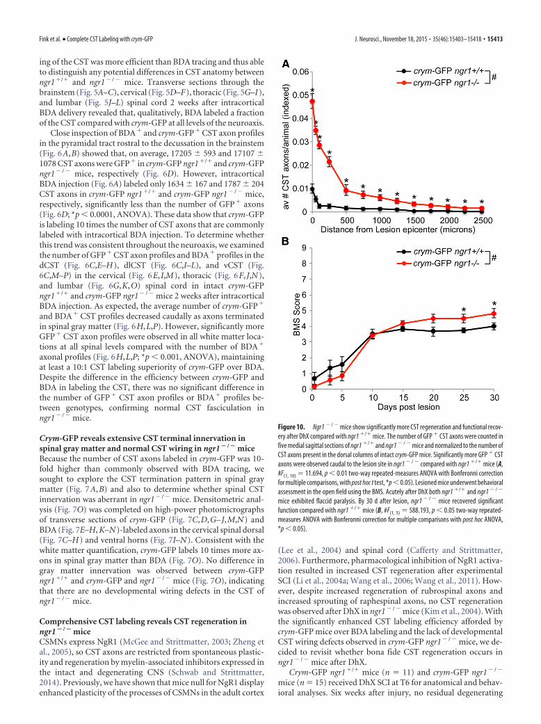
Figure 10. Ngr1 � / � mice show significantly more CST regeneration and functional recov-ery after DhX compared with ngr1 �/� mice. The number of GFP � CST axons were counted infive medial sagittal sections of ngr1 �/� and ngr1 � / � mice and normalized to the number ofCST axons present in the dorsal columns of intact crym-GFP mice. Significantly more GFP � CSTaxons were observed caudal to the lesion site in ngr1 � / � compared with ngr1 �/� mice (A,#F(1, 10) � 11.694, p 0.01 two-way repeated-measures ANOVA with Bonferonni correctionfor multiple comparisons, with post hoc t test, *p 0.05). Lesioned mice underwent behavioralassessment in the open field using the BMS. Acutely after DhX both ngr1 �/� and ngr1 � / �
mice exhibited flaccid paralysis. By 30 d after lesion, ngr1 � / � mice recovered significantfunction compared with ngr1 �/� mice (B, #F(1, 5) � 588.193, p 0.05 two-way repeated-measures ANOVA with Bonferonni correction for multiple comparisons with post hoc ANOVA,*p 0.05).
Fink et al. • Complete CST Labeling with crym-GFP J. Neurosci., November 18, 2015 • 35(46):15403–15418 • 15413
crym-GFP axons were seen caudal to the lesion site in either ge-notype (Figs. 8, 9). GFP� CST axons can be seen regenerating upto the lesion site in crym-GFP ngr1�/� mice, with very few, if any,regenerating CST axons observed caudal to the lesion site (Figs.8A, 9A). However, crym-GFP ngr1� / � mice exhibit significantregeneration of crym-GFP CST axons through and caudal to thelesion site (Figs. 8B–D, 9B–D). Figures 8 and 9 exhibit compara-ble spinal sections from six independent examples that demon-strate CST regeneration in ngr1� / � mice is robust andreproducible. Crym-GFP� regenerating axons were quantified atseveral distances caudal to the lesion epicenter (Fig. 10A).Ngr1� / � mice exhibited significantly greater CST regenerationcompared with ngr1�/� mice (Fig. 10A; #p 0.01, two-wayANOVA with repeated measures). Ngr1� / � mice also exhibitedenhanced locomotor recovery in the open field (Fig. 10B; #p
0.05, two-way ANOVA with repeated measures), consistent withour previous data (Kim et al., 2004).
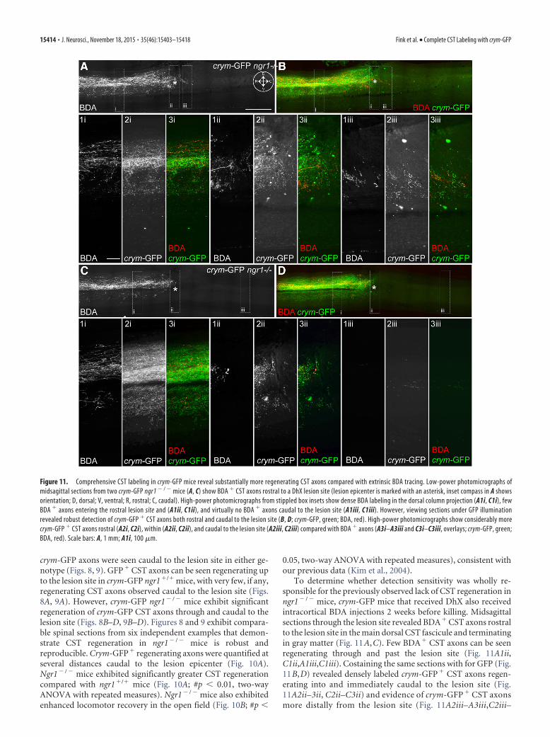
To determine whether detection sensitivity was wholly re-sponsible for the previously observed lack of CST regeneration inngr1� / � mice, crym-GFP mice that received DhX also receivedintracortical BDA injections 2 weeks before killing. Midsagittalsections through the lesion site revealed BDA� CST axons rostralto the lesion site in the main dorsal CST fascicule and terminatingin gray matter (Fig. 11A,C). Few BDA� CST axons can be seenregenerating through and past the lesion site (Fig. 11A1ii,C1ii,A1iii,C1iii). Costaining the same sections with for GFP (Fig.11B,D) revealed densely labeled crym-GFP� CST axons regen-erating into and immediately caudal to the lesion site (Fig.11A2ii–3ii, C2ii–C3ii) and evidence of crym-GFP � CST axonsmore distally from the lesion site (Fig. 11A2iii–A3iii,C2iii–
Figure 11. Comprehensive CST labeling in crym-GFP mice reveal substantially more regenerating CST axons compared with extrinsic BDA tracing. Low-power photomicrographs ofmidsagittal sections from two crym-GFP ngr1 � / � mice (A, C) show BDA � CST axons rostral to a DhX lesion site (lesion epicenter is marked with an asterisk, inset compass in A showsorientation; D, dorsal; V, ventral; R, rostral; C, caudal). High-power photomicrographs from stippled box insets show dense BDA labeling in the dorsal column projection (A1i, C1i), fewBDA � axons entering the rostral lesion site and (A1ii, C1ii), and virtually no BDA � axons caudal to the lesion site (A1iii, C1iii). However, viewing sections under GFP illuminationrevealed robust detection of crym-GFP � CST axons both rostral and caudal to the lesion site (B, D; crym-GFP, green; BDA, red). High-power photomicrographs show considerably morecrym-GFP � CST axons rostral (A2i, C2i), within (A2ii, C2ii), and caudal to the lesion site (A2iii, C2iii) compared with BDA � axons (A3i–A3iii and C3i–C3iii, overlays; crym-GFP, green;BDA, red). Scale bars: A, 1 mm; A1i, 100 �m.
15414 • J. Neurosci., November 18, 2015 • 35(46):15403–15418 Fink et al. • Complete CST Labeling with crym-GFP
C3iii). These data further suggest that inefficient BDA labelingobscures the comprehensive CST regenerative phenotype inngr1 � / � mice.
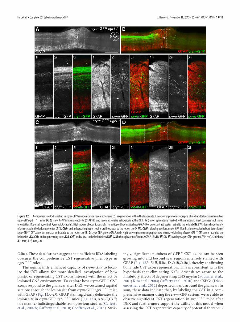
The significantly enhanced capacity of crym-GFP to local-ize the CST allows for more detailed investigation of howplastic or regenerating CST axons interact with the intact orlesioned CNS environment. To explore how crym-GFP � CSTaxons respond to the glial scar after DhX, we costained sagittalsections through the lesion site from crym-GFP ngr1 � / � micewith GFAP (Fig. 12A–D). GFAP staining clearly delineates thelesion site in crym-GFP ngr1 � / � mice (Fig. 12 A, A1ii,C,C1ii)in a manner indistinguishable from previous studies (Caffertyet al., 2007b; Cafferty et al., 2010; Geoffroy et al., 2015). Strik-
ingly, significant numbers of GFP � CST axons can be seengrowing into and beyond scar regions intensely stained withGFAP (Fig. 12B, B3ii, B3iii, D, D3ii,D3iii), thereby confirmingbona fide CST axon regeneration. This is consistent with thehypothesis that eliminating NgR1 desensitizes axons to theinhibitory effects of degenerating CNS myelin (Fournier et al.,2001; Kim et al., 2004; Cafferty et al., 2010) and CSPGs (Dick-endesher et al., 2012) deposited in and around the glial scar. Insum, these data indicate that, by labeling the CST in a com-prehensive manner using the crym-GFP system, we are able toobserve significant CST regeneration in ngr1 � / � mice afterDhX and furthermore support the utility of this model whenassessing the CST regenerative capacity of potential therapeu-
Figure 12. Comprehensive CST labeling in crym-GFP transgenic mice reveal extensive CST regeneration within the lesion site. Low-power photomicrographs of midsagittal sections from twocrym-GFP ngr1 � / � mice (A, C) show GFAP immunoreactivity (GFAP-IR) and reveal extensive astrogliosis at the DhX site (lesion epicenter is marked with an asterisk, inset compass in A showsorientation: D, dorsal; V, ventral; R, rostral; C, caudal). High-power photomicrographs from stippled box insets show GFAP-IR of quiescent astrocytes rostral to the lesion (A1i, C1i), dense hypertrophyof astrocytes in the lesion epicenter (A1ii, C1ii), and a decreasing hypertrophic profile caudal to the lesion site (A1iii, C1iii). Viewing sections under GFP illumination revealed robust detection ofcrym-GFP � CST axons both rostral and caudal to the lesion site (B, D; crym-GFP, green; GFAP, red). High-power photomicrographs show extensive labeling of crym-GFP � CST axons rostral to thelesion site (A2i, C2i), and regenerating into (A2ii, C2ii) and caudal to the lesion site (A2iii, C2iii) through areas of intense GFAP-IR (A3i-iii, C3i-iii, overlays; crym-GFP, green; GFAP, red). Scale bars:A, 1 mm; A1i, 100 �m.
Fink et al. • Complete CST Labeling with crym-GFP J. Neurosci., November 18, 2015 • 35(46):15403–15418 • 15415

tic interventions, thereby minimizing anatomical variationswithin experiments and between laboratories.
DiscussionTo date, comprehensive assessment of axon regeneration afterSCI has been fraught by the need to use inefficient and inconsis-tent extrinsic labeling methodologies (Tuszynski and Steward,2012). Here, we introduce a model in which the CST is compre-hensively and reliably genetically labeled via expression of a flu-orescent reporter controlled by the crym promoter. In this study,we: (1) show that crym-GFP mice label 10 times the number ofCST axons at all levels of the neuroaxis compared with the an-terograde CST tracer BDA, (2) reveal the comprehensive spinalgray matter termination pattern of the CST, and (3) demonstratethe streamlined utility of this transgenic line for in vivo SCI ex-periments for crossing onto knock-out strains. In addition, bycompleting DhX SCI on crym-GFP ngr1�/� and crym-GFPngr1� / � mice, we can draw several conclusions regarding thecapacity of the CST to mount a regenerative response in the pres-ence and absence of NgR1. First, ngr1� / � mice have indistin-guishable CST white and gray matter wiring compared withngr1�/� mice. Second, crym-GFP ngr1�/� reveal that the CSMNsmount a modest regenerative response after DhX, with a smallnumber of CST axons regenerating up to and into the lesion site.Third, crym-GFP ngr1� / � mice show significant CST axon re-generation after DhX compared with ngr1�/� mice, providingfurther evidence that NgR1 activation is a significant abortivesignal for regeneration of adult CST axons.
Inefficiency of extrinsic axon tracing methodologyAbortive axon regeneration in the adult CNS remains a signifi-cant barrier to functional SCI recovery (Filli and Schwab, 2012).To test the functional efficacy of proregeneration therapeutics invivo, investigators routinely deliver subcomplete lesions to thedorsal spinal cord and monitor functional recovery over time.These data are then correlated with anatomy in fixed tissue sec-tions at the end of the experiment. This approach is preferredfrom a functional perspective as these lesions are moderately easyto complete; interrupt a number of crucial descending spinalmotor tracts, including the CST; and result in a reproducible lossof motor function that can be evaluated using standardized tests(Basso et al., 1995; Basso et al., 2006). However, assessing axonregeneration in these models is complicated because it requiresdiscrete delivery of anterograde tracers to axotomized somata.BDA is commonly used for this purpose via cortical microinjec-tions into layer V of M1. Although BDA is absorbed and trans-ported by a number of CSMNs, its delivery requires an additionalsurgery and multiple injection sites and therefore routinely re-sults in varied, inconsistent, and incomplete filling of regenerat-ing axons in experimental animals within and between groups(Steward et al., 2008b).
Transgenic labeling of the CST with crym-GFP is superiorto BDAWe sought to identify an alternative method for labeling CSMNsthat would obviate the need for inefficient extrinsic tracer deliv-ery and more comprehensively label the CST. To this end, weexplored the GENSAT catalog and found the crym BAC trans-genic line that appeared to express GFP specifically in CSMNs.Close investigation revealed robust and comprehensive GFP ex-pression in CSMNs, their axons, and their terminals. We injectedBDA into M1 of crym-GFP mice to compare directly the effi-ciency of these two approaches to label the CST. We found that
crym-GFP mice label 10 times the number of CST axons in spinalwhite matter and CST terminals in spinal gray matter comparedwith BDA (Figs. 6, 7). Previous studies have also reported thebenefits of using transgenic fluorescent reporter lines to studyCST anatomy; however, none has directly compared labeling ef-ficiency directly with BDA (Bareyre et al., 2005; Carter et al.,2008). The YFP-H line expresses YFP under the thy1 promoterand therefore labels, not only CSMNs, but also other classes ofcortical, subcortical, and spinal neurons (Carter et al., 2008). Thisline has myriad benefits for studying anatomy ex vivo (Chung andDeisseroth, 2013) and in vivo (Holtmaat et al., 2012; Akbik et al.,2013); however, the stochastic nature of YFP expression (Feng etal., 2000) renders it inappropriate for CST SCI studies (Carter etal., 2008). A more refined approach to labeling the CST wasachieved by Bareyre et al. (2005), who developed a double trans-genic line by crossing thy1-STOP-YFP mice with Emx1-cre mice.Progeny of this cross (CST-YFP mice) show robust expression ofYFP in CSMNs. The number of YFP-labeled CST axons in thedCST, dlCST, and vCST in the cervical, thoracic, and lumbarspinal cord in CST-YFP mice were comparable to our counts inthe crym-GFP line. However, despite equivalent labeling of CSTaxons in the spinal cord, �90% of layer V pyramidal neuronswere labeled in the CST-YFP line; however, only 30% of layer Vneurons were labeled in our crym-GFP line. This suggests thatYFP expression in CST-YFP mice is not restricted to spinallyprojecting CSMNs, but also labels layer V neurons, sending pro-jections intracortically and corticofugally. Our data show thatcrym-GFP exclusively labels spinally projecting CSMNs because�100% of CSMNs retrogradely traced via intraspinal delivery ofFB were crym-GFP� (Fig. 4). Furthermore, differential gene ex-pression analysis comparing corticospinal, cortico– cortico, andcorticofugal CSMNs revealed that crym was enriched in the cor-ticospinal class (Arlotta et al., 2005), thus confirming crym as anideal candidate to target reporter expression to the CST.
CST axon regeneration in ngr1 �/� and ngr1 � / � miceThe single transgenic approach presented by crym-GFP line miceallows for streamlined breeding compared with double trans-genic CST-YFP mice onto existing knock-out and knock-in lines.We crossed crym-GFP mice with ngr1� / � mice to revisit CSTaxon regeneration in these knock-out mice. Abundant geneticreports support a critical role for NgR1 in transducing inhibitorysignals in the intact and lesioned CNS that prevent axon regen-eration (Li et al., 2005), sprouting (Cafferty and Strittmatter,2006; Geoffroy et al., 2015; Siegel et al., 2015), and plasticity (Leeet al., 2004; McGee et al., 2005; Akbik et al., 2013). Furthermore,pharmacological inhibition of NgR1 signaling also results in sig-nificant CST axon regeneration (Li et al., 2004b; Wang et al.,2006; Wang et al., 2011). Therefore, it was puzzling that we didnot detect CST axon regeneration after DhX in our previousstudy (Kim et al., 2004) despite the emergence of locomotor re-covery. The discrepancy in axon regeneration phenotype be-tween constitutive knock-out and pharmacological inhibition ofNgR1 may have arisen due aberrant CST wiring in ngr1� / � com-pared with ngr1�/� mice or the incomplete tracing of regenerat-ing CST axons with BDA. We addressed both of these possibilitiesusing crym-GFP line mice. By crossing ngr1� / � with crym-GFP,we were able to complete careful anatomical analysis of crym-GFP� and BDA� CST axons in the same animals in both crym-GFP ngr1�/� and crym-GFP ngr1� / � mice. Despite crym-GFPlabeling being 10 times more sensitive than BDA tracing, we didnot find a difference in the number of crym-GFP� or BDA� CSTaxons in white matter (Fig. 6) or terminals in gray matter (Fig. 7)
15416 • J. Neurosci., November 18, 2015 • 35(46):15403–15418 Fink et al. • Complete CST Labeling with crym-GFP

between ngr1�/� and ngr1� / � mice. Therefore, using a moresensitive measure of CST wiring, we can confirm our earlier re-port that constitutive ngr1� / � mice have normal CST patterning(Kim et al., 2004; Cafferty and Strittmatter, 2006). Next, wesought to determine whether the significantly enhanced sensitiv-ity of CST labeling afforded by crym-GFP would yield a deeperinsight into the regenerative capacity of axotomized CST axons.Indeed, inspection of sagittal sections through the lesion site 6weeks after DhX in crym-GFP ngr1�/� mice showed that largenumbers of GFP� CST axons had regenerated up to the lesionsite, with a few axons observed caudal to the lesion epicenter(Figs. 8A,9A). Significantly more GFP� CST axons were ob-served regenerating caudal to the lesion site in crym-GFPngr1� / � mice (Figs. 8B–D,9B–D). A percentage of regeneratingGFP� CST axons were also BDA� (Fig. 11); no axons wereBDA� and GFP�. The majority of GFP�-regenerating CST ax-ons in crym-GFP ngr1� / � mice were BDA�, further underscor-ing the need for more comprehensive CST labeling techniques tosubvert underinterpretation or overinterpretation of regenera-tion data. From these data, we conclude that the CST mounts asignificant regenerative response in constitutive ngr1� / � mice.
In summary, incomplete CST labeling may in part be respon-sible for the difficulty in reproducing SCI experiments (Stewardet al., 2012). Our current data demonstrate that BDA labels�10% of a mixed population of corticospinal, cortico– cortico,and corticofugal CSMN axons. This paltry percentage allows forsignificant variation in region and density of layer V neurons inM1 that are labeled between animals, surgical days, surgeons, andlaboratories. Comprehensive, robust, and invariant CST labelingin transgenic crym-GFP mice allows for the standardization ofanatomical analyses of CST regeneration after SCI and provides aframework with which to study the molecular mechanisms thatrestrict and enhance CST axon plasticity, sprouting, andregeneration.
ReferencesAkbik FV, Bhagat SM, Patel PR, Cafferty WB, Strittmatter SM (2013) Ana-
tomical plasticity of adult brain is titrated by Nogo Receptor 1. Neuron77:859 – 866. CrossRef Medline
Akbik F, Cafferty WB, Strittmatter SM (2012) Myelin associated inhibitors:a link between injury-induced and experience-dependent plasticity. ExpNeurol 235:43–52. CrossRef Medline
Arlotta P, Molyneaux BJ, Chen J, Inoue J, Kominami R, Macklis JD (2005)Neuronal subtype-specific genes that control corticospinal motor neurondevelopment in vivo. Neuron 45:207–221. CrossRef Medline
Atwal JK, Pinkston-Gosse J, Syken J, Stawicki S, Wu Y, Shatz C, Tessier-Lavigne M (2008) PirB is a functional receptor for myelin inhibitors ofaxonal regeneration. Science 322:967–970. CrossRef Medline
Bareyre FM, Kerschensteiner M, Misgeld T, Sanes JR (2005) Transgenic la-beling of the corticospinal tract for monitoring axonal responses to spinalcord injury. Nat Med 11:1355–1360. CrossRef Medline
Bartus K, James ND, Didangelos A, Bosch KD, Verhaagen J, Yanez-Munoz RJ,Rogers JH, Schneider BL, Muir EM, Bradbury EJ (2014) Large-scalechondroitin sulfate proteoglycan digestion with chondroitinase genetherapy leads to reduced pathology and modulates macrophage pheno-type following spinal cord contusion injury. J Neurosci 34:4822– 4836.CrossRef Medline
Basso DM, Beattie MS, Bresnahan JC (1995) A sensitive and reliable loco-motor rating scale for open field testing in rats. J Neurotrauma 12:1–21.CrossRef Medline
Basso DM, Fisher LC, Anderson AJ, Jakeman LB, McTigue DM, Popovich PG(2006) Basso Mouse Scale for locomotion detects differences in recoveryafter spinal cord injury in five common mouse strains. J Neurotrauma23:635– 659. CrossRef Medline
Blackmore MG, Wang Z, Lerch JK, Motti D, Zhang YP, Shields CB, Lee JK,Goldberg JL, Lemmon VP, Bixby JL (2012) Kruppel-like Factor 7 engi-neered for transcriptional activation promotes axon regeneration in the
adult corticospinal tract. Proc Natl Acad Sci U S A 109:7517–7522.CrossRef Medline
Bradbury EJ, Moon LD, Popat RJ, King VR, Bennett GS, Patel PN, FawcettJW, McMahon SB (2002) Chondroitinase ABC promotes functional re-covery after spinal cord injury. Nature 416:636 – 640. CrossRef Medline
Cafferty WB, Strittmatter SM (2006) The Nogo-Nogo receptor pathwaylimits a spectrum of adult CNS axonal growth. J Neurosci 26:12242–12250. CrossRef Medline
Cafferty WB, Kim JE, Lee JK, Strittmatter SM (2007a) Response to corre-spondence: Kim et al., “axon regeneration in young adult mice lackingNogo-A/B.” Neuron 38, 187–199. Neuron 54:195–199. CrossRef Medline
Cafferty WB, Yang SH, Duffy PJ, Li S, Strittmatter SM (2007b) Functionalaxonal regeneration through astrocytic scar genetically modified to digestchondroitin sulfate proteoglycans. J Neurosci 27:2176 –2185. CrossRefMedline
Cafferty WB, Duffy P, Huebner E, Strittmatter SM (2010) MAG and OMgpsynergize with Nogo-A to restrict axonal growth and neurological recov-ery after spinal cord trauma. J Neurosci 30:6825– 6837. CrossRef Medline
Carter LM, Starkey ML, Akrimi SF, Davies M, McMahon SB, Bradbury EJ(2008) The yellow fluorescent protein (YFP-H) mouse reveals neuropro-tection as a novel mechanism underlying chondroitinase ABC-mediatedrepair after spinal cord injury. J Neurosci 28:14107–14120. CrossRefMedline
Chung K, Deisseroth K (2013) CLARITY for mapping the nervous system.Nat Methods 10:508 –513. CrossRef Medline
Dickendesher TL, Baldwin KT, Mironova YA, Koriyama Y, Raiker SJ, AskewKL, Wood A, Geoffroy CG, Zheng B, Liepmann CD, Katagiri Y, BenowitzLI, Geller HM, Giger RJ (2012) NgR1 and NgR3 are receptors for chon-droitin sulfate proteoglycans. Nat Neurosci 15:703–712. CrossRefMedline
Dimou L, Schnell L, Montani L, Duncan C, Simonen M, Schneider R, Lieb-scher T, Gullo M, Schwab ME (2006) Nogo-A-deficient mice revealstrain-dependent differences in axonal regeneration. J Neurosci 26:5591–5603. CrossRef Medline
Feng G, Mellor RH, Bernstein M, Keller-Peck C, Nguyen QT, Wallace M,Nerbonne JM, Lichtman JW, Sanes JR (2000) Imaging neuronal subsetsin transgenic mice expressing multiple spectral variants of GFP. Neuron28:41–51. CrossRef Medline
Festing MF, Altman DG (2002) Guidelines for the design and statisticalanalysis of experiments using laboratory animals. ILAR J 43:244 –258.CrossRef Medline
Filli L, Schwab ME (2012) The rocky road to translation in spinal cord re-pair. Ann Neurol 72:491–501. CrossRef Medline
Fisher D, Xing B, Dill J, Li H, Hoang HH, Zhao Z, Yang XL, Bachoo R,Cannon S, Longo FM, Sheng M, Silver J, Li S (2011) Leukocyte commonantigen-related phosphatase is a functional receptor for chondroitin sul-fate proteoglycan axon growth inhibitors. J Neurosci 31:14051–14066.CrossRef Medline
Fournier AE, GrandPre T, Strittmatter SM (2001) Identification of a recep-tor mediating Nogo-66 inhibition of axonal regeneration. Nature 409:341–346. CrossRef Medline
Fry EJ, Chagnon MJ, Lopez-Vales R, Tremblay ML, David S (2010) Corti-cospinal tract regeneration after spinal cord injury in receptor proteintyrosine phosphatase sigma deficient mice. Glia 58:423– 433. Medline
Geoffroy CG, Lorenzana AO, Kwan JP, Lin K, Ghassemi O, Ma A, Xu N,Creger D, Liu K, He Z, Zheng B (2015) Effects of PTEN and Nogo code-letion on corticospinal axon sprouting and regeneration in mice. J Neu-rosci 35:6413– 6428. CrossRef Medline
Gong S, Zheng C, Doughty ML, Losos K, Didkovsky N, Schambra UB, NowakNJ, Joyner A, Leblanc G, Hatten ME, Heintz N (2003) A gene expressionatlas of the central nervous system based on bacterial artificial chromo-somes. Nature 425:917–925. CrossRef Medline
GrandPre T, Li S, Strittmatter SM (2002) Nogo-66 receptor antagonist pep-tide promotes axonal regeneration. Nature 417:547–551. CrossRefMedline
Hallen A, Cooper AJ, Jamie JF, Haynes PA, Willows RD (2011) Mammalianforebrain ketimine reductase identified as mu-crystallin; potential regu-lation by thyroid hormones. J Neurochem 118:379 –387. CrossRefMedline
Heintz N (2004) Gene expression nervous system atlas (GENSAT). NatNeurosci 7:483. CrossRef Medline
Holtmaat A, de Paola V, Wilbrecht L, Trachtenberg JT, Svoboda K, Portera-
Fink et al. • Complete CST Labeling with crym-GFP J. Neurosci., November 18, 2015 • 35(46):15403–15418 • 15417

Cailliau C (2012) Imaging neocortical neurons through a chronic cra-nial window. Cold Spring Harb Protoc 2012:694 –701. Medline
Kempf A, Tews B, Arzt ME, Weinmann O, Obermair FJ, Pernet V, Zagrebel-sky M, Delekate A, Iobbi C, Zemmar A, Ristic Z, Gullo M, Spies P, DoddD, Gygax D, Korte M, Schwab ME (2014) The sphingolipid receptorS1PR2 is a receptor for Nogo-a repressing synaptic plasticity. PLoS Biol12:e1001763. CrossRef Medline
Kim JE, Li S, GrandPre T, Qiu D, Strittmatter SM (2003) Axon regenerationin young adult mice lacking Nogo-A/B. Neuron 38:187–199. CrossRefMedline
Kim JE, Liu BP, Park JH, Strittmatter SM (2004) Nogo-66 receptor preventsraphespinal and rubrospinal axon regeneration and limits functional re-covery from spinal cord injury. Neuron 44:439 – 451. CrossRef Medline
Kim RY, Gasser R, Wistow GJ (1992) mu-crystallin is a mammalian homo-logue of Agrobacterium ornithine cyclodeaminase and is expressed inhuman retina. Proc Natl Acad Sci U S A 89:9292–9296. CrossRef Medline
Lang BT, Cregg JM, DePaul MA, Tran AP, Xu K, Dyck SM, Madalena KM,Brown BP, Weng YL, Li S, Karimi-Abdolrezaee S, Busch SA, Shen Y, SilverJ (2015) Modulation of the proteoglycan receptor PTPsigma promotesrecovery after spinal cord injury. Nature 518:404 – 408. CrossRef Medline
Lee JK, Kim JE, Sivula M, Strittmatter SM (2004) Nogo receptor antago-nism promotes stroke recovery by enhancing axonal plasticity. J Neurosci24:6209 – 6217. CrossRef Medline
Lee JK, Geoffroy CG, Chan AF, Tolentino KE, Crawford MJ, Leal MA, Kang B,Zheng B (2010) Assessing spinal axon regeneration and sprouting inNogo-, MAG-, and OMgp-deficient mice. Neuron 66:663– 670. CrossRefMedline
Li S, Liu BP, Budel S, Li M, Ji B, Walus L, Li W, Jirik A, Rabacchi S, Choi E,Worley D, Sah DW, Pepinsky B, Lee D, Relton J, Strittmatter SM (2004a)Blockade of nogo-66, myelin-associated glycoprotein, and oligodendro-cyte myelin glycoprotein by soluble nogo-66 receptor promotes axonalsprouting and recovery after spinal injury. J Neurosci 24:10511–10520.CrossRef Medline
Li S, Kim JE, Budel S, Hampton TG, Strittmatter SM (2005) Transgenicinhibition of Nogo-66 receptor function allows axonal sprouting andimproved locomotion after spinal injury. Mol Cell Neurosci 29:26 –39.CrossRef Medline
Li W, Walus L, Rabacchi SA, Jirik A, Chang E, Schauer J, Zheng BH, BenedettiNJ, Liu BP, Choi E, Worley D, Silvian L, Mo W, Mullen C, Yang W,Strittmatter SM, Sah DW, Pepinsky B, Lee DH (2004b) A neutralizinganti-Nogo66 receptor monoclonal antibody reverses inhibition of neuriteoutgrowth by central nervous system myelin. J Biol Chem 279:43780 –43788. CrossRef Medline
Liebscher T, Schnell L, Schnell D, Scholl J, Schneider R, Gullo M, Fouad K,Mir A, Rausch M, Kindler D, Hamers FP, Schwab ME (2005) Nogo-Aantibody improves regeneration and locomotion of spinal cord-injuredrats. Ann Neurol 58:706 –719. CrossRef Medline
Liu K, Tedeschi A, Park KK, He Z (2011) Neuronal intrinsic mechanisms ofaxon regeneration. Annu Rev Neurosci 34:131–152. CrossRef Medline
McGee AW, Strittmatter SM (2003) The Nogo-66 receptor: focusing myelininhibition of axon regeneration. Trends Neurosci 26:193–198. CrossRefMedline
McGee AW, Yang Y, Fischer QS, Daw NW, Strittmatter SM (2005)Experience-driven plasticity of visual cortex limited by myelin and Nogoreceptor. Science 309:2222–2226. CrossRef Medline
Schnell L, Schwab ME (1990) Axonal regeneration in the rat spinal cordproduced by an antibody against myelin-associated neurite growth inhib-itors. Nature 343:269 –272. CrossRef Medline
Schwab ME, Strittmatter SM (2014) Nogo limits neural plasticity and re-covery from injury. Curr Opin Neurobiol 27:53– 60. CrossRef Medline
Shen Y, Tenney AP, Busch SA, Horn KP, Cuascut FX, Liu K, He Z, Silver J,Flanagan JG (2009) PTPsigma is a receptor for chondroitin sulfate pro-
teoglycan, an inhibitor of neural regeneration. Science 326:592–596.CrossRef Medline
Siegel CS, Fink KL, Strittmatter SM, Cafferty WB (2015) Plasticity of intactrubral projections mediates spontaneous recovery of function after cor-ticospinal tract injury. J Neurosci 35:1443–1457. CrossRef Medline
Simonen M, Pedersen V, Weinmann O, Schnell L, Buss A, Ledermann B,Christ F, Sansig G, van der Putten H, Schwab ME (2003) Systemic dele-tion of the myelin-associated outgrowth inhibitor Nogo-A improvesregenerative and plastic responses after spinal cord injury. Neuron 38:201–211. CrossRef Medline
Steward O, Zheng B, Tessier-Lavigne M (2003) False resurrections: distin-guishing regenerated from spared axons in the injured central nervoussystem. J Comp Neurol 459:1– 8. CrossRef Medline
Steward O, Zheng B, Banos K, Yee KM (2007) Response to: Kim et al., “axonregeneration in young adult mice lacking Nogo-A/B.” Neuron 54:191–195. CrossRef
Steward O, Sharp K, Yee KM, Hofstadter M (2008a) A re-assessment of theeffects of a Nogo-66 receptor antagonist on regenerative growth of axonsand locomotor recovery after spinal cord injury in mice. Exp Neurol209:446 – 468. CrossRef Medline
Steward O, Zheng B, Tessier-Lavigne M, Hofstadter M, Sharp K, Yee KM(2008b) Regenerative growth of corticospinal tract axons via the ventralcolumn after spinal cord injury in mice. J Neurosci 28:6836 – 6847.CrossRef Medline
Steward O, Popovich PG, Dietrich WD, Kleitman N (2012) Replication andreproducibility in spinal cord injury research. Exp Neurol 233:597– 605.CrossRef Medline
Tuszynski MH, Steward O (2012) Concepts and methods for the study ofaxonal regeneration in the CNS. Neuron 74:777–791. CrossRef Medline
Vie MP, Evrard C, Osty J, Breton-Gilet A, Blanchet P, Pomerance M, RougetP, Francon J, Blondeau JP (1997) Purification, molecular cloning, andfunctional expression of the human nicodinamide-adenine dinucleotidephosphate-regulated thyroid hormone-binding protein. Mol Endocrinol11:1728 –1736. Medline
Wang X, Baughman KW, Basso DM, Strittmatter SM (2006) Delayed Nogoreceptor therapy improves recovery from spinal cord contusion. AnnNeurol 60:540 –549. CrossRef Medline
Wang X, Duffy P, McGee AW, Hasan O, Gould G, Tu N, Harel NY, Huang Y,Carson RE, Weinzimmer D, Ropchan J, Benowitz LI, Cafferty WB, Strit-tmatter SM (2011) Recovery from chronic spinal cord contusion afterNogo receptor intervention. Ann Neurol 70:805– 821. CrossRef Medline
Wang Z, Reynolds A, Kirry A, Nienhaus C, Blackmore MG (2015) Overex-pression of Sox11 promotes corticospinal tract regeneration after spinalinjury while interfering with functional recovery. J Neurosci 35:3139 –3145. CrossRef Medline
Willenberg R, Steward O (2015) Nonspecific labeling limits the utility ofCre-Lox bred CST-YFP mice for studies of corticospinal tract regenera-tion. J Comp Neurol. In press.
Woolf CJ (2003) No Nogo: now where to go? Neuron 38:153–156. CrossRefMedline
Yasvoina MV, Genc B, Jara JH, Sheets PL, Quinlan KA, Milosevic A, ShepherdGM, Heckman CJ, Ozdinler PH (2013) eGFP expression under UCHL1promoter genetically labels corticospinal motor neurons and a subpopu-lation of degeneration-resistant spinal motor neurons in an ALS mousemodel. J Neurosci 33:7890 –7904. CrossRef Medline
Yiu G, He Z (2006) Glial inhibition of CNS axon regeneration. Nat RevNeurosci 7:617– 627. CrossRef Medline
Zheng B, Atwal J, Ho C, Case L, He XL, Garcia KC, Steward O, Tessier-Lavigne M (2005) Genetic deletion of the Nogo receptor does not re-duce neurite inhibition in vitro or promote corticospinal tractregeneration in vivo. Proc Natl Acad Sci U S A 102:1205–1210. CrossRefMedline
15418 • J. Neurosci., November 18, 2015 • 35(46):15403–15418 Fink et al. • Complete CST Labeling with crym-GFP