Developmental Expression in Leishmania donovani: Cloning
10
MOLECULAR AND CELLULAR BIOLOGY, May 1994, p. 2975-2984 Vol. 14, No. 5 0270-7306/94/$04.00+0 Copyright X 1994, American Society for Microbiology Developmental Gene Expression in Leishmania donovani: Differential Cloning and Analysis of an Amastigote-Stage- Specific Gene HUGUES CHAREST AND GREG MATLASHEWSKI* Institute of Parasitology of McGill University, Macdonald Campus, Ste-Anne de Bellevue, Quebec, Canada H9X 3V9 Received 17 November 1993/Returned for modification 10 January 1994/Accepted 31 January 1994 Leishmania protozoans are the causative agents of leishmaniasis, a major parasitic disease in humans. During their life cycle, Leishmania protozoans exist as flagellated promastigotes in the sand fly vector and as nonmotile amastigotes in the mammalian hosts. The promastigote-to-amastigote transformation occurs in the phagolysosomal compartment of the macrophage cell and is a critical step for the establishment of the infection. To study this cytodifferentiation process, we differentially screened an amastigote cDNA library with life cycle stage-specific cDNA probes and isolated seven cDNAs representing amastigote-specific transcripts. Five of these were closely related (A2 series) and recognized, by Northern (RNA) blot analyses, a 3.5-kb transcript in amastigotes and in amastigote-infected macrophages. Expression of the amastigote-specific A2 gene was induced in promastigotes when they were transferred from culture medium at 26°C and pH 7.4 to medium at 37°C and pH 4.5, conditions which mimic the macrophage phagolysosomal environment. A2 genes are clustered in tandem arrays, and a 6-kb fragment corresponding to a unit of the cluster was cloned and partially sequenced. An open reading frame found within the A2-transcribed region potentially encoded a 22-kDa protein containing repetitive sequences. The recombinant A2 protein produced in Escherichia coli cells was specifically recognized by immune serum from a patient with visceral leishmaniasis. The A2 protein repetitive element has strong homology with an S antigen of Plasmodium falciparum, the protozoan parasite responsible for malaria. Both the A2 protein of Leishmania donovani and the S antigen ofP.falciparum are stage specific and developmentally expressed in mammalian hosts. Leishmania protozoans are the causative agents of human leishmaniasis, which includes a spectrum of diseases ranging from self-healing skin ulcers to fatal visceral infections. Leish- mania donovani causes visceral leishmaniasis, also known as kala-azar, which has a high mortality rate if not treated. The Leishmania protozoans exist as extracellular flagellated pro- mastigotes in the alimentary tract of the sand fly and are transmitted to the mammalian hosts through the bite of the insect. Once injected through the skin, the promastigotes are taken up by macrophages, rapidly differentiate into amasti- gotes, and start to multiply within the phagolysosomal com- partment. As the infected cells rupture, amastigotes subse- quently infect other macrophages, giving rise to the various symptoms associated with leishmaniasis (23). While in the midgut of the insect, newly transformed promastigotes, functionally avirulent, progressively acquire capacity for infection and migrate to the mouthparts (30, 32). This process, termed metacyclogenesis, is concurrent with the differential expression of major surface glycoconjugates which mediate the migration of promastigotes in the alimentary tract and prepare the organism for the serum environment (27, 28, 31, 33). In comparison, the promastigote-to-amastigote cyto- differentiation is a profound morphological and physiological transformation. During differentiation, the parasite loses its flagellum, rounds up, changes its glycoconjugate coat (13, 21, 22, 39), and starts to express a set of metabolic enzymes optimally active at low pHs (14, 25). The survival of the parasite inside the macrophage phagolysosome is associated with its ability to down-regulate many effector and accessory * Corresponding author. Mailing address: Institute of Parasitology of McGill University, Macdonald Campus, 21,111 Lakeshore Rd., Ste-Anne de Bellevue, Quebec, Canada H9X 3V9. functions of its host cell, including oxygen metabolite-mediated killing and the capacity of the macrophage to act as an efficient antigen-presenting cell (5, 20). Glycoconjugates that coat the promastigote have been shown to play a critical role in the mechanism of entry of the parasite into its host cell and in the modulation of the host immune response (reviewed in refer- ences 24 and 38). Because of difficulties in obtaining a sufficient number of pure and viable amastigotes, the biology of this form of the parasite has not been well characterized. The importance of the promastigote-amastigote cytodifferentiation for the estab- lishment of infection in the mammalian host has prompted us to identify molecular events involved in this process. Our approach was to identify Leishmania genes developmentally expressed during the amastigote stage. Our assumption was that genes expressed only by amastigotes were likely to be essential for the survival of the parasite in its human host. In this study, we have characterized an amastigote-stage- specific gene (A2 gene) and determined the physiological conditions required to induce its expression. We also demon- strate that the A2 gene product is recognized by serum from a kala-azar patient and has identity with a major antigen devel- opmentally expressed by the pathologic forms of the malaria- associated protozoan Plasmodium falciparum. These results suggest that the Leishmania and Plasmodium parasites, phylo- genetically distant parasitic protozoans, have evolved the A2- like genes to carry out similar functions associated with infecting their human hosts. MATERIALS AND METHODS Leishmania strains and media. Amastigotes of the L. dono- vani infantum Ethiopian LV9 strain were harvested from 2975 on January 28, 2019 by guest http://mcb.asm.org/ Downloaded from
Transcript of Developmental Expression in Leishmania donovani: Cloning
MOLECULAR AND CELLULAR BIOLOGY, May 1994, p. 2975-2984 Vol. 14, No. 50270-7306/94/$04.00+0Copyright X 1994, American Society for Microbiology
Developmental Gene Expression in Leishmania donovani:Differential Cloning and Analysis of an Amastigote-Stage-
Specific GeneHUGUES CHAREST AND GREG MATLASHEWSKI*
Institute of Parasitology ofMcGill University, Macdonald Campus, Ste-Anne de Bellevue, Quebec, Canada H9X 3V9
Received 17 November 1993/Returned for modification 10 January 1994/Accepted 31 January 1994
Leishmania protozoans are the causative agents of leishmaniasis, a major parasitic disease in humans.During their life cycle, Leishmania protozoans exist as flagellated promastigotes in the sand fly vector and asnonmotile amastigotes in the mammalian hosts. The promastigote-to-amastigote transformation occurs in thephagolysosomal compartment of the macrophage cell and is a critical step for the establishment of theinfection. To study this cytodifferentiation process, we differentially screened an amastigote cDNA library withlife cycle stage-specific cDNA probes and isolated seven cDNAs representing amastigote-specific transcripts.Five of these were closely related (A2 series) and recognized, by Northern (RNA) blot analyses, a 3.5-kbtranscript in amastigotes and in amastigote-infected macrophages. Expression of the amastigote-specific A2gene was induced in promastigotes when they were transferred from culture medium at 26°C and pH 7.4 tomedium at 37°C and pH 4.5, conditions which mimic the macrophage phagolysosomal environment. A2 genesare clustered in tandem arrays, and a 6-kb fragment corresponding to a unit of the cluster was cloned andpartially sequenced. An open reading frame found within the A2-transcribed region potentially encoded a22-kDa protein containing repetitive sequences. The recombinant A2 protein produced in Escherichia coli cellswas specifically recognized by immune serum from a patient with visceral leishmaniasis. The A2 proteinrepetitive element has strong homology with an S antigen of Plasmodium falciparum, the protozoan parasiteresponsible for malaria. Both the A2 protein ofLeishmania donovani and the S antigen ofP.falciparum are stagespecific and developmentally expressed in mammalian hosts.
Leishmania protozoans are the causative agents of humanleishmaniasis, which includes a spectrum of diseases rangingfrom self-healing skin ulcers to fatal visceral infections. Leish-mania donovani causes visceral leishmaniasis, also known askala-azar, which has a high mortality rate if not treated. TheLeishmania protozoans exist as extracellular flagellated pro-mastigotes in the alimentary tract of the sand fly and aretransmitted to the mammalian hosts through the bite of theinsect. Once injected through the skin, the promastigotes aretaken up by macrophages, rapidly differentiate into amasti-gotes, and start to multiply within the phagolysosomal com-partment. As the infected cells rupture, amastigotes subse-quently infect other macrophages, giving rise to the varioussymptoms associated with leishmaniasis (23).While in the midgut of the insect, newly transformed
promastigotes, functionally avirulent, progressively acquirecapacity for infection and migrate to the mouthparts (30, 32).This process, termed metacyclogenesis, is concurrent with thedifferential expression of major surface glycoconjugates whichmediate the migration of promastigotes in the alimentary tractand prepare the organism for the serum environment (27, 28,31, 33). In comparison, the promastigote-to-amastigote cyto-differentiation is a profound morphological and physiologicaltransformation. During differentiation, the parasite loses itsflagellum, rounds up, changes its glycoconjugate coat (13, 21,22, 39), and starts to express a set of metabolic enzymesoptimally active at low pHs (14, 25). The survival of theparasite inside the macrophage phagolysosome is associatedwith its ability to down-regulate many effector and accessory
* Corresponding author. Mailing address: Institute of Parasitologyof McGill University, Macdonald Campus, 21,111 Lakeshore Rd.,Ste-Anne de Bellevue, Quebec, Canada H9X 3V9.
functions of its host cell, including oxygen metabolite-mediatedkilling and the capacity of the macrophage to act as an efficientantigen-presenting cell (5, 20). Glycoconjugates that coat thepromastigote have been shown to play a critical role in themechanism of entry of the parasite into its host cell and in themodulation of the host immune response (reviewed in refer-ences 24 and 38).
Because of difficulties in obtaining a sufficient number ofpure and viable amastigotes, the biology of this form of theparasite has not been well characterized. The importance ofthe promastigote-amastigote cytodifferentiation for the estab-lishment of infection in the mammalian host has prompted usto identify molecular events involved in this process. Ourapproach was to identify Leishmania genes developmentallyexpressed during the amastigote stage. Our assumption wasthat genes expressed only by amastigotes were likely to beessential for the survival of the parasite in its human host.
In this study, we have characterized an amastigote-stage-specific gene (A2 gene) and determined the physiologicalconditions required to induce its expression. We also demon-strate that the A2 gene product is recognized by serum from akala-azar patient and has identity with a major antigen devel-opmentally expressed by the pathologic forms of the malaria-associated protozoan Plasmodium falciparum. These resultssuggest that the Leishmania and Plasmodium parasites, phylo-genetically distant parasitic protozoans, have evolved the A2-like genes to carry out similar functions associated withinfecting their human hosts.
MATERIALS AND METHODS
Leishmania strains and media. Amastigotes of the L. dono-vani infantum Ethiopian LV9 strain were harvested from
2975
on January 28, 2019 by guesthttp://m
cb.asm.org/
Dow
nloaded from
2976 CHAREST AND MATLASHEWSKI
spleens of infected female gold Syrian hamsters and purified asdescribed by Reiner (29). Briefly, parasites were released fromtissue with a homogenizer, the mixture was centrifuged threetimes at 100 x g to remove cellular debris, and amastigoteswere pelleted at 1,500 x g. The pellet was resuspended in 0.17M sodium acetate to lyse contaminating erythrocytes. Theamastigotes were recovered by centrifugation and were incu-bated at 37°C in complete RPMI medium (RPMI 1640 sup-plemented with 10% endotoxin-free heat-inactivated fetal bo-vine serum, 10 ml of 1 M N-2-hydroxyethylpiperazine-N'-2-ethanesulfonic acid [HEPES; pH 7.3], 100 U of penicillin perml, 100 U of streptomycin per ml) for 18 h prior to RNAextractions. In preliminary analyses, we observed that a periodof incubation of 18 h and multiple washes were necessary toallow the amastigote to dislodge completely from host celldebris and then to obtain amastigote RNA samples relativelyfree of mammalian RNA contamination. To obtain promasti-gotes, LV9 strain amastigotes were allowed to differentiate incomplete RPMI medium at 26°C and cultured for at least 7days in the same medium before use. Promastigotes of the L.donovani Sudanese strain 1S2D (wt) were cultivated andpassaged in complete RPMI medium at 26°C. Amastigote-likeorganisms of the 1S2D strain were cultivated by proceduresdescribed by Doyle et al. (12).The Sudanese strains 1S2D and 1S2D (wt) were a gift from
S. Turco (University of Kentucky, Lexington). The 1S2D (wt)promastigotes were adapted to grow in axenic conditions andlost the ability to transform into infective promastigotes (37b).
Nucleic acid preparations. Total RNA of amastigotes andpromastigotes was prepared by the guanidinium isothiocyanatemethod with RNAzol (Cinna/Biotecx Laboratories Interna-tional, Inc., Friendswood, Tex.); poly(A)+ RNA was selectedby oligo(dT)-cellulose chromatography (grade 7; Pharmacia)as described by Sambrook et al. (34). Leishmania total DNAwas extracted from promastigotes by the methods describedfor mammalian cells by Sambrook et al. (34).
Construction and screening of an amastigote cDNA library.A total of 10 ,ug of amastigote mRNA was used to construct anEcoRI-XhoI unidirectional cDNA library of 106 clones in the AZAP II vector (Stratagene); hemimethylated cDNA was pro-duced with manufacturer-supplied reagents and protocols.About 40,000 clones of the primary library were screeneddifferentially with stage-specific probes; the cDNA probes wereprepared with oligo(dT)12-18 primer (Pharmacia) and Molo-ney murine leukemia virus reverse transcriptase (BethesdaResearch Laboratories [BRL]) by protocols described by Sam-brook et al. (34). Duplicate filters were hybridized with eachprobe for 18 h at 42°C in 50% formamide-6 x SSC-5 xDenhardt's solution (1 x SSC consists of 0.15 M NaCl plus0.015 M sodium citrate, pH 7.0; 1 x Denhardt's solution is0.02% polyvinylpyrrolidone, 0.02% Ficoll, and 0.02% bovineserum albumin)-5% dextran sulfate. Membranes were thenwashed twice at room temperature in 1 x SSC for 20 min,washed twice at 55°C in 1 x SSC-0.1% sodium dodecyl sulfate(SDS), and then autoradiographed on Kodak X-Omat filmswith an intensifying screen for 18 to 72 h. Areas on the platescontaining putative clones of interest were picked, and thephage pools were submitted to a second round of screening.Only clones which were hybridizing with the amastigote cDNAprobe, but not with the promastigote probe, were considered.For each cDNA clone isolated, a Bluescript plasmid derivativewas excised from theA ZAP II recombinant phages in vivo withthe helper phage R-408 (Stratagene protocol).
Northern and Southern blot analysis. Northern (RNA) blotanalyses were carried out as previously described (11); unlessspecified otherwise, 10 p,g of total RNA was loaded in each
lane. For Southern blot analysis, 10 ,ug of total DNA wasdigested to completion with restriction enzymes (BRL), andfragments were separated on 0.7% agarose gels and trans-ferred to nylon membranes (Hybond-N; Amersham) as de-scribed by Sambrook et al. (34). Membranes were prehybrid-ized for a few hours in the hybridization buffer (50%formamide, 6 x SSC, 5 x Denhardt's solution, 0.1% SDS, 200,ug of denatured salmon sperm DNA per ml) before theaddition of the probes. Hybridizations were carried out for 18h at 42°C. The membranes were washed twice in 1 x SSC for15 min at room temperature, washed twice in 2 to 0.1 xSSC-0.1% SDS at 55 to 60°C for 15 min, and autoradio-graphed on Kodak X-Omat films with intensifying screens.DNA probes were prepared with agarose gel-purified restric-tion fragments labelled to high specificity with [at-32P]dCTP(ICN; 3,000 Ci/mmol) by nick translation (Amersham kit andprotocol).
Isolation of genomic clones. A 6- to 10-kb EcoRI fragmentpartial genomic library was constructed in the X ZAP II vector(Stratagene). More than 2,000 clones were screened on dupli-cate filters with nick-translated probes prepared with the A2cDNA by techniques and hybridization conditions describedfor the differential screening. Eight clones were isolated andpurified. Bluescript plasmid derivatives were excised fromrecombinant X phages as for cDNA clones.DNA sequencing. The A2 cDNA sequence was determined
with oligonucleotide DNA primers. The 5' and 3' ends of A3to All were obtained with common or A2 cDNA-specificprimers. For the 1.9-kb XhoI-EcoRI portion of the genomicclone GECO 90, the fragment was subcloned in the Bluescriptphagemids KS' and KS-. Nested deletions were carried outon plasmids with exonuclease III and S1 nuclease (double-stranded Nested Deletion Kit; Pharmacia). Sequencing reac-tions were performed on single-stranded DNA templates(prepared with the M13K07 helper phage and by the methodof Sambrook et al. [34]) or double-stranded DNA plasmidswith the Deaza T7 sequencing mixes (Pharmacia) and labellednucleotides (35S-ATP or 35S-CTP; 600 Ci/mmol; Amersham).Reactions were analyzed on 6% denaturing polyacrylamidegels.
Subcloning of the ORFII fragment. Synthetic oligonucleo-tides containing tails with BamHI restriction sites were used toamplify the ORFII coding region from the 1.9-kb XhoI-EcoRIfragment subcloned in a Bluescript KS' subclone (5' primer,ctcgaggatccgatgaagatccgcagcgtgc; 3' primer, tagagaggatccgcatctagatggcttg [underlined sequences correspond to the cloningtails]). Reactions were performed with the Tth DNA poly-merase (Pharmacia) and the GeneAmp PCR system 9600(Perkin-Elmer Cetus). After 3 min at 95°C to denature thelinearized template, the reaction mixture was first incubatedfor 15 cycles at 55, 75, and 95°C for 2 min, 3 min, and 30 s,respectively, and the annealing temperature was raised from 55to 65°C for 20 subsequent cycles. The PCR product (1.1 kb)was first inserted in the PCR II cloning vector (T/A cloningsystem; Invitrogen) and then subcloned with the BamHIrestriction sites in the pET16b expression vector (Novagen), inframe with the histidine tag (HT).
Immunoprecipitations and western blot analysis. TheHT-A2 fusion protein was synthesized in vitro with T7 RNApolymerase and rabbit reticulocyte lysate (TNT assay; Pro-mega) and labelled with L-[355]methionine (ICN; >1,000 Ci/mmol). A human p53 cDNA construct was used as a positivecontrol for the in vitro transcription-translation assay, and thenthe labelled p53 protein was used as a negative control for theimmunoprecipitations. Precleared protein samples were di-luted with 1 volume of NET/Gel (50 mM Tris [pH 7.5], 150
MOL. CELL. BIOL.
on January 28, 2019 by guesthttp://m
cb.asm.org/
Dow
nloaded from
L. DONOVANI AMASTIGOTE-STAGE-SPECIFIC GENE 2977
IA 1A2 (probes) _ A6
2 3 -' 6 6-- 11 2S3 6 8 9 11
B2 3 5 6 8 9 11
- 3.0-<- 2.0
.K- 3.0 ---
.k- 2.0 35kb.-.. s1..6 -4- 1.6
t'i''tt't<-~1.0 I*° -.0
0.5 . 0.5>
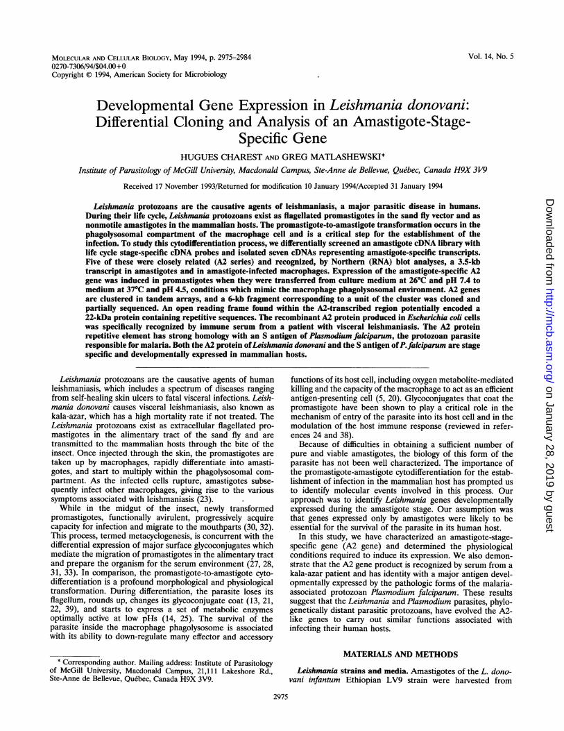
FIG. 1. (A) Southern blot analysis of amastigote-specific cDNA clones. Panel 1, Bluescript SK recombinant plasmids (A2-A11) were digestedwith EcoRI and XhoI to excise the cDNA inserts. Fragments were separated on an agarose gel (1%) and revealed by ethidium bromide staining;cDNA inserts varied from 0.5 kb (A5) to 1.8 kb (A8; contained an internal EcoRI site). Panel 2, Restriction fragments were transferred to nylonmembranes, and duplicates were hybridized with a cDNA clone A2 (0.9 kb)- or A6 (0.6 kb)-specific probe. The A2 probe recognized five cDNAs(A2, A3, A8, A9, and Al1); the A6 cDNA hybridized only to itself. (B) Differential expression of A2-related genes in promastigotes andamastigote-infected macrophages. Total RNA (15 jig) extracted from bone marrow-derived macrophages (BMM), L. donovani LV9-infectedBMM (IBMM), and L. donovani LV9 promastigotes (PRO) was analyzed by Northern blot analysis with an A2 cDNA-specific probe (0.9 kb).Murine bone marrow-derived macrophage cultures and L. donovani amastigote in vitro infections were carried out as previously described (11).Total RNA samples were stained with ethidium bromide on the gel prior to the transfer (right panel). Note that Leishmania rRNA runs as threebands which are present in the promastigote lane and the amastigote-infected macrophage lane (41).
mM NaCl, 5 mM EDTA, 0.05% Nonidet P-40, 0.25% gelatin)and allowed to react with 3 ,l of each serum sample for 2 h atroom temperature; immune complexes were precipitated with1 volume of prepared Staphylococcus aureus Cowan 1-fixedcells (Immuno-precipitin; BRL). Pellets were washed threetimes with 1 ml of NET/Gel, and samples were analyzed onSDS-polyacrylamide gels (19). Gels were treated with Amplifyreagent (Amersham), dried, and autoradiographed for 18 h onKodak X-Omat film. The kala-azar immune serum and thenegative control serum were provided by the National Centerfor Parasitology (Serology). Preparation of E. coli total pro-teins and Western blot (immunoblot) analysis were carried outas described previously (19); a 1/100 dilution of the kala-azarLi serum was used for first antibodies, a biotinylated goatanti-human immunoglobulin G Fc (BRL) was used as thesecond. Bound antibodies were detected with streptavidin-conjugated horseradish peroxidase (Amersham) and 3,3'-dia-minobenzidine (Sigma) in a 0.03% H202 solution.
Processing of the in vitro-synthesized A2 protein. For cleav-age of the HT sequence from the HT-A2 fusion protein,aliquots of the TNT reaction samples (2.5 RI) were added to17.5 RI of factor Xa reaction buffer (20 mM Tris-Cl [pH 8.0],100 mM NaCl, 2 mM CaCl2) containing 1 Rig of factor Xaprotease (NEB). Samples were incubated at 37°C for 90 min;the reaction was stopped by the addition of 10 RI of 3 x SDSloading buffer, and samples were boiled for 10 min. The invitro processing of the A2 protein was analyzed with caninepancreatic microsomal membranes (CPMM; Promega) and invitro transcription-translation assays; 2.5 ,lI of CPMM wasadded to the TNT reactions prior to the addition of T7 RNApolymerase (Promega protocol); proteins were labelled with[35S]cysteine (ICN; >1,000 Ci/mmol) or L-[14C]leucine (Am-ersham; 317 mCi/mmol). The plasmid template used in theseassays (pET16b/ORFIINX) was constructed by deleting theNcoI-XhoI fragment (70 bp) containing the HT coding se-quence from the pET16b/ORFII plasmid; the original ATG ofthe A2 protein replaced the ATG of the HT-A2 fusion proteinnear the Shine-Dalgarno sequence (see Fig. 4, lower panel).
After 90 min of incubation at 30°C, the microsomes werepelleted (10 min at 16,000 x g and 4°C) and washed threetimes with 100 pAl of phosphate-buffered saline (PBS) (pH 7.3).The microsomal fraction was resuspended in 20 ,lI of PBS; asample consisted of 12 pAl of microsomes added to 6 pI of 3 xSDS loading buffer and denatured by boiling.
RESULTS
Isolation of cDNA clones representing amastigote-specifictranscripts. The morphological and physiological changes ofthe Leishmania parasite which occur following its transfer fromthe sand fly to a mammalian host suggest a rapid modulation ofthe expression of numerous genes. To identify molecularevents associated only with the amastigote stage, our approachwas to compare the gene expression of both forms of theparasite and characterize transcripts developmentally ex-pressed in amastigotes. We constructed an amastigote cDNAlibrary from amastigotes purified from infected animals anddifferentially screened about 40,000 clones with stage-specificcDNA probes. This strategy yielded seven independent cloneswhich hybridized only with the amastigote-specific probes. Foreach of these, a pBluescript plasmid derivative was excisedfrom the A ZAP II cloning vector. The cDNAs were sized onagarose gels and compared with each other by Southern blotanalysis. As shown in Fig. 1A, five of these cDNA clones (A2,A3, A8, A9, and All) were recognized by the A2 cDNA probeand, therefore, represented the same or closely related tran-scripts. The A6 and A5 cDNAs were the only representativesof their respective genes.We verified the developmental expression of the transcripts
in the amastigote stage by Northern blot analysis. A probeprepared with the cDNA insert of the A2 plasmid (0.9 kb)recognized predominantly a 3.5-kb transcript which waspresent in amastigote-infected macrophages but not in pro-mastigotes or in noninfected macrophages (Fig. 1B). Thisconfirmed the developmental expression of the A2-relatedgenes in amastigote-infected macrophages. On longer expo-
VOL. 14, 1994
on January 28, 2019 by guesthttp://m
cb.asm.org/
Dow
nloaded from
2978 CHAREST AND MATLASHEWSKI
C
E S X C E S X26/45 i 37/4.5 37/7.4a b c d a b c d a b c d
LV91S2D,T-I
a
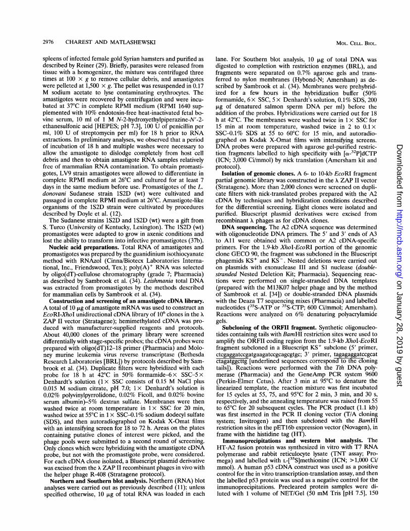
FIG. 2. (A) Induction of A2 gene expression in promastigotes.Promastigotes of the L. donovani Sudanese 1S2D (wt) strain cultivatedin complete RPMI medium at 26°C and pH 7.4 were transferred intomedia at pH 4.5 (HEPES in complete RPMI was replaced by 20 mMsuccinic acid) and incubated at 26 or 37°C or were maintained inmedium at pH 7.4 but incubated at 37°C, as indicated above the lanes.Total RNA was extracted after 3, 6, 10, and 30 h following the transfer(lanes a, b, c, and d, respectively). (B) Induction of A2 gene expressionin in vitro-cultured amastigote-like organisms (L. donovani Sudanstrain 1M2D). Amastigotes were transferred in fetal bovine serum
supplemented with 20 mM succinate for 18 h to reduce the pH (L') or
were maintained under neutral pH conditions (L). Both L and L' cellswere incubated at 37°C. Lanes P and A, RNA samples from L.donovani Ethiopian LV9 promastigotes and amastigotes isolated frominfected hamsters, respectively. For both panels, RNA samples were
analyzed by Northern blot with A2 cDNA-specific probes (0.9 kb).Equal loading of RNA in each lane was verified by ethidium stainingof the agarose gel prior to the Northern blot.
sure, however, weak hybridization bands were observed for thepromastigote RNA, indicating that expression was not totallyshut off in promastigotes (data not shown). The A5 and A6cDNAs also recognized RNAs present only in amastigotes; AShybridized with a high-molecular-weight transcript (8.0 kb),and A6 hybridized with a 1.0-kb RNA (not shown). ThesecDNAs have not as yet been characterized.
Physicochemical factors triggering the expression of theamastigote-specific genes. The promastigote-to-amastigote cy-todifferentiation process is thought to be triggered by temper-ature and pH shifts occurring during the passage from insectvector to mammalian host. The insect alimentary tract is at27°C and neutral pH, whereas the macrophage phagolysosomecompartment is at 37°C and pH 4.5 (2). We investigated theimportance of both of these physicochemical parameters on
the induction of expression of the A2 transcripts. We at-tempted to induce the expression of the amastigote-specific A2gene in cultured promastigotes by shifting the temperature andpH of the culture media. Promastigotes of L. donovani Su-danese strain 1S2D (wt) cultured at 26°C and at pH 7.4 were
transferred into media at pH 4.5 and/or 37°C, and total RNAwas extracted 3, 6, 10, and 30 h after the transfer. As shown inFig. 2A, it was possible to induce A2 expression in promasti-gotes with a combination of temperature and pH shifts. Similarresults were observed for the L. donovani LV9 strain (notshown). Furthermore, A2 gene expression was inducible inamastigote-like organisms of the Sudanese 1S2D strain by a
pH shift (Fig. 2B). These amastigote-like organisms cultivatedin 100% fetal bovine serum at 37°C (at neutral pH) were
shown to share some metabolic and physiologic characteristics
(kb)10
6.0>
4.0 >
3.0 >
2.0 >
1.66
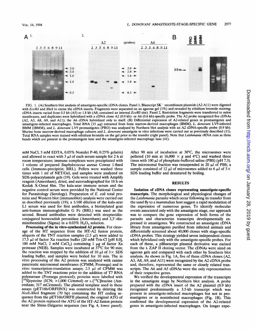
FIG. 3. Genomic organization of the A2 gene family. In each lane,10 ,ug of total DNA extracted from L. donovani LV9 promastigoteswas digested to completion with different restriction endonucleases (E,EcoRI; S, Sall; X, XbaI; and C, Clal). For double digests, the DNA was
first cut to completion with ClaI, and the fragments were thenprecipitated and resuspended in the appropriate buffer for the seconddigest. Restriction fragments were analyzed by Southern blot analysiswith a PstI-XhoI fragment of the A2 cDNA insert (0.5 kb).
of intracellular amastigotes (12). On the same panel, thispattern of differential expression was compared with the one
obtained with L. donovani LV9 promastigotes and amasti-gotes. Taken together, these data indicate that the pH shift isthe major trigger for the developmental expression of the A2gene but that a combination of pH and temperature shifts isneeded for induction of expression.A2 transcripts are encoded by a multigene family. The
genomic organization of the A2 genes was analyzed by South-ern blot analyses. Total DNA was digested to completion withseveral restriction endonucleases, separated on an agarose gel,and hybridized with a probe prepared with the A2 cDNA insert(the A2 cDNA did not contain sites for these restrictionenzymes). As shown in Fig. 3, the hybridization pattern in eachlane displayed a series of bands of different intensities, clearlyshowing that many copies of the gene were present in thegenome. However, common bands for the EcoRI, XbaI, andSall digests (between 6 and 10 kb) suggested an arrangementin tandem array. Indeed, if each of these restriction sites isunique in the A2 gene, then copies clustered in tandem arraywould display fragments of the same length for each digest. Inorder to verify this assumption and isolate a genomic fragmentcarrying the complete coding region of the A2 transcript, weisolated and characterized genomic clones identified by thescreening of an EcoRI partial genomic library with an A2cDNA probe. The inserts were mapped with several restrictionenzymes and displayed similar patterns. One of these clones,GECO 90, was kept for detailed characterization. Figure 4(upper panel) shows the restriction map of the fragment andhow it corresponds to the A2 cDNA series. It contained uniquesites for Sall and XbaI but no ClaI site, and this was consistentwith the Southern blot analysis shown in Fig. 3. The DNA
A. B.
MOL. CELL. BIOL.
on January 28, 2019 by guesthttp://m
cb.asm.org/
Dow
nloaded from
L. DONOVANI AMASTIGOTE-STAGE-SPECIFIC GENE 2979
1IkbI I
p P PO s p 0 repeat X EI I 11 I I 1.I_I I 11 I I I'
GECO 90cDNAs
H A 8 H--------------------------------*A9
- > All
GECO 90/1.8+
N OB
HT -[~H
ORFH
1.1 kb
B
AXG
X B
-. pET 16b/ORFII
X B
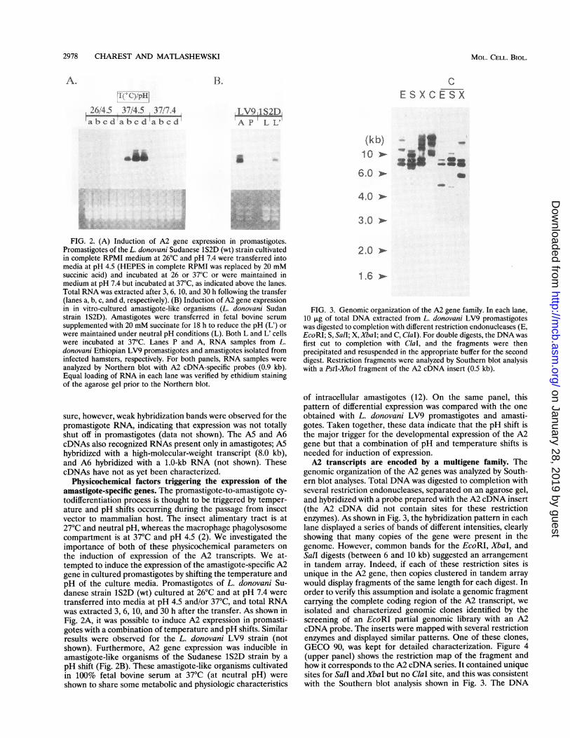
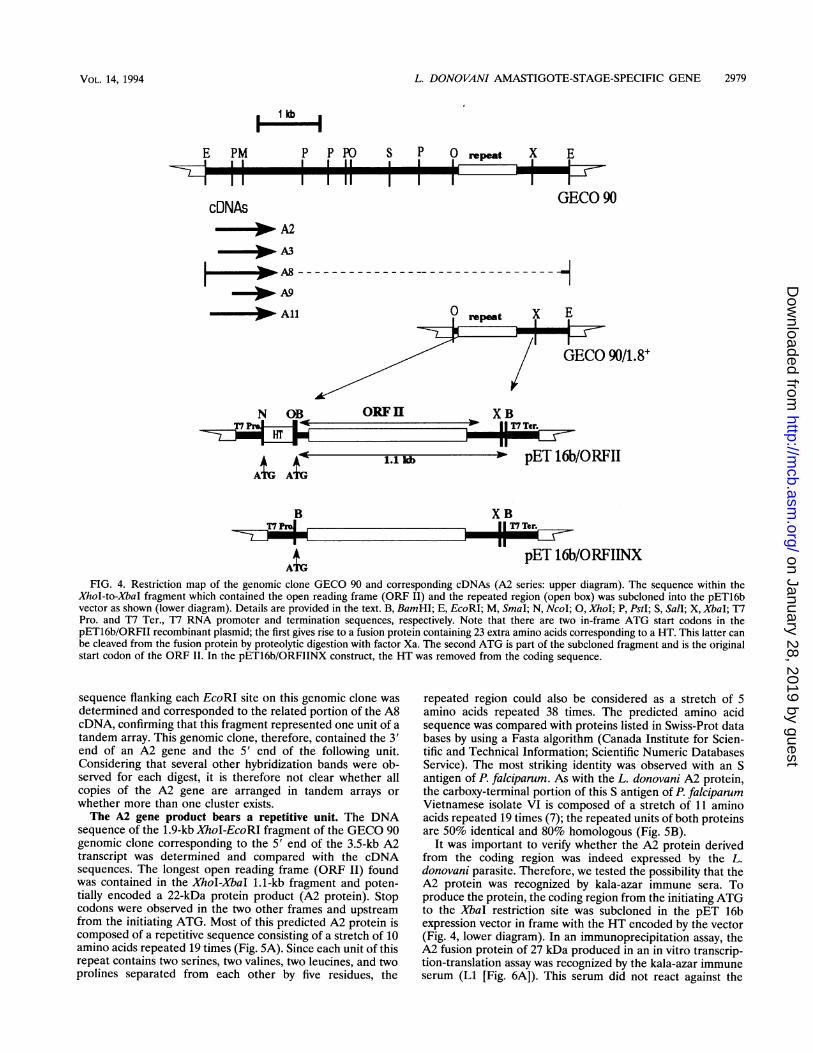
pET 16b/ORFIINXFIG. 4. Restriction map of the genomic clone GECO 90 and corresponding cDNAs (A2 series: upper diagram). The sequence within the
XhoI-to-XbaI fragment which contained the open reading frame (ORF II) and the repeated region (open box) was subcloned into the pET16bvector as shown (lower diagram). Details are provided in the text. B, BamHI; E, EcoRI; M, SmaI; N, Ncol; 0, XhoI; P, PstI; S, Sall; X, XbaI; T7Pro. and T7 Ter., T7 RNA promoter and termination sequences, respectively. Note that there are two in-frame ATG start codons in thepET16b/ORFII recombinant plasmid; the first gives rise to a fusion protein containing 23 extra amino acids corresponding to a HT. This latter canbe cleaved from the fusion protein by proteolytic digestion with factor Xa. The second ATG is part of the subcloned fragment and is the originalstart codon of the ORF II. In the pET16b/ORFIINX construct, the HT was removed from the coding sequence.
sequence flanking each EcoRI site on this genomic clone wasdetermined and corresponded to the related portion of the A8cDNA, confirming that this fragment represented one unit of atandem array. This genomic clone, therefore, contained the 3'end of an A2 gene and the 5' end of the following unit.Considering that several other hybridization bands were ob-served for each digest, it is therefore not clear whether allcopies of the A2 gene are arranged in tandem arrays orwhether more than one cluster exists.The A2 gene product bears a repetitive unit. The DNA
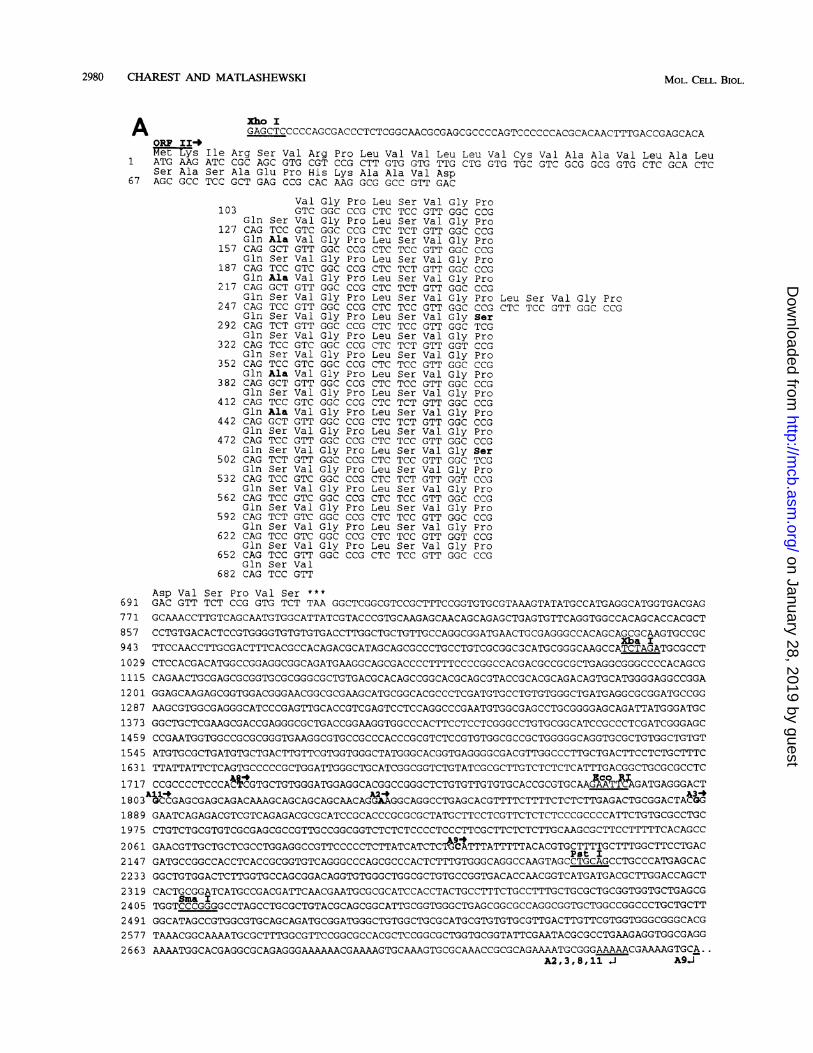
sequence of the 1.9-kb XhoI-EcoRI fragment of the GECO 90genomic clone corresponding to the 5' end of the 3.5-kb A2transcript was determined and compared with the cDNAsequences. The longest open reading frame (ORF II) foundwas contained in the XhoI-XbaI 1.1-kb fragment and poten-tially encoded a 22-kDa protein product (A2 protein). Stopcodons were observed in the two other frames and upstreamfrom the initiating ATG. Most of this predicted A2 protein iscomposed of a repetitive sequence consisting of a stretch of 10amino acids repeated 19 times (Fig. 5A). Since each unit of thisrepeat contains two serines, two valines, two leucines, and twoprolines separated from each other by five residues, the
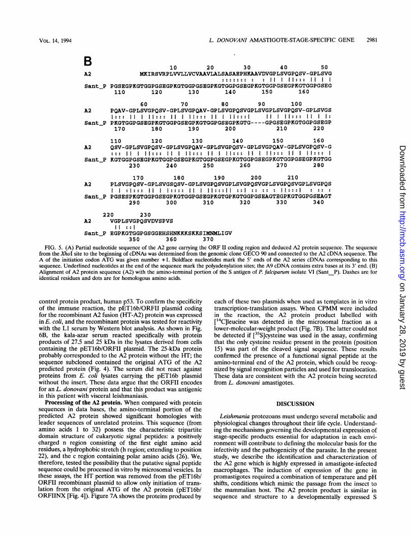
repeated region could also be considered as a stretch of 5amino acids repeated 38 times. The predicted amino acidsequence was compared with proteins listed in Swiss-Prot databases by using a Fasta algorithm (Canada Institute for Scien-tific and Technical Information; Scientific Numeric DatabasesService). The most striking identity was observed with an Santigen of P. falciparum. As with the L. donovani A2 protein,the carboxy-terminal portion of this S antigen of P. falciparumVietnamese isolate VI is composed of a stretch of 11 aminoacids repeated 19 times (7); the repeated units of both proteinsare 50% identical and 80% homologous (Fig. 5B).
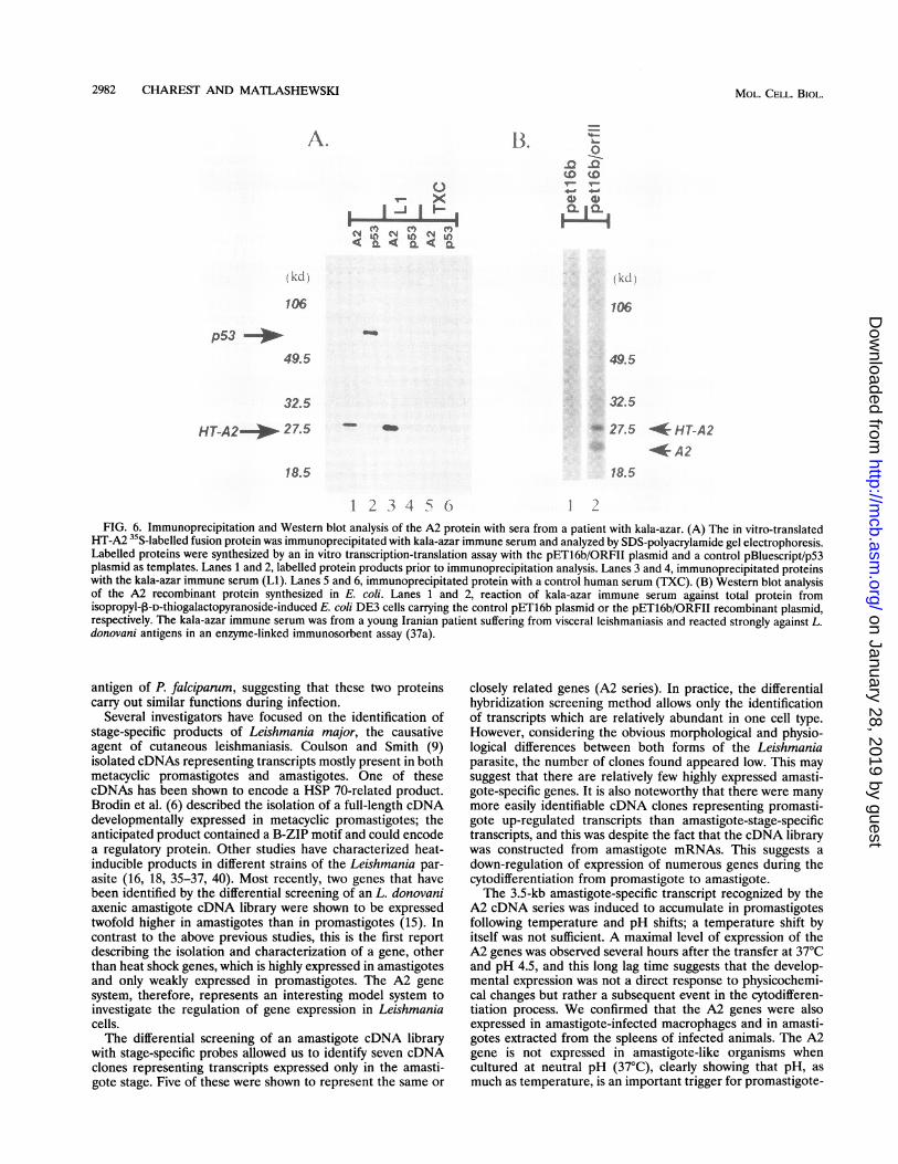
It was important to verify whether the A2 protein derivedfrom the coding region was indeed expressed by the L.donovani parasite. Therefore, we tested the possibility that theA2 protein was recognized by kala-azar immune sera. Toproduce the protein, the coding region from the initiating ATGto the XbaI restriction site was subcloned in the pET 16bexpression vector in frame with the HT encoded by the vector(Fig. 4, lower diagram). In an immunoprecipitation assay, theA2 fusion protein of 27 kDa produced in an in vitro transcrip-tion-translation assay was recognized by the kala-azar immuneserum (Li [Fig. 6A]). This serum did not react against the
E PMI I I
--,-? I I
VOL. 14, 1994
on January 28, 2019 by guesthttp://m
cb.asm.org/
Dow
nloaded from
2980 CHAREST AND MATLASHEWSKI
AORF II4Met Lys
1 ATG AAGSer Ala
67 AGC GCC
Xho IGAGCTCCCCCAGCGACCCTCTCGGCAACGCGAGCGCCCCAGTCCCCCCACGCACAACTTTGACCGAGCACA
Ile Arg Ser Val Arg Pro Leu Val Val Leu Leu Val Cys Val Ala Ala Val Leu Ala LeuATC CGC AGC GTG CGT CCG CTT GTG GTG TTG CTG GTG TGC GTC GCG GCG GTG CTC GCA CTCSer Ala Glu Pro His Lys Ala Ala Val AspTCC GCT GAG CCG CAC AAG GCG GCC GTT GAC
Val GlyGTC GGC
Gln Ser Val GlyCAG TCC GTC GGCGln Ala Val GlyCAG GCT GTT GGCGln Ser Val GlyCAG TCC GTC GGCGln Ala Val GlyCAG GCT GTT GGCGln Ser Val GlyCAG TCC GTT GGCGln Ser Val GlyCAG TCT GTT GGCGln Ser Val GlyCAG TCC GTC GGCGln Ser Val GlyCAG TCC GTC GGCGln Ala Val GlyCAG GCT GTT GGCGln Ser Val GlyCAG TCC GTC GGCGln Ala Val GlyCAG GCT GTT GGCGln Ser Val GlyCAG TCC GTT GGCGln Ser Val GlyCAG TCT GTT GGCGln Ser Val GlyCAG TCC GTC GGCGln Ser Val GlyCAG TCC GTC GGCGln Ser Val GlyCAG TCT GTC GGCGln Ser Val GlyCAG TCC GTC GGCGln Ser Val GlyCAG TCC GTT GGCGln Ser ValCAG TCC GTT
ProCCGProCCGProCCGProCCGProCCGProCCGProCCGProCCGProCCGProCCGProCCGProCCGProCCGProCCGProCCGProCCGProCCGProCCGProCCG
LeuCTCLeuCTCLeuCTCLeuCTCLeuCTCLeuCTCLeuCTCLeuCTCLeuCTCLeuCTCLeuCTCLeuCTCLeuCTCLeuCTCLeuCTCLeuCTCLeuCTCLeuCTCLeuCTC
Ser Val Gly ProTCC GTT GGC CCGSer Val Gly ProTCT GTT GGC CCGSer Val Gly ProTCC GTT GGC CCGSer Val Gly ProTCT GTT GGC CCGSer Val Gly ProTCT GTT GGC CCGSer Val Gly ProTCC GTT GGC CCGSer Val Gly SerTCC GTT GGC TCGSer Val Gly ProTCT GTT GGT CCGSer Val Gly ProTCC GTT GGC CCGSer Val Gly ProTCC GTT GGC CCGSer Val Gly ProTCT GTT GGC CCGSer Val Gly ProTCT GTT GGC CCGSer Val Gly ProTCC GTT GGC CCGSer Val Gly SerTCC GTT GGC TCGSer Val Gly ProTCT GTT GGT CCGSer Val Gly ProTCC GTT GGC CCGSer Val Gly ProTCC GTT GGC CCGSer Val Gly ProTCC GTT GGT CCGSer Val Gly ProTCC GTT GGC CCG
Leu Ser Val Gly ProCTC TCC GTT GGC CCG
Asp Val Ser Pro Val Ser ***691 GAC GTT TCT CCG GTG TCT TAA GGCTCGGCGTCCGCTTTCCGGTGTGCGTAAAGTATATGCCATGAGGCATGGTGACGAG771 GCAAACCTTGTCAGCAATGTGGCATTATCGTACCCGTGCAAGAGCAACAGCAGAGCTGAGTGTTCAGGTGGCCACAGCACCACGCT857 CCTGTGACACTCCGTGGGGTGTGTGTGACCTTGGCTGCTGTTGCCAGGCGGATGAACTGCGAGGGCCACAGCAGCGCAAGTGCCGC
Xba I943 TTCCAACCTTGCGACTTTCACGCCACAGACGCATAGCAGCGCCCTGCCTGTCGCGGCGCATGCGGGCAAGCCATCTAGATGCGCCT1029 CTCCACGACATGGCCGGAGGCGGCAGATGAAGGCAGCGACCCCTTTTCCOCGGOCACGACGCCGCGCTGAGGCGGGCCCCACAGCG1115 CAGAACTGCGAGCGCGGTGCGCGGGCGCTGTGACGCACAGCCGGCACGCAGCGTACCGCACGCAGACAGTGCATGGGGAGGCCGGA1201 GGAGCAAGAGCGGTGGACGGGAACGGCGCGAAGCATGCGGCACGCCCTCGATGTGCCTGTGTGGGCTGATGAGGCGCGGATGCCGG1287 AAGCGTGGCGAGGGCATCCCGAGTTGCACCGTCGAGTCCTCCAGGCCCGAATGTGGCGAGCCTGCGGGGAGCAGATTATGGGATGC1373 GGCTGCTCGAAGCGACCGAGGGCGCTGACCGGAAGGTGGCCCACTTCCTCCTCGGGCCTGTGCGGCATCCGCCCTCGATCGGGAGC1459 CCGAATGGTGGCCGCGCGGGTGAAGGCGTGCCGCCCACCCGCGTCTCCGTGTGGCGCCGCTGGGGGCAGGTGCGCTGTGGCTGTGT1545 ATGTGCGCTGATGTGCTGACTTGTTCGTGGTGGGCTATGGGCACGGTGAGGGGCGACGTTGGCCCTTGCTGACTTCCTCTGCTTTC1631 TTATTATTCTCAGTGCCCCCGCTGGATTGGGCTGCATCGGCGGTCTGTATCGCGCTTGTCTCTCTCATTTGACGGCTGCGCGCCTC
1717 CCGCCCCTCCCAt6CGTGCTGTGGGATGGAGGCACGGCCGGGCTCTGTGTTGTGTGCACCGCGTGCAAGOAATGAGATGAGGGACT1803 GCCGAGCGAGCAGACAAAGCAGCAGCAGCAACAGGAAGGCAGGCCTGAGCACGTTTTCTTTTCTCTCTTGAGACTGCGGACTACGG1889 GAATCAGAGACGTCGTCAGAGACGCGCATCCGCACCCGCGCGCTATGCTTCCTCGTTCTCTCTCCCGCCCCATTCTGTGCGCCTGC1975 CTGTCTGCGTGTCGCGAGCGCCGTTGCCGGCGGTCTCTCTCCCCTCCCTTCGCTTCTCTCTTGCAAGCGCTTCCTTTTTCACAGCC2061 GAACGTTGCTGCTCGCCTGGAGGCCGTTCCCCCTCTTATCATCTCTGCATTTATTTTTACACGTGCTTTTGCTTTGGCTTCCTGAC
Pat2147 GATGCCGGCCACCTCACCGCGGTGTCAGGGCCCAGCGCCCACTCTTTGTGGGCAGGCCAAGTAGCCTGCAGCCTGCCCATGAGCAC2233 GGCTGTGGACTCTTGGTGCCAGCGGACAGGTGTGGGCTGGCGCTGTGCCGGTGACACCAACGGTCATGATGACGCTTGGACCAGCT
2319 CACTGCGGATCATGCCGACGATTCAACGAATGCGCGCATCCACCTACTGCCTTTCTGCCTTTGCTGCGCTGCGGTGGTGCTGAGCGSma
2405 TGGTCCCGGGGCCTAGCCTGCGCTGTACGCAGCGGCATTGCGGTGGGCTGAGCGGCGCCAGGCGGTGCTGGCCGGCCCTGCTGCTT2491 GGCATAGCCGTGGCGTGCAGCAGATGCGGATGGGCTGTGGCTGCGCATGCGTGTGTGCGTTGACTTGTTCGTGGTGGGCGGGCACG2577 TAAACGGCAAAATGCGCTTTGGCGTTCCGGCGCCACGCTCCGGCGCTGGTGCGGTATTCGAATACGCGCCTGAAGAGGTGGCGAGG2663 AAAATGGCACGAGGCGCAGAGGGAAAAAACGAAAAGTGCAAAGTGCGCAAACCGCGCAGAAAATGCGGGAAAAACGAAAAGTGCA -.
A2,3,8,11 J A94
103
127
157
187
217
247
292
322
352
382
412
442
472
502
532
562
592
622
652
682
MOL. CELL. BIOL.
on January 28, 2019 by guesthttp://m
cb.asm.org/
Dow
nloaded from
L. DONOVANI AMASTIGOTE-STAGE-SPECIFIC GENE 2981
BA2
Sant_P
10 20 30 40 50MKIRSVRPLVVLLVCVAAVLALSASAEPHKAAVDVGPLSVGPQSV-GPLSVG
:::::::: : 11 I II::: 11PC R(aPKGTGGPaRaPKUTaPaGSaPRaTaaPaSEGPKGTGGPGSEGPKGTGGPGSEG110 120 130 140 150 160
60 70 80 90 100A2 PQAV-GPLSVGPQSV-GPLSVGPQAV-GPLSVGPQSVGPLSVGPLSVGPQSV-GPLSVGS
1::: 11 I II::: 11 I II::: 11 I 11:::1 11 1 11::: 11 I 1:Sant_P PKGTGGPGSEGPKGTGGPGSEGPKGTGGPGSEGPKGTG ---- GPGSEGPKGTGGPGSEGP
170 180 190 200 210 220
A2
Sant_P
110 120 130 140 150 160QSV-GPLSVGPQSV-GPLSVGPQAV-GPLSVGPQSV-GPLSVGPQAV-GPLSVGPQSV-G
11111::: 11 1 11::: 11 1 11::: 11 1 11::: 11 1 11:::KGTGGPGSEGPKGTGGPGSEGPKGTGGPGSEGPKGTGGPGSEGPKGTGGPGSEGPKGTGG
230 240 250 260 270 280
170 180 190 200 210A2 PLSVGPQSV-GPLSVGSQSV-GPLSVGPQSVGPLSVGPQSVGPLSVGPQSVGPLSVGPQS
I I :1::: 11 1:::: 11 I 11:::II ::I :: ::: 11:::I : :::Sant_P PGSESPKGTGGPGSEGPKGTGGPGSEGPKGTGPKGTGGPGSEAGTEGPKGTGGPGSEAGT
290 300 310 320 330 340
220 230A2 VGPLSVGPQSVDVSPVS
11 ::ISant_P EGPKGTGGPGSGGEHSHNKKKSKKSIMNMLIGV
350 360 370FIG. 5. (A) Partial nucleotide sequence of the A2 gene carrying the ORF II coding region and deduced A2 protein sequence. The sequence
from the XhoI site to the beginning of cDNAs was determined from the genomic clone GECO 90 and connected to the A2 cDNA sequence. TheA of the initiation codon ATG was given number +1. Boldface nucleotides mark the 5' ends of the A2 series cDNAs corresponding to thissequence. Underlined nucleotides at the end of the sequence mark the polyadenylation sites; the A9 cDNA contains extra bases at its 3' end. (B)Alignment of A2 protein sequence (A2) with the amino-terminal portion of the S antigen of P. falciparum isolate VI (Sant_P). Dashes are foridentical residues and dots are for homologous amino acids.
control protein product, human p53. To confirm the specificityof the immune reaction, the pET16b/ORFII plasmid codingfor the recombinant A2 fusion (HT-A2) protein was expressedin E. coli, and the recombinant protein was tested for reactivitywith the Li serum by Western blot analysis. As shown in Fig.6B, the kala-azar serum reacted specifically with proteinproducts of 27.5 and 25 kDa in the lysates derived from cellscontaining the pET16b/ORFII plasmid. The 25-kDa proteinprobably corresponded to the A2 protein without the HT; thesequence subcloned contained the original ATG of the A2predicted protein (Fig. 4). The serum did not react againstproteins from E. coli lysates carrying the pET16b plasmidwithout the insert. These data argue that the ORFII encodesfor an L. donovani protein and that this product was antigenicin this patient with visceral leishmaniasis.
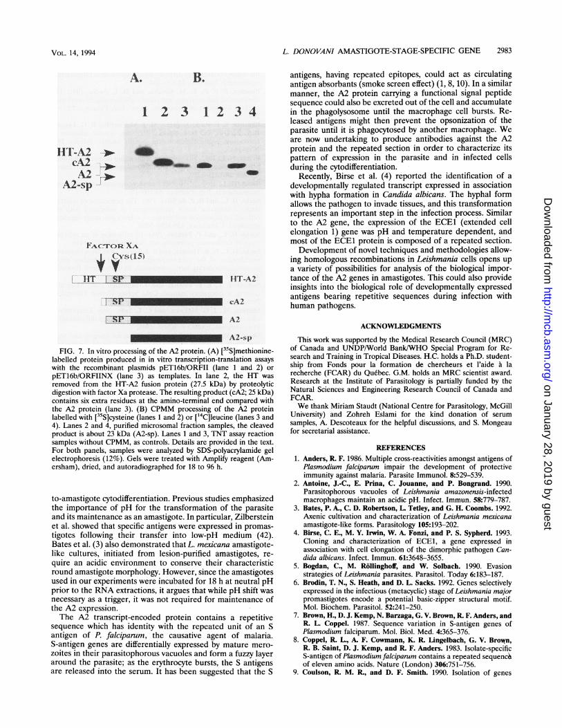
Processing of the A2 protein. When compared with proteinsequences in data bases, the amino-terminal portion of thepredicted A2 protein showed significant homologies withleader sequences of unrelated proteins. This sequence (fromamino acids 1 to 32) possess the characteristic tripartitedomain structure of eukaryotic signal peptides: a positivelycharged n region consisting of the first eight amino acidresidues, a hydrophobic stretch (h region; extending to position22), and the c region containing polar amino acids (26). We,therefore, tested the possibility that the putative signal peptidesequence could be processed in vitro by microsomal vesicles. Inthese assays, the HT portion was removed from the pET16b/ORFII recombinant plasmid to allow only initiation of trans-lation from the original ATG of the A2 protein (pET16b/ORFIINX [Fig. 4]). Figure 7A shows the proteins produced by
each of these two plasmids when used as templates in in vitrotranscription-translation assays. When CPMM were includedin the reaction, the A2 protein product labelled with["4C]leucine was detected in the microsomal fraction as alower-molecular-weight product (Fig. 7B). The latter could notbe detected if [35S]cysteine was used in the assay, confirmingthat the only cysteine residue present in the protein (position15) was part of the cleaved signal sequence. These resultsconfirmed the presence of a functional signal peptide at theamino-terminal end of the A2 protein, which could be recog-nized by signal recognition particles and used for translocation.These data are consistent with the A2 protein being secretedfrom L. donovani amastigotes.
DISCUSSION
Leishmania protozoans must undergo several metabolic andphysiological changes throughout their life cycle. Understand-ing the mechanisms governing the developmental expression ofstage-specific products essential for adaptation in each envi-ronment will contribute to defining the molecular basis for theinfectivity and the pathogenicity of the parasite. In the presentstudy, we describe the identification and characterization ofthe A2 gene which is highly expressed in amastigote-infectedmacrophages. The induction of expression of the gene inpromastigotes required a combination of temperature and pHshifts, conditions which mimic the passage from the insect tothe mammalian host. The A2 protein product is similar insequence and structure to a developmentally expressed S
VOL. 14, 1994
on January 28, 2019 by guesthttp://m
cb.asm.org/
Dow
nloaded from
2982 CHAREST AND MATLASHEWSKI
A.
0
v- xu(
(kLI j
106
p53 --ii
49.5
32.5
18.5
13. o'-o¶Q Q
__
I CLI C
I A >
106
49.5
32.5
- 27.5 * HT-A2
* A218.5
1 2 3 4 5 () 1 2FIG. 6. Immunoprecipitation and Western blot analysis of the A2 protein with sera from a patient with kala-azar. (A) The in vitro-translated
HT-A2 35S-labelled fusion protein was immunoprecipitated with kala-azar immune serum and analyzed by SDS-polyacrylamide gel electrophoresis.Labelled proteins were synthesized by an in vitro transcription-translation assay with the pET16b/ORFII plasmid and a control pBluescript/p53plasmid as templates. Lanes 1 and 2, labelled protein products prior to immunoprecipitation analysis. Lanes 3 and 4, immunoprecipitated proteinswith the kala-azar immune serum (Li). Lanes 5 and 6, immunoprecipitated protein with a control human serum (TXC). (B) Western blot analysisof the A2 recombinant protein synthesized in E. coli. Lanes 1 and 2, reaction of kala-azar immune serum against total protein fromisopropyl-13-D-thiogalactopyranoside-induced E. coli DE3 cells carrying the control pET16b plasmid or the pET16b/ORFII recombinant plasmid,respectively. The kala-azar immune serum was from a young Iranian patient suffering from visceral leishmaniasis and reacted strongly against L.donovani antigens in an enzyme-linked immunosorbent assay (37a).
antigen of P. falciparum, suggesting that these two proteinscarry out similar functions during infection.
Several investigators have focused on the identification ofstage-specific products of Leishmania major, the causativeagent of cutaneous leishmaniasis. Coulson and Smith (9)isolated cDNAs representing transcripts mostly present in bothmetacyclic promastigotes and amastigotes. One of thesecDNAs has been shown to encode a HSP 70-related product.Brodin et al. (6) described the isolation of a full-length cDNAdevelopmentally expressed in metacyclic promastigotes; theanticipated product contained a B-ZIP motif and could encodea regulatory protein. Other studies have characterized heat-inducible products in different strains of the Leishmania par-asite (16, 18, 35-37, 40). Most recently, two genes that havebeen identified by the differential screening of an L. donovaniaxenic amastigote cDNA library were shown to be expressedtwofold higher in amastigotes than in promastigotes (15). Incontrast to the above previous studies, this is the first reportdescribing the isolation and characterization of a gene, otherthan heat shock genes, which is highly expressed in amastigotesand only weakly expressed in promastigotes. The A2 genesystem, therefore, represents an interesting model system toinvestigate the regulation of gene expression in Leishmaniacells.The differential screening of an amastigote cDNA library
with stage-specific probes allowed us to identify seven cDNAclones representing transcripts expressed only in the amasti-gote stage. Five of these were shown to represent the same or
closely related genes (A2 series). In practice, the differentialhybridization screening method allows only the identificationof transcripts which are relatively abundant in one cell type.However, considering the obvious morphological and physio-logical differences between both forms of the Leishmaniaparasite, the number of clones found appeared low. This maysuggest that there are relatively few highly expressed amasti-gote-specific genes. It is also noteworthy that there were manymore easily identifiable cDNA clones representing promasti-gote up-regulated transcripts than amastigote-stage-specifictranscripts, and this was despite the fact that the cDNA librarywas constructed from amastigote mRNAs. This suggests adown-regulation of expression of numerous genes during thecytodifferentiation from promastigote to amastigote.The 3.5-kb amastigote-specific transcript recognized by the
A2 cDNA series was induced to accumulate in promastigotesfollowing temperature and pH shifts; a temperature shift byitself was not sufficient. A maximal level of expression of theA2 genes was observed several hours after the transfer at 370Cand pH 4.5, and this long lag time suggests that the develop-mental expression was not a direct response to physicochemi-cal changes but rather a subsequent event in the cytodifferen-tiation process. We confirmed that the A2 genes were alsoexpressed in amastigote-infected macrophages and in amasti-gotes extracted from the spleens of infected animals. The A2gene is not expressed in amastigote-like organisms whencultured at neutral pH (370C), clearly showing that pH, asmuch as temperature, is an important trigger for promastigote-
MOL. CELL. BIOL.
HT-A2--)p- 2 7.5
on January 28, 2019 by guesthttp://m
cb.asm.org/
Dow
nloaded from
L. DONOVANI AMASTIGOTE-STAGE-SPECIFIC GENE 2983
A. B.
1 2 3 12 3
IIT-A2c.A2A2
A2-sp
4b
FACrOR XA
Cvs(-5)1lT.
R_P cA2
A2-sp
FIG. 7. In vitro processing of the A2 protein. (A) [35S]methionine-labelled protein produced in in vitro transcription-translation assayswith the recombinant plasmids pET16b/ORFII (lane 1 and 2) orpET16b/ORFIINX (lane 3) as templates. In lane 2, the HT wasremoved from the HT-A2 fusion protein (27.5 kDa) by proteolyticdigestion with factor Xa protease. The resulting product (cA2; 25 kDa)contains six extra residues at the amino-terminal end compared withthe A2 protein (lane 3). (B) CPMM processing of the A2 proteinlabelled with [35S]cysteine (lanes 1 and 2) or ['4C]leucine (lanes 3 and4). Lanes 2 and 4, purified microsomal fraction samples, the cleavedproduct is about 23 kDa (A2-sp). Lanes 1 and 3, TNT assay reactionsamples without CPMM, as controls. Details are provided in the text.For both panels, samples were analyzed by SDS-polyacrylamide gelelectrophoresis (12%). Gels were treated with Amplify reagent (Am-ersham), dried, and autoradiographed for 18 to 96 h.
to-amastigote cytodifferentiation. Previous studies emphasizedthe importance of pH for the transformation of the parasiteand its maintenance as an amastigote. In particular, Zilbersteinet al. showed that specific antigens were expressed in promas-tigotes following their transfer into low-pH medium (42).Bates et al. (3) also demonstrated that L. mexicana amastigote-like cultures, initiated from lesion-purified amastigotes, re-quire an acidic environment to conserve their characteristicround amastigote morphology. However, since the amastigotesused in our experiments were incubated for 18 h at neutral pHprior to the RNA extractions, it argues that while pH shift wasnecessary as a trigger, it was not required for maintenance ofthe A2 expression.The A2 transcript-encoded protein contains a repetitive
sequence which has identity with the repeated unit of an Santigen of P. falciparum, the causative agent of malaria.S-antigen genes are differentially expressed by mature mero-zoites in their parasitophorous vacuoles and form a fuzzy layeraround the parasite; as the erythrocyte bursts, the S antigensare released into the serum. It has been suggested that the S
antigens, having repeated epitopes, could act as circulatingantigen absorbants (smoke screen effect) (1, 8, 10). In a similarmanner, the A2 protein carrying a functional signal peptidesequence could also be excreted out of the cell and accumulate
4 in the phagolysosome until the macrophage cell bursts. Re-leased antigens might then prevent the opsonization of theparasite until it is phagocytosed by another macrophage. Weare now undertaking to produce antibodies against the A2protein and the repeated section in order to characterize itspattern of expression in the parasite and in infected cellsduring the cytodifferentiation.
Recently, Birse et al. (4) reported the identification of adevelopmentally regulated transcript expressed in associationwith hypha formation in Candida albicans. The hyphal formallows the pathogen to invade tissues, and this transformationrepresents an important step in the infection process. Similarto the A2 gene, the expression of the ECE1 (extended cellelongation 1) gene was pH and temperature dependent, andmost of the ECE1 protein is composed of a repeated section.Development of novel techniques and methodologies allow-
ing homologous recombinations in Leishmania cells opens upa variety of possibilities for analysis of the biological impor-
-A2 tance of the A2 genes in amastigotes. This could also provideinsights into the biological role of developmentally expressedantigens bearinz renetitive seauences durinz infection withhuman pathogens.
ACKNOWLEDGMENTS
This work was supported by the Medical Research Council (MRC)of Canada and UNDP/World Bank/WHO Special Program for Re-search and Training in Tropical Diseases. H.C. holds a Ph.D. student-ship from Fonds pour la formation de chercheurs et l'aide a larecherche (FCAR) du Quebec. G.M. holds an MRC scientist award.Research at the Institute of Parasitology is partially funded by theNatural Sciences and Engineering Research Council of Canada andFCAR.We thank Miriam Staudt (National Centre for Parasitology, McGill
University) and Zohreh Eslami for the kind donation of serumsamples, A. Descoteaux for the helpful discussions, and S. Mongeaufor secretarial assistance.
REFERENCES1. Anders, R. F. 1986. Multiple cross-reactivities amongst antigens of
Plasmodium falciparum impair the development of protectiveimmunity against malaria. Parasite Immunol. 8:529-539.
2. Antoine, J.-C., E. Prina, C. Jouanne, and P. Bongrand. 1990.Parasitophorous vacuoles of Leishmania amazonensis-infectedmacrophages maintain an acidic pH. Infect. Immun. 58:779-787.
3. Bates, P. A., C. D. Robertson, L. Tetley, and G. H. Coombs. 1992.Axenic cultivation and characterization of Leishmania mexicanaamastigote-like forms. Parasitology 105:193-202.
4. Birse, C. E., M. Y. Irwin, W. A. Fonzi, and P. S. Sypherd. 1993.Cloning and characterization of ECE1, a gene expressed inassociation with cell elongation of the dimorphic pathogen Can-dida albicans. Infect. Immun. 61:3648-3655.
5. Bogdan, C., M. Rollinghoff, and W. Solbach. 1990. Evasionstrategies of Leishmania parasites. Parasitol. Today 6:183-187.
6. Brodin, T. N., S. Heath, and D. L. Sacks. 1992. Genes selectivelyexpressed in the infectious (metacyclic) stage of Leishmania majorpromastigotes encode a potential basic-zipper structural motif.Mol. Biochem. Parasitol. 52:241-250.
7. Brown, H., D. J. Kemp, N. Barzaga, G. V. Brown, R. F. Anders, andR. L. Coppel. 1987. Sequence variation in S-antigen genes ofPlasmodium falciparum. Mol. Biol. Med. 4:365-376.
8. Coppel, R. L., A. F. Cowmann, K. R. Lingelbach, G. V. Brown,R. B. Saint, D. J. Kemp, and R. F. Anders. 1983. Isolate-specificS-antigen of Plasmodium falciparum contains a repeated sequenc6of eleven amino acids. Nature (London) 306:751-756.
9. Coulson, R. M. R., and D. F. Smith. 1990. Isolation of genes
VOL. 14, 1994
on January 28, 2019 by guesthttp://m
cb.asm.org/
Dow
nloaded from
2984 CHAREST AND MATLASHEWSKI
showing increased or unique expression in the infective promas-tigotes of Leishmania major. Mol. Biochem. Parasitol. 40:63-76.
10. Culvenor, J. G., and P. Crewther. 1990. S-antigen localization inthe erythrocyte stages of Plasmodium falciparum. J. Protozool.37:59-65.
11. Descoteaux, A., and G. Matlashewski. 1989. c-fos and tumornecrosis factor gene expression in Leishmania donovani-infectedmacrophages. Mol. Cell. Biol. 9:5223-5227.
12. Doyle, P. S., J. C. Engel, P. F. P. Pimenta, P. da Silva, and D. M.Dwyer. 1991. Leishmania donovani: long-term culture of axenicamastigotes at 37°C. Exp. Parasitol. 73:326-334.
13. Glaser, T. A., S. F. Moody, E. Handmann, A. Bacic, and T. W.Spithill. 1991. An antigenically distinct lipophosphoglycan onamastigotes of Leishmania major. Mol. Biochem. Parasitol. 45:337-344.
14. Glaser, T. A., and A. J. Mukkada. 1992. Proline transport inLeishmania donovani amastigotes: dependence on pH gradientsand membrane potential. Mol. Biochem. Parasitol. 51:1-8.
15. Joshi, M., D. M. Dwyer, and H. L. Nakhasi. 1993. Cloning andcharacterization of differentially expressed genes from in vitrogrown "amastigotes" of Leishmania donovani. Mol. Biochem.Parasitol. 58:345-354.
16. Kidane, G. Z., N. Samaras, and T. W. Spithill. 1989. Cloning ofdevelopmentally regulated gene from Leishmania major and ex-pression fallowing heat induction. J. Biol. Chem. 264:4244-4250.
17. Laemmli, U. K. 1970. Cleavage of structural proteins during theassembly of the head of bacteriophage T4. Nature (London)227:680-685.
18. MacFarlane, J., M. L. Blaxter, R. P. Bishop, M. A. Miles, and J. M.Kelly. 1990. Identification and characterization of a Leishmaniadonovani antigen belonging to the 70 kDa heat-shock proteinfamily. Eur. J. Biochem. 190:377-384.
19. Matlashewski, G., L. Banks, D. Pim, and L. Crawford. 1986.Analysis of human p53 proteins and mRNA levels in normal andtransformed cells. Eur. J. Biochem. 154:665-672.
20. Mauel, J. 1990. Macrophage-parasite interactions in Leishmaniainfections. J. Leukocyte Biol. 47:187-193.
21. McConville, M. J., and J. M. Blackwell. 1991. Developmentalchanges in the glycosylated phosphatidylinositols of Leishmaniadonovani: characterization of the promastigote and amastigoteglycolipids. J. Biol. Chem. 266:15170-15179.
22. Medina-Acosta, E., R. E. Karess, H. Schwartz, and D. G. Russell.1989. The promastigote surface protease (gp63) is expressed butdifferentially processed and localized in the amastigote stage. Mol.Biochem. Parasitol. 37:263-274.
23. Molyneux, D. H., and R. Killick-Kendrick. 1987. Morphology,ultrastructure and life cycles, p. 121-176. In W. Peters and R.Killick-Kendrick (ed.), The leishmaniases in biology and medicine,vol. 1. Biology and epidemiology. Academic Press, Inc., New York.
24. Moody, S. F. 1993. Molecular variation in Leishmania. Acta Trop.53:185-204.
25. Mukkada, A. J., J. C. Meade, T. A. Glaser, and P. F. Bonventre.1985. Enhanced metabolism of Leishmania donovani amastigotesat acid pH: an adaptation for intracellular growth. Science 229:1099-1101.
26. Nothwehr, S. F., and J. I. Gordon. 1990. Targeting of proteins into
the eukaryotic secretory pathway: signal peptide structure/functionrelationship. Bioessays 12:479-484.
27. Pimenta, P. F. P., E. M. Saraiva, and D. L. Sacks. 1991. Thecomparative fine structure and surface glycogonjugate expressionof three life stages of Leishmania major. Exp. Parasitol. 72:191-204.
28. Pimenta, F. F. P., S. J. Turco, M. J. McConville, P. G. Lawyer,P. V. Perkins, and D. L. Sacks. 1992. Stage-specific adhesion ofLeishmania promastigotes to the sandfly midgut. Science 256:1812-1815.
29. Reiner, N. E. 1982. Host-parasite relationship in murine leishma-niasis: pathophysiological and immunological changes. Infect.Immun. 38:1223-1230.
30. Sacks, D. L. 1989. Minireview: metacyclogenesis in Leishmaniapromastigotes. Exp. Parasitol. 69:100-103.
31. Sacks, D. L., and R. P. da Silva. 1987. The generation of infectivestages Leishmania major promastigotes is associated with thecell-surface expression and release of a developmentally regulatedglycolipid. J. Immunol. 139:3099-3106.
32. Sacks, D. L., and P. Perkins. 1984. Identification of an infectivestage of Leishmania promastigotes. Science 223:1417-1419.
33. Sacks, D. L., N. B. Thomas, and S. J. Turco. 1990. Developmentalmodification of the lipophosphoglycan from Leishmania majorpromastigotes during metacyclogenesis. Mol. Biochem. Parasitol.42:225-234.
34. Sambrook, J., E. F. Fritsch, and T. Maniatis. 1989. Molecularcloning: a laboratory manual. Cold Spring Harbor LaboratoryPress, Cold Spring Harbor, N.Y.
35. Shapira, M., J. G. McEwen, and C. L. Jaffe. 1988. Temperatureeffects on molecular processes which lead to stage differentiationin Leishmania. EMBO J. 7:2895-2901.
36. Shapira, M., and G. Pedraza. 1990. Sequence analysis and tran-scriptional activation of heat-shock protein 83 of Leishmaniamexicana amazonensis. Mol. Biochem. Parasitol. 42:247-256.
37. Shapira, M., and E. Pinelli. 1989. Heat-shock protein 83 ofLeishmania mexicana amazonensis is an abundant cytoplasmicprotein with a tandemly repeated genomic arrangement. Eur. J.Biochem. 185:231-236.
37a.Staudt, M., and Z. Eslami. Personal communication.37b.Turco, S. Personal communication.38. Turco, S. J., and A. Descoteaux. 1992. The lipophosphoglycan of
Leishmania parasites. Annu. Rev. Microbiol. 46:65-94.39. Turco, S. J., and D. L. Sacks. 1991. Expression of a stage specific
lipophosphoglycan in Leishmania major amastigotes. Mol. Bio-chem. Parasitol. 45:91-100.
40. Van der Ploeg, L. H. T., S. H. Giannini, and C. R. Cantor. 1985.Heat shock genes: regulatory role for differentiation in parasiticprotozoa. Nature (London) 228:1443-1446.
41. Vedel, M., and M. Robert-Gero. 1987. Characterization of RNAfrom Leishmania tropica and Leishmania d. donovani promasti-gotes. Mol. Biochem. Parasitol. 24:81-87.
42. Zilberstein, D., N. Blumenfeld, V. Liveanu, A. Gepstein, and C. L.Jaffe. 1991. Growth at acidic PH induces an amastigote stage-specific protein in Leishmania promastigotes. Mol. Biochem.Parasitol. 45:175-178.
MOL. CELL. BIOL.
on January 28, 2019 by guesthttp://m
cb.asm.org/
Dow
nloaded from