Autism Spectrum Disorders (ASDs), a.k.a. Pervasive Developmental Disorders (PDDs):

1
This chapter covers a varied group of disorders that develop as a consequence of genetic abnormalities, infections, ischemia, or uncertain etiology. All lead to abnor-mal development of the nervous system. Some disorders have been selected
because of the challenge they pose in diagnosis and management, and others because theyillustrate important concepts in neurodevelopment or pathophysiology.
Advances in molecular genetics and neuroimaging play an ever-increasing role infurthering our understanding of congenital central nervous system malformations [1,2]. Athorough general physical examination provides valuable diagnostic information in infants and children who present with seizures and developmental delay. Nowhere is this principlemore important than in connection with the phakomatoses, clues to the specifi c diagnosis of which become apparent during a careful general physical examination. We now also have a better understanding of the mechanisms underlying abnormal cell proliferation,angiogenesis, and synaptogenesis in neurocutaneous syndromes such as tuberous sclerosis and neurofi bromatosis [3]. As a general rule, the more severe the disturbance of brain development, the earlier the onset of clinical symptoms. Thus, seizures, microcephaly, macrocephaly, hemiparesis, external defects, and developmental delay regularly accompanydevelopmental disorders presenting in infancy or early childhood.
Suresh Kotagal, Alma R. Bicknese, Marthand Eswara, Glen A. Fenton, Thomas J. Geller, Dorothy K. Grange, David S. Martin,
Michael A. Nigro, and Thomas Pittman
Developmental Disorders
CHAPTER
© Springer Science+Business Media LLC 2009R. N. Rosenberg (ed.), Atlas of Clinical Neurology
Current Medicine Group LLC, part of

2 Atlas of Clinical Neurology
MYELOMENINGOCELE
Research on the effect of maternal folicacid supplementation offers some hope for prevention [4], but presently the dis-order is continuing to occur in the UnitedStates at an incidence of 0.5 to 1 per 1000 pregnancies [5].
ANENCEPHALY
Figure 1-2. Lateral view of an infant with anencephaly showing lack of normal develop-ment of the brain, skull, and scalp. Anencephaly and other neural tube defects can bediagnosed prenatally through maternal serum alpha-fetoprotein screening and fetal ultrasonography. The defect results from failed closure of the anterior neuropore of theneural tube. All cases of anencephaly should be detectable by ultrasonography by 14weeks of gestation with state-of-the-art equipment [6]. After the first affected offspring, the recurrence risk for any neural tube defect in a subsequent pregnancy is approximate-ly 4%. Neural tube defects are multifactorial in etiology, with a group of genes inherited from each parent acting in association with environmental factors to cause the defect [7]. Maternal folic acid deficiency can contribute to the incidence of neural tube defects. For women who have had a previous pregnancy with a neural tube defect, the consumptionof folic acid, 4 mg/d, can reduce the recurrence risk to 0.5% to 1% [4]. To reduce the over-all incidence of neural tube defects, it is now recommended that all women of childbear-ing age ingest folic acid, 0.4 mg/d [5]. Since the average intake of dietary folic acid is about0.2 mg/d, government agencies are recommending periconceptional maternal folic acidsupplementation [8]. The genes mutated in several mouse models of neural tube defects
Figure 1-3. Newborn infant with a massive occipital encephalo-cele. An encephalocele is a neural tube defect in which there is extrusion of cranial contents through a bony defect in the skull [10]. The pathogenesis is poorly understood but most likely involves defective development of the skull base [11]. Encepha-loceles can be located anywhere over the cranium, althoughmost appear in the occipital (70%–80%) or frontal locations [7,10]. Parietal, nasal, and nasopharyngeal lesions may occur as well. Temporal lesions are the least common. Most defects areskin covered, although some have only a thin membranous cov-ering that can rupture during delivery or with manipulation. The clinical consequences and prognosis are related directly to thecontents of the encephalocele sac rather than the size of the
Continued on the next page
ENCEPHALOCELE
Figure 1-1. A myelomeningocele. About 1 child in 1000 is born with a myelomeningo-cele. Symptoms vary depending on the level of the lesion. Bowel and bladder dysfunc-tion are nearly universal, but motor disability is specific to the functional level; thatis, children with thoracic lesions have flaccid paraplegia, whereas those with lumbarlesions have various degrees of lower extremity weakness. Almost all affected children have hydrocephalus; many have macrocephaly apparent at birth, but others do notdevelop signs of intracranial hypertension until the back has been surgically closed.Over 90% of patients with myelomeningocele have asymptomatic type II Chiari mal-formations that require no treatment.
The treatment of children with myelomeningoceles that require interventioninvolves back closure, usually performed within the first 24 hours of life; delay beyondthat time increases the likelihood of infection. A ventriculoperitoneal shunt can be placed at the same time, but many neurosurgeons prefer to perform the two proce-dures separately. Many children will have problems with bony deformity and contrac-tures, and almost all with higher lesions ultimately develop scoliosis. Recurrent urinary tract infections, vesicoureteral reflux associated with a spastic bladder, and problems with continence and sexual function are also common. Sleep-disordered breathing,consisting of a combination of hypoventilation and central and obstructive sleep apnea, may lead to daytime fatigue and pulmonary hypertension. Finally, as they grow, some patients with myelomeningoceles become symptomatic from tethering of the spinalcord that is manifested by back pain, progressive weakness, and changes in bowel andbladder habits. Symptomatic tethering usually requires operative intervention.
involve actin regulation, supporting the postulation that actin plays a key role inneurulation [9].

Developmental Disorders 3Figure 1-3. (Continued) defect. The infant shown had severe microcephaly with a bony defect in the occipital region ofthe skull; most of the brain tissue was contained within the encephalocele sac. Approximately 20% of affected children are mentally retarded or have neurologic abnormalities [7]. There is a high frequency of associated anomalies of the brain, such as neuronal migrational defects, absent corpus callosum, and hydrocephalus, and posterior fossa anomalies, includingDandy-Walker and Arnold-Chiari malformations [7]. Extracranialanomalies occur more often with encephalocele than with oth-
er neural tube defects. All infants with encephaloceles should be examined carefully for additional anomalies, because a significant number of recognizable genetic syndromes include encephaloceles. The presence of a specific syndrome wouldalter the recurrence risk figures for future pregnancies.Encephaloceles occur in 1 in 5000 to 1 in 10,000 births [7]. The recurrence risk for future pregnancies after the first affectedchild is about 6%. As with other neural tube defects, maternal use of folic acid before and during pregnancy may reduce the risk of recurrence [12].
CAUDAL REGRESSION SEQUENCE
Figure 1-4. Frontal view of an infant with caudal regression sequence showing a “frog leg” appearance of the lower extremi-ties. There are abduction and flexion deformities of the hips aswell as popliteal webs, a talipes equinovarus deformity of the left foot, and a calcaneovalgus deformity of the right foot. The upper body appears normal, but there is marked hypoplasia of the lower body. Primary neurulation occurs during embryonicdays 18 to 27 and involves the formation of the neural plate, neural tube, and spinal cord. Secondary neurulation occurs during embryonic days 28 to 48 and results in formation of thespinal cord below the lumbosacral junction [13]. The pairedsomites, derived from mesoderm, develop along the spinalcord. The vertebral segments form from a portion of each somite. The caudal eminence, or tailbud, gives rise to the ter-minal spinal cord, the caudal notochord, vertebral segment S-2 through the last coccygeal segment, and parts of the hindgutand urogenital system. Thus, an insult to the caudal eminencemay cause malformations in any of the structures normally derived from it, and might result in agenesis of sacral and coc-cygeal vertebrae and in lower gastrointestinal and urogenitalanomalies [13,14]. (From Gellis et al. [15]; with permission.)
Figure 1-5. Posterior view of the same infant as in Figure 1-4,showing flat buttocks and sacral dimples, as well as a spinal projection of the lower back. Radiographic examination showed absence of the sacrum and lumbar vertebrae, fused iliac bones, and hypoplastic femurs. Hydronephrosis was present.
Caudal regression sequence is a developmental field defect with absence or defects of structures derived from the embry-onic caudal axis [14,16]. Sacral agenesis and variable abnormali-ties of the lumbar vertebrae are commonly seen. Hypoplasia of the sacrum leads to flattening of the buttocks, shortening of the intergluteal cleft, or dimpling of the buttocks. There is frequent-ly severe lack of growth in the caudal region. Sensory sparingis characteristic and suggests a relative preservation of neural crest cells. There may be abnormalities of the distal spinal cordwith neurologic impairment [13]. Other anomalies include imperforate anus or rectal agenesis, hypoplasia of the external genitalia, and renal anomalies or agenesis [17].
Caudal regression has been previously grouped with sireno-melia, or sympodia, in which the lower extremities are fused,with posterior alignment of the knees and feet; these defects are now thought by some investigators to be pathogeneticallydifferent, with sirenomelia being caused by vitelline artery steal [17]. Caudal regression sequence should also be distinguishedfrom isolated sacral agenesis with or without spina bifida, whichis probably a separate autosomal dominant condition [18]. (FromGellis et al. [15]; with permission.)

4 Atlas of Clinical Neurology
Figure 1-6. Radiograph showing an infant with severe caudalregression sequence. The lumbar spine and sacrum are com-pletely absent, as are several of the lower thoracic vertebrae,and there are associated rib anomalies. The iliac wings arehypoplastic and medially displaced. The infant had hypoplasiaof the external genitalia and an imperforate anus. There was severe hypoplasia of the lower extremities with popliteal web-bing. The infant was stillborn to a mother with type 2 diabetes mellitus. Most cases of caudal regression sequence are sporadic with an unknown etiology, although there have been reports of mendelian inheritance in some families [18]. Maternal diabetes is thought to be responsible for at least 16% of cases; however,only about 1% of diabetic mothers have offspring with caudalregression [16]. (From Gellis et al. [15]; with permission.)
HOLOPROSENCEPHALY SEQUENCE
Figure 1-7. displaying semilobar holoprosencephaly. Note the incomplete differentiation of the two lateral ventricles and absence of the septum pellucidum. The interhemispheric fissure is rudimentary in its anterior aspect but is better developed in the occipital regions. Holo-prosencephaly occurs in approximately 1 in 13,000 live births, but the incidence is 50-fold greater in spontaneously aborted embryos. It lies at the severe end of the spectrumof disorders of prosencephalic development. Normally, neural tube closure is accom-plished by embryonic day 28, followed by division into three distinct segments: froma rostral to caudal direction, these segments are termed the prosencephalon, mesen-cephalon, and rhombencephalon. The prenotochordal mesoderm then induces the ven-tral aspect of the prosencephalon to form the paired cerebral hemispheres, the lateralventricles, and the diencephalon, as well as the midline portion of the face. Inductionconsists of a group of cells secreting chemical signals that cause the surrounding tissueto change. Holoprosencephaly is characterized by noncleavage of the prosencephalonowing to failure of this normal inductive process. The timing of the insult is invariably prior to the 5th or 6th week of embryonic life. The failure of the telencephalon to cleaveinto the two cerebral hemispheres may be incomplete or complete. A single midline ventricle replaces the paired lateral ventricles. The cytoarchitecture of the cerebral cortex surrounding this single ventricle resembles that of the limbic cortex. Neuronalmigration abnormalities are also visible. Holoprosencephaly is invariably associatedwith arhinencephaly (failure of the olfactory bulbs and tracts to develop). The corpuscallosum is also usually absent, as the presence of an interhemispheric fissure seems essential for the formation of the corpus callosum. Most patients are microcephalic, but patients with macrocephaly from associated hydrocephalus have also been described. Associated facial anomalies include a single median incisor in the upper jaw, ocularhypotelorism, a single nostril, median cleft lip and palate, and a hypoplastic philtrum.Facial anomalies generally parallel the brain anomalies in severity. When associated witha single median eye, the disorder is termed cyclopia. Up to a third of the patients havenormal facial features, however. About three fourths of the patients have additional con-genital anomalies involving the cardiovascular, genitourinary, or gastrointestinal system[19]. Lesser degrees of abnormalities in prosencephalic development may lead to mid-line disorders such as agenesis of the corpus callosum (onset no later than 9 to 20 weeks of gestation), absence of the septum pellucidum, and septo-optic dysplasia (unilateralor bilateral optic nerve hypoplasia, absence of the septum pellucidum, hypothalamic dysfunction, and various degrees of cortical dysfunction in the form of seizures or intel-lectual deficit). Even milder forms of the holoprosencephaly sequence are characterizedby subtle midfacial abnormalities such as single midline incisor or arhinencephaly.
Antenatal diagnosis of the more severe forms can be established in the first andsecond trimesters using cranial ultrasound, which shows fusion of the cerebral hemi-spheres and thalami, and a single lateral ventricle [20]. Cranial ultrasonography, CT, orMRI can establish the diagnosis after birth, with the MRI ideal for revealing the associ-ated cortical migration abnormalities.

Developmental Disorders 5
Figure 1-8. Etiology of holoprosencephaly. From conventional chromosomal studies, approximately half of the patients show nor-mal karyotypes, whereas the remaining have trisomy 13-15, mosaictrisomy 13-15, trisomy 18, or deletion or ring abnormalities of chro-mosome 18. High-resolution banding and molecular studies mayreveal chromosomal abnormalities not otherwise visualized using conventional cytogenetic methods. A mouse model of holopros-encephaly, created by maternal exposure to alcohol during early pregnancy, demonstrates a midline anterior neural plate deficiency that leads to positioning of the olfactory placodes too close to the midline and other secondary changes [21]. Retinoic acid admin-istration has also been implicated in the pathogenesis in someanimal models of holoprosencephaly [22]. Sporadic and autosomal dominant forms have been associated with mutations of the sonic hedgehog gene located on the 7q36 region, designated HPE3 [23].
ACHONDROPLASIA
Chromosomal anomalies
Chromosome 13: trisomy 13-15, trisomy 13-15 mosaicism,ring 13, deletion 13
Chromosome 18: trisomy 18, ring 18, deletion 18q
Chromosome 2, 3, 7, and 21 deletions, trisomies
Triploidy 69,XX
Familial, without overt chromosomal anomalies
Autosomal dominant
Autosomal recessive
X-linked recessive
In association with normal karyotype and family history
Figure 1-9. Frontal view of a 9-month-old girl with typical achondroplasia. Achondro-plasia is the most common skeletal dysplasia and occurs in approximately 1 of 16,000 to 1 of 35,000 newborns [24]. It is characterized by significant macrocephaly, with headcircumference well above normal for the age, a relatively normal trunk size, shortstature, and rhizomelic shortening of the extremities with redundant folds of skin andsoft tissue. Gross motor developmental milestones in infancy and early childhood aredelayed because of hypotonia and short extremities [25]. Individuals with achondropla-sia, however, have normal intelligence and can attain normal development within thelimits of their short stature. Potential medical problems include an increased risk for respiratory disturbances, obstructive sleep apnea, and even sudden infant death dueto upper cervical spinal cord and medullary compression caused by narrowing of the foramen magnum [26,27]. Mild enlargement of the ventricles frequently occurs with true megalencephaly, but frank hydrocephalus requiring treatment occurs only in approxi-mately 5% of patients. Therefore, baseline MRI and serial brain ultrasonography until the patient is 6 months old have been recommended [28]. Kyphosis, lordosis, and gib-bus formation may develop. Lordosis and spinal stenosis with spinal cord compressionsymptoms may develop in adults with achondroplasia. Deformities of the lower extremi-ties, such as genu valgum or varus, may require orthopedic intervention.
The phenotype is almost invariable from patient to patient, and the physical fea-tures are usually obvious at birth. Some cases can be detected by prenatal ultrasonog-raphy in the third trimester of pregnancy. The diagnosis is confirmed by radiographic examination. The interpedicular spaces narrow progressively in the lumbar spine, the sacrum is narrow and oriented horizontally, and the pelvis is short and broad. Thevertebral bodies are concave posteriorly, and there may be anterior wedging of some vertebral bodies, especially at the thoracolumbar junction. The bones show rhizomelicshortening (proximal greater than distal), especially in the upper extremities. The fibula may be longer than normal at the distal end in relation to the tibia.
Achondroplasia is an autosomal dominant disorder with complete penetrance,although at least 80% of cases represent new mutations [24]. The molecular geneticbasis was discovered in 1994. Achondroplasia is caused by mutations in the gene forfibroblast growth factor receptor 3 (FGFR3), which is located on chromosome 4p16.3[29,30]. Greater than 95% of all patients have either a G-to-A or a G-to-C point muta-tion in nucleotide 1138 that results in an amino acid change from glycine to argininein the transmembrane portion of the molecule at position 380. Given that most casesare due to new mutations, nucleotide 1138 is the most highly mutable nucleotide cur-rently known in the human genome. Several atypical cases of achondroplasia, and therelated conditions of hypochondroplasia and thanatophoric dysplasia, have different mutations in the FGFR3 gene.

6 Atlas of Clinical Neurology
MIGRATIONAL DEFECTS
MZ
CPstage
a
b
cd
e
f
g
PPZstage
VZstage
CP
SP
IZ
Figure 1-10. Formation of early neocorti-cal layering and the cortical plate. A, Thecourse of early neocortical layering. The preplate zone (PPZ), marginal zone (MZ),and subplate (SP) are highlighted by color. An idealized dorsolateral wall ofthe telencephalic vesicle is shown, andfor convenience, three stages of earlycortical development are represented. This compression is indicated by thebroken lines at the pial (upper) and ven-tricular (lower) borders of the drawing. Inthe ventricular zone (VZ) stage, processesof dividing cells (a) and presumptiveradial glia extend from pial to ventricular surfaces. In the PPZ stage (b), postmitotic neurons collect and extend axons. These cells later form the MZ and the subplate (c). The subplate is a precocious neuro-nal organization, complete with synaptic connections and long axons. The preplateand subplate neurons achieve a high levelof morphologic maturity at early cortical stages. Subplate cells form local circuitsand interconnections. The subplatereceives the earliest afferent connections from outside the cortex, and its axonsform the earliest efferent connections from the cortex to subcortical sites. If the subplate is destroyed, normal innervation and patterning of the cortex do not occur.
The cortical plate (CP) cells that are generated later collect between the MZand SP in an inside-out manner (d, e). In contrast, neuroblasts of the cortical plateare formed in proliferative regions lining the lumen of the neural tube, and postmi-totic cells must migrate over long dis-tances from these regions to the surface of the forebrain. Specialized radial glialcells seem essential to this migratory pro-cess. They are elongated glial cells thatare anchored on both the pial and the ventricular surfaces. As the cortical wall expands, the radial glia lengthen. Thesespecialized glia provide radial scaffoldingfor the cortical plate neuroblasts, whichappear to climb ameoba-like up the radial glia until reaching their correct position in the cortex. Neuroblasts secrete extra-cellular proteins such as astrotactin andreelin, which provide a receptor system for migration. Later, thalamic axons selec-tively extend in the SP (f, g).
Continued on the next page
B
Marginal zonePial surface
Radial glialprocess
Migratingneuron
Cell body
Cell body ofradial glial cell
Cortical plate
IIIIIVVIV
Subplate
Ventricular surface
10 m

Developmental Disorders 7Figure 1-10. (Continued) B, Cortical plate formation. Radial glia span the cortical wall. Neurons generated in the ventricularzone migrate up radial glia into position in the cortex. The corti-cal plate is formed in an inside-out manner. Layers IV and V areformed fi rst, and more superfi cial neurons migrate past these to
form the upper layers of the cortex. Cortical plate cells form lay-ers II to VI of the cortex. The radial glia can be considered cellsof the original columnar epithelium that became stretched as the cortical wall thickened. (A, adapted fromA Bicknese et al. [31]; B, adapted from Rakic [32].)
A
Figure 1-11. A normal cerebral cortex compared with a classiclissencephalic cerebral cortex. A, Normal six-layer cortex. Lissen-cephaly implies a smooth brain and represents a failure to form normal convolutions, resulting in a smooth cortical surface. Corti-cal development proceeds to cleavage into two hemispheres andthe formation of the sylvian fi ssure, but normal migration fails. B,Classic lissencephalic cortex, with four layers: a thick deep layerof cells arrested in migration; a relatively cell-free layer; a layerof disorganized early migrating cells; and a molecular layer. In allforms of lissencephaly, there is an outer layer that may representthe preplate. However, the remaining neurons do not form the normal six-layered cortex and have been arrested or stopped dur-ing migration. Often the lissencephalic cortex has large, simple
gyri called pachygyria. Lissencephaly may be associated with focalareas of pachygyria. Advances in genetics have improved both theclassifi cation and understanding of the pathogenesis of lissen-cephaly. C, Type II lissencephaly: thickened meninges obscure themolecular layer in some places. Gliomesenchymal bundles isolateneuronal heterotopias.
Several classifi cation systems have been used for lissenceph-aly. In the most commonly used system, lissencephaly is classed as type I, classic or Miller-Dieker lissencephaly; type II, Walker-Warburg syndrome; and sporadic lissencephaly [33]. The dif-ferent forms of lissencephaly show differences in severity and microscopic anatomy, and they appear to result from defects inseparate factors. (Adapted from Aicardi [34].)
CCB
LISSENCEPHALY
MILLER-DIEKER LISSENCEPHALY
A B
Figure 1-12. Thickened simple cortexshowing type I lissencephaly (Miller-Diekerlissencephaly or classic lissencephaly) [35]. On gross inspection much of the corticalsurface is smooth and agyric (A and B). Theseverity of the agyria varies. Many brains have pachygyria, particularly on the inferiorand frontal surfaces of the cortex. Becauseof the association with pachygyria, someauthorities have called type I lissencephalythe agyria-pachygyria complex [34].
Continued on the next page

8 Atlas of Clinical Neurology
C
Figure 1-13. The facial features of Miller-Dieker syndrome. Phenotypic features include an upturned nose, microgna-thia, and bitemporal hollowing [38]. Atbirth the head circumference may be normal, but as the growth rate falls off,microcephaly develops. Most affectedindividuals are hypotonic and severely developmentally delayed, and haveintractable epilepsy, including infantilespasms and feeding difficulties. Sur-vival is often short, with many children dying by 5 to 10 years of age. Classiclissencephaly occurs in Miller-Diekersyndrome. Because of this association,classic lissencephaly is sometimes calledMiller-Dieker lissencephaly. Not all clas-sic lissencephaly patients have Miller-Dieker syndrome, although affected individuals usually have some of thephenotypic features.
A B
Figure 1-14. Fluorescence in situ hybridization (FISH) probes of chromosomes of anormal and a type I lissencephaly patient. Initially described by Miller in 1963 andDieker in 1969, type I lissencephaly was at first attributed to a familial autosomal reces-sive disorder with occasional sporadic or isolated cases. High-resolution chromosomebanding techniques demonstrated deletion of band 17p13 in Miller-Dieker syndrome.Development of FISH probes into the lissencephaly region demonstrated deletionsin the 17p13.3 region in the majority of both Miller-Dieker patients and patients withisolated lissencephaly. The LIS1 gene has been localized to this region [39]. A, Chromo-somes in metaphase are counterstained with a chromosome 17–specific centromeric
satellite probe (green). A cosmid probe from within the smallest deletion intervalfor lissencephaly is detected with rhodamine (red). In a normal individual, the cosmidprobe shows two positive signals on each chromosome 17 (yellow filled arrowheads). B,In a lissencephaly patient with submicroscopic deletion there is one normal chromo-some 17 (filled arrowhead), and the other homologue shows no hybridization to the cosmid probe (open arrowhead). The failure of labeling in the lissencephaly patient indicates a deletion in the 17p13 region. Most, if not all, familial cases are secondaryto balanced translocations in one of the parents. Analysis of parental chromosomes is necessary, and will assist in predicting the likelihood of recurrence in a given family. Additional types of lissencephaly include X-linked lissencephaly related to mutationsin the doublecortin (DCX) gene, and X-linked lissencephaly with absent corpus callo-sum and abnormal genitalia that is related to mutations in the ARX gene [40].
Figure 1-12. (Continued) C, T1-weighted MRI of type I lissencephaly. Because the syl-vian fissure has formed, transverse sections give a “figure 8” appearance to the cor-tex. This characteristic shape of the telencephalon may be seen on either CT or MRI.The ventricles keep their fetal shape and thus appear large, with occipital dilatation or colpocephaly. The hippocampus is small and may be simple. Often the brainstemis hypoplastic with heterotopia in the olivary nuclei. Usually there is a corpus cal-losum, although the body may be short or hypoplastic. Myelination of white matterand the corpus callosum occurs at developmentally normal times [36]. Heterotopia may appear along the ventricle within the band of white matter [37].

Developmental Disorders 9
X-LINKED LISSENCEPHALY AND SUBCORTICAL BAND HETEROTOPIAS
Figure 1-15. lissencephaly has been linked to the syndrome of subcortical band heterotopia or doublecortex. These syndromes are the sex-dependent expression of the same gene [40,41]. Hemizygous males have classic lissencephaly, whereas females express band heterotopia. Females may be asymptomatic, but frequently have severe seizures and may be mentallyretarded. MRI scans are the diagnostic procedure of choice. Recurrence risk in carriers is high, with half of boys having lissencephaly and half of girls having a double cortex. Thegene regulating the expression of doublecortin is localized on chromosome Xq21-24. Itcontains a tyrosine kinase phosphorylation site, which is important for signal transduction and, hence, neuronal migration [42]. (From Altman et al. [43]; with permission.)
COBBLESTONE COMPLEX
BA
Figure 1-16. Pathologic specimen and MRI showing lissenceph-aly type II, now called cobblestone complex [33]. A and B, The irregular surface of the cortex, with thickened meninges, is froma patient who had cobblestone complex. Cobblestone complexoccurs in Walker-Warburg syndrome (WWS). The typical presenta-tion is severe, with macrocephaly secondary to hydrocephalus,weakness, and seizures. Although the cortex has areas of agyria, other pathologic changes clearly separate this from other typesof lissencephaly. Four abnormalities are required for the diag-nosis of this disorder: lissencephaly, cerebellar malformation, retinal malformation, and congenital muscular dystrophy. WWSis a lethal autosomal recessive disorder with a 25% recurrencerisk [44]. The facial appearance varies but usually includes a highforehead and facial weakness. Ocular abnormalities may includemicrophthalmia, cataracts, congenital glaucoma, coloboma, and
optic nerve hypoplasia. Retinal dysplasia and detachments occurin all patients. Unlike classic lissencephaly, areas of polymicrogyria and pachygyria may “cobblestone” the surface of the brain. The cerebella are small and aplastic, the vermi absent or hypoplastic, and the folia small with microgyria and rosette formations [45].Agenesis of the corpus callosum is common. C, MRI scan of WWS. Neuroimaging of type II lissencephaly demonstrates lissencephaly,a varying degree of hydrocephalus, and a Dandy-Walker malfor-mation. Posterior encephaloceles are common. The meninges are thickened and appear inflamed. Leptomeningeal neuronal andglial heterotopia may partly obstruct the subarachnoid space and cause fusion of the two cerebral hemispheres. Whether ventriculardilation is secondary to meningeal adhesion, the Dandy-Walker malformation, or aqueductal stenosis is unclear. (Courtesy ofWilliam Dobyns, MD, University of Chicago, Chicago, IL.)
C

10 Atlas of Clinical Neurology
Figure 1-17. Neuroimages and a gross specimen showing pachy-gyria. A, T2-weighted MRI of left occipital pachygyria. Pachygyriarefers to a cerebral cortex developing overly large gyri, instead of the normal development of several smaller gyri. Usually diagnosed on MRI finding, pachygyria may be generalized across the cortexor lateralized or focal over one hemisphere. Pachygyria are part oftype I lissencephaly. Some cases of generalized pachygyria have
the lissencephaly deletion on chromosome 17p13 [43]. B, A single photon emission CT (SPECT) scan showing decreased perfusion inthe area of pachygyria. SPECT scanning may detect subtle areas of cortical malformation and is performed in many medical centers prior to epilepsy surgery. C, Gross specimen of the parieto-occipi-tal region of the brain demonstrating pachygyria (arrow). (w C, fromCByrd et al. [46]; with permission.)
A B C
PACHYGYRIA
A B C
POLYMICROGYRIA
Figure 1-18. A, T2-weighted MRI of polymicrogyria. Polymicrogyria occurs in focal areas or is generalized across the cortex. Polymicrogyria is linked todisruptions in cortical genesis after migration has begun. Four-layer polymicrogyria is attributed to laminar necrosis of layers V and VII. One-layer microgyria is generally disorganized andis attributed to cell loss in weeks 15 to 17 of pregnancy [47].
Superficially, this MRI appears to show pachygyria and ventriculomegaly, but closer inspection of the surface revealspolymicrogyria. The nubby surface appearance helps to distinguish polymicrogyria from pachygyria. Generalized
polymicrogyria has been linked to congenital cytomegalo-virus infection [48]. B, T1-weighted MRI of perisylvian poly-microgyria (arrow). C, Pathologic specimen of perisylvian polymicrogyria. This disorder was recognized after neuro-imaging became a common procedure in cases of epilepsy.Unlike generalized polymicrogyria, symmetric polymicrogy-ria recurs within families and is believed to have a geneticorigin [49]. Symmetric polymicrogyria appears less commonlyin the frontal and parietal lobes. (A and B, courtesy of Wil-liam Dobyns, MD, University of Chicago, Chicago, IL; C, fromBecker et al. [50]; with permission.)
Developmental Disorders 11
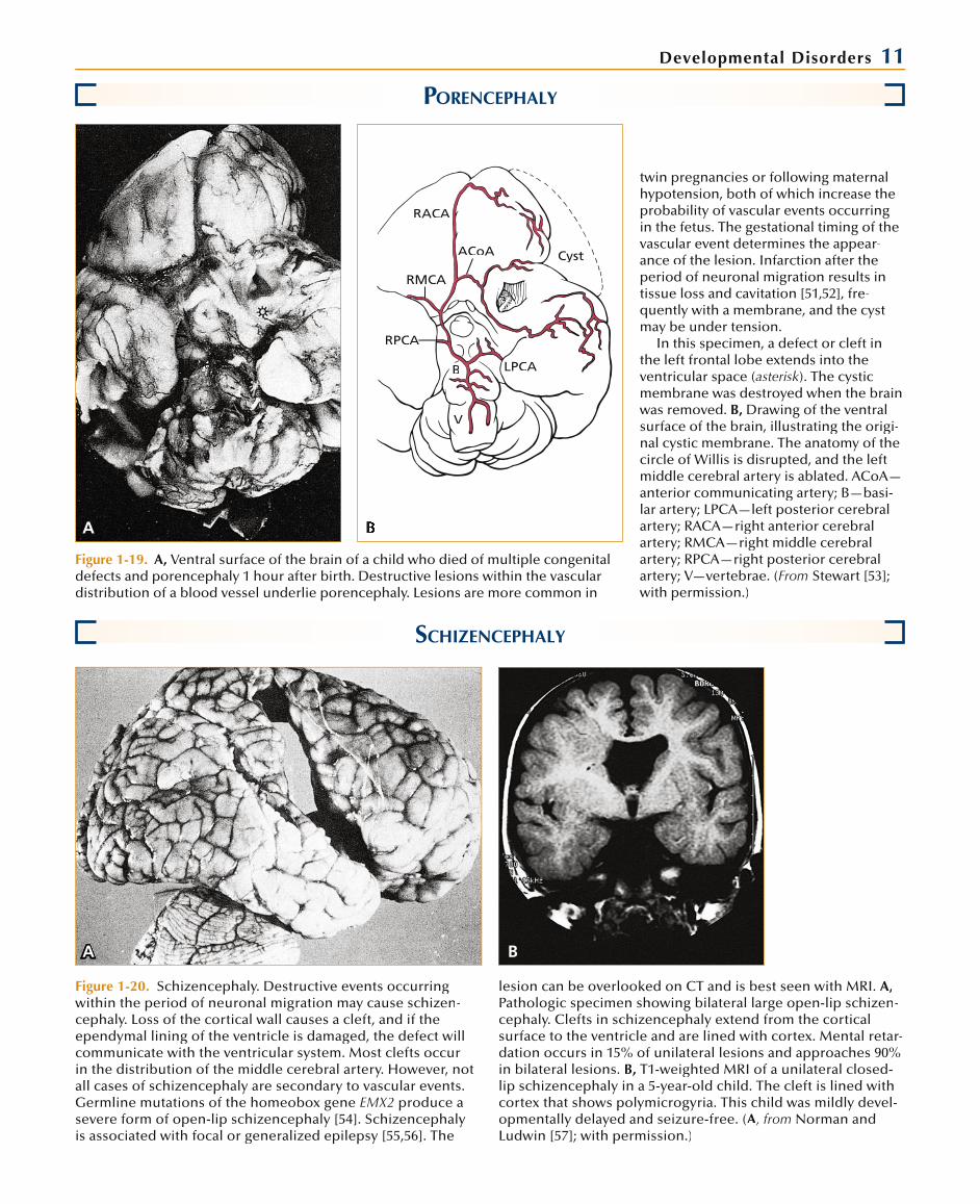
PORENCEPHALY
RACA
RMCA
ACoA
RPCAA
Cyst
B
V
LPCA
A
Figure 1-19. A, Ventral surface of the brain of a child who died of multiple congenitaldefects and porencephaly 1 hour after birth. Destructive lesions within the vascular distribution of a blood vessel underlie porencephaly. Lesions are more common in
SCHIZENCEPHALY
Figure 1-20. Schizencephaly. Destructive events occurring within the period of neuronal migration may cause schizen-cephaly. Loss of the cortical wall causes a cleft, and if the ependymal lining of the ventricle is damaged, the defect willcommunicate with the ventricular system. Most clefts occur in the distribution of the middle cerebral artery. However, not all cases of schizencephaly are secondary to vascular events. Germline mutations of the homeobox gene EMX2 produce a severe form of open-lip schizencephaly [54]. Schizencephaly is associated with focal or generalized epilepsy [55,56]. The
lesion can be overlooked on CT and is best seen with MRI. A,Pathologic specimen showing bilateral large open-lip schizen-cephaly. Clefts in schizencephaly extend from the cortical surface to the ventricle and are lined with cortex. Mental retar-dation occurs in 15% of unilateral lesions and approaches 90% in bilateral lesions. B, T1-weighted MRI of a unilateral closed-lip schizencephaly in a 5-year-old child. The cleft is lined withcortex that shows polymicrogyria. This child was mildly devel-opmentally delayed and seizure-free. (A, from Norman and Ludwin [57]; with permission.)
BA
twin pregnancies or following maternalhypotension, both of which increase theprobability of vascular events occurringin the fetus. The gestational timing of thevascular event determines the appear-ance of the lesion. Infarction after the period of neuronal migration results intissue loss and cavitation [51,52], fre-quently with a membrane, and the cyst may be under tension.
In this specimen, a defect or cleft inthe left frontal lobe extends into theventricular space (asterisk). The cysticmembrane was destroyed when the brainwas removed. B, Drawing of the ventralsurface of the brain, illustrating the origi-nal cystic membrane. The anatomy of thecircle of Willis is disrupted, and the left middle cerebral artery is ablated. ACoA—anterior communicating artery; B—basi-lar artery; LPCA—left posterior cerebralartery; RACA—right anterior cerebralartery; RMCA—right middle cerebralartery; RPCA—right posterior cerebralartery; V—vertebrae. (From Stewart [53];with permission.)

12 Atlas of Clinical Neurology
FOCAL CORTICAL DYSPLASIA
Figure 1-21. Surgical specimen of focal cortical dysplasia. Heterotopia appear betweenthe cortical wall and the pial surface. The defect is easily recognized using high-resolu-tion MRI. These defects occur on a limited portion of the cortical wall, and appear tohave a high association with epilepsy. Extratemporal seizure foci are most commonly observed. The seizures are frequently refractory to antiepileptic drug therapy, and canbe partial motor, partial complex, or secondarily generalized in type. Histologically there is localized disruption of the cortical laminae, and large, bizarre neurons withastrocytosis. (From Layton [58]; with permission.)
HYDRANENCEPHALY
Figure 1-22. -tic disorder in which nearly the entire telencephalon is destroyed, presumably by disrup-tion of bilateral carotid arterial flow. The vertebrobasilar circulation is preserved, which is reflected in the relative preservation of the brainstem and cerebellum, as well as occasion-al portions of the occipital and inferior temporal cortex. This is a disorder of destruction of previously formed tissue, with onset early enough in gestation to produce cavitation but with a minimum of reactive gliosis. The hemispheres are replaced by fluid-filled cavitieslined with a thin membrane that abuts the dura mater [59]. The etiology of the vascular dis-ruption varies and includes infections with cytomegalovirus, syphilis, toxoplasmosis, andinfluenza [60]. Attempted abortions, radiation exposure, and, rarely, embryonal tumors of the hemispheres have also been associated with the disorder [61].
Clinically, the child may appear normal if the hypothalamus is intact, and the headsize and shape may be normal at birth but grow rapidly thereafter. Lethargy and extreme thermoregulatory instability may be observed in those with impaired dience-phalic function. Transillumination of the skull demonstrates the large fluid sacs withsmall overlying vessels. The patients have a poor prognosis, with anticipated survivalof only a few years. They often lack normal diurnal sleep-wake rhythm organization,which complicates their care [62]. Radiographically, the skull and falx appear normallyformed, but the hemispheres are replaced by large cysts. Electrophysiologically, corti-cal evoked potentials are absent and the electroencephalogram is flat, or nearly so.Brainstem responses are generally intact, however. The left arrow indicates the intact thalamus, and the right arrow indicates a segment of the deep venous sinus system.w
WILLIAMS SYNDROME
AAAAAAAAAAAAAAAAAAAAAAAAAAAAAAAAAAA BBBBBB
Figure 1-23. Williams syndrome in an 8-year-old child. A, Williams syndrome is a rela-tively common genetic disorder that causes mild to moderate developmental disabil-ity. It is easily diagnosed because of characteristic facies and a recognizable behavioralphenotype [63]. B, This patient shows epicanthal folds, flattened bridge of the nose,short nose with upturned nares, relatively long philtrum, and prominent lips that are
characteristic of the elfin facial featuresassociated with Williams syndrome. Ocular features include epicanthal folds, medial eyebrow flare, strabismus, andperiorbital fullness. Individuals with light-colored eyes often demonstrate a stellate pattern of the iris.The patient also exem-plifies the behavioral phenotype called“cocktail party chatter,” characterized by inappropriately friendly and loquaciousspeech. This feature often persists into adulthood, with many patients describ-ing themselves as too trusting and easilytaken advantage of. The relative preserva-tion of verbal skills in the face of impairedreasoning and poor visual motor integra-tion often leads to unrealistic expecta-tions and frustration for the family until the diagnosis has been established.
Continued on the next page
Developmental Disorders 13Figure 1-23. (Continued) Medical problems in infancy include irritability, feeding diffi culty, failure to thrive, constipation,and hypercalcemia. The hypercalcemia rarely persists beyondinfancy, but may occasionally require control measures such asadministration of calcitonin or intravenous fl uids and diuret-ics. Cardiovascular anomalies include supravalvular aortic or pulmonic stenosis, but ventricular septal defects and patent ductus arteriosus may also be seen. Hypertension, with or with-out renal anomalies, may be encountered; indeed, some infants with Williams syndrome are diagnosed upon presentation withhypertensive encephalopathy. A history of hyperacusis is oftenelicited on direct questioning of the parents.
Adults with Williams syndrome are often moderately mentally retarded and found in sheltered work environments. They oftenhave health problems related to hypertension, genitourinary anomalies (multiple urinary tract infections and nephrocalci-
nosis), or gastrointestinal problems such as ulcer disease orconstipation. The facial features change from the elfi n appear-ance during early childhood to a relatively coarse visage. Otherproblems sometimes encountered include obesity, diabetes, jointcontractures, and premature graying [64]. Most instances are spo-radic, but there is clear evidence now for autosomal dominantinheritance (the sporadic cases would represent new dominant mutations) [65]. Deletions in the elastin gene on the long arm ofchromosome 7 account for at least part of the observed pheno-type. Clearly, other genes in the region are implicated in someof the other characteristics, such as the developmental profi leand the hypercalcemia; therefore Williams syndrome is best considered a contiguous gene syndrome with many of the otherinvolved genes yet to be isolated. When the diagnosis is suspect-ed, a fl uorescent in situ hybridization study using a DNA probederived from the elastin gene is usually diagnostic [66].
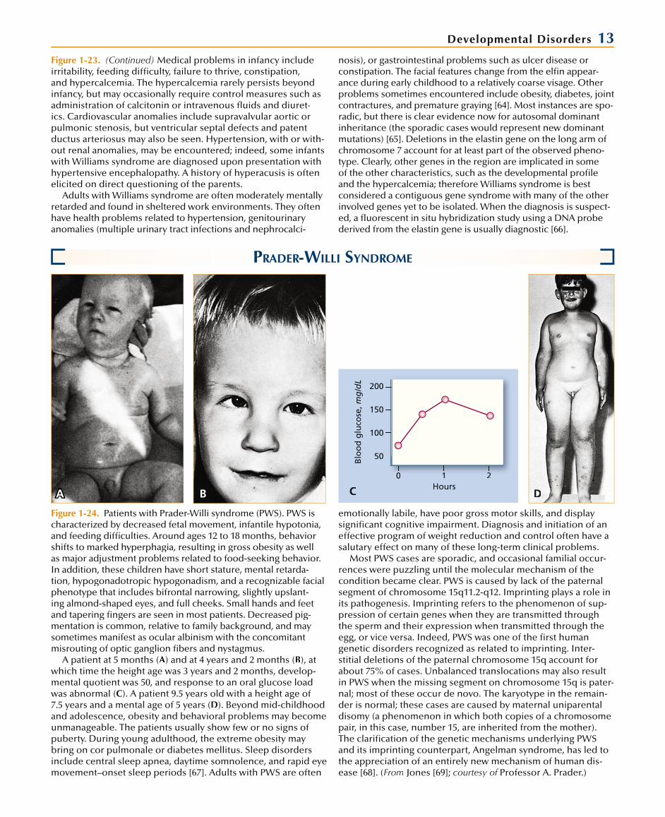
PRADER-WILLI SYNDROME
B C1100
Hours
Blo
od
glu
cose
, mg
/dL
200
150
50
100
22
DDDDDDDDDDDDDDDDDA
Figure 1-24. Patients with Prader-Willi syndrome (PWS). PWS ischaracterized by decreased fetal movement, infantile hypotonia,and feeding diffi culties. Around ages 12 to 18 months, behavior shifts to marked hyperphagia, resulting in gross obesity as well as major adjustment problems related to food-seeking behavior. In addition, these children have short stature, mental retarda-tion, hypogonadotropic hypogonadism, and a recognizable facialphenotype that includes bifrontal narrowing, slightly upslant-ing almond-shaped eyes, and full cheeks. Small hands and feet and tapering fi ngers are seen in most patients. Decreased pig-mentation is common, relative to family background, and maysometimes manifest as ocular albinism with the concomitantmisrouting of optic ganglion fi bers and nystagmus.
A patient at 5 months (A) and at 4 years and 2 months (B), atwhich time the height age was 3 years and 2 months, develop-mental quotient was 50, and response to an oral glucose loadwas abnormal (C). A patient 9.5 years old with a height age of7.5 years and a mental age of 5 years (D). Beyond mid-childhood and adolescence, obesity and behavioral problems may becomeunmanageable. The patients usually show few or no signs ofpuberty. During young adulthood, the extreme obesity maybring on cor pulmonale or diabetes mellitus. Sleep disordersinclude central sleep apnea, daytime somnolence, and rapid eyemovement–onset sleep periods [67]. Adults with PWS are often
emotionally labile, have poor gross motor skills, and display signifi cant cognitive impairment. Diagnosis and initiation of an effective program of weight reduction and control often have a salutary effect on many of these long-term clinical problems.
Most PWS cases are sporadic, and occasional familial occur-rences were puzzling until the molecular mechanism of thecondition became clear. PWS is caused by lack of the paternal segment of chromosome 15q11.2-q12. Imprinting plays a role in its pathogenesis. Imprinting refers to the phenomenon of sup-pression of certain genes when they are transmitted throughthe sperm and their expression when transmitted through the egg, or vice versa. Indeed, PWS was one of the fi rst human genetic disorders recognized as related to imprinting. Inter-stitial deletions of the paternal chromosome 15q account forabout 75% of cases. Unbalanced translocations may also result in PWS when the missing segment on chromosome 15q is pater-nal; most of these occur de novo. The karyotype in the remain-der is normal; these cases are caused by maternal uniparental disomy (a phenomenon in which both copies of a chromosome pair, in this case, number 15, are inherited from the mother). The clarifi cation of the genetic mechanisms underlying PWS and its imprinting counterpart, Angelman syndrome, has led tothe appreciation of an entirely new mechanism of human dis-ease [68]. (From Jones [69]; courtesy of Professor A. Prader.)f

14 Atlas of Clinical Neurology
ANGELMAN SYNDROME
Figure 1-25. An individual with Angelman syndrome (AS). AS is characterized by severe intellectual and motor deficits, seizures,ataxia, and minimal to no speech. The facial features include a prominent jaw (A) and an open-mouthed expression (B)and repeated tongue thrusting (C). There are also jerky, ataxicmovements along with excessive, inappropriate laughter. Otherfeatures include an occipital groove, possibly related to cerebel-lar atrophy, abnormal choroidal pigmentation, and an electro-encephalogram pattern consisting of symmetric, synchronous,high-amplitude spike and wave activity. Many patients have hypopigmentation relative to family members, a feature also noted in patients with Prader-Willi syndrome (PWS) [70].
AS is difficult to diagnose in the first 2 years of life [71], and clinical suspicion is usually delayed until the appearance of the
signs described above. Most cases of AS occur sporadically. Mul-tiple instances of recurrence in siblings have been reported, but inheritance of AS is not typically mendelian. The puzzling pattern of inheritance began to yield to molecular genetic understandingafter the phenomena of uniparental disomy and imprinting were discovered in the 1980s. Deletions of the long arm of chromo-some 15q11.2 have been noted repeatedly in cases of AS. Incontrast to PWS, the origin of the deleted chromosome in AS is maternal. An alternative mechanism (in cases with no cytogenetic or molecular deletion detectable) is paternal uniparental disomy. The critical region involved in both AS and PWS has been workedout. One of the candidate genes is a ubiquitin protein ligase; there may also be additional genes [72]. (From Williams and Frias [73]; with permission.)
A B C
ARACHNOID CYSTS
Figure 1-26. An unenhanced CT of an arachnoid cyst of characteristic shape and masseffect, located in the temporal fossa and extending into the sylvian fissure. Arachnoidcysts are congenital lesions that occur intracranially or within the spinal canal. They are cavities filled with cerebrospinal fluid (CSF) and lined by arachnoidal cells and col-lagen fibers. They comprise about 1% of all space-occupying intracranial masses. Mostarachnoid cysts are clinically asymptomatic and are discovered incidentally. Infre-quently, they may come to attention because of mass effect; symptoms are relatedeither to local pressure or to hydrocephalus from compression along the CSF pathway. Macrocephaly is especially common in children with cysts who are less than 2 years of age; these patients are also more likely to require shunts for treatment [74]. Arach-noid cysts occur in boys slightly more often than in girls. Patients usually have solitaryarachnoid cysts. Familial occurrence is rare. About two thirds of the cysts occur in the supratentorial space. The middle cranial fossa is the most common location for theselesions, but they may also occur over the suprasellar region, within the ventricles, between the cerebral hemispheres, and over the cerebral convexities [75].
Most lesions are benign and asymptomatic. Infrequently, there may be headache,macrocephaly, nausea, vomiting, and papilledema. A small group of patients presentwith spontaneous hemorrhage into the cyst. Consequently a preexisting arachnoidcyst should be considered in patients who develop subdural hematomas after minimaltrauma. Spinal arachnoid cysts may lead to a myelopathy from spinal cord compression.
MRI is the imaging modality of choice. The cysts appear as circumscribed, T2-intense lesions without enhancement. Their shape depends on their location: middlefossa cysts are frequently trapezoidal and splay the sylvian fissure; suprasellar cystscan bulge into the third ventricle, infrequently obstructing both foramina of Monro,giving the appearance of “Mickey Mouse ears.”
Most arachnoid cysts are found incidentally and do not require treatment. When associated with mass effect, they may be decompressed by either a cystoperitoneal shunt or fenestration of the cyst wall. A shunt is a simpler procedure but carries with itthe risks of indwelling hardware and shunt failure. Fenestration avoids these problemsbut requires a craniotomy and is not uniformly successful [76].
Developmental Disorders 15
DERMAL SINUS TRACTS, DERMOIDS, AND EPIDERMOIDS
A B
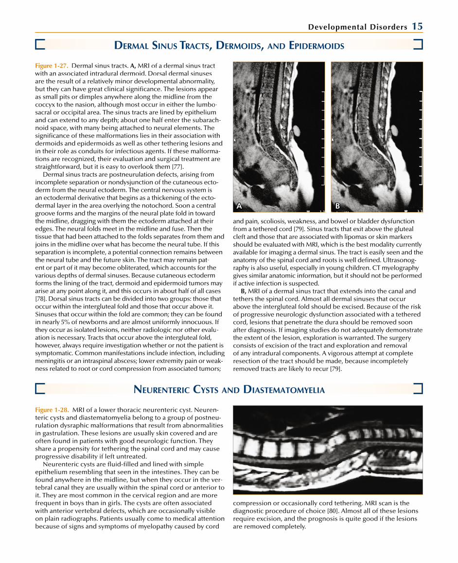
Figure 1-27. Dermal sinus tracts. A, MRI of a dermal sinus tract with an associated intradural dermoid. Dorsal dermal sinuses are the result of a relatively minor developmental abnormality, but they can have great clinical significance. The lesions appearas small pits or dimples anywhere along the midline from thecoccyx to the nasion, although most occur in either the lumbo-sacral or occipital area. The sinus tracts are lined by epitheliumand can extend to any depth; about one half enter the subarach-noid space, with many being attached to neural elements. Thesignificance of these malformations lies in their association with dermoids and epidermoids as well as other tethering lesions and in their role as conduits for infectious agents. If these malforma-tions are recognized, their evaluation and surgical treatment are straightforward, but it is easy to overlook them [77].
Dermal sinus tracts are postneurulation defects, arising from incomplete separation or nondysjunction of the cutaneous ecto-derm from the neural ectoderm. The central nervous system isan ectodermal derivative that begins as a thickening of the ecto-dermal layer in the area overlying the notochord. Soon a central groove forms and the margins of the neural plate fold in toward the midline, dragging with them the ectoderm attached at their edges. The neural folds meet in the midline and fuse. Then thetissue that had been attached to the folds separates from them and joins in the midline over what has become the neural tube. If this separation is incomplete, a potential connection remains between the neural tube and the future skin. The tract may remain pat-ent or part of it may become obliterated, which accounts for the various depths of dermal sinuses. Because cutaneous ectodermforms the lining of the tract, dermoid and epidermoid tumors may arise at any point along it, and this occurs in about half of all cases[78]. Dorsal sinus tracts can be divided into two groups: those that occur within the intergluteal fold and those that occur above it. Sinuses that occur within the fold are common; they can be foundin nearly 5% of newborns and are almost uniformly innocuous. If they occur as isolated lesions, neither radiologic nor other evalu-ation is necessary. Tracts that occur above the intergluteal fold,however, always require investigation whether or not the patient is symptomatic. Common manifestations include infection, includingmeningitis or an intraspinal abscess; lower extremity pain or weak-ness related to root or cord compression from associated tumors;
NEURENTERIC CYSTS AND DIASTEMATOMYELIA
Figure 1-28. MRI of a lower thoracic neurenteric cyst. Neuren-teric cysts and diastematomyelia belong to a group of postneu-rulation dysraphic malformations that result from abnormalities in gastrulation. These lesions are usually skin covered and are often found in patients with good neurologic function. They share a propensity for tethering the spinal cord and may causeprogressive disability if left untreated.
Neurenteric cysts are fluid-filled and lined with simple epithelium resembling that seen in the intestines. They can befound anywhere in the midline, but when they occur in the ver-tebral canal they are usually within the spinal cord or anterior toit. They are most common in the cervical region and are morefrequent in boys than in girls. The cysts are often associated with anterior vertebral defects, which are occasionally visible on plain radiographs. Patients usually come to medical attention because of signs and symptoms of myelopathy caused by cord
and pain, scoliosis, weakness, and bowel or bladder dysfunction from a tethered cord [79]. Sinus tracts that exit above the glutealcleft and those that are associated with lipomas or skin markers should be evaluated with MRI, which is the best modality currently available for imaging a dermal sinus. The tract is easily seen and the anatomy of the spinal cord and roots is well defined. Ultrasonog-raphy is also useful, especially in young children. CT myelography gives similar anatomic information, but it should not be performed if active infection is suspected.
B, MRI of a dermal sinus tract that extends into the canal and tethers the spinal cord. Almost all dermal sinuses that occur above the intergluteal fold should be excised. Because of the risk of progressive neurologic dysfunction associated with a tethered cord, lesions that penetrate the dura should be removed soon after diagnosis. If imaging studies do not adequately demonstratethe extent of the lesion, exploration is warranted. The surgery consists of excision of the tract and exploration and removalof any intradural components. A vigorous attempt at completeresection of the tract should be made, because incompletely removed tracts are likely to recur [79].
compression or occasionally cord tethering. MRI scan is the diagnostic procedure of choice [80]. Almost all of these lesions require excision, and the prognosis is quite good if the lesions are removed completely.

16 Atlas of Clinical Neurology
Figure 1-29. MRI of diastematomyelia with a bony septum. Diastematomyeliadescribes a malformation characterized by duplication of the spinal cord with varieddural and bony anatomy. Some patients have two hemicords separated by fi brous sep-tae that share the same dural tube. Other patients have two dural tubes with bone orcartilage between. These lesions occur most frequently in the lumbar spine and oftenhave cutaneous markers; a hair-bearing patch is most common. Patients are generallyneurologically normal at birth, and impairment usually progresses slowly, although several reports of acute decompensation associated with excessive or forced fl exionhave appeared. Pain and weakness are the most common complaints, but bowel orbladder dysfunction may be the only symptoms of a tethered cord [81]. An MRI is often suffi cient to defi ne the lesion, but bony or cartilaginous septae can be over-looked, and for that reason CT myelography is valuable. Repair is complicated because both cords are usually functional, but owing to the risk of neurologic deteriorationassociated with tethering lesions, surgery is recommended in all patients with diaste-matomyelia [81]. (From Dias and Pang [81]; with permission.)
Cranial
Cauda
l
Prochordal plateAbnormal adhesion betweenectoderm and endoderm
AdhesionEctoderm
Endoderm
Developing notochordalprocess
Cloacalmembrane
Primitive pit
a
a
A
Figure 1-30. Split notochord theory. Although a common defect in embryogenesis is thought to cause both neurenteric cysts and diastematomyelia, the details of the embryopathy are not known.It seems most likely that in these lesions duplication of part ofthe notochord leads to duplication of various parts of the spinalcord and its surrounding structures. This would potentially allowdirect communication between the endoderm and the ectodermin the area between the split notochords. Notochordal abnor-
mality could have several causes. A, An adhesion between the ectoderm and endoderm of the two-layered embryonic diskmay occur, cranial to the developing notochordal process (cross-sectional view a). B, The notochordal process is forced to dividearound the adhesion, creating two notochordal processes. Twoneural plates may be induced (cross-sectional view b), which ultimately could form an area of diastematomyelia.
Continued on the next page
AdhesionTwo neural plates
Two notochordalprocesses
Notochordal processbifid around adhesion
b
b
B
Developmental Disorders 17
Skin
Gut
Dual notochord anddiastematomyelia
Combined spina bifida C
Skin
Gut
Diastematomyeliawith septumD
Neurenteric cyst
Gut
Prevertebral cyst
Cleft vertebrae
Anterior spinal mass
Skin
E
Figure 1-30. (Continued) C, An enterocutaneous fi stula may penetrate through a vertebral body cleft, diastematomyelia, andposterior spina bifi da to create a combined anterior-posterior spina bifi da. The theories invoking an abnormality of the neur-enteric canal are attractive.
D, Healing of the anterior portions of the fi stula could lead to diastematomyelia with septum, which is usually accompanied byposterior element and vertebral body abnormalities. E, Healingof the posterior and middle portions of the fi stula could lead toneurenteric cyst, vertebral body anomalies, and various entericmalformations. (Adapted from Beardmore and Wiggelsworth [82].)
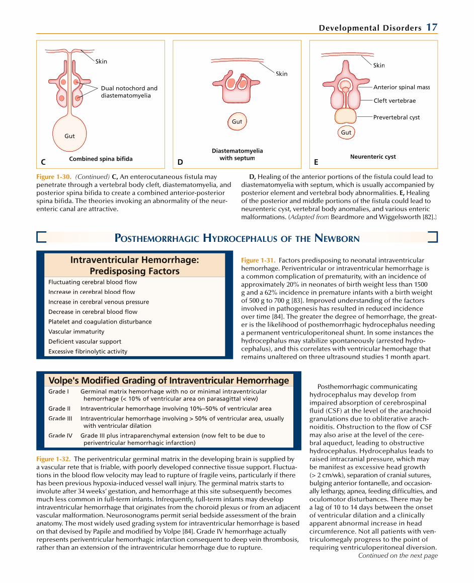
POSTHEMORRHAGIC HYDROCEPHALUS OF THE NEWBORN
Figure 1-31. Factors predisposing to neonatal intraventricular hemorrhage. Periventricular or intraventricular hemorrhage isa common complication of prematurity, with an incidence of approximately 20% in neonates of birth weight less than 1500g and a 62% incidence in premature infants with a birth weightof 500 g to 700 g [83]. Improved understanding of the factors involved in pathogenesis has resulted in reduced incidence over time [84]. The greater the degree of hemorrhage, the great-er is the likelihood of posthemorrhagic hydrocephalus needinga permanent ventriculoperitoneal shunt. In some instances the hydrocephalus may stabilize spontaneously (arrested hydro-cephalus), and this correlates with ventricular hemorhage thatremains unaltered on three ultrasound studies 1 month apart.
Intraventricular Hemorrhage: Predisposing Factors
Fluctuating cerebral blood fl ow
Increase in cerebral blood fl ow
Increase in cerebral venous pressure
Decrease in cerebral blood fl ow
Platelet and coagulation disturbance
Vascular immaturity
Defi cient vascular support
Excessive fi brinolytic activity
Volpe's Modifi ed Grading of Intraventricular HemorrhageGrade I Germinal matrix hemorrhage with no or minimal intraventricular
hemorrhage (< 10% of ventricular area on parasagittal view)
Grade II Intraventricular hemorrhage involving 10%–50% of ventricular area
Grade III Intraventricular hemorrhage involving > 50% of ventricular area, usually with ventricular dilation
Grade IV Grade III plus intraparenchymal extension (now felt to be due to periventricular hemorrhagic infarction)
Figure 1-32. The periventricular germinal matrix in the developing brain is supplied by a vascular rete that is friable, with poorly developed connective tissue support. Fluctua-tions in the blood fl ow velocity may lead to rupture of fragile veins, particularly if therehas been previous hypoxia-induced vessel wall injury. The germinal matrix starts toinvolute after 34 weeks’ gestation, and hemorrhage at this site subsequently becomesmuch less common in full-term infants. Infrequently, full-term infants may developintraventricular hemorrhage that originates from the choroid plexus or from an adjacentvascular malformation. Neurosonograms permit serial bedside assessment of the brainanatomy. The most widely used grading system for intraventricular hemorrhage is based on that devised by Papile and modifi ed by Volpe [84]. Grade IV hemorrhage actually represents periventricular hemorrhagic infarction consequent to deep vein thrombosis,rather than an extension of the intraventricular hemorrhage due to rupture.
Posthemorrhagic communicating hydrocephalus may develop fromimpaired absorption of cerebrospinalfl uid (CSF) at the level of the arachnoidgranulations due to obliterative arach-noiditis. Obstruction to the fl ow of CSF may also arise at the level of the cere-bral aqueduct, leading to obstructive hydrocephalus. Hydrocephalus leads to raised intracranial pressure, which maybe manifest as excessive head growth (> 2 cm/wk), separation of cranial sutures, bulging anterior fontanelle, and occasion-ally lethargy, apnea, feeding diffi culties, andoculomotor disturbances. There may bea lag of 10 to 14 days between the onsetof ventricular dilation and a clinicallyapparent abnormal increase in headcircumference. Not all patients with ven-triculomegaly progress to the point ofrequiring ventriculoperitoneal diversion.
Continued on the next page
18 Atlas of Clinical Neurology
Figure 1-32. (Continued) Pharmacologic measures to reduce CSF production (acetazolamide and furosemide) may be helpful in con-trolling hydrocephalus [85] but frequently cause systemic metabolicderangements. Repeated lumbar punctures have also been used as an interim measure with variable results. A large multicenter European study failed to show its benefit in either arresting hydro-cephalus and thus avoiding shunting, or on long-term neurode-velopmental outcome [86]. Placement of a ventriculoperitonealor ventriculoatrial shunt is fraught with complications (high rates ofinfection and shunt blockage) when performed early. The tempo-rary use of a subcutaneous ventricular catheter reservoir [87,88] is
safe and effective until the patient’s weight reaches 2 kg and CSF protein falls below 200 mg/dL (factors associated with better shuntsuccess rate). Ventriculoscopic methods of manipulating the clot and instillation of fibrinolytic agents into the ventricle [89] showpromise but require further study before their routine use can bejustified. The outcome in patients with intraventricular hemorrhage varies based on the severity of the hemorrhage. The incidence of major neurologic sequelae is 5% to 15% following low-grade hemorrhage, 35% in grade III hemorrhage, and 90% in those with an intraparenchymal component (100% in those with an extensiveintraparenchymal component) [84]. (Adapted from(( Volpe [84].)
A B
Figure 1-33. Coronal cranial ultrasound images from a17-day-old premature baby born at 28 weeks’ gesta-tion who had a grade III intraventricular hemorrhage.A and B, Dilation of frontal and temporal horns of the lateral ventricles and of the third ventricle withresidual blood clots (arrows). This patient was treated with serial lumbar punctures to remove cerebrospi-nal fluid (CSF) until his weight reached 2 kg and CSF protein fell to less than 1 g/dL, at which time a ven-triculoperitoneal shunt was placed. Shunt revisionwas necessary at 17 months. At 30 months of age thepatient showed mild global developmental delay.
DANDY-WALKER MALFORMATION
Syndromes Associated WithDandy-Walker Malformation
Aase-Smith Jaeken
Aicardi Joubert
Cerebro-oculo-muscular Meckel-Gruber
Coffin-Siris Oral-facial-digital types II and III
Cornelia de Lange Ruvalcaba-Myhre-Smith
Dekaban Smith-Lemli-Opitz
Ellis–van Creveld Warburg
Fraser/cryptophthalmos
Goldenhar
Figure 1-34. Syndromes associated with Dandy-Walker malfor-mation (DWM). DWM is a developmental disorder characterizedby a cystic enlargement of the fourth ventricle, partial or com-plete agenesis of the cerebellar vermis, hydrocephalus, andenlargement of the posterior fossa. Several conditions mas-querade as DWM (mega cisterna magna, arachnoid cyst, and other variants marked by cerebellar vermis agenesis) but can beexcluded based on the preceding criteria. DWM accounts forapproximately 3% of cases of hydrocephalus and is estimated to occur in 1 per 25,000 to 30,000 births [90]. Although a variablefemale preponderance has been noted in several reports, it isnot statistically significant [91]. Most cases are sporadic. Thosewith cerebellar vermis agenesis and extraneural anomalies mayrepresent specific syndromes with known inheritance patterns, making their distinction important.
Central Nervous System Anomalies Associated With
Dandy-Walker MalformationAnomalies of cerebellar folia
Cerebral and cerebellar hamartomas
Corpus callosum agenesis
Malformation of olivary and dentate nuclei
Microgyria/polymicrogyria
Nonspecific gyral disturbances
Occipital encephalocele
Pachygyria
Uncrossed pyramidal tracts
Figure 1-35. Central nervous system anomalies associated withDandy-Walker malformation (DWM). With advances in prenatal and neonatal ultrasonography, DWM is increasingly being diag-nosed at birth. The diagnosis is made by 1 year of age in approxi-mately 80% of cases [90], the most common presenting symptombeing macrocephaly with a prominent occiput. Hypotonia andsigns of hydrocephalus may be present. Associated brain anoma-lies are present in 68% of cases [91]. Anomalous developmentof other organ systems (cardiac, genitourinary, skeletal, ocular, facial) frequently coexists.
The etiopathogenesis has been debated widely. DWM is regard-ed as a developmental disorder of the midline central nervoussystem with marked genetic and etiologic heterogeneity [92]. A localized defect in differentiation of the roof of the newly closedneural tube in the hindbrain [93] results in absence of the foramenof Magendie, a consistent finding during surgical intervention [90].
Continued on the next page

Developmental Disorders 19Figure 1-35. (Continued) Lack of cerebrospinal fluid outflow pro-duces cystic enlargement of the fourth ventricle. Hirsch et al. [90]have estimated that the development of DWM occurs in the 3rdpostovulatory week. This would explain the associated cardiovascu-lar and facial anomalies. Hart et al. [91] reviewed pathologic materialand believe that the disturbance occurs by the end of month 2.
The management of DWM has changed and is still contro-versial. Initially, excision of the cyst membrane was proposed,
but this showed a high failure rate. Currently favored optionsinclude shunting the ventricular system, the cyst, or both. Theoutcome depends on the presence of coexistent neural and extraneural malformations, in addition to the timing and effec-tiveness of the surgical intervention. The percentage of patientswith DWM who possess an intelligence quotient of 80 or abovevaries from 29% to 67% [90].
A B
Figure 1-36. T1-weighted MRIs in the midline sagittal (A) and transaxial sec-tion at the level of the third ventricle (B) of a 7-day-old infant with Dandy-Walkermalformation. The sagittal view shows thetypical cystic enlargement of the fourth ventricle (large arrow), with anterior dis-placement of the brainstem (small arrow),aplasia of the cerebellar vermis, and elevation of the tentorium (curved arrow), as well as the confluence of sinuses. Thetransaxial view shows the fourth ven-tricular cyst (white arrow) with cerebellar aplasia and dilation of the temporal horns of the lateral ventricles (arrowheads) and third ventricle (curved arrow). This patientwas treated with a diversion procedure in which both the fourth ventricular cyst
CHIARI MALFORMATIONS
AAAAAAAAAAAAAAAAAAAAAAA B
Figure 1-37. Type II Chiari malformations. A, MRI of a type II Chiari malformation, the lesion most commonly associated with a myelomeningocele. B, Pathologic specimen of the Chiari typeII lesion. The extent of the cerebellar displacement is easilyseen. The medulla is elongated and kinked and the fourth ven-tricle can be seen to extend well into the cervical spine.
In patients with type I Chiari malformation, the cerebellar ton-sils extend below the plane of the foramen magnum, but the posi-tion of the brainstem and midline cerebellar structures remains normal. Syringomyelia develops occasionally in these patients [94]. A majority of type I Chiari malformations are clinically asymp-tomatic. When symptoms do develop, they are often in adults, in the form of headaches, neck pain, upper extremity weakness, and unsteady gait. Only rarely do children have symptomatic lesions;those who do often present with scoliosis, a complication of themalformation that rarely occurs in adults [95]. Diagnosis is mademost easily with MRI. Treatment is surgical decompression of the lesion. The best way to treat patients with associated syringomy-elia is still uncertain. Some favor decompression alone, whereas
others recommend more extensive procedures [96]. Interestingly, iatrogenic symptomatic type I Chiari malformations have been reported as a result of lumboperitoneal shunts [97].
In type II Chiari malformations, both the cerebellar vermis and the tonsils are displaced into the cervical canal. In addition, thebrainstem and fourth ventricle are elongated and displaced. These lesions appear almost exclusively in children with myelomenin-goceles, in whom they are often associated with a constellation of other anomalies, most notably hydrocephalus, cortical hetero-topia, and a small posterior fossa. Chiari malformations may be symptomatic at birth, with stridor, respiratory abnormalities, and dysphagia. The problems usually resolve with placement of a ven-triculoperitoneal shunt. Most patients with type II Chiari malforma-tions become symptomatic in infancy or early childhood. Manyhave upper extremity pain, incoordination, or weakness. Lower cranial neuropathies are also common, as is headache. Diagno-sis is made by MRI and the treatment is surgical decompression.Because the site of compression generally lies within the cervical canal, the bony decompression is performed primarily at that level.
Continued on the next page
and the hydrocephalus were decompressed using a single catheter. Postoperatively, the patient is stable, but development is significantly delayed.

20 Atlas of Clinical Neurology
Figure 1-37. (Continued) Shunt malfunction may mimicsymptomatic Chiari malformation and should be excluded prior to surgery.
The pathogenesis of Chiari malformation is uncertain. The type I anomaly may be acquired rather than congenital. In those born with the lesion, the developmental abnormality most likely occurs late in embryogenesis, because the cerebellar hemi-
sphere and the deep nuclei are normal. The pathogenic mecha-nism of the type II malformation probably operates earlier indevelopment. Current theories focus on mechanical or hydro-dynamic effects of the accompanying myelomeningocele orprimary dysgenetic mechanisms as the cause of the lesion [98].Research is ongoing, and mouse models of abnormal neurula-tion may improve understanding of this issue.
CHOROID PLEXUS PAPILLOMA
Figure 1-38. Choroid plexus neoplasms are the only disorderassociated with hydrocephalus as a result of cerebrospinal fluid (CSF) overproduction. They account for up to 6% of childhood intracranial neoplasms. They can present at any age, 40% beingdiagnosed in children less than 15 years old. Most childhood series have noted a tendency in children under 2 years. Cases presenting in adults tend to favor the fourth ventricular loca-tion, whereas in the pediatric population 80% are located in the lateral ventricle, 16% in the fourth ventricle, and 4% in thethird ventricle. The most common presenting symptoms are increased intracranial pressure (irritability, lethargy, vomiting), intellectual dysfunction, and seizures. Examination reveals anenlarged head size, and a CT or MRI may show an enhancingmass within the enlarged ventricular system. Uniform enhance-ment of a lobulated mass that does not infiltrate the paren-chyma favors diagnosis of choroid plexus papilloma (CPP), whereas a variegated appearance with local invasion suggestsmalignancy (carcinoma). MR spectroscopy may be useful indistinguishing choroid plexus papilloma from carcinoma, with the former showing increased peaks of myoinositol, and the latter increased peaks of choline [99]. The cytologic features of
Microscopic Features of MalignantChoroid Plexus Tumors
Invasion
Loss of differentiation
Nuclear pleomorphism
Increased mitoses
Necrosis
CPP are similar to those of normal choroid plexus. The papillaeare usually composed of a single layer of columnar or cuboidalepithelium supported by a stroma of vascularized connective tissue [100]. Although histologic examination distinguishes papilloma from carcinoma [101], not all papillomas behave in abenign fashion. Ploidy analysis seems to allow greater predic-tion of neoplastic behavior, because aneuploid choroid plexus neoplasms (whether papilloma or carcinoma) have a signifi-cantly increased mortality rate (67%) compared with diploidpapillomas (9%) [102]. In addition to choroid plexus carcinoma, the differential diagnosis of CPP includes medulloepithelioma, teratoma belonging to the embryonal carcinoma or endodermal sinus tumor group, metastatic adenocarcinoma from elsewhere in the body, and villous hypertrophy of the choroid plexus [100].
The treatment typically consists of alleviation of hydrocephaluswith either external temporary or internal permanent CSF shunt-ing, followed by removal of the tumor. Not all patients requirepermanent ventricular diversion once the tumor has been removed completely [103]. Removal of these highly vascularizedtumors is frequently complicated by bleeding. The optimal meth-od of tumor removal entails reaching the arterial feeder(s) first, although this is not always possible [103]. Carcinomas require additional therapy. Complete resection of the carcinoma may be curative but is not always possible. Irradiation of the neuraxis is recommended owing to the high rate of leptomeningeal metasta-ses. Unfortunately, irradiation in the developing infant may cause intellectual deterioration, endocrinopathies, and spinal growthfailure. Postoperative chemotherapy, using vincristine, cyclo-phosphamide, cisplatinum, and etoposide, has shown promise instabilizing the disease and delaying the need for irradiation [104]. (From Ho et al. [101]; with permission.)
A B
Figure 1-39. CTs from a 10-month-oldinfant who presented with enlarging head size and symptoms of increasing intracra-nial pressure. The noncontrast scan (A) shows a mass in the left lateral ventricle (large arrow) causing localized enlarge-ment of the left lateral ventricle (small arrow) and diffuse hydrocephalus. Follow-ing contrast infusion (B), the typical lobu-lated appearance becomes more obvious,and uniform enhancement (arrow) sug-gests choroid plexus papilloma, which in this case was proven histologically following removal of the tumor.

Developmental Disorders 21
KLIPPEL-FEIL ANOMALY
Classification of Klippel-Feil AnomalyType I Complete fusion of cervical vertebrae into blocks;
may include upper thoracic vertebrae
Type II Involvement at a few cervical levels with hemiver-tebrae and atlanto-occipital fusion
Type III Cervical fusions plus lower thoracic or lumbar fusions
Figure 1-40. The Klippel-Feil anomaly consists of congenital fusion of the cervical vertebrae. It occurs between day 21 and 37of gestation through failure of segmentation of adjacent sclero-tomes in the fetus [105]. The genetic basis of the defect is not yet known, but the processes of appropriate segmentation andsomite differentiation appear to be under the control of a group of paired homeobox genes that have been conserved throughthe genomes of many species and are normally expressed in a specific anteroposterior sequence. At present no specific Hoxdefects have been detected in patients with the Klippel-Feilsyndrome, but it is likely that such a relationship eventually willbe found [106,107]. The anatomic feature that defines Klippel-Feil
syndrome is fusion of vertebral bodies, laminae, pedicles, and posterior spinous processes. The classification system devel-oped by Lemire et al. [108] divides the syndrome into three types.Type I anomalies include those with massive fusion of multiplecervical and high thoracic vertebrae into solid blocks of bone. Despite the fusion, intervertebral foramina remain patent andthe brachial plexus formation is preserved. Clinically, the head is slightly retroflexed or tilted, and appears to rest on the shoulders.In type II Klippel-Feil anomaly, only one or two cervical vertebrae are fused, although hemivertebrae and fusion of the atlas andoccipital bone often occur. Patients with this most common form of the anomaly have less restriction of motion but may developcomplications such as scoliosis, spinal stenosis, and subluxation.They may exhibit other musculoskeletal anomalies such as absentribs, Sprengel’s deformity (congenital raised scapula), kyphosis, torticollis, microtia, or conductive hearing deficits. Diplomyelia,hydromyelia, and malformations of the urinary tract and other organs have been also reported [109,110]. The type III anomaly is characterized by fusion of the thoracic or lumbar spine with associated type I or type II anomalies in the cervical region.
Figure 1-41. A 15-year-old patient with type II Klippel-Feil anomaly who has a short neck, low hairline, and conductive deafness (A). The patient’s neck mobility was impaired, especially lateral rota-tion (B). These clinical findings are characteristic of the Klippel-Feil anomaly. Neurologic findings include radicular pain and paresthe-sias, particularly about the roots that are not involved in fusion,where open disk spaces allow excessive compensatory mobilityand eventual osteoarthritic spurring. Vertebral artery compres-sion may develop in older patients via a similar mechanism and
cause posterior circulation symptoms [111,112]. Cervical instability can precipitate symptoms and signs of cord compression, includ-ing transient quadriparesis, spastic gait, reflex asymmetry, and Lhermitte’s paresthesia. The association of synkinesia or mirrormovements with the Klippel-Feil syndrome is ascribed to insuf-ficient decussation of the pyramidal tracts. Special considerationand precautions should be taken to prevent traction on the neckduring anesthesia and intubation in Klippel-Feil patients. Participa-tion in gymnastic activities such as tumbling should be avoided.
BBBBBBBBBBBBBBBBBBBBBBBBBBBBBBBBBBBBBBBBBBBBBBBBBBBBBBBBBBBBBBBBBBBBBBBAAAAAAAAAAAAAAAAAAAAAAAAAAAAAAAAAAAAAAAAAAAAAAAAAAAAAAAAAAAA

22 Atlas of Clinical Neurology
NEUROFIBROMATOSIS
Two or More Criteria Necessary for Diagnosis of Neurofibromatosis Type 1
Five or more café-au-lait macules > 5 mm in diameter in a prepubertal child or >15 mm in a postpubertal child
Two or more cutaneous neurofibromas or one plexiform neurofibroma
Axillary or inguinal freckles
Two or more iris (Lisch) nodules
Osseous lesions—sphenoid wing hypoplasia, thinning of long bone cortex, or pseudoarthrosis
Bilateral optic pathway glioma
First-degree relative with neurofibromatosis type 1
Figure 1-42. Neurofibromatosis type 1 (NF1) is the most com-mon neurocutaneous syndrome, with an incidence of about 1 in 3000 [113]. Although the nervous system, bone, and skinare most often affected, other organ systems might also beinvolved. The diagnosis is based on clinical findings, generally when two or more of the classic features of NF1 are present.
Café-au-lait spots are often present at birth. They increase in pigmentation, size, and number with age. Five or more café-au-lait spots (of > 5 mm diameter in prepubertal children and >15 mm diameter after puberty) is one of the diagnostic criteria. Axillary or inguinal freckles, which a complexion identical tocafé-au-lait spots, are also an important manifestation. Sessile,pedunculated, or flat cutaneous neurofibromas may range insize from several millimeters to centimeters. Less often seen are hypopigmented macules. Iris hamartomas (Lisch nodules)evolve progressively with age, their prevalence rising from10% in the first decade to 100% by the sixth decade. These iris hamartomas are yellowish brown in color and usually bilateral.Skull abnormalities include absence of the sphenoid wing(harlequin appearance on plain radiographs), enlarged sella tur-cica, and middle cranial fossa arachnoid cysts. Spine anomalies include scoliosis, enlarged vertebral foramina, and a widenedspinal canal [114]. Most often spinal cord compression results from adjacent neurofibromas (dumbbell lesions) or vertebralcompression. Intrinsic spinal cord tumors are relatively benignastrocytomas [115]. The optic chiasm and optic radiations mayalso be affected by hamartomas. The cerebral astrocytomas are pilocytic, usually benign with rare grade IV tumors, andare located in the hypothalamus, brainstem, and cerebellum[116]. Ependymomas and medulloblastoma are somewhat more common. Ventricular dilation is usually asymptomatic, althoughacute hydrocephalus associated with aqueductal stenosis hasbeen reported. Some patients have megalencephaly. Fibromus-cular dysplasia of carotid vessels may be associated with acute and progressive cerebrovascular accidents. Moyamoya syn-drome may be present in rare instances. Short stature occurred in 27% of North’s series [117]. Neurofibromas can affect nervesat any level—small distal radicles, larger nerves, trunks, andplexuses. Plexiform neurofibromas can be massive and aremore prone to undergo malignant transformation to neurofi-brosarcomas. Orbital and periorbital plexiform neurofibromasmay be particularly deforming. Pheochromocytoma occurs in
less than 1% of NF1 patients. Hypertension may be related topheochromocytoma or secondary to renal artery stenosis from fibromuscular dysplasia. Rhabdomyosarcoma may evolve from preexisting plexiform neurofibromas. Gastrointestinal involve-ment was estimated at 10% by Hochberg et al. [118] and includ-ed neurofibromas, leiomyomas, and sarcomas predominantlyaffecting the serosa and extending from stomach to colon.
The diagnosis is made by clinical examination. Molecularanalysis provides confirmation in questionable cases, defines sporadic and familial forms, and allows prenatal screening ifdesired. Clinical monitoring and spine MRI studies are needed for monitoring insidious progression in spinal cord lesions. Cra-nial MRI studies help in evaluating megalencephaly, and also in defining asymptomatic lesions that warrant close clinical assess-ment, such as optic pathway tumors. Conventional radiographs are warranted for skeletal and skull lesions. Serial MRI scans of the plexiform lesions help define the size, complexity, and pro-gression. Visual and auditory evoked responses help in defining the significance of neuroimaging abnormalities and in detectingfunctional impairment. Genetics consultation may help deter-mine the mode of inheritance (dominant with variable expres-sivity or spontaneous mutation). Parents should be aware that cutaneous neurofibromas can enlarge in adolescence, becomepainful, and require removal. Ultimately, therapy will need tobe aimed at somehow minimizing the effects of neurofibro-min loss. Psychometric testing is helpful in determining theextent of cognitive impairment, central processing dysfunction,and behavioral problems that might affect learning and social adjustment. Families should be referred to the National Neuro-fibromatosis Foundation for information about the disease andavailable resources [119].
Genetically, this autosomal dominant disorder exhibits com-plete penetrance and variable expressivity and has an approxi-mate 50% mutation rate. An NF1 gene microdeletion is the mostcommon mutation, and correlates with younger age of onsetand a substantially larger risk of developing neurofibromas over the lifetime. The NF1 gene resides on chromosome 17q11.2 [120]. This defective gene, a truncated polypeptide, can be usedin the molecular detection of approximately 70% of mutationsresponsible for NF1. The gene spans 350 kb of genomic DNA, encodes an mRNA of 11 kb to 13 kb, and is organized into 50exons. The gene product, neurofibromin, is ubiquitous, with itsgreatest levels occurring in the brain. One role of neurofibro-min appears to be inactivation of ras, a protooncogene protein.Guttman and Collins [121] postulated that a mutational lossof neurofibromin could result in a loss of down-regulation ofras, leading to cell proliferation and transformation. A portion of neurofibromin accelerates the hydrolysis of ras-guanidine triphosphate (GTP) to ras-guanidine diphosphate (GDP) andconverts ras from the inactive to active forms. Further proofwas provided by increased ras-GTP levels with reduced neuro-fibromin levels in neurofibrosarcoma cell lines [121]. Mutationsaffecting mRNA splicing appear to be the most common molec-ular defect in NF1 [122]. Analysis of gene function has become more feasible with primary Schwann cell cultures from NF1 patients, the development of mouse models of the disease, andproteomic technologies [123].
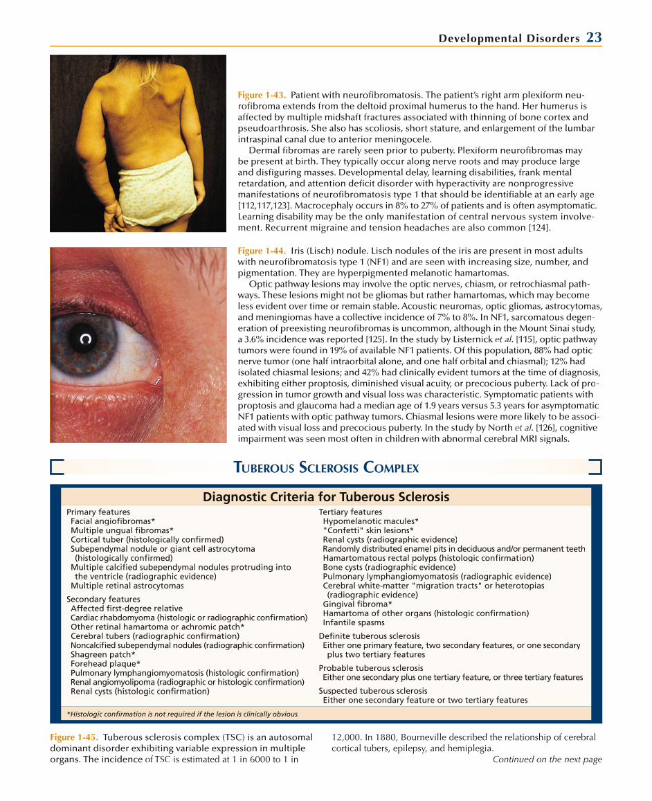
Developmental Disorders 23
Figure 1-43. Patient with neurofibromatosis. The patient’s right arm plexiform neu-rofibroma extends from the deltoid proximal humerus to the hand. Her humerus is affected by multiple midshaft fractures associated with thinning of bone cortex and pseudoarthrosis. She also has scoliosis, short stature, and enlargement of the lumbar intraspinal canal due to anterior meningocele.
Dermal fibromas are rarely seen prior to puberty. Plexiform neurofibromas maybe present at birth. They typically occur along nerve roots and may produce largeand disfiguring masses. Developmental delay, learning disabilities, frank mental retardation, and attention deficit disorder with hyperactivity are nonprogressive manifestations of neurofibromatosis type 1 that should be identifiable at an early age[112,117,123]. Macrocephaly occurs in 8% to 27% of patients and is often asymptomatic.Learning disability may be the only manifestation of central nervous system involve-ment. Recurrent migraine and tension headaches are also common [124].
Figure 1-44. Iris (Lisch) nodule. Lisch nodules of the iris are present in most adultswith neurofibromatosis type 1 (NF1) and are seen with increasing size, number, and pigmentation. They are hyperpigmented melanotic hamartomas.
Optic pathway lesions may involve the optic nerves, chiasm, or retrochiasmal path-ways. These lesions might not be gliomas but rather hamartomas, which may becomeless evident over time or remain stable. Acoustic neuromas, optic gliomas, astrocytomas,and meningiomas have a collective incidence of 7% to 8%. In NF1, sarcomatous degen-eration of preexisting neurofibromas is uncommon, although in the Mount Sinai study, a 3.6% incidence was reported [125]. In the study by Listernick et al. [115], optic pathway tumors were found in 19% of available NF1 patients. Of this population, 88% had opticnerve tumor (one half intraorbital alone, and one half orbital and chiasmal); 12% hadisolated chiasmal lesions; and 42% had clinically evident tumors at the time of diagnosis,exhibiting either proptosis, diminished visual acuity, or precocious puberty. Lack of pro-gression in tumor growth and visual loss was characteristic. Symptomatic patients withproptosis and glaucoma had a median age of 1.9 years versus 5.3 years for asymptomatic NF1 patients with optic pathway tumors. Chiasmal lesions were more likely to be associ-ated with visual loss and precocious puberty. In the study by North et al. [126], cognitive impairment was seen most often in children with abnormal cerebral MRI signals.
TUBEROUS SCLEROSIS COMPLEX
Diagnostic Criteria for Tuberous SclerosisPrimary featuresFacial angiofibromas*Multiple ungual fibromas*Cortical tuber (histologically confirmed)Subependymal nodule or giant cell astrocytoma (histologically confirmed)
Multiple calcified subependymal nodules protruding intothe ventricle (radiographic evidence)
Multiple retinal astrocytomas
Tertiary featuresHypomelanotic macules*"Confetti" skin lesions*Renal cysts (radiographic evidence)Randomly distributed enamel pits in deciduous and/or permanent teethHamartomatous rectal polyps (histologic confirmation)Bone cysts (radiographic evidence)Pulmonary lymphangiomyomatosis (radiographic evidence)Cerebral white-matter "migration tracts" or heterotopias (radiographic evidence)Gingival fibroma*Hamartoma of other organs (histologic confirmation)Infantile spasms
Secondary featuresAffected first-degree relativeCardiac rhabdomyoma (histologic or radiographic confirmation)Other retinal hamartoma or achromic patch*Cerebral tubers (radiographic confirmation)Noncalcified subependymal nodules (radiographic confirmation)Shagreen patch*Forehead plaque*Pulmonary lymphangiomyomatosis (histologic confirmation)Renal angiomyolipoma (radiographic or histologic confirmation)Renal cysts (histologic confirmation)
Definite tuberous sclerosisEither one primary feature, two secondary features, or one secondaryplus two tertiary features
Probable tuberous sclerosisEither one secondary plus one tertiary feature, or three tertiary features
Suspected tuberous sclerosisEither one secondary feature or two tertiary features
*Histologic confirmation is not required if the lesion is clinically obvious.
Figure 1-45. Tuberous sclerosis complex (TSC) is an autosomaldominant disorder exhibiting variable expression in multipleorgans. The incidence of TSC is estimated at 1 in 6000 to 1 in
12,000. In 1880, Bourneville described the relationship of cerebralcortical tubers, epilepsy, and hemiplegia.
Continued on the next page

24 Atlas of Clinical Neurology
Figure 1-45. (Continued) Subsequently, numerous authors have described involvement of skin, retina, lung, heart, kidney, and other organs in TSC, along with variable degrees of mental and behavioral disturbance. The term phakoma was coined for TSCbecause of the lentil-shaped retinal lesions, and then applied toother neurocutaneous disorders as the phakomatoses. Clinicaldiagnostic criteria for TSC were established by a consensus con-ference of the Diagnostic Criteria Committee of the National Tuberous Sclerosis Association [127]. Clinical heterogeneity is common within the same pedigree. Clinical manifestation of TSC may be present at birth in the form of cardiac rhabdomyo-mas or neurologic involvement. Rhabdomyomas of spontane-ous mutational origin are also commonly seen in asymptomaticnewborns and may be the first clue in the diagnosis of TSC.
The advantage of CT imaging is the identification of calcifiedlesions, which may appear in later childhood. Cranial MRI is thepreferred diagnostic study, because it allows for the detailed assessment of both cortical and subcortical lesions. Sulcal
islands, gyral cores, radial migration lines, wedges, and subepen-dymal nodules have been described as MRI imaging abnormali-ties of tuberous sclerosis [128]. On rare occasions the diagnosisis made during the evaluation of learning disability or polycystic kidney disease. Echocardiography is helpful in detecting cardiac rhabdomyomas. Renal angiomyolipomas, usually asymptomaticexcept for microscopic hematuria, can be detected on ultra-sound studies. Renal cysts are also usually asymptomatic. Both renal lesions are readily identified by ultrasonography. Polycystic kidneys may be associated with progressive renal disease that requires transplantation. Pulmonary lymphangiomyomatosis may result in adult-onset pulmonary disease in women. Gastrointes-tinal hamartomas may produce recurring pain and bleeding but are usually asymptomatic. Dental enamel pitting is a commonfinding in 90% of patients. It becomes more apparent in adultsubjects. Hamartomas of the liver are seen in 25% of children and may be clinically silent. Bone cysts are small and clinically insig-nificant. TSC most often presents as seizures.
Figure 1-46. Patient with tuberous sclerosis complex (TSC). In this patient, a large forehead plaque was present at birth. She also exhibited asymptomatic rhabdomyomaand at 3 years of age had microscopic hematuria and renal angioleiomyoma. The facial lesions that started to develop at age 3 years were angiofibromas. Though the seizures were initially responsive to adenocorticotropic hormone (ACTH), the patient ultimate-ly required a combination of vigabatrin and carbamazepine for seizure control.
In the majority of patients, seizures are present from infancy and occur in 84% ofall TSC patients. The emergence of infantile spasms between 2 and 11 months of age is common, with peak incidence between 4 and 6 months. Atypical features of infan-tile spasms of TSC include the appearance of lateralizing features, such as asymmetrictonic and atonic components, unilateral grimacing, head turning, eye deviation, andunequal limb movements. Multifocal electroencephalographic epileptiform abnor-malities may be seen. Progression from multifocal spikes to generalized spike andwave discharges on electroencephalogram, with episodes of sudden, brief voltageattenuation, is consistent with hypsarrhythmia. The emergence in time of infan-tile spasms in TSC relates to the development of dysplastic neuronal populationsand expanded dendritic arborization, providing an enhanced connective pathwayto propagate epileptiform abnormalities, from focal and multifocal to generalized.Although an infant with TSC and partial epilepsy may appear to be developing nor-mally, regression of cognitive, behavioral, and motor skills commonly accompaniesthe onset of infantile spasms, and severity parallels the location and number of corti-cal tubers. Children with multiple tubers and spasms that are resistant to antiepilepticdrugs are more likely to be permanently impaired, sometimes developing autism. Thereported excellent response of TSC infantile spasms to vigabatrin is probably relatedto their focal origin. When seizures are intractable despite aggressive antiepilepticdrug therapy, surgery for epilepsy must be considered [129,130]. Even in the presence of multiple cortical tubers or astrocytic hamartomas, if the seizure onset can be con-sistently localized to a dominant cerebral lesion, after resection of the primary focusthe less prominent or secondary foci may gradually diminish [131]. Mental subnor-mality occurs in approximately 80% of TSC patients and is evident in early childhood [131]. Intractable seizures and multiple bilateral tubers are more commonly associ-ated with mental retardation [132]. Although a high incidence of autism has beendescribed in TSC studies [133], a prospective comprehensive behavioral analysis of a large unselected TSC population is warranted [133]. Developmental evaluation andpsychometric studies are warranted in all patients with TSC to define learning dis-abilities, mental impairment, and behavioral disorders.
TSC is an autosomal dominant condition with variable expression, and two thirdsof cases are new mutations. Linkage studies have identified loci on chromosome 9q34(TSC1 gene, encoding for hamartin) [134] and 16p13 (TSC2 gene, encoding for tuberin)[135]. Locus heterogeneity may also explain the clinical heterogeneity. Linkage analysismay determine whether the TSC patient’s genetic abnormality is linked to chromosome
9 or 16. In a new mutation, the risk ofanother sibling being affected is 2% to 5%. Mutations in either the TSC1 or the TSC2gene may lead to decreased expression ofthe gene products, tuberin and hamartin [136]. To identify an asymptomatic TSC parent, examination should include care-ful cutaneous examination, neuroimaging, and ophthalmologic and renal evaluations. Rarely, an asymptomatic parent may be identified, redefining the genetic risk at50%. Coexistence of neurofibromatosis type 1 and TSC in a patient has also beenreported [137]. Genetic counseling and,when applicable, linkage analysis are nec-essary after evaluation of family members. In the case of a new mutation, the risk foranother affected sibling is 2% to 5%. TheNational Tuberous Sclerosis Association is a valuable resource for patients, families,and professionals [138].

Developmental Disorders 25
HYPOMELANOSIS OF ITO
Figure 1-48. A patient with hypomelanosis of Ito. Hypomelanosis of Ito (HOI), or incontinentia pigmentiachromians, was described by Minor Ito in 1951. The disorder is characterized by variable cutaneousmacular, hypopigmented whorls, streaks, and patches that usually occur on the trunk and the limbs, and to a lesser extent over the face [139]. Neurologic involvement is reported at 40% to 100%. The sever-ity of neuropathologic involvement is variable. Hypopigmentation may be apparent at birth, but as intuberous sclerosis the skin lesions may become obvious only with increasing skin pigmentation afterseveral months of age. Ocular abnormalities include choroidal atrophy and corneal opacities. Anoma-lies of teeth and facial bones, scoliosis, limb length irregularities, strabismus, macrocephaly, craniofacial dysmorphism, and syndactyly have been described. In this patient a large hypopigmented patch becameevident after 12 months of age. The hypomelanotic lesions had irregular borders, and biopsy of the skinlesions revealed decreased melanocytes and melanin deposition. Wood’s lamp (ultraviolet) examination can be helpful in identifying the hypopigmented whorls in the younger, fair-skinned infant. This patientalso had macrocephaly and learning disabilities but no focal deficit or epilepsy.
Familial case reports suggest that HOI is a dominantly inherited disorder with variable expressionand a high mutation rate, which would explain the frequency of sporadic cases. Trisomy 14 mosa-icism seen in cultured fibroblasts may underlie some of the pigmentation abnormalities [140]. No single, specific chromosomal abnormality, however, explains all cases of HOI. Mosaicism of various genes, sometimes apparent only on skin biopsy, seems the likely basis for the disorder [141].
WYBURN-MASON SYNDROME
Figure 1-49. Wyburn-Mason syndrome (WBS), or retinocephalicvascular malformation, a spontaneous, nonfamilial neuro-cutaneous disorder characterized by ocular and intracranialarteriovenous malformations (AVMs). Although the conditionwas described by others earlier, in 1943 Wyburn-Mason [142]
Figure 1-47. T1-weighted transaxial MRI of a large right foramen of Monro astrocytic hamartoma with a central cystic component. This lesion was enlarging and causing progressive ventriculomegaly. Rightfrontal cortical and left parietal cortical tubers with gyral cores were associated with multifocal epilep-tiform activity. Partial seizures followed infantile spasms. The ventricular mass was resected without complications. Language was delayed and affect was appropriate.
Cortical tubers occur most often in the cerebrum and to a lesser extent in the cerebellum. Thefrontal and parietal lobes are involved most often. Macroscopically, the tubers are firm, flat, or slightly elevated and paler than the surrounding cortex. On microscopic examination, normal cytoarchitecture is lost, with enlarged multinucleate glial cells and enlarged neurons that are irregularly distributed.The cortical gray-white interface shows hypomyelination. Rarely, the cortical tubers undergo necrosis. Subependymal nodules typically occur in the body of the lateral ventricles and are composed of largemultinucleated astrocytes and large spindle cells. Microscopic mineralization is seen in at least 50%of cases. Nodules in the region of the foramen of Monro may grow to greater than 3 cm and causeobstructive hydrocephalus. Approximately 6% of patients may experience malignant tranformation oflesions, but the actual frequency of this neoplastic change is poorly defined. DiMario [128] has report-ed brainstem tubers and cerebellar dysgenesis with calcification in a patient with central apnea.
reviewed the available literature on retinal AVMs and pointedout the association in 22 of 27 patients of intracranial vascular malformations, particularly of the midbrain. Subsequent reportsof WBS have included patients with optic atrophy without reti-nal vascular malformation but exhibiting intraorbital or chiasmalAVMs and associated cerebral AVMs [143,144]. Additionally,vascular malformations of the face and mandible have beenassociated with WBS. From these descriptions, it is apparentthat facial, ocular, and intracranial AVMs may be heterogeneousin their clinical presentation. The cerebral and ocular malfor-mation has its origin in the first trimester of fetal development.WBS and Sturge-Weber syndrome have been reported in thesame patient [145].
Retinal lesions can be seen on funduscopy. The vessels are tortuous and dilated, and arteriovenous shunts may be identi-fied. The retinal lesions are generally unilateral. Intracranial AVMs are typically ipsilateral to the retinal lesion and lie deepwithin cerebral parenchyma, involving the midbrain, thala-mus, optic chiasm, or the basal ganglia. Patel and Gupta [146] described a child with bilateral neuroretinal involvement.
Continued on the next page

26 Atlas of Clinical Neurology
Figure 1-49. (Continued) Nonneural involvement is also fre-quent and includes facial angioma and palatal, oral mucosal, mandibular, and maxillary vascular malformations in up to 50%of patients. Vision is usually impaired, but this may be difficult to assess in the young child. Proptosis, nystagmus, impairedpupillary reaction, conjunctival hyperemia, and variable degrees of visual loss are common. Pyramidal tract involvement may be unilateral or bilateral, depending on the location of the lesion.Cranial nerves III, IV, VI, VII, and VIII may be also be affected.
WBS should be considered when retinal, facial, mandibu-lar, or maxillary vascular malformations are present. Retinal
examination reveals AVMs or optic atrophy. Also, the exam-iner should auscultate for cranial or orbital bruits. Con-trast-enhanced cerebral and orbital CT or MR angiography or conventional contrast angiography reveals the extent ofintraocular and intracranial involvement. Selective angiogra-phy is necessary to delineate the origin and extent of the AVM. Definitive treatment is determined by the extent and location of the AVMs. Depending on the individual circumstances,intravascular occlusive therapy, gamma irradiation, direct surgical intervention, or no intervention at all may be to thepatient’s advantage.
STURGE-WEBER SYNDROME
Figure 1-50. Patient with Sturge-Weber syndrome (SWS). SWS is a nonfamilial, spon-taneous neurocutaneous disorder affecting all ethnic groups. It has onset in infancy in most patients, with a high frequency of partial seizures, hemiparesis, and devel-opmental delay. Sujansky and Conradi [147] analyzed data on 171 patients with SWSand provided valuable information on clinical expression of the disease. Glaucoma is a common feature and is usually ipsilateral to the nevus. Because of variable clini-cal expression, Roach described three forms of SWS: type I has associated facial and leptomeningeal angiomas, often accompanied by glaucoma; type II has facial angioma without cerebral leptomeningeal angioma and may have accompanying glaucoma; type III has cerebral leptomeningeal angioma without facial nevus. An extensivecapillary venous angioma overlies the affected cerebral tissue; typically it affects the occipital region but can involve the entire hemisphere. The leptomeninges are thick-ened, containing extensive thin-walled capillaries and veins that often exhibit hyaline thickening of their walls. Cerebral cortical vascular changes are similar, and in time,calcification of the gyral angiomas produces railroad-track calcifications. Progressivecerebral atrophy and external hydrocephalus are long-term effects of SWS. The cere-bral lesions are unilateral in 85% of patients.
Facial nevi, present at birth, may also involve the oral and nasopharyngeal mucosa.Port wine stains appear on the trunk and limbs of 44% of SWS patients. Facial port wine stains occur along the distribution of the fifth cranial nerve (V) as follows: 16% in V1 (ophthalmic) alone; 18% in V1 plus V2 (maxillary); 38% in V1, V2, and V3 (man-dibular); and the remainder exhibiting various degrees of bilateral involvement. Theport wine nevus is present at birth, and the extent of involvement impacts the degree of neurologic involvement. Seizures occur in 80% of patients, with 75% experiencing onset in the first year of life and 95% by 5 years [148]. Partial and secondarily general-ized seizures are typical, and initially may be less responsive to aggressive antiepi-leptic drug (AED) therapy than in the chronic epileptic disorder of SWS. This maypartly be caused by relatively acute neuropathologic changes such as cerebral corticalmicroinfarction owing to progressive venous occlusive phenomena. Ocular choroid membrane angioma is the cause of glaucoma, with 48% of patients affected, two thirdsunilateral and one third bilateral. Approximately 60% of patients exhibit glaucoma by 1 year of age [147]. It is more common when the port wine stain involves both the upper and lower eyelids [149].
Developmental delay and subsequent mental impairment occur in approximately50% of SWS patients. The greatest risk for retardation occurs in children with bilateral cerebral abnormalities and those with seizure onset in the first year of life, especially if the epilepsy remains poorly responsive even to aggressive anticonvulsant therapy.Progressive intellectual decline with motor impairment in the presence of widespreadhemispheric angioma and intractable seizures is considered to be a potentially pre-ventable condition responsive to epilepsy surgery [150]. Progressive hemiplegia withaccompanying hemianopia and specifically localized impairments can be attributed toocclusive venous infarction and cerebral atrophy. Symptomatic external hydrocephalus
with macrocephaly may further compli-cate the condition. If seizures begin after4 years of age, intellectual impairment isgenerally infrequent, although seizuresmay be less responsive to AED therapy.Cerebral involvement is best defined bygadolinium-enhanced MRI and MR angi-ography. Somatic cell mosaicism seems to underlie most cases of SWS [151].
Electroencephalographic (EEG) find-ings are reflective of the focal structuralabnormalities. Epileptiform abnormalities may extend beyond the extent of angio-matosis, calcification, and atrophy. This patient showed facial angioma port winenevus involving V1, V2, and V3 distribu-tion with conjunctival and choroidal angioma and ipsilateral glaucoma. The child experienced complex partial sei-zures and had ipsilateral focal occipitalcortical venous angioma. Skull radiogra-phy revealed railroad-track calcifications in the affected occipital region, and the EEG was abnormal due to frequent epi-leptiform discharges in the left occipitaland parietal regions.
Developmental Disorders 27
INCONTINENTIA PIGMENTI
Figure 1-51. A patient with incontinentia pigmenti (IP). Mul-tiple verrucous lesions are present on her upper and lower limbs. They consist of papules and hyperkeratotic streaks and thick crusts. They were preceded by bullae that began 2 weeks after the onset of partial seizures at 6 weeks of age. During the verrucous stage, the patient exhibited recurrent complexpartial seizures and regression in milestones.
IP is a unique progressive genodermatosis that occurs almost exclusively in girls, with one third exhibiting neurologic compli-cations [152]. Most patients develop evolving cutaneous lesions(stage 1, bullae; stage 2, verrucae) in the first year of life, although the cutaneous manifestations may appear at single or multiplestages at birth with accompanying neurologic symptoms. Overall systemic involvement occurs in 80% of patients who manifest cutaneous lesions. The female to male ratio is 35 to 1. X-linkeddominant inheritance is the likely basis for the predominanceof affected girls and presumed lethality in affected boys. In ourown experience, fathers with cutaneous lesions of IP had noneurologic symptoms, and the literature indicates that survivingaffected males have no greater degree of neurologic involve-ment. The disorder is related to mutations in the inhibitor ofkappa light polypeptide gene enhancer located on Xq28 [153].Traupe and Vehring [154] attribute mosaic disease expression ofIP in males to an unstable premutation, which ordinarily remains silent in males during embryogenesis. Surviving affected infantboys have a mosaic state of full and premutation alleles.
Acute neurologic manifestations may be present at birthwhen stages 1 and 2 (skin bullae and verrucae) are also pres-ent [155,156]. Seizures may precede the skin lesions by days orweeks. Significant neurologic impairment occurs more often with neonatal or infantile onset of symptoms. Mental retarda-tion occurs in approximately 12%, epilepsy in 13%, and spastic cerebral palsy in 11%. Behavioral problems and learning disabili-ties are also seen in children with less severe neurologic involve-ment. Neuropathologic features include acute hemorrhagic encephalopathy of white matter with associated edema and chronic effects similar to residua of encephalitis, including neu-ronal loss, gliosis, ventriculomegaly, and cortical atrophy [155]. Lee et al. described MRI and magnetic resonance spectroscopy findings indicative of small and large vessel cerebrovascularocclusive disease with probably shared pathophysiologic mecha-nisms of retinal vasculopathy of IP [157]. Neuroimaging abnor-malities are determined by the acute or chronic phase of thedisease. At the onset of central nervous system (CNS) symptoms, cerebral edema is suggested by loss of gray-white interface, effacement of lateral ventricles, and increased MRI signal in the affected areas [155]. Acute cerebrovascular abnormalities appear as focal ischemic lesions. Chronic effects include focal and dif-fuse cortical atrophy, ventriculomegaly, and microcephaly.
Dental abnormalities include adontia, late dentition, hypodentia, and abnormal (notched and conical) tooth for-mation [152]. Ocular abnormalities occur in up to 35% of IPcases. Seen in decreasing frequency are strabismus, blind-ness, congenital cataracts, microphthalmia, nystagmus, irispigmentation, uveitis, chorioretinitis, and papillitis. Wald et al. reported four patients with traction retinal detachments[158]. Retinal vascular abnormalities suggest retinal arteriolarocclusion with ischemic infarction. Eosinophilia occurs dur-ing the acute phase in two thirds of patients; it ranges from5% to 65% and subsides with resolution of the CNS symp-toms and cutaneous lesions [152].
Four Stages in the Evolution of the Cutaneous Lesions in Incontinentia PigmentiStage 1 Vesicular lesions may be present at birth in 50% of patients. They are usually erythematous and occur on the extremities
and trunk and usually resolve over 1 to 2 weeks, although rarely they recur over several months. Ninety percent of patientsmanifest vesicles by 1 year of age. Microscopically, the vesicles contain and are surrounded by numerous eosinophils with extensive intercellular edema.
Stage 2 Verrucae usually follow vesicles and rarely occur simultaneously with them. The lesions are wartlike and have papularhyperkeratotic formation. These occur in 70% of patients and only on the extremities. Dyskeratosis is prominent; basalcells exhibit vacuolization with loss of pigment.
Stage 3 This is the pigmentary phase. Lesions are present over the trunk or limbs and are irregular, whorling, marbled, or reticulated. Although the hyperpigmented stage may occur at birth, it is usually evident at 3 to 6 months of age. There is a subtle fadingof the pigmented streaks in the late teens. Microscopically, melanin is heavily deposited in melanophages in the upper dermis.
Stage 4 Atrophic scars occur in one third of cases and follow the fading hyperpigmented lesions. Ultimately, the scars flatten. Althoughthe cutaneous lesions of incontinentia pigmenti appear to evolve from one stage to the next, different stages may appear simultaneously, and in time the following stage may appear at a different site.
Figure 1-52. Four stages in the evolution of the cutaneous lesions in incontinentia pigmenti. They usually occur in sequence,
although two stages may be present simultaneously. The stages are vesicular lesions, verrucae, a pigmentary phase, and atrophic scars.

28 Atlas of Clinical Neurology
SECONDARY MICROCEPHALY
Characteristics of Primary and Secondary MicrocephalyPrimary Microcephaly Secondary Microcephaly
Etiology Gene loci on 8p22/19q13/9q34 Environmental, toxic, or infectious cause
Pathology Abnormally shaped skull; primary migrational disturbances of microgyria, heterotopias
Destructive lesions; cortical atrophy, focal necrosis, mineralizing angiopathy
Neuroimaging Heterotopias, hypoplasia or aplasia of cerebellar vermis and corpus callosum, periventricular T2 hyperintense lesions
Calcifi cations, cystic encephalomalacia, relative ventriculomegaly, thinned cortex
Figure 1-53. Characteristics of secondary microcephaly. When microcephaly is one component of a constellation of abnormali-ties, it can be characterized as secondary microcephaly. It maybe due to a number of insults to the developing brain, including chromosomal anomalies; malnutrition; malformations such aslissencephaly, holoprosencephaly, and micropolygyria; intra-uterine exposure to toxins such as alcohol; exposure to radia-tion; and exposure to congenital intrauterine infections. Theinfectious agents associated with microcephaly that is apparent at birth include toxoplasmosis, cytomegalovirus, rubella, herpessimplex type II, and HIV [159,160]. The cortex demonstrates diffuse atrophy, focal necrosis, or gliosis. Deeper structures are frequently affected by degeneration of blood vessel walls, resulting in mineralizing microangiopathy and calcifi cation thatis detectable on neuroimaging studies [108]. The mechanism bywhich viral infections affect specifi c immature cell populations leading to microcephaly with disordered cell migration, micro-gyria, and changes of encephalomalacia is not absolutely clear.
However, one theory relates to the role of vasoactive intestinal peptide (VIP) in neuron differentiation in cell cultures [161]. VIPcauses glia to release molecules that promote mitosis of astro-cytes and neuroblasts. All of the neuropathologic features of microcephaly can be induced in mice following prenatal admin-istration of a specifi c VIP antagonist (neurotensin-VIP 7-28) intra-peritoneally between the 9th and 12th gestational days.
Clinically, patients with secondary microcephaly tend tohave more neurologic handicaps and greater intellectualdysfunction compared with those with primary microcephaly, who may present more commonly with mental retardationwith or without seizures [162]. Because congenital infections can involve the developing optic nerve and auditory system,patients with microcephaly should be evaluated thoroughly for deafness and optic atrophy as well. A careful family historyshould be taken to rule out familial (recessive or dominant forms) of microcephaly. In some instances, the microcephaly is complex and polygenic.
Figure 1-54. CT scan of microcephaly due to cytomegalovirus infection, demonstratingperiventricular mineralizing angiopathy as well as diffuse atrophy. Maternal viremia allows transplacental passage of the virus into the most vascularized tissues of the fetus. This allows rapid proliferation of the virus in the subependymal germinal matrix, resulting in the periventricular distribution of calcifi cation that is evident on this scan.CT is helpful in detecting gross brain abnormalities. An MRI scan, however, enables better visualization of associated gyral malformations, the corpus callosum, and neu-ronal heterotopias. If a suggestion of calcifi c lesions is present on the MRI scan based on low T1 and T2 signals from specifi c regions, spin-echo sequences can be used toconfi rm calcifi cation. Alternatively, an unenhanced CT scan can be performed.
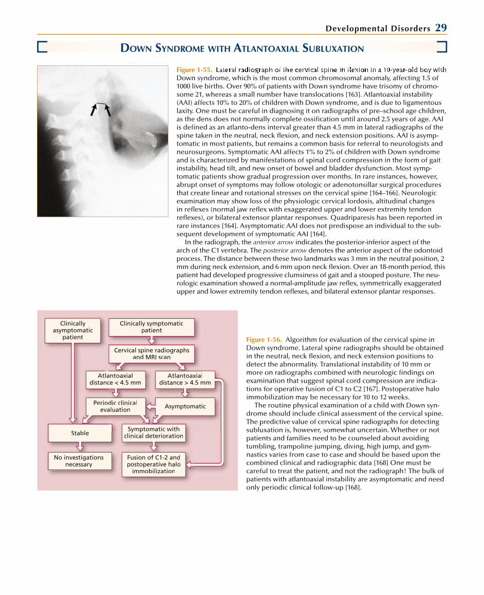
Developmental Disorders 29
DOWN SYNDROME WITH ATLANTOAXIAL SUBLUXATION
Figure 1-55. Down syndrome, which is the most common chromosomal anomaly, affecting 1.5 of 1000 live births. Over 90% of patients with Down syndrome have trisomy of chromo-some 21, whereas a small number have translocations [163]. Atlantoaxial instability (AAI) affects 10% to 20% of children with Down syndrome, and is due to ligamentous laxity. One must be careful in diagnosing it on radiographs of pre–school age children,as the dens does not normally complete ossifi cation until around 2.5 years of age. AAIis defi ned as an atlanto-dens interval greater than 4.5 mm in lateral radiographs of the spine taken in the neutral, neck fl exion, and neck extension positions. AAI is asymp-tomatic in most patients, but remains a common basis for referral to neurologists and neurosurgeons. Symptomatic AAI affects 1% to 2% of children with Down syndrome and is characterized by manifestations of spinal cord compression in the form of gait instability, head tilt, and new onset of bowel and bladder dysfunction. Most symp-tomatic patients show gradual progression over months. In rare instances, however,abrupt onset of symptoms may follow otologic or adenotonsillar surgical proceduresthat create linear and rotational stresses on the cervical spine [164–166]. Neurologicexamination may show loss of the physiologic cervical lordosis, altitudinal changesin refl exes (normal jaw refl ex with exaggerated upper and lower extremity tendonrefl exes), or bilateral extensor plantar responses. Quadriparesis has been reported inrare instances [164]. Asymptomatic AAI does not predispose an individual to the sub-sequent development of symptomatic AAI [164].
In the radiograph, the anterior arrow indicates the posterior-inferior aspect of the warch of the C1 vertebra. The posterior arrow denotes the anterior aspect of the odontoidwprocess. The distance between these two landmarks was 3 mm in the neutral position, 2mm during neck extension, and 6 mm upon neck fl exion. Over an 18-month period, this patient had developed progressive clumsiness of gait and a stooped posture. The neu-rologic examination showed a normal-amplitude jaw refl ex, symmetrically exaggerated upper and lower extremity tendon refl exes, and bilateral extensor plantar responses.
Atlantoaxialdistance < 4.5 mm
Atlantoaxialdistance > 4.5 mm
Periodic clinicalevaluation
Symptomatic withclinical deterioration
Asymptomatic
Cervical spine radiographsand MRI scan
No investigationsnecessary
Stable
Fusion of C1-2 andpostoperative halo
immobilization
Clinicallyasymptomatic
patient
Clinically symptomaticpatient
Figure 1-56. Algorithm for evaluation of the cervical spine inDown syndrome. Lateral spine radiographs should be obtainedin the neutral, neck fl exion, and neck extension positions todetect the abnormality. Translational instability of 10 mm ormore on radiographs combined with neurologic fi ndings onexamination that suggest spinal cord compression are indica-tions for operative fusion of C1 to C2 [167]. Postoperative halo immobilization may be necessary for 10 to 12 weeks.
The routine physical examination of a child with Down syn-drome should include clinical assessment of the cervical spine. The predictive value of cervical spine radiographs for detecting subluxation is, however, somewhat uncertain. Whether or not patients and families need to be counseled about avoidingtumbling, trampoline jumping, diving, high jump, and gym-nastics varies from case to case and should be based upon thecombined clinical and radiographic data [168] One must be careful to treat the patient, and not the radiograph! The bulk ofpatients with atlantoaxial instability are asymptomatic and needonly periodic clinical follow-up [168].

30 Atlas of Clinical Neurology
DYSOSTOSIS MULTIPLEX SECONDARY TO LYSOSOMAL STORAGE DISEASE
Syndromes with Dysostosis Multiplex
Disorder Deficient EnzymeElevated Urinary Mucopolysaccharide Inheritance Severity of Skeletal Changes
Hurler (MPS I) -Iduronidase Yes AR Severe
Hunter (MPS II) Iduronate sulfatase Yes X-linked Severe
Sanfilippo (MPS III)
Type A Heparan-N-sulfatase Yes/no AR Mild
Type B -N-acetylglucosaminidase Yes/no AR Mild
Type C Acetyl CoA: -glucosaminide acetyltransferase
Yes/no AR Mild
Type D N-acetylglucosamine-6-sulfatase Yes/no AR Mild
Morquio (MPS IV)
Type A Galactosamine-6-sulfatase Yes AR Severe, plus additional changes from other MPS
Type B -Galactosidase Yes AR Severe
Maroteaux-Lamy (MPS VI) Arylsulfatase B Yes AR Severe
Sly (MPS VII) -Glucuronidase Yes AR Severe
I cell disease (ML II) N-acetylglucosaminephosphotransferase
No AR Severe
Pseudo-Hurler polydystrophy (ML III)
N-acetylglucosaminephosphotransferase
No AR Moderate
-Mannosidosis -Mannosidase No AR Severe
-Mannosidosis -Mannosidase No AR Mild/absent
Sialidosis type II Sialidase No AR Severe
Aspartylglucosaminuria Aspartylglucosaminidase No AR Mild
Fucosidosis -Fucosidase No AR Moderate
Carbohydrate-deficient glycoprotein syndrome type Ia
Phosphomannomutase No AR Moderate to severe
Figure 1-57. Storage disorders in which dysostosis multiplexcan be seen [159–161]. Dysostosis multiplex is used to describeskeletal abnormalities seen in lysosomal storage diseases, particularly the mucopolysaccharidoses and mucolipidoses[169–171]. These bony changes are not present at birth, butmay evolve over the first 6 to 12 months of life and may be the first manifestation of the disease. The ribs become thick-ened anteriorly. The long tubular bones become broad and irregular, especially at the metaphyses [172,173]. The longbones develop multiple linear streaks in the trabecular pat-tern and lose normal tubulation. The skull becomes enlargedand thickened; frequently, dolichocephaly or scaphocephaly
may occur. The sella turcica may be J-shaped. The vertebral bodies develop anterior beaking and wedge deformities,leading to gibbus formation in the back. The metacarpals may become broad and irregular, with conical deformities of theproximal metaphyses. Accumulation of storage material in theflexor tendons and sheaths leads to a clawlike deformity of thehands. Dysostosis multiplex is one component of the clinicaldata collected in the global registry for monitoring patients with mucopolysaccharidosis (MPS) type I, or Hurler’s disease[174]. This disorder is treatable using bone marrow transplan-tation or lysosomal enzyme replacement [175]. AR—autosomal recessive; ML—mucolipidosis.

Developmental Disorders 31
A BBBBBBBBBB
Figure 1-58. Radiographs of a boy with Hurler syndrome. A, The hand shows bony changes of dysostosis multiplex.The second through fourth metacarpalsare broad and irregular, with cone-shapedand pinched proximal epiphyses. The phalanges are short, thickened, andirregular with a bulletlike configuration.The distal radius and ulna tilt toward eachother. B, Lateral spine radiograph show-ing anterior beaking and hooklike projec-tions of the vertebral bodies. This patienthas retrolisthesis of T12 on L1 and focalkyphosis of T12–L1. Many patients withdysostosis multiplex develop a gibbus ofthe lumbar spine.
REFERENCES
1. Burnelli S, Faiella A, Capra V, et al.: Germline mutations in the homeo-box gene EMX2 in patients with severe schizencephaly. Nat Genet 1996,t12:94–96.
2. Dobyns W, Reiner O, Carrozzo R, Ledbetter D: Lissencephaly: a human brain malformation associated with deletion of the LIS-1 gene locatedon chromosome 17p13. JAMA 1993, 270:2838–2842.
3. Guttman RH, Collins FS: Recent progress toward understanding the molecular biology of von Recklinghausen’s neurofibromatosis. AnnNeurol 1992, 31:555–561. l
4. Use of folic acid for prevention of spina bifida and other neural tube defects: 1983–1991. MMWR Morb Mortal Wkly Rep 1991, 40:513–516.
5. Shaer CM, Chescheir N, Schulkin J: Myelomeningocele: a review of theepidemiology, genetics, risk factors for conception, prenatal diagnosis, and prognosis for affected individuals. Obstet Gynecol Surv 2007, 62:471–479.v
6. Watson WJ, Chescheir NC, Katz VL, Seeds JW: The role of ultrasound in evaluation of patients with elevated maternal serum -fetoprotein: a review. Obstet Gynecol 1991, 78:123–128.l
7. Hunter AGW: Brain and spinal cord. In Human Malformations and Related Anomalies, vol 2. Edited by Stevenson RE, Hall JG, Goodman RM.New York: Oxford University Press; 1993:109–137.
8. Romano PS, Waitzman NJ, Scheffler RM, Randy D: Folic acid fortification of grain: an economic analysis. Am J Public Health 1995, 85:667–676.
9. Juriloff DM, Harris MJ: Mouse models for neural tube closure defects. Hum Mol Genet 2000, 9:993–1000.t
10. Shapiro K: Encephalocele. In Birth Defects Encyclopedia. Edited by Buyse ML. Dover, MA: Center for Birth Defects Information Services in asso-ciation with Blackwell Scientific Publications; 1990:614–615.
11. Marin-Padilla M: Cephalic axial skeletal-neural dysraphic disorders:embryology and pathology. Can J Neurol Sci 1991, 18:153–169.i
12. Daly LE, Kirke PN, Molloy A, et al.: Folate levels and neural tube defects: implications for prevention. JAMA 1995, 274:1698–1702.
13. Estin D, Cohen AR: Caudal agenesis and associated caudal spinal cord malformations. Neurosurg Clin North Am 1995, 6:377–391.
14. Johnston MC: Understanding human embryonic development. In Human Malformations and Related Anomalies, vol 1. Edited by Stevenson RE, HallJG, Goodman RM. New York: Oxford University Press; 1993:31–63.
15. Gellis SS, Feingold M, Tunnessen WW Jr, Raettig JA: Caudal regressionsyndrome: picture of the month. Am J Dis Child 1968, 116:407–408. d
16. Escobar LF, Weaver DD: Caudal regression syndrome. In Birth Defects Encyclopedia. Edited by Buyse L. Dover, MA: Center for Birth Defects Information Services in association with Blackwell Scientific Publica-tions; 1990:296–297.
17. Jones KL: Smith’s Recognizable Patterns of Human Malformation, edn 4.Philadelphia: WB Saunders; 1988.
18. Welch JP, Aterman K: The syndrome of caudal dysplasia: a review, including etiologic considerations and evidence of heterogeneity.Pediatr Pathol 1984, 2:313–327.l
19. Volpe JJ, ed: Neurology of the Newborn, edn 3. Philadelphia: WBSaunders; 1994.
20. Hamada H, Oki A, Tsunoda H, Kubo T: Prenatal diagnosis of holopros-encephaly by transvaginal ultrasonography in the first trimester. AsiaOceania J Obstet Gynaecol 1992, 18:125–129.l
21. Johnston MC, Bronsky PT: Prenatal cranio-facial development: new insights on normal and abnormal mechanisms. Crit Rev Oral Biol Med1995, 6:25–79.
22. Johnston MC, Bronksy PT: Animal models of human cranio-facial mal-formations. J Craniofac Genet Dev Biol 1991, 11:277–291.l
23. Roessler E, Ward DE, Gaudenz K, et al.: Cytogenetic rearrangements involving the loss of the Sonic Hedgehog gene at 7q36 cause holopros-encephaly. Hum Genet 1997, 100:172–181.t
24. Gorlin RJ, Cohen MM Jr, Levin LS: Syndromes of the Head and Neck. NewYork: Oxford University Press; 1990.
25. Todorov AB, Scott CI Jr, Warren AE, Leeper JD: Developmental screening tests in achondroplasia. Am J Med Genet 1981, 9:19–23. t
26. Pauli RM, Scott CI, Wassman ER, et al.: Apnea and sudden unexpecteddeath in infants with achondroplasia. J Pediatr 1984, 104:342–348.r
27. Reid CS, Pyeritz RE, Kopits SE, et al.: Cervicomedullary compression in young patients with achondroplasia: value of comprehensive neuro-logic and respiratory evaluation. J Pediatr 1987, 110:522–530. r
28. Seashore MR, Cho S, Desposito F, et al.: Health supervision for children with achondroplasia. Pediatrics 1995, 95:443–451.

32 Atlas of Clinical Neurology
29. Bellus GA, Hefferon TW, Ortiz de Luna RI, et al.: Achondroplasia is defined by recurrent G380R mutations of FGFR3. Am J Hum Genet1995, 56:368–373.
30. Horton WA, Hall JG, Hect JT: Achondroplasia. Lancet 2007, 370:162–172.
31. Bicknese AR, Sheppard AM, O’Leary DDM, Pearlman AL: Thalamocortical afferents extend along a chondroitin sulfate proteoglycan-enriched pathway coincident with the neocortical subplate and distinct from the efferentpathway. J Neurosci 1994, 14:3500–3510. i
32. Rakic P: Mode of cell migration to the superficial layers of fetal monkey neocortex. J Comp Neurol 1972, 145:61–83. l
33. Dobyns WB, Gilbert EF, Opitz JM: Further comments on the lissencephaly syndromes. Am J Med Genet 1985, 22:197–211.t
34. Aicardi J: The agyria-pachygyria complex: a spectrum of cortical malfor-mations. Brain Dev 1991, 13:1–8.
35. Dobyns W, Truwit C: Lissencephaly and other malformations of cortical development: 1995 update. Neuropediatrics 1995, 26:132–147.
36. Barkovich A, Koch T, Carrol C: The spectrum of lissencephaly: report often patients analysed by magnetic resonance imaging. Ann Neurol 1991, l30:139–146.
37. Dieker H, Edwards R, ZuRhein G, et al.: The lissencephaly syndrome. Birth Defects 1969, 5:53–64.
38. Dobyns W, Reiner O, Carrozzo R, Ledbetter D: Lissencephaly: a human brain malformation associated with deletion of the LIS1 gene located at chromosome 17p13. JAMA 1993, 270:2838–2842.
39. Reiner O, Albrecht U, Gordon M, et al.: Lissencephaly gene (LIS1)expression in the CNS suggests a role in neuronal migration. J Neurosci1995, 15:3730–3738.
40. Verloes A, Elmaleh M, Gonzalez M, et al.: Genetic and clinical aspects oflissencephaly. Rev Neurol (Paris) 2007, 163:533–547.
41. Pinard J, Motte J, Chiron C, et al.: Subcortical laminar heterotopia and lissencephaly in two families: a single X linked dominant gene. J Neurol Neurosurg Psychiatry 1994, 57:914–920.
42. Gleeson JG, Allen KM, Fox JW, et al.: Doublecortin, a brain-specific gene mutated in human X-linked lissencephaly and double cortex syndrome,encodes a putative signaling protein. Cell 1998, 92:63–72.l
43. Altman N, Palasis S, Pacheco-Jacome E: Advanced magnetic resonanceimaging of disorders of neuronal migration and sulcation. Int Pediatr 1995, r10(Suppl 1):16–25.
44. Dobyns W: The neurogenetics of lissencephaly. Neurol Clin 1989, 7:89–105.
45. Gelot A, Billette de Villemeur T, Bordarier C, et al.: Developmental aspects of type II lissencephaly: comparative study of dysplastic lesionsin fetal and postnatal brains. Acta Neuropathol 1995, 89:72–84. l
46. Byrd S, Osborn R, Radkowski M: The MR evaluation of pachygyria and associated syndromes. Eur J Radiol 1991, 12:53–59. l
47. Barkovich A, Rowley H, Bollen A: Correlation of prenatal events with the development of polymicrogyria. Am J Neuroradiol 1995, 16:822–827.l
48. Hayward JC, Titelbaum DS, Clancy RR, Zimmerman RA: Lissencephaly-pachygyria associated with congenital cytomegalovirus infection. J Child Neurol 1991, 6:109–114. l
49. Kuzniecky R, Andermann F, Guerrino R: Infantile spasms: an earlyepileptic manifestation in some patients with the congenital bilateralperisylvian syndrome. J Child Neurol 1994, 9:420–423. l
50. Becker P, Dixon A, Troncoso J: Bilateral opercular polymicrogyria. Ann Neurol 1989, 25:90–92.l
51. Myers R: Cerebral ischemia in the developing primate fetus. Biomed Biochim Acta 1989, 48:S137–S142.
52. Dominguez R, Aguirre Vila-Coro A, Slopis J, Bohan T: Brain and ocularabnormalities in infants with in utero exposure to cocaine and other street drugs. Am J Dis Child 1991, 145:688–695.d
53. Stewart R: Ventral porencephaly: a posterior cerebral defect associated with multiple congenital anomalies. Acta Neuropathol 1978, 42:231–235.l
54. Brunelli S, Faiella A, Capra V, et al.: Germline mutating in the homebox geneEMX2 in patients with severe schizencephaly. Nat Genet 1996, 12:94–96. t
55. Robinson R: Familial schizencephaly. Dev Med Child Neurol 1991, l33:1010–1012.
56. Kuzniecky RI: Malformations of cortical development, part I: diagnosisand classification scheme. Rev Neurol Dis 2006, 3:151–162.
57. Norman M, Ludwin S: Congenital malformations of the nervous system. In Textbook of Neuropathology, edn 2. Edited by Davis R, Robertson D. Baltimore: Williams and Wilkins; 1991:207–280.
58. Layton DD: Heterotopic cerebral gray matter as an epileptogenic focus.J Neuropathol Exp Neurol 1962, 21:244–249. l
59. Lemire RJ, Loeser JD, Leech RW, Alvord EC: Forebrain cortex. In Normal and Abnormal Development of the Nervous System. New York: Harper & Row; 1975:251–253.
60. Kubo S, Kishino T, Sutuke N, et al.: A neonatal case of hydranencephalycaused by obstruction of the aortic arch. J Perinatol 1994, 14:483–486.l
61. Velasco ME, Brown JA, Kini J, Ruppert ES: Primary congenital rhabdoid tumor of the brain with neoplastic hydranencephaly. Childs Nerv Syst1993, 9:185–190.
62. Hashimoto T, Fukuda K, Endo S, et al.: Circadian rhythm in patients with hydranencephaly. J Child Neurol 1992, 7:188–194.l
63. Williams JC, Barratt-Boyes BG, Lowe JB: Supravalvular aortic stenosis. Circulation 1961, 24:1311–1318.
64. Morris CA, Demsey SA, Leonard CO, et al.: Natural history of Williams syndrome: physical characteristics. J Pediatr 1988, 113:318–326. r
65. Sadler LS, Robinson LK, Verdaasdonk KR, Gingell R: The Williams syn-drome: evidence for possible autosomal dominant inheritance. Am J Med Genet 1993, 47:468–470.t
66. Osborne LR, Joseph-George AM, Scherer SW: Williams-Beuren syndrome:diagnosis using fluorescence in situ hybridization. Methods Mol Med 2006, d126:113–128.
67. Festen DA, Weavers M, De Weerd AW, et al.: Psychomotor developmentin infants with Prader-Willi syndrome and associations with sleep-relatedbreathing disorders. Pediatr Res 2007, 62:221–224.
68. Nicholls RD, Knoll JHM, Butler MG, et al.: Genetic imprinting suggestedby maternal heterodisomy in non-deletion Prader-Willi syndrome.Nature 1989, 342:281–285.
69. Jones KL: Smith’s Recognizable Patterns of Human Malformation, edn 4.Philadelphia: WB Saunders; 1988:170–173.
70. Clayton-Smith J: Clinical research on Angelman syndrome in the United Kingdom: observations on 82 affected individuals. Am J Med Genet 1993,t46:12–15.
71. Fryburg JS, Breg WR, Lindgren V: Diagnosis of Angelman syndrome in infants. Am J Med Genet 1991, 38:58–64. t
72. Lalande M, Calciano MA: Molecular epigenetics of Angelman syndrome. Cell Mol Life Sci 2007, 64:947–960.i
73. Williams CA, Frias JL: The Angelman (“Happy Puppet”) syndrome. Am J Med Genet 1982, 11:453–460.t
74. Zada G, Krieger MD, McNatt SA, et al.: Pathogenesis and treatment ofintracranial arachnoid cysts in pediatric patients younger than 2 years ofage. Neurosurg Focus 2007, 22:E1.
75. Oberbauer RW, Haase J, Pucher R: Arachnoid cysts in children: a European co-operative study. Childs Nerv Sys 1992, 8:281–286.
76. Ciricillo SF, Cogen PH, Harsh GR, Edwards MSB: Intracranial arachnoidcysts in children: a comparison of the effects of fenestration and shunting.J Neurosurg 1991, 74:230–235.g
77. McLone DG, Naidich TP: The tethered spinal cord. In Disorders of the Developing Nervous System: Diagnosis and Treatment. Boston: Blackwell Scientific Publications; 1986:71–96.
78. Lemire RJ, Loeser JD, Leech RW, et al.: Normal and Abnormal Development of the Human Nervous System. New York: Harper & Row; 1975:71–83.

Developmental Disorders 3379. Kanev PM, Park TS: Dermoids and dermal sinus tracts of the spine.
Neurosurg Clin North Am 1995, 6:359–366.
80. Steinbok P: Dysraphic lesions of the cervical spinal cord. Neurosurg Clin North Am 1995, 6:367–376.
81. Dias MS, Pang D: Split cord malformations. Neurosurg Clin North Am1995, 6:339–358.
82. Beardmore HE, Wiggelsworth FW: Vertebral anomalies and alimentary duplications. Pediatr Clin North Am 1958, 5:457–474.
83. Pulido Rivas P, Martinez Sarries FJ, Ochoa M, Sola RG: Treatment of hydrocephalus secondary to intraventricular haemorrhage in preterminfants: a review of the literature. Rev Neurol 2007, 44:616–624. l
84. Volpe JJ: Neurology of the Newborn. Philadelphia: WB Saunders;1995:403–463.
85. Shinnar S, Gammon K, Bergman EW, et al.: Management of hydrocephalusin infancy: use of acetazolamide and furosemide to avoid cerebrospinalfluid shunts. J Pediatr 1985, 107:31–37.r
86. Ventriculomegaly Trial Group: Randomised trial of early tapping in neonatal posthaemorrhagic ventricular dilatation: results at 30 months. Arch Dis Child 1994, 70:F129–F136. d
87. Papile L, Burstein J, Burstein R, Koffler H: Incidence and evolution of subependymal and intraventricular hemorrhage: a study of infantswith birth weights less than 1500 gm. J Pediatr 1978, 92:529–534. r
88. Anwar M, Doyle AJ, Kadam S, et al.: Management of posthemor-rhagic hydrocephalus in the preterm infant. J Pediatr Surg 1986, 21:334–337.
89. Whitelaw A, Rivers RPA, Creighton L, Gaffney P: Low dose intraventricularfibrinolytic treatment to prevent posthaemorrhagic hydrocephalus. ArchDis Child 1992, 67:12–14. d
90. Hirsch JF, Pierre-Kahn A, Renier D, et al.: The Dandy-Walker malformation: a review of 40 cases. J Neurosurg 1984, 61:515–522. g
91. Hart MN, Malamud N, Ellis WG: The Dandy-Walker syndrome: a clini-copathological study based on 28 cases. Neurology 1972, 22:771–780. y
92. Pascual-Castroviejo I, Velez A, Pascual-Pascual SI, et al.: Dandy-Walker malformation: analysis of 38 cases. Childs Nerv Syst 1991, 7:88–97.t
93. Volpe JJ: Neural tube formation and prosencephalic development. InNeurology of the Newborn. Edited by Volpe JJ. Philadelphia: WB Saunders; 1995:34–36.
94. Tubbs RS, Lyerly MJ, Loukas M, et al.: The pediatric Chiari malformation:a review. Childs Nerv Syst 2007, 23:1239–1250.t
95. Dauser RC, DiPietro MA, Venes JL: Symptomatic Chiari I malforma-tion in childhood: a report of 7 cases. Pediatr Neurosci 1988, 14:184–190.
96. Levy WJ, Mason L, Hahn JF: Chiari malformation presenting in adults: a surgical experience in 127 cases. Neurosurgery 1983, 12:377–390.
97. Sullivan LP, Stears JC, Ringel SP: Resolution of syringomyelia and Chiarimalformation by ventriculoatrial shunting in a patient with pseudotumor cerebri and a lumboperitoneal shunt. Neurosurgery 1991, 22:744–747.
98. McClone DG, Knepper PA: The cause of Chiari II malformation: a unifiedtheory. Pediatr Neurosci 1989, 15:1–12.i
99. Krieger MD, Panigrahy A, McComb JG, et al.: Differentiation of choroid plexus tumors by advanced magnetic spectroscopy. Neurosurg Focus2005, 18:E4.
100. Russell DS, Rubinstein LJ: Pathology of Tumors of the Nervous System.Baltimore: Williams & Wilkins; 1989:394–404.
101. Ho DM, Wong TT, Liu HC: Choroid plexus tumors in childhood: histo-pathologic study and clinico-pathological correlation. Childs Nerv Syst1991, 7:437–441.
102. Qualman SJ, Shannon BT, Boesel CP, et al.: Ploidy analysis and cerebro-spinal fluid nephelometry as measures of clinical outcome in child-hood choroid plexus neoplasia. Pathol Annu 1992, 27:305–320.
103. Yettou H, Marchal JC, Vinikoff L, et al.: Choroid plexus papillomas inchildhood: report of 11 cases. Neurochirurgie 1994, 40:227–232.
104. Duffner PK, Kun LE, Burger PC, et al.: Postoperative chemotherapy and delayed radiation in infants and very young children with choroidplexus carcinomas. Pediatr Neurosurg 1995, 22:189–196.g
105. Greipp M: Klippel-Feil syndrome. Orthoped Nurs 1992, 11:13–18.
106. Burke A, Nelson C, Morgan B, Tabin C: Hox genes and the evolution ofvertebrate axial morphology. Development 1995, 121:333–346.
107. Smith C, Tuan R: Human PAX gene expression and development of thevertebral column. Clin Orthop 1994, 302:241–250.
108. Lemire R, Loeser J, Leach R, Alvord E: Suprasegmental structures,CNS associated structures and related considerations. In Normal and Abnormal Development of the Human Nervous System. Hagerstown, MD:Harper & Row; 1975:239–392.
109. da Silva E: Preaxial polydactyly and other defects associated withKlippel-Feil anomaly. Hum Hered 1993, 43:371–374.
110. Dubey S, Ghosh L: Klippel-Feil syndrome with congenital conductivedeafness: report of a case and review of literature. Int J Pediatr Otorhi-nolaryngol 1993, 25:201–208.l
111. Erbengi A, Oge H: Congenital malformations of the craniovertebral junction: classification and surgical treatment. Acta Neurochir 1994, r127:180–185.
112. Ritterbusch J, McGinty L, Spar J, Orrison W: Magnetic resonanceimaging for stenosis and subluxation in Klippel-Feil syndrome.Spine 1991, 16:S539–S541.
113. Rubenstein AH: Neurofibromatosis: a review of the clinical problem inneurofibromatosis. Ann N Y Acad Sci 1986, 486:1–13.
114. Crawford AH: Neurofibromatosis in children. Acta Orthoped Scand1986, 57(Suppl 218):9–60.
115. Listernick P, Chauren J, Greenwald M, Mets M: Natural history of optic pathway tumors in children with neurofibromatosis type I: a longitudinalstudy. J Pediatr 1994, 125:63–66. r
116. Nelson JS: The neuropathology of selected neurocutaneous dis-eases in seminars in pediatric neurology. Pediatr Neuropathol 1995,l2:192–199.
117. North K: Neurofibromatosis type I: review of the first 200 patients in an Australian clinic. J Child Neurol 1993, 8:395–402.l
118. Hochberg FH, Da Silva AB, Galbadini J, et al.: Gastrointestinal involve-ment in von Recklinghausen’s neurofibromatosis. Neurology 1974,24:1144–1151.
119. National Neurofibromatosis Foundation, 95 Pine Street, 16th Floor,New York, NY 10005; 212-344-6633, 212-747-0004, 800-323-7938; [email protected]; web page http:\\www.nf.org.
120. Heim RA, Silverman LM, Farber RA, et al.: Screening for truncated NF-1proteins. Nat Genet 1994, 8:218–219.t
121. Guttman RH, Collins FS: Recent progress toward understanding themolecular biology of von Recklinghausen’s neurofibromatosis. Ann Neurol 1992, 31:555–561.l
122. Ars E, Serra E, Garcia J, et al.: Mutations affecting mRNA splicing arethe most common molecular defects in patients with neurofibromatosis type 1 [published correction appears in Hum Mol Genet 2000, 9:659].Hum Mol Genet 2000, 9:237–247.t
123. Lee MJ, Stephenson DA: Recent developments in NF1. Curr Opin Neurol2007, 20:135–141.
124. DiMario FJ Jr, Langshur S: Headaches in patients with neurofibromatosis-1. J Child Neurol 2000, 15:235–238.l
125. Riccardi VM: Neurofibromatosis. Neurol Clin 1987, 5:341–349.
126. North K, Joy P, Yuille D, et al.: Specific learning disabilities in children with neurofibromatosis type I: significance of MRI abnormalities. Neurology 1994, 44:878–883.y
127. Roach ES, Smith M, Huttenlocher P, et al.: Diagnostic criteria: tuberous sclerosis complex. Diagnostic Criteria Committee of the National Tuberous Sclerosis Association. J Child Neurol 1992, 7:221–224.l
128. DiMario FT: Brainstem tubers presenting as disordered breathing in tuberous sclerosis complex. J Child Neurol 1995, 10:407–409.l

34 Atlas of Clinical Neurology
129. Roach ES: International tuberous sclerosis conference. J Child Neurol1990, 5:269–272.
130. Bebin EM, Kelly PJ, Gomez MR: Surgical treatment for epilepsy in cerebraltuberous sclerosis. Epilepsia 1993, 4:651–657.
131. Webb SW, Osborne JP, Fryer AE: On the incidence of fits and mental retardation in tuberous sclerosis. J Med Genet 1991, 28:385–388.t
132. Shepherd CW, Houser OW, Gomez MR: MR findings in tuberoussclerosis complex and correlation with seizure development andmental impairment. AJNR Am J Neuroradiol 1995, 16:149–155. l
133. Hunt A, Shephard C: A prevalence study of autism in tuberous sclerosis. J Autism Dev Disord 1993, 23:323–329.d
134. Janssen B, Sampson J, Vander Est M, et al.: Refined localization of TSC by combined analysis of 9q34 and 16p13 in 14 tuberous sclerosis families.Hum Genet 1994, 887:1–4.t
135. The European Chromosome 16 Tuberous Sclerosis Consortium:Identification and characterization of the tuberous sclerosis gene on chromosome 16. Cell 1993, 7:1305–1315.
136. Schwartz RA, Fernandez G, Kotuska K, Jozwiak S: Tuberous sclerosis complex: advances in diagnosis, genetics, and management. J Am Acad Dermatol 2007, 57:189–202.l
137. Lee TC, Sung ML, Chen JS: Tuberous sclerosis associated with neurofi-bromatosis: report of a case. J Formos Med Assoc 1994, 9:797–801.
138. National Tuberous Sclerosis Association, 8181 Professional Place, Suite110, Landover, MD 20785; 800-225-NTSA; e-mail [email protected]; websitehttp://www.sky.net/adamse/TS.htm.
139. Ito M: A singular case of naevus depigmentosus systematicus bilateralis. Jpn J Dermatol 1951, 61:131.l
140. Lenzini E, Bertoli P, Artifoni L, et al.: Hypomelanosis of Ito: involvement of the chromosome aberrations in this syndrome. Ann Genet 1991, 34:30–32.t
141. Taibjee SM, Bennett DC, Moss C: Abnormal pigmentation in hypomel-anosis of Ito and pigmentary mosaicism: the role of pigmentary genes.Br J Dermatol 2004, 151:269–282.l
142. Wyburn-Mason R: Arteriovenous aneurysm of midbrain, retina, facial naevi and mental changes. Brain 1943, 66:163.
143. Davis R, Appen RE: Optic atrophy and the Wyburn-Mason syndrome. J Clin Neuro-Ophthalmol 1984, 4:91–95. l
144. Brom DG, Hilal SK, Tenner MS: Wyburn-Mason syndrome: report of two cases without retinal involvement. Arch Neurol 1973, 28:67–68. l
145. Ward JB, Katz NN: Combined phakomatoses: a case report of Sturge-Weber and Wyburn-Mason syndrome occurring in the same individual.Ann Ophthalmol 1983, 15:1112–1116. l
146. Patel U, Gupta SC: Wyburn-Mason syndrome: a case report and review of the literature. Neuroradiol 1990, 31:544–546.
147. Sujansky E, Conradi S: Sturge-Weber syndrome: age of onset of seizures and glaucoma and the prognosis for affected children. J Child Neurol1995, 10:49–58.
148. Arzimanoglou A, Aicardi J: The epilepsy of Sturge-Weber syndrome: clinical features and treatment in 23 patients. Acta Neurol Scand 1992, d140(Suppl):18–22.
149. van Emelen C, Goethals M, Dralands L, Casteels I: Treatment of glau-coma in children with Sturge-Weber syndrome. J Pediatr Ophthalmol Strabismus 2000, 37:29–34.
150. Roach ES, Riela AR, Chugani HT, et al.: Sturge-Weber syndrome: recom-mendations for surgery. J Child Neurol 1994, 9:190–192. l
151. Huq AH, Chugani DC, Hukku B, Serajee FJ: Evidence of somatic mosa-icism in Sturge Weber syndrome. Neurology 2002, 59:780–782.y
152. Cohen BA: Incontinentia pigmenti. Neurol Clin North Am 1987, 5:361–377.
153. Aradhya S, Woffendin H, Jakins T, et al.: A recurrent deletion in the ubiquitously expressed NEMO (IKK-gamma) gene accounts for the vast majority of incontinentia pigmenti mutations. Hum Mol Genet 2001, t10:2171–2179.
154. Traupe H, Vehring KH: Unstable premutation may explain mosaic disease expression of incontinentia pigmenti in males. Am J Med Genet1994, 49:397–398.
155. Chatkupt S, Gozo AO, Wolansky LJ, Sun S: Characteristic MR findings in a neonate with incontinentia pigmenti. AJR Am J Roentgenol 1993,l372–374.
156. Yang JH, Ma SY, Tsai CH: Destructive encephalopathy in incontinentia pigmenti: a case report. J Dermatol 1995, 22:340–343.
157. Lee AG, Goldberg MF, Gillard JH: Intracranial assessment of inconti-nentia pigmenti using magnetic resonance imaging angiography and spectroscopic imaging. Arch Pediatr Adolesc Med 1995, 149:573–580.
158. Wald KJ, Mehta MC, Katsumi O, et al.: Retinal detachments in inconti-nentia pigmenti. Arch Ophthalmol 1993, 111:614–617. l
159. Barton L, Budd S, Morfitt W, et al.: Congenital lymphocytic choriomen-ingitis virus infection in twins. Pediatr Infect Dis J 1993, 12:942–946. J
160. Perlman J, Argyle C: Lethal cytomegalovirus infection in preterm infants:clinical, radiological and neuropathological findings. Ann Neurol 1992,l31:64–68.
161. Gressens P, Hill J, Paindaveine B, et al.: Severe microcephaly induced by blockade of vasoactive intestinal peptide function in the primitive neuroepithelium of the mouse. J Clin Invest 1994, 94:2020–2027.t
162. Sugimoto T, Yashuhara A, Nishida N, et al.: MRI of the head in the evaluation of microcephaly. Neuropediatrics 1993, 24:4–7.
163. McKusick VA, Tiller GE, Hamosh A, et al.: Down syndrome. In Online Mendelian Inheritance in Man. Baltimore: Johns Hopkins University.[MIM no. 190685.]
164. Committee on Sports Medicine & Fitness, American Academy ofPediatrics: Atlantoaxial instability in Down syndrome: subject review.Pediatrics 1995, 96:151–154.
165. Harley EH, Collins MD: Neurologic sequelae secondary to atlanto-axial instability in Down syndrome. Arch Otolaryngol Head Neck Surg 1994,g120:159–165.
166. Chaudhry V, Sturgeon C, Gates AJ, Myers G: Symptomatic atlanto-axialdislocation in Down syndrome. Ann Neurol 1987, 21:606–609. l
167. Fusion of the upper cervical spine in children and adolescents: ananalysis of 17 patients. Spine 1991, 16:695–701.
168. Cohen WI: Current dilemmas in Down syndrome clinical care: coeliac disease, thyroid disorders, and atlanto-axial instability. Am J Med Genet Part C Semin Med Genet 2006, 142C:141–148.t
169. Neufeld EF, Muenzer J: The mucopolysaccharidoses. In The Metabolic and Molecular Bases of Inherited Disease. Edited by Scriver CR, Beaudet AL, Sly WS, Valle D. New York: McGraw-Hill; 1995:2465–2494.
170. Kornfeld S, Sly WS: I-cell disease and pseudo-Hurler polydystrophy: disorders of lysosomal enzyme phosphorylation and localization. In TheMetabolic and Molecular Bases of Inherited Disease. Edited by Scriver CR, Beaudet AL, Sly WS, Valle D. New York: McGraw-Hill; 1995:2495–2508.
171. Thomas GH, Beaudet AL: Disorders of glycoprotein degradation andstructure: -mannosidosis, -mannosidosis, fucosidosis, sialidosis, aspartylglucosaminuria, and carbohydrate-deficient glycoprotein syn-drome. In The Metabolic and Molecular Bases of Inherited Disease. Edited by Scriver CR, Beaudet AL, Sly WS, Valle D. New York: McGraw-Hill;1995:2529–2561.
172. Silverman FN, Kuhn JP: Caffey’s Pediatric X-Ray Diagnosis: An Integrated Imaging Approach. St Louis: CV Mosby; 1993.
173. Taybi H, Lachman RS: Radiology of Syndromes, Metabolic Disorders, and Skeletal Dysplasias. Chicago: Year Book Medical Publishers; 1990.
174. Pastores GM, Arn P, Beck M, et al.:The MPS I registry: design, method-ology, and early findings of a global registry for monitoring patientswith mucopolysaccharidosis type I. Mol Genet Metab 2007, 91:37–47.
175. Wraith JE, Beck M, Lane R, et al.: Enzyme replacement therapy in patients who have mucopolysaccharidosis I and are younger than 5 years: results of a multinational study of recombinant human alpha-L-iduronidase (laronidase). Pediatrics 2007, 120:e37–e46.