Developmental biology
25
Page | 1 D E V E L O P M E N T A L B I O L O G Y ORIGIN OF SEXUAL REPRODUCTION • Reproduction • The goal of reproduction, for any organism, is to ensure the survival of its genetic lineage Two ways to do this: • ASEXUAL: offspring are exact (almost) genetic copies of a single parent • SEXUAL: chromosomes of two parents are segregated and recombined so that no two offspring are identical to each other or to either parent • Most organisms are sexual • Of the 1.8 million known species only 2000 of them are totally asexual Why did sex evolve? • Life originated without sex (as best we can tell) so sexual reproduction is something that had to evolve • There are a large number of disadvantages to sexual reproduction which makes the evolution of sex a conundrum • Ancient asexuals: Bdelloid rotifers • Bdelloid rotifers date back ~100 million years • Despite bdelloids' asexuality, they've diversified into 380 species The Cost of Sex • The cost of males • The cost of recombination • The cost of mating • The Cost of Males Passing on genes is like tossing coins Two copies exist for each gene Whether you pass on a certain copy of a gene is an independent event for each child If you have two children, sometimes you will pass on the same copy to both children (leaving the second copy passed on to neither child) FITNESS: o the number of offspring an individual produces that survive to reproduce themselves o Fitness = 1.0 means that individuals of this phenotype are successfully passing on 100% of their genes, on average
-
Upload
aftab-badshah -
Category
Science
-
view
115 -
download
6
Transcript of Developmental biology

P a g e | 1
D E V E L O P M E N T A L B I O L O G Y
ORIGIN OF SEXUAL REPRODUCTION
• Reproduction
• The goal of reproduction, for any organism, is to ensure the survival of its genetic lineage
Two ways to do this:
• ASEXUAL: offspring are exact (almost) genetic copies of a single parent
• SEXUAL: chromosomes of two parents are segregated and recombined so that no two offspring are identical to each other or to either parent
• Most organisms are sexual
• Of the 1.8 million known species only 2000 of them are totally asexual
Why did sex evolve?
• Life originated without sex (as best we can tell) so sexual reproduction is something that had to evolve
• There are a large number of disadvantages to sexual reproduction which makes the evolution of sex a
conundrum
• Ancient asexuals: Bdelloid rotifers
• Bdelloid rotifers date back ~100 million years
• Despite bdelloids' asexuality, they've diversified into 380 species
The Cost of Sex
• The cost of males
• The cost of recombination
• The cost of mating
• The Cost of Males
Passing on genes is like tossing coins
Two copies exist for each gene
Whether you pass on a certain copy of a gene is an independent event for each child
If you have two children, sometimes you will pass on the same copy to both children (leaving the second copy passed on to neither child)
FITNESS:
o the number of offspring an individual produces that survive to reproduce themselves
o Fitness = 1.0 means that individuals of this phenotype are successfully passing on 100% of their genes, on average

P a g e | 2
How is fitness calculated
Fitness = the number of genes passed on to the next generation
Because diploid organisms (I.e., most organisms) only pass on half of their genes to each child, they must have two offspring living to reproductive age to have Fitness = 1
Fitness = 1 does not exactly mean that you have passed on 100% of your genes to the next generation
(Remember: sometimes you send two copies of the same gene and zero copies of the other)
Cost of recombination
Asexual Sexual
F F
F F F F M M
Fitness of females 2 1
The Cost of Mating
• Cost of sexual mechanisms
– Chemical attractants
– Sexual organs
• Cost of mating behaviour
– Courtship is costly
– Potential exposure to predators
• Disease Transmission
Sexuality must have its advantages
• Hardly any asexual lineages seem old, and fossil evidence has suggested that asexuality is a dead end
• The prevalence of sexuality amongst species is caused not because asexual species don't evolve, but because
they don't last
Red Queen Hypothesis
The Red Queen Hypothesis was first suggested by Leigh Van Valen .
According to this theory, organisms have to run fast-just to stay still! That is to say, they constantly have to “run to try to improve” (and the development of sex would be one way of accomplishing that).
An animal constantly must run to chase its prey, elude predators, and resist infection from disease-causing organisms
Evidence for Red Queen Hypothesis
In top minnows, sexual and asexual lineages coexist
Sexual lineages are the least susceptible to parasites
Genetic variation needed to keep up with evolution of parasites
Why are babies born young?
Somatic cells die, but the germ line seems to be practically immortal

P a g e | 3
In a landmark article published in 1989, Bernstein, Hopf, and Michod suggested that they had discovered the answer:
‘We argue that the lack of ageing of the germ line results mainly from repair of the genetic material by
meiotic recombination during the formation of germ cells.
Muller’s Ratchet hypothesis
• Vast majority of mutations are detrimental
• Mutation acquisition is a one-way process in the genomes of asexuals
• In Salmonella typhimurium 444 lineages started from a single colony
• After 1700 generations, 1% of lineages showed decrease in fitness (growth rate) but no lineages showed increased fitness
Sex increases variation
Genes from maternal and paternal parent get “shuffled up” when gametes are made
Causes some gametes to have “superfit” genotypes and others to have “superunfit” genotypes
Theory for the Origin of Sexual Reproduction
Viral Eukaryogenesis: Eukaryotic cells arose from a combination of a DNA virus and a bacterium. The virus
incorporated genetic material from the bacterium and took over the role of information storage for the cell. Meiosis
arises because of selective pressure on
the virus to spread horizontally
throughout the population by cell-to-cell
fusion. Two cells infected with related
but different viruses fused because they
recognised each other as uninfected.
After the fusion of the two cells,
incompatibilities between the two viruses result in a meiotic-like cell division.
SPERMATOGENESIS
Spermatozoa develop from male primordial germ cells called spermatogonia.
During embryonic life these cells originate in the endoderm of the yolk sac and migrate to the region where testes are to develop. here they increase in number by mitotic division and finally settle down in the developing seminiferous tubules.
TESTES

P a g e | 4
Spermatogonia remain dormant during infancy and childhood of the individual.
When the person achieves puberty ,the interstitial cells of the testes begin to secrete testosterone,under the influence of this hormone the process of spermatogenesis is occur.
With the onset of puberty the spermatogonia begin to divide mitotically and soon three varieties of male primordial germ cells can be identified in the seminiferous tubules:
(i) Type A dark spermatogonia
(ii) Type A pale spermatogonia
(iii) Type B spermatogonia
Type A dark spermatogonia
These cells are considered to be reserve cells.they occasionlly divide mitotically to maintain their own number and to give rise to type A pale spermatogonia.
Type A pale spermatogonia
These cells divide by mitosis regularly to give rise to other type A pale spermatogonia as well as type B spermatogonia.
Type B spermatogonia
Each type B spermatogonium divides mitotically a few times to produce a number of daughter cells which do not divide further but enlarge to become primary spermatocytes.
The spermatogonia and the early spermatocytes have the same chromosomal configuration as that of all ordinary somatic cell.
Each primary spermatocyte replicates its DNA and duvides by meiosis-I to give rise to two daughter cells called secondary spermatocytes.
Each secondary spermatocyte undergoes meiosis-II to produce two daughter cells called spermatids. The spermatids come to lie within grooves on the luminal surface of sertoli cells where they enter the next
stage of spermatogenesis,which is called spermiogenesis.

P a g e | 5
Spermiogenesis.
The process by which spermatids are transformed into spermatozoa is called spermiogenesis.
Spermiogenesis include:
condensation of nuclear chromatin Formation of acrosome
Development of a flagellum
Loss of excess cytoplasm o During spermiogenesis no further chromosomal change buried in the grooves on the surface of the sertoli
cells of the seminiferous tubules occur but a great modification in the shape of the cell takes place. o The spermatids lie buried in grooves on the surface of the sertoli cells of the seminiferous tubules. o A spermatid is a small,round cell containing a centrally located spherical nucleus. o Important organelles are also present: o There is a prominent golgi apparatus o A pair of centriole o Numerous mitochondria
The formation of spermatozoon involves elaborate changes in all these cellular elements
The golgi apparatus give rise to a large vesicle,called acrosome,which come to lie at the anterior pole of the nucleus in the form of cap.
The acrosomal material is rich in carbohydrates and contains hydrolytic enzymes.
The nucleus itself become progressively condensed and assume a highly flattened and elongated shape. The area occupied by the nucleus and acrosomal cap becomes te head region of the developing
spermatozoon.
As the acrosome is formng ,the two centrioles move to the caudal pole of the nucleus. One of the centriole give rise to flagellum which form the tail of the developing spermatozoon.
The cell gradually elongates;this elongation occur due to displacement of the cytoplasm caudally.
The mitochondria gradually move toward the caudal region of the cell.the area containing the mitochondria is known as middle piece of the spermatozoon.
In the final stages of the maturation of the spermatid,the residual cytoplasm is partitioned off from the remainder of the spermatid.
When the sperm is released from the seminiferous epithelium,the excess cytoplasm becomes detached from the sperm asmembrane-bound structure called residual body,which is phagocytosed by the sertoli cells.

P a g e | 6
The net result of these changes is that a fully developed spermatozoon
i.attain a verysmall size
ii.is capable of attaining motality
Iii.attains only those cellular structures which are essential to make it capable of fertilizing an ovum.
OOGENESIS
The process of formation and development of ova is called oogenesis.
Like spermatogonia ,the oogonia also originate in the endoderm of the yolk sac during development.
From here they migrate to and settle down in the developing ovaries.
In the ovaries the oogonia pass through a phase of proliferation in which they increase in number by a series of mitotic division.
Later great numbers of these oogonia degenerate and disappear.
By the 12th week of the development all of the persisting oogonia enlarge in size to become primary oocytes,after which no proliferation occurs.the primary oocytes become surrounded by a single layer of follicular cells to form spherical or ovoid structure called ovarian follicles.
A primary oocyte contains a relatively large,vesicular,eccentrally situated nucleus with a prominent nucleolus.
The oocyte nucleus in many species increases significantly in volume and becomes very active in RNA synthesis.
Cytoplasmic organelles are much more abundant in oocytes.
P a g e | 7
For instance a fully grown Xenopus oocyte contains 200,000 times as many ribosomes as an average somatic
cell.
Animal eggs contain proteins,lipids and glycogen to nourish the developing embryo.these materials which are collectively called yolk,accumulate in the egg cytoplasm.
The amounts of yolk are minimum in mammalian eggs,which have to sustain embryogenesis only to a very immature stage.
In contrast the large eggs of birds and reptiles have large amount of yolk to support embryonic development to advanced stages.
In the course of embryogenesis,yolk is broken down into small molecules,such as amino acids,which are then used by embyonic cells in their synthetic processes.
Like RNA,yolk accumulates in the oocyte during meiotic arrest and contributes to the oocyte’s enormous growth.
Major egg proteins are called vittelins,which are formed from precursor called vitellogenins.
In vertebrates vitellogenin synthesis occur in liver.
In insects the major production site is the fat body.
Initially the plasmalemma of the oocyte is smooth and in close apposition with the surrounding follicular cells.however, as the oocyte enlarges and the zona pellucida forms around it,a uniform cover of microvilli develops over the oolemma.
The golgi apparatus produces a special type of granules called cortical granules.they are bounded by a unit membrane and contain enzymes which play a very important role in blocking polyspermy.
At the end of the growth period,all the primary oocytes replicate their DNA and then enter the prophase of mieosis-I,but the completion of the prophase of this division does not occur durinng the rest of the intra-uterine life.during this period and for a long time after birth,these cells remain arrested in the prophase of meiosis-I.this prolonged period of suspended prophase persists for several years.
Eventually, with the onset of puberty the process of oogenesis is reactivated.
During the follicular phase of each ovarian cycle many ovarian follicles grow under the influence of FSH but only one of them ovulates,others degenerates.
Shortly before ovulation,the primary oocyte completes meiosis-I and divide into two daughter cells.
Unlike spermatogenesis,the division of cytoplasm is markedly unequal.
one of the daughter cells recieves most of the cytoplasm and is known as secondary oocyte; the other cell called first polar body recieves very little cytoplasm.it is a small non-functional cell which may divide again but eventually undergoes degeneration.
As soon as meiosis-I is completed,the secondary oocyte enters the meiosis-II but the division progresses only upto the metaphase when the process is once again arrested.this so called ovum is released at ovulation.
The second meiotic division will only be completed if the ovum is fertilized by the sperm.
Eggs of different animal species are ovulated and fertilized at different stages of mieosis.
As soon as the cell membrane of the ovum is penetrated by a sperm,the oocyte completes the second meiotic division and two daughter cells are produced.once again,most of the cytoplasm goes to one cell which is called ootid.the other cell called second polar body,is small and fuctionanless that soon degenerates.
Eggs are protected by elaborate envelopes.
The plasma membrane of egg cell is covered by a glycoprotein layer,called zona pellucida in mammals and generally referred to as vitelline envelope which plays an important role in fertilization.

P a g e | 8
In contrast ,many types of eggs deposited on land,such as those of reptiles and birds,have hard shells.for
instance the yolky chicken egg is surrounded initially by a fragile vitelline envelope.above this envelope several layers of “egg white” consisted of ovalbumin is deposited above which shell membranes are added.
Fertilization
Fertilization is the process whereby two sex cells (gametes) fuse together to create a new individual .
Fertilization accomplishes two separate ends: sex (the combining of genes derived from the two parents) and reproduction (the creation of new organisms).
Thus, the first function of fertilization is to transmit genes from parent to offspring, and the seco nd is to
initiate in the egg cytoplasm those reactions that permit development to proceed.
Although the details of fertilization vary from species to species, conception generally consists of four major events
1. Recognition between sperm and egg.
2. Regulation of sperm entry into the egg.
3. Fusion of the genetic material of sperm and egg.
4. Activation of egg metabolism to start development.

P a g e | 9
Recognition of sperm and egg
The interaction of sperm and egg generally proceeds according to five basic steps
1. The chemoattraction of the sperm to the egg by soluble molecules secreted by the egg
2. The exocytosis of the acrosomal vesicle to release its enzymes
3. The binding of the sperm to the extracellular envelope (vitelline layer or zona pellucida) of the egg
4. The passing of the sperm through this extracellular envelope
5. Fusion of egg and sperm cell plasma membranes
Enclosing the cytoplasm is the egg plasma membrane. Outside the plasma membrane is the vitelline
envelope. This envelope contains glycoproteins and is often involved in sperm-egg recognition. It is
supplemented by extensions of membrane glycoproteins from the plasma membrane and by proteinaceous
vitelline posts that adhere the vitelline envelope to the membrane. The vitelline envelope is essential for the species-specific binding of sperm.
In mammals, the vitelline envelope is a separate and thick extracellular matrix called the zona pellucida.
The mammalian egg is also surrounded by a layer of cells called the cumulus , which is made up of the
ovarian follicular cells that were nurturing the egg at the time of its release from the ovary.Mammalian
sperm have to get past these cells to fertilize the egg. The innermost layer of cumulus cells,immediately adjacent to the zona pellucida,is called the corona radiata.
External fertilization in sea urchins
Many marine organisms release their gametes into the environment.
These organisms are faced with two problems: How can sperm and eggs meet in such a dilute concentration, and how can sperm be prevented from trying to fertilize eggs of another species?
Two major mechanisms have evolved to solve these problems:
species specific attraction of sperm and species-specific sperm activation.
Sperm attraction:
One chemotactic molecule, called resact, has been isolated from the egg jelly of the sea urchin Arbacia
punctulata .
Resact diffuses readily in seawater and has a profound effect at very low concentrations when added to a suspension of Arbacia sperms.
Resact is specific for A.punctulata and does not attract sperm of other species. A. punctulata sperm have
receptors in their plasma membranes that bind resact and can swim up a concentration gradient of this
compound until they reach the egg.
The acrosomal reaction in sea urchins
Acrosomal reaction in sea urchins is initiated by contact of the sperm with the egg jelly.
In sea urchins, the acrosomal reaction is thought to be initiated by a fucose -containing polysaccharide in the egg jelly that binds to the sperm and allows calcium to enter into the sperm head.
In most marine invertebrates, the acrosomal reaction has two components:
the fusion of the acrosomal vesicle with the sperm plasma membrane and
the extension of the acrosomal process

P a g e | 10
Species-specific recognition in sea urchins
Once the sea urchin sperm has penetrated the egg jelly, the acrosomal process of the sperm contacts the
surface of the egg . A major species-specific recognition step occurs at this point. The acrosomal protein mediating this recognition is called bindin.
it is capable of binding to dejellied eggs of the same species.
Thus, species-specific recognition of sea urchin gametes occurs at the levels of sperm attraction, sperm activation, and sperm adhesion to the egg surface.
Once the sea urchin sperm has penetrated the egg jelly, the acrosomal process of the sperm contacts the
surface of the egg.
Gamete Fusion and the Prevention of Polyspermy
1) Fusion of the egg and sperm plasma membranes
o Recognition of sperm by the vitelline envelope is followed by the lysis of that portion of the envelope of the
sperm head by the acrosomal enzyme. This lysis is followed by the fusion of the sperm plasma membrane with the plasma membrane of the egg.
2) The prevention of polyspermy
o The fast block to poly-spermy is achieved by changing the electric potential of the egg plasma membrane.
o This membrane provides a selective barrier between the egg cytoplasm and the outside environment,and
the ionic concentration of the egg differs greatly from that of its surroundings.
o Within 1 3 seconds after the binding of the first sperm, the membrane potential shifts to a positive level,
about +20 mV . This change is caused by a small influx of sodium ions into the egg.
3) Slow block to polyspermy
o The slow block to polyspermy.
o Directly beneath the sea urchin egg plasma membrane are about 15,000 cortical granules.
o Upon sperm entry, these cortical granules fuse with the egg plasma membrane and release their contents
into the space between the plasma membrane and the vitelline envelope proteins.

P a g e | 11
Several proteins are released by this cortical granule exocytosis. The first are proteases. These enzymes
dissolve the protein posts that connect the vitelline envelope proteins to the cell membrane, and they clip
off the bindin receptor and any sperm attached to it .
Mucopolysaccharides released by the cortical granules produce an osmotic gradient that causes
water to rush into the space between the plasma membrane and the vitelline envelope, causing the
envelope to expand and become the fertilization envelope. A third protein released by the cortical granules, a peroxidase enzyme, hardens the fertilization envelope.
Finally, a fourth cortical granule protein, hyalin, forms a coating around the egg .
The egg extends elongated microvilli whose tips attach to this hyaline layer. This layer provides support for
the blastomeres during cleavage.
Fusion of genetc material
In sea urchins, the sperm nucleus enters the egg perpendicular to the egg surface. After fusion of the sperm
and egg plasma membranes, the sperm nucleus and its centriole separate from the mitochondria and the
flagellum. The mitochondria and the flagellum disintegrate inside the egg, so very few, if any, sperm-derived mitochondria are found in developing or adult organisms.
Thus, although each gamete contributes a haploid genome to the zygote, the mitochondrial genome is
transmitted primarily by the maternal parent.
After the sea urchin sperm enters the egg cytoplasm, the male pronucleus rotates 180° so that the sperm
centriole is between the sperm pronucleus and the egg pronucleus. The sperm centriole then acts as a
microtubule organizing center, extending its own microtubules and integrating them with egg microtubules
to form an aster.
These microtubules extend throughout the egg and contact the female pronucleus,and the two pronuclei migrate toward each other.
Their fusion forms the diploid zygote nucleus .
The Activation of Egg Metabolism
Although fertilization is often depicted as merely the means to merge two haploid nuclei,it has an equally
important role in initiating the processes that begin development.These events happen in cytoplasm and
occur without the involvement of the parental nuclei.
The release of calcium ions that occurs when the sperm enters the egg is critical f or activating the egg’s metabolism and initiating development.
Calcium ions release the inhibitors from maternally stored messages,allowing these mRNAs to be translated.
They also release the inhibition of nuclear division,thereby allowing cleavage to occur.
IP3 Pathway
The membrane phospoholipid phosphotidylinositol 4,5-bisphosphate is split by the enzyme phospholipase C to yield two active compounds i.e IP3(inositol 1,4,5-triphosphate) and DAG(diacylglycerol).
IP3 is able to release calcium ions into cytoplasm by opening the calcium channels of endoplasmic reticulum.
DAG activates protein kinase C which inturn activate Na+/H+ exchange pump.
These processes result in the liberation of Ca2+ and the alkalinization of egg and therefore areinvolved in the
development of egg.

P a g e | 12
CLASSIFICATION OF EGGS
The Structure of a typical Ovum
• Ovum is the female gamete. lt stores food required for the entire process of development in the form of yolk. lt has three important f unctions: 1. lt supplies a haploid set of chromosomes to the future embryo. 2. lt contributes almost all cytoplasm to the zygote. 3. lt supplies food to the developing embryo
Shape and Size
• Typically, the eggs are spherical or ovoid in shape. But in a few animals like insects, the eggs are elongated and cylindrical . Eggs are generally larger than the sperms and average somatic cells. The egg is covered externally by a plasma membrane or plasmalemma. Within the plasma membrane is the granular cytoplasm.
Organization of Egg Cytoplasm
• The cytoplasm of egg is known as ooplasm. lt is granular and contains in addition to the usual cellular organelles certain other inclusions Iike yolk and cortical granules. The peripheral layer of ooplasm is more viscous and gelatinous. lt is known as the egg cortex which is provided with many microvilli and cortical granules. The microvilli are formed by the outpushings of the plasmalemma and they help in transportation of substances from the outside into the ooplasm during the development of egg.
• They are membrane bound and are formed from golgi complex. They contain homogeneous and granular polysaccharides. Cortical granules are present in the eggs of sea urchins, frogs, fishes, bivalve molluscs, several annelids and certain mammals.
Yolk:
• Nutritive substances are stored in the cytoplasm of egg in the form of yolk . This stored food is utilized by the embryo for its early development. The process of formation of yolk is known as vitellogenesis. The yolk is a complex material consisting of proteins, fats, carbohydrates etc. The yolk may be called "protein yolk" when it has more proteins than lipids, or " fatty yolk" when it has more fat contents than the proteins. Most animal eggs contain both kinds of yolk.
Classification of Egg
• 1. On the Basis of the Amount of yolk
• 2. On the Basis of the distribution of yolk

P a g e | 13
• 3. On the basis of potentialities of egg for further development
Classification of egg on the basis of amount of yolk
• Eggs are grouped into four types on the basis of the amount of yolk present in them.
a. Alecithal egg
b.Microlecithal egg
c.Mesolecithal egg
d.Macrolecithal egg
1) Alecithal egg :
When the egg contains negligible amount of yolk , it is called alecithal egg. Eg. The eggs of Eutherian mammals
2)Microlecithal egg :
When the egg contain small amount of yolk it is said to be microlecithal.
Romer and Balinsky named these eggs as oligolecithal eggs.
Eg.Amphioxus, Tunicates
3)Mesolecithal egg:
When the amount of yolk present is moderate and is not high, these eggs are also named as mesolecithal eggs
e.g amphibian, Dipnoi , xenopus and Petromyzon
4)Macrolecithal egg:
When the egg contains large amount of yolk it is said to be macrolecithal or megalecithal egg.
It is also called Polylecithal egg.
Eg. bony fishes' amphibians,reptiles and birds. Etc
Alecithal egg microlecithal egg mesolecithal egg macrolecithal egg
On the Basis of the distribution of yolk
a. Isolecithal egg
b. Telolecithal egg
c. Centrolecithal egg
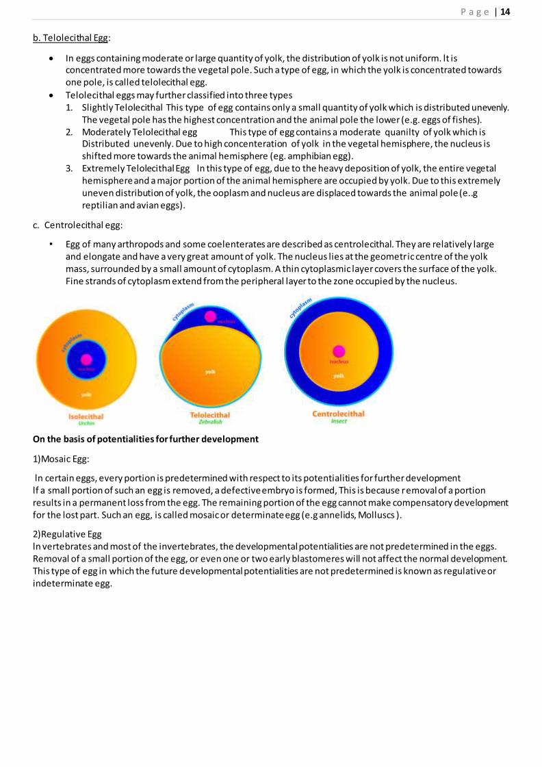
a. Isolecithal or Homolecithal Egg:
ln isolecithal eggs, the very little amount of yolk present is uniformly distributed throughout the ooplasm
(e.g echinoderms, Amphioxus, mammals). This condition is usually observed in eggs with very little amount of yolk.
P a g e | 14
b. Telolecithal Egg:
In eggs containing moderate or large quantity of yolk, the distribution of yolk is not uniform. lt is concentrated more towards the vegetal pole. Such a type of egg, in which the yolk is concentrated towards one pole, is called telolecithal egg.
Telolecithal eggs may further classified into three types 1. Slightly Telolecithal This type of egg contains only a small quantity of yolk which is distributed unevenly.
The vegetal pole has the highest concentration and the animal pole the lower (e.g. eggs of fishes). 2. Moderately Telolecithal egg This type of egg contains a moderate quanilty of yolk which is
Distributed unevenly. Due to high concenteration of yolk in the vegetal hemisphere, the nucleus is shifted more towards the animal hemisphere (eg. amphibian egg).
3. Extremely Telolecithal Egg ln this type of egg, due to the heavy deposition of yolk, the entire vegetal hemisphere and a major portion of the animal hemisphere are occupied by yolk. Due to this extremely uneven distribution of yolk, the ooplasm and nucleus are displaced towards the animal pole (e..g reptilian and avian eggs).
c. Centrolecithal egg:
• Egg of many arthropods and some coelenterates are described as centrolecithal. They are relatively large and elongate and have a very great amount of yolk. The nucleus lies at the geometric centre of the yolk mass, surrounded by a small amount of cytoplasm. A thin cytoplasmic layer covers the surface of the yolk. Fine strands of cytoplasm extend from the peripheral layer to the zone occupied by the nucleus.
On the basis of potentialities for further development
1)Mosaic Egg:
ln certain eggs, every portion is predetermined with respect to its potentialities for further development lf a small portion of such an egg is removed, a defective embryo is formed, This is because removal of a portion results in a permanent loss from the egg. The remaining portion of the egg cannot make compensatory development for the lost part. Such an egg, is called mosaic or determinate egg (e.g annelids, Molluscs ).
2)Regulative Egg ln vertebrates and most of the invertebrates, the developmental potentialities are not predetermined in the eggs. Removal of a small portion of the egg, or even one or two early blastomeres will not affect the normal development. This type of egg in which the future developmental potentialities are not predetermined is known as regulative or indeterminate egg.
P a g e | 15
Cleavage
Cleavage is a rapid series of mitotic divisions that occur just after fertilization.
There are two critical reasons why cleavage is so important:
Generation of a large number of cells that can undergo differentiation and gastrulation to form organs. Increase in the nucleus / cytoplasmic ratio. Eggs need a lot of cytoplasm to support embryogenesis. It is
difficult or impossible for one nucleus to support a huge cytoplasm, and oocytes are one of the largest cells that exist. One small nucleus just cannot transcribe enough RNA to meet the needs of the huge cytoplasm.
A larger nucleus to cytoplasmic ratio is optimal for cell function. Cell division occurs rapidly after fertilization to correct this problem.
Cleavage differs from normal mitoses in 2 respects
1. Blastomeres do not grow in size between successive cell divisions as they do in most cells. This leads to a
rapid increase in the nucleus/ cytoplasmic ratio. Cells undergoing cleavage have mainly S and M phases of
the cell cycle (little or no G1 or G2).
2. Cleavage occurs very rapidly, and mitosis and cytokinesis in each round of cell division are complete within
an hour. Typical somatic cells divide much more slowly (several hours to days) and even the fastest cancer cells divide much slower than occurs in a zygote during cleavage.
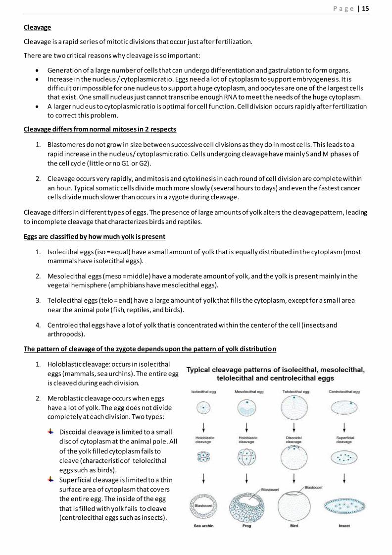
Cleavage differs in different types of eggs. The presence of large amounts of yolk alters the cleavage pattern, leading
to incomplete cleavage that characterizes birds and reptiles.
Eggs are classified by how much yolk is present
1. Isolecithal eggs (iso = equal) have a small amount of yolk that is equally distributed in the cytoplasm (most mammals have isolecithal eggs).
2. Mesolecithal eggs (meso = middle) have a moderate amount of yolk, and the yolk is present mainly in the vegetal hemisphere (amphibians have mesolecithal eggs).
3. Telolecithal eggs (telo = end) have a large amount of yolk that fills the cytoplasm, except for a small area
near the animal pole (fish, reptiles, and birds).
4. Centrolecithal eggs have a lot of yolk that is concentrated within the center of the cell (insects and arthropods).
The pattern of cleavage of the zygote depends upon the pattern of yolk distribution
1. Holoblastic cleavage: occurs in isolecithal
eggs (mammals, sea urchins). The entire egg
is cleaved during each division.
2. Meroblastic cleavage occurs when eggs
have a lot of yolk. The egg does not divide completely at each division. Two types:
Discoidal cleavage is limited to a small
disc of cytoplasm at the animal pole. All
of the yolk filled cytoplasm fails to
cleave (characteristic of telolecithal
eggs such as birds).
Superficial cleavage is limited to a thin
surface area of cytoplasm that covers
the entire egg. The inside of the egg
that is filled with yolk fails to cleave (centrolecithal eggs such as insects).

P a g e | 16
Sea Urchins Have Isolecithal Eggs And Undergo Holoblastic Cleavage
Cleavage plane: this is the plane in which cleavage occurs. It is oriented at right angles to the metaphase
plate. In sea urchins, the first cleavage is meridional.
Meridional cleavage runs from one pole to another (top to bottom), like the meridian on a globe.
The second cleavage is also meridional.
Equatorial cleavage encircles the zygote like the equator on the globe. The third cleavage in the sea urchin is equatorial. This creates an animal and vegetal half.
The fourth cleavage is unique. Equal cytokinesis occurs in the four blastomeres of the animal pole, giving rise
to 8 mesomeres (all the same size).
Unequal cytokinesis occurs in the vegetal pole. This causes 4 large macromeres and 4 small micromeres
The 5th cleavage is meridional. All mesomeres divide equally as do the macromeres.
As cleavage progresses, all blastomeres adhere at the outer surface, but attachment is lost at the inner
surface. The blastocoel is a cavity formed due to the unequal adherence of blastomeres .
Amphibian eggs have moderate amount of yolk, however, they are still able to undergo holoblastic cleavage.
The 1st cleavage is meridional, as is the 2nd. The 3rd cleavage is equatorial. The cleavage is displaced toward the
animal pole due to the yolk. This results in 4 small animal blastomeres and 4 large vegetal blastomeres.
Morula (morum = mulberry) at the 16 to 32 cell stage the embryo is called a morula because it looks like a mulberry.

P a g e | 17
The blastocoel is displaced to the animal pole in amphibians
Blastula = from the 128 cell stage onward the
amphibian embryo is a blastula. The outer surface of
the amphibian blastula has cells connected by
specialized cell junctions.
Tight junctions create a seal that isolates the outside
of an embryo from the inner layer. Tight junctions
polarize the apical and basal surfaces. The basal
portions of cells start secreting into the blastocoel.
Desmosomes attach the
blastomeres together on the
outside.
Gap junctions connect all
surface blastomeres.
Mammalian eggs have rotational cleavage that is holoblastic
The mammalian egg is a little slow. It begins to cleave in the oviduct and continues until it implants in the wall of the uterus (1 cleavage / 24 hr).
Asynchronous cleavage: mammalian embryos are unusual in that they have asynchronous cleavage. Not all blastomeres divide at the same time.
The first cleavage is meridional, and the second cleavage is rotational. The 2 blastomeres divide in different planes (one is equatorial and one is meridional.
Mammalian embryos undergo compaction at the 8 cell stage
At first, the blastomeres of mammalian embryos have a loose arrangement, and touch only at the basal surfaces.
After compaction, blastomeres adhere tightly, maximizing the area of contact.
During compaction, each blastomere undergoes polarization. Tight junctions develop on the outer surface,

P a g e | 18
allowing proteins to specialize.Cells take up fluids from the uterine environment and Secrete into the blastocoel.
Gap junctions form on the outer cells to aid in intercellular communication.
A blastocoel develops as cleavage proceeds to the 32-64 cell stage
After compaction at the 8-16 cell stage, there are 2 types of blastomeres. Outside blastomeres are tightly joined and number about 9-14. They surround 2-7 inside blastomeres that are loosely joined.
Cavitation: the outside blastomeres start to take up fluid from the uterus and pump it into the center, creating the blastocoel. The blastocyst is the characteristic of early embryonic development in mammals.
Inner cell mass: this gives rise to the embryo, and develops from the inside blastomeres
Trophoblast: a structure consisting of outside blastomeres, this contributes to forming the placenta.
Embryonic stem cells can be cultured from the inner cell mass
Cells in the inner cell mass are undifferentiated, they multiply indefinitely, and are known as embryonic stem cells. Stem cells are totipotent = they have the potential to form any tissue. These cells are of great scientific and medical importance.
They can be removed from the embryo, genes can be introduced into the cells, and then they can be placed back in the blastocyst. This is how one constructs transgenic or “knock out” mice.
The embryonic stem cells are also used to grow certain types of tissue in culture. Theoretically, it should be possible to grow structures such as ears, muscles, nerves, and skin for transplantation to sick individuals.
Interestingly, if you inject adult, differentiated cells back into the environment of the morula or blastula, they become undifferentiated, and they can redifferentiate to form many parts of the body.
Development of monozygotic or identical twins
Monozygotic twins develop from one zygote by splitting at various stages of development (from the 2 cell to the
blastocyst stage).
The stage of splitting effects the overall structure of the embryo and extra embryonic membranes.Conjoined twins
• are identical twins who develop from a single fertilized ovum.
• are always the same sex and race.
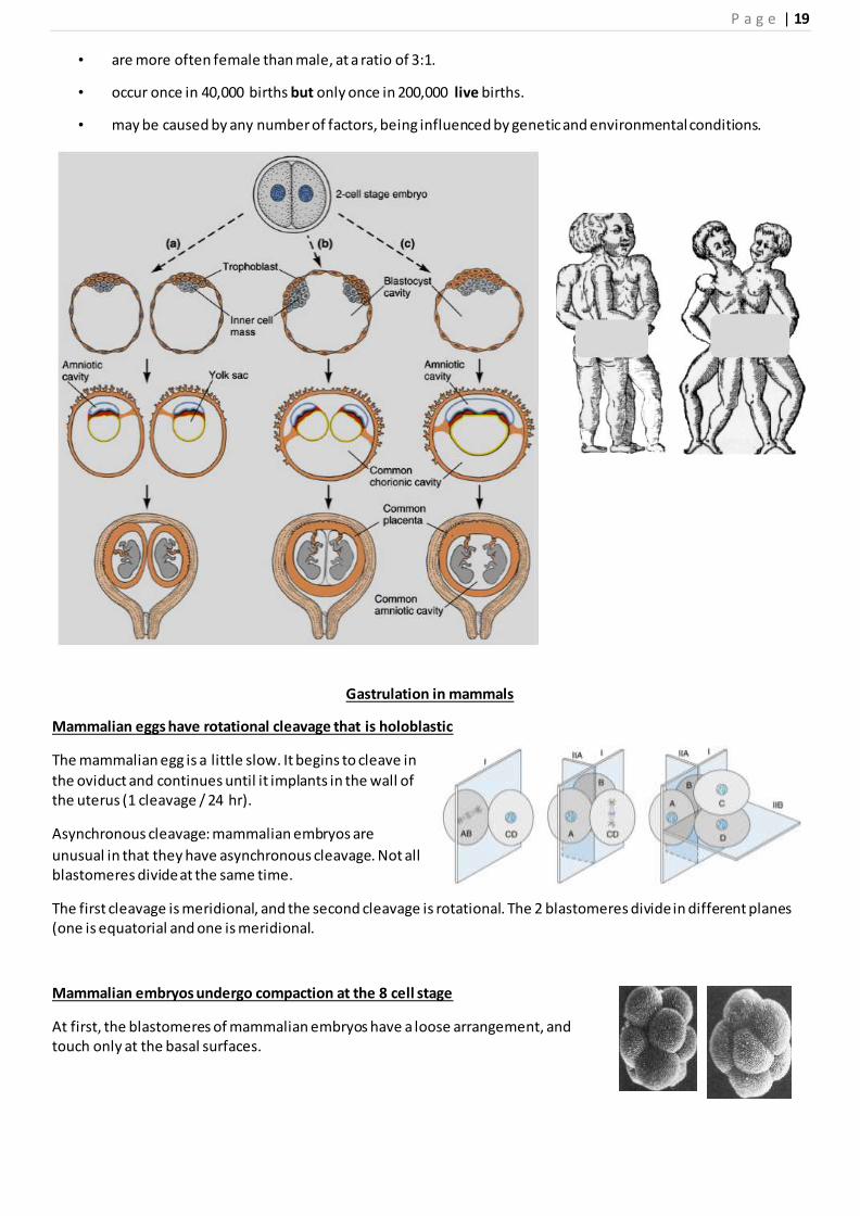
P a g e | 19
• are more often female than male, at a ratio of 3:1.
• occur once in 40,000 births but only once in 200,000 live births.
• may be caused by any number of factors, being influenced by genetic and environmental conditions.
Gastrulation in mammals
Mammalian eggs have rotational cleavage that is holoblastic
The mammalian egg is a little slow. It begins to cleave in
the oviduct and continues until it implants in the wall of the uterus (1 cleavage / 24 hr).
Asynchronous cleavage: mammalian embryos are
unusual in that they have asynchronous cleavage. Not all blastomeres divide at the same time.
The first cleavage is meridional, and the second cleavage is rotational. The 2 blastomeres divide in different planes (one is equatorial and one is meridional.
Mammalian embryos undergo compaction at the 8 cell stage
At first, the blastomeres of mammalian embryos have a loose arrangement, and touch only at the basal surfaces.

P a g e | 20
After compaction, blastomeres adhere tightly, maximizing the area of contact. During compaction, each blastomere undergoes polarization.
Tight junctions develop on the outer surface, allowing proteins to specialize.
Cells take up fluids from the uterine environment and Secrete into the blastocoel.
Gap junctions form on the outer cells to aid in intercellular communication.
A blastocoel develops as cleavage proceeds to the 32-64 cell stage
After compaction at the 8-16 cell stage, there are 2 types of blastomeres. Outside
blastomeres are tightly joined and number about 9-14. They surround 2-7 inside blastomeres that are loosely joined.
Cavitation: the outside blastomeres start to take up fluid from the uterus and
pump it into the center, creating the blastocoel. The blastocyst is the
characteristic of early embryonic development in mammals.
Inner cell mass: this gives rise to the embryo, and develops from the inside blastomeres
Trophoblast: a structure consisting of outside blastomeres, this contributes to forming the placenta.
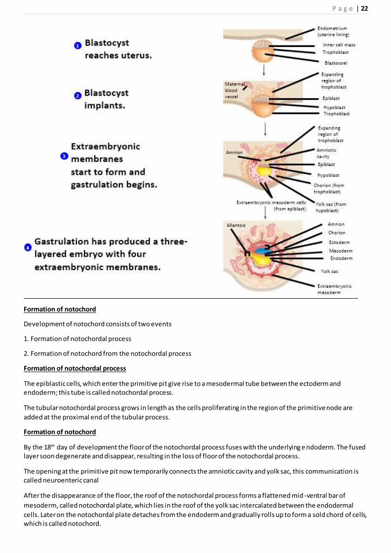
Beginning of implantation
The blastocyst lies free in the uterine secretions for about 2 days.
On the 6th day of development the zona pellucida disappears and the conceptus begins to implant into endometrium.
The trophoblast begin to differentiate into two layers
i. the outer layer called syncytiotrophoblast, consists of multinucleated protoplasmic mass in which no cell
boundaries can be distinguished
Ii .the inner layer, termed cytotrophoblast, is made up of mono-nucleated cells with distinct boundaries
The syncytiotrophoblast possess active invasive properties because it secrete hydrolytic enzymes
Completion of implantation
On day 7 the blastocyst become tightly adherent to the epithelium of the endometrium due to invasive activity of the syncytiotrophoblast.
The cells of endometrium respond by differentiating into large polyhedral cells, which are laden with lipids and glycogen.
The endometrium is now referred to as decidua.
As implantation progresses, the expanding syncytiotrophoblast gradually spreads over the outer surface of the
blastocyst and by day 9 the blastocyst is almost completely covered by a thick layer of syncytiotrophoblast.
Formation of bilaminar germ disc
With the beginning of the second week, the cells of the embryoblast become differentiated into two layers,
Epiblast and hypoblast
Formation of amniotic cavity
Development of primary yolk sac

P a g e | 21
Gastrulation
Gastrulation in mammals begins with the appearance of primitive streak on the dorsal surface of the embryonic disc
On day 15 of development the primitive streak can be seen in the longitudinal midline of the caudal part of the embryonic disc as a narrow groove with slightly elevated margins.
These elevations are produced by proliferation and accumulation of epiblastic cells.
The groove within the primitive streak is called primitive groove.
By the 16th day a small circular swelling appears at the cranial end of the primitive streak. This swelling called
primitive node, shows a small depression called primitive pit.
At its cranial end, the primitive groove become continuous with the primitive pit.
After formation of the primitive streak and appearance of the primitive groove, the epiblastic cells surrounding the primitive streak begin to proliferate, flatten and lose their connections with each other.
These epiblastic cells migrate towards the primitive streak
As the primitive groove is arrived, these cells slip through the groove into space between the epiblast and hypoblast.this inward movement of the epiblastic cells is called invagination.
Those epiblastic cells which enter the cranial part of the primitive streak invade the hypoblast and displace its cells, so that eventually the hypoblast becomes completely replaced by a new layer of cells called definitive endoderm.
The definitive endoderm forms the roof of the yolk sac.
Other epiblastic cells which invaginate through the primitive streak come to lie between the definitive endoderm
and the epiblast. These cells spread laterally and forwards to form a new layer known as embryonic mesoderm also called intraembryonic mesoderm.
After formation of the embryonic mesoderm, those epiblastic
cells which remain on the dorsal surface of the embryonic disc
now constitute embryonic ectoderm.
Under the embryonic mesoderm lies the definitive endoderm
which is now referred to as embryonic endoderm or simply endoderm.
Thus, the two-layered (bilaminar) embryonic disc (consisting
of epiblast and hypoblast) has now become three-layered (trilaminar).
The three layers of the trilaminar embryonic disc (ectoderm, mesoderm and endoderm) are called primary germ
layers. All tissues and organs of the human body will derive from these three layers.
P a g e | 22
Formation of notochord
Development of notochord consists of two events
1. Formation of notochordal process
2. Formation of notochord from the notochordal process
Formation of notochordal process
The epiblastic cells, which enter the primitive pit give rise to a mesodermal tube between the ectoderm and endoderm; this tube is called notochordal process.
The tubular notochordal process grows in length as the cells proliferating in the region of the primitive node are
added at the proximal end of the tubular process.
Formation of notochord
By the 18th day of development the floor of the notochordal process fuses with the underlying e ndoderm. The fused layer soon degenerate and disappear, resulting in the loss of floor of the notochordal process.
The opening at the primitive pit now temporarily connects the amniotic cavity and yolk sac, this communication is called neuroenteric canal
After the disappearance of the floor, the roof of the notochordal process forms a flattened mid -ventral bar of
mesoderm, called notochordal plate, which lies in the roof of the yolk sac intercalated between the endodermal
cells. Later on the notochordal plate detaches from the endoderm and gradually rolls up to form a sold chord of cells, which is called notochord.
P a g e | 23
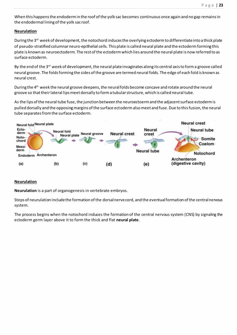
When this happens the endoderm in the roof of the yolk sac becomes continuous once again and no gap remains in the endodermal lining of the yolk sac roof.
Neurulation
During the 3rd week of development, the notochord induces the overlying ectoderm to differentiate into a thick plate
of pseudo-stratified columnar neuro-epithelial cells. This plate is called neural plate and the ectoderm forming this
plate is known as neuroectoderm. The rest of the ectoderm which lies around the neural plate is now referred to as surface ectoderm.
By the end of the 3rd week of development, the neural plate invaginates along its central axis to form a groove called
neural groove. The folds forming the sides of the groove are termed neural folds. The edge of each fold is known as neural crest.
During the 4th week the neural groove deepens, the neural folds become concave and rotate around the neural groove so that their lateral lips meet dorsally to form a tubular structure, which is called neural tube.
As the lips of the neural tube fuse, the junction between the neuroectoerm and the adjacent surface ectoderm is
pulled dorsally and the opposing margins of the surface ectoderm also meet and fuse. Due to this fusion, the neural tube separates from the surface ectoderm.
Neurulation
Neurulation is a part of organogenesis in vertebrate embryos.
Steps of neurulation include the formation of the dorsal nerve cord, and the eventual formation of the central nervous system.
The process begins when the notochord induces the formation of the central nervous system (CNS) by signaling the ectoderm germ layer above it to form the thick and flat neural plate.

P a g e | 24
The neural plate folds in upon itself to form the neural tube, which will later differentiate into the spinal cord and the brain, eventually forming the central nervous system.
Primary neurulation
Induction
Primary neurulation occurs in response to
soluble growth factors secreted by
the notochord. Ectodermal cells are induced
to form neuroectoderm from a variety of
signals. Ectoderm sends and receives signals
of BMP4 (bone morphogenic protein) and
cells which receive BMP4 signal develop into
epidermis.
The inhibitory
signals chordin, noggin and follistatin are needed to form neural plate.
These inhibitory signals are created and
emitted by the notochord. Cells which do
not receive BMP4 signaling due to the
effects of the inhibitory signals will develop
into the anterior neuroectoderm cells of the
neural plate.
Shape Change
The cells of the neural plate are signaled to
become high-columnar and can be identified
through microscopy as different from the surrounding epiblastic ectoderm. The cells move laterally and away from
the central axis and change into a truncated pyramid shape.
Folding
The process of the flat neural plate folding into the cylindrical neural tube is termed primary neurulation
As a result of the cellular shape changes, the neural plate forms the medial hinge point (MHP). The expanding
epidermis puts pressure on the MHP and causes the neural plate to fold resulting in neural folds and the creation of the neural groove. The neural folds meet and fuse at the midline.
The folding of the neural tube to form an actual tube does not occur all at once.
The lateral edges of the neural plate touch in the midline and join together. This continues both cranially and caudally.
The openings that are formed at the cranial and caudal regions are termed the cranial and caudal neuropores. In human embryos, the cranial neuropore closes approximately on day 25 and the caudal neuropore on day 27.

P a g e | 25
Secondary Neurulation
In secondary neurulation, the neural ectoderm and some
cells from the endoderm form the medullary cord. The
medullary cord condenses, separates and then forms
cavities. These cavities then merge to form a single tube.
Secondary Neurulation occurs in the posterior section of
most animals but it is better expressed in birds. Tubes from
both primary and secondary neurulation eventually connect.
Early brain development
The anterior segment of the neural tube forms the three
main parts of the brain: the forebrain, midbrain, and the hindbrain. Formation of these structures begins with a
swelling of the neural tube. Ion pumps are used to increase the fluid pressure within the tube and create a bulge. A
blockage between the brain and the spinal cord prevents the fluid accumulation from leaking out. These brain regions
further divide into sub-regions. The neural tube becomes the germinal neuro-epithelium and serves as a source of new
neurons during brain development.
Non-neural ectoderm tissue
Mesoderm surrounding the notochord at the sides will devel op into the somites (future muscles, bones, and contributes to the formation of limbs of the vertebrate).
Neural crest cells
Masses of tissue called the neural crest that are located at the very edges of the lateral plates of the folding neural
tube separate from the neural tube and migrate to become a variety of cell populations, including the cells of the
peripheral nervous system.