Developmental Biology – Biology 4361pschoff/documents/EndodermalandMesodermalOrg… · ...
29
Endodermal and Mesodermal Organs Developmental Biology – Biology 4361 December 5, 2005 Part 1
Transcript of Developmental Biology – Biology 4361pschoff/documents/EndodermalandMesodermalOrg… · ...

Endodermal and Mesodermal Organs
Developmental Biology – Biology 4361
December 5, 2005
Part 1

Mammalian gastrulation

Figure 14.2
Gut formation – lateral folding
the gut closes by lateral folding
Mammals endoderm is open to the adjacent yolk or to the yolk sac

Gut formation – craniocaudal flextion
Figure 14.3
Lateral folding is enhanced by craniocaudial flexion: rapid extension of neural plate bends embryo
The wide opening between gut and yolk sac is reduced to narrow duct = vitelline duct
18 d 24 d
30 d
21 d
28 d 18 d

4 weeks 5 weeks
Endodermal derivatives
Figure 14.4
pharyngeal pouches trachea rudiment connects to the lungs liver and pancreas rudiments urinary bladder (with allantois)
Gut derivatives:
mesenchymal (mesoderm) cells will surround the tube to form muscles, connective tissue, cartilage
endodermal cells generate only the lining of the digestive tube Gut sections: pharynx – forgut – midgut – hindgut

Pharyngeal pouch formation
endoderm displaces mesoderm endoderm induces cleft mesenchyme forms arches
Figure 14.5
generalized vertebrate embryo shark adult
in fish pharyngeal pouches (arches) develop into spiracle, gills
also jaws! (primitive fish)

Pharyngeal arch formation
Adapted from Gilbert 1994, p. 284
neural crest cell migration
Figure 14.1 Human embryo 31 d
pharyngeal arch: endoderm mesoderm

Pharyngeal arches
A. Arches 1, 2; buccopharyngeal membrane remnant (arrow) B. Mesoderm core lined by ectoderm (arrows), endoderm (arrowheads)
A B
Phayrngeal arch: precartilage cells (NC) premuscle mesenchyme blood vessel cranial nerve
Figure 14.1 Human embryo 31 d

stomatodeum
cardiac bulge
Pharyngeal arches – frontal view

Phylotypic Stage Vertebrates
Figure 14.9
developmental stage that is very similar for all species of a phylum
Phylotypic stage:
notochord brain rudiments sense organs gut heart
Similar body plans:
Why?

Class Agnatha – jawless fishes

Figure 14.6
Pharyngeal arch cartilage
develop into gills & jaws in primitive fish have acquired other functions in terrestrial vertebrates
Pharyngeal arches
Developing shark skull


Mammalian jaw and ossicle development
Figure 14.7 1st pharyngeal arch cartilage forms primary jaw joint (vertebrates, exc. mammals)
Mammals secondary jaw joint replaces primary jaw joint; formed by dentary and squamosal bones.
Parts of primary jaw cartilage persists as middle ear bones: mandibular cartilage → malleus (hammer); quadrate cartilage → incus (anvil)
also hyomandibular cartilage (anchors jaw to braincase) → stapes
10 week fetus adult
secondary
primary

Figure 15.23 in SF Gilbert: Developmental Biology 7 th ed, 2003; see Figure 14.8 in Kalthoff
Pharyngeal pouch derivatives human
1st → auditory cavities of middle ear and Eustachian tubes
2nd → walls of the tonsils (gland = lymphoid tissue)
(NOTE thyroid gland forms from an unpaired thyroid primordium between the 2nd pair of pouches)
3rd → dorsal lower (inferior) parathyroid glands
→ thymus
4th → upper (superior) parathyroid glands
→ ultimobranchial (postbranchial) body

4 weeks 5 weeks
Endodermal derivatives
Figure 14.4
pharyngeal pouches trachea rudiment – forms and connects to lungs liver and pancreas rudiments urinary bladder (with allantois)
Gut derivatives:

Figure 14.11
Hindgut/cloaca human

tracheal rudiment forms branches three on right two on left (humans)
tracheal rudiment forms bronchi
bronchi form bronchioli
alveoli form at ends of bronchi
Endoderm forms the inner epithelium of trachea, bronchi & lungs
Mesodermal forms connective tissue, blood vessels, cartilage of the trachea and lungs
Lung development
Figure 14.10
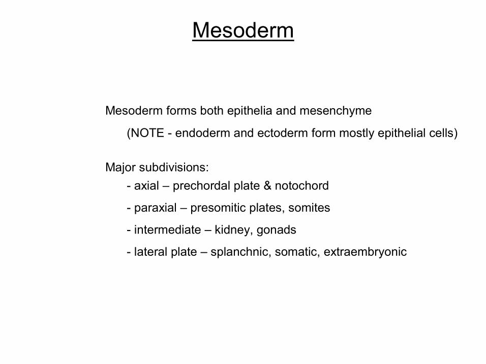
Mesoderm
Mesoderm forms both epithelia and mesenchyme
(NOTE endoderm and ectoderm form mostly epithelial cells)
Major subdivisions: axial – prechordal plate & notochord
paraxial – presomitic plates, somites
intermediate – kidney, gonads
lateral plate – splanchnic, somatic, extraembryonic

SF Gilbert: Developmental Biology 7 th ed, 2003
Major lineages of the mesoderm

Mesoderm differentiation
Figure 14.12

SF Gilbert: Developmental Biology 7 th ed, 2003
Major lineages of the mesoderm axial

Figure 14.13
Axial mesoderm – notochord formation
in vertebrates (exc. mammals) – notochord replaced by vertebral column
in mammals – notochord remnants remain in intervertebral discs
notochord posterior head, in neck, trunk and tail dorsal rod of cartilagelike connective tissue persists in primitive marine Chordata
urochordates (tunicates) cephalochordates (brachiostomes)
prechordal plate (anterior head region) forms mesenchyme contributes to cranial cartilage
Axial mesoderm forms along dorsal midline
tunicate larva, adult
notochord

SF Gilbert: Developmental Biology 7 th ed, 2003
Major lineages of the mesoderm paraxial

Paraxial mesoderm – somite formation
Figure 14.14
presomitic plates form somitomeres somitomeres “whorls” of mesenchymal cells
paraxial mesoderm forms somites
presomitic plates form as node regresses
somitomeres mature into somites

Regulation of somite formation
Hairy1 expression wave
Hairy1
periodic expression of Eph receptor tyrosine kinases periodic expression of ephrins (Eph ligands)
Eph targets include Notch signaling pathway
Notch also regulates the periodic expression of Hairy1 ~ 90 min periodicity
Notch directs the placement of somite borders
Eph/ephrins involved in cellcell repulsion
Figure 14.6 in SF Gilbert: Developmental Biology 7 th ed, 2003

Epithelialization of somites
Epithelialization: solid mesenchymal mesoderm transforms into hollow epithelial ball
synthesize extracellular matrix proteins; e.g. fibronectin, Ncadherin (see above) cells form tight junctions between basal lamina cells polarize: subapical surface (inward); basal membrane (outside)
Figure 14.16

(a) somite – forms epithelial sac
(c) epaxial & hypaxial myotome form myotome epaxial – dorsal trunk muscles hypaxial – detach from myotome; form
limb muscles, ventral trunk muscles
(d) dermatome cells become mesenchymal; form dermis
(b) ventral & medial cells separate; migrate towards notochord, neural tube = sclerotome; cartilage precursors of vertebrae remainder of somite = dermamyotome
dorsomedial marginal lip = epaxial myotome ventrolateral marginal lip = hypaxial myotome
Somite derivations
Figure 14.17

Somite Patterning
Figure 14.18
Somites are exposed to signals from surrounding organ rudiments:
ventralizing: sonic hedgehog (Shh) also medialize dosedependent:
Shh high = sclerotome Shh lower = myotome
dorsalizing: Wnt family antagonizes Shh
lateralizing: bone mophogenic protein (BMP4) inhibits dorsalizing signal (noggin?)