Development, validation and regulatory acceptance of ...
11
METHODOLOGY Open Access Development, validation and regulatory acceptance of improved purification and simplified quality control of [ 13 N] Ammonia Daniel L. Yokell 1,2* , Peter A. Rice 1 , Ramesh Neelamegam 1,2 and Georges El Fakhri 1,2 * Correspondence: dyokell@mgh. harvard.edu 1 Department of Radiology, Gordon Center for Medical Imaging, Massachusetts General Hospital, 55 Fruit Street, Edwards 019B, Boston, MA 02114, USA 2 Department of Radiology, Harvard Medical School, Boston, MA, USA Abstract Background: [ 13 N]Ammonia is a cyclotron produced myocardial perfusion imaging agent. With the development of high-yielding [ 13 N]ammonia cyclotron targets using a solution of 5 mM ethanol in water, there was a need to develop and validate an automated purification and formulation system for [ 13 N]ammonia to be in a physiological compatible formulation of 0.9% sodium chloride since there is no widely available commercial system at this time. Due to its short half-life of 10 min, FDA and USP regulations allow [ 13 N]ammonia to be tested in quality control (QC) sub-batches with limited quality control testing performed on the sub-batches for patient use. The current EP and the original USP method for the determination of the radiochemical purity and identity of [ 13 N]ammonia depended on an HPLC method using a conductivity detector and a solvent free of other salts. This HPLC method created issues in a modern cGMP high volume PET manufacturing facility where the HPLC is used with salt containing mobile phase buffers for quality control analysis of other PET radiopharmaceuticals. Flushing of the HPLC system of residual salt buffers which may interfere with the [ 13 N]ammonia assay can take several hours of instrument time. Since there are no mass limits on [ 13 N]ammonia, a simplified TLC assay to determine radiochemical identity and purity could be developed to simplify and streamline QC. Results: We have developed and validated a streamlined automated synthesis for [ 13 N]ammonia which provides the drug product in 8 mL of 0.9% sodium chloride for injection. A novel radio-TLC method was developed and validated to demonstrate feasibility to quantitate [ 13 N]ammonia and separate it from all known radiochemical impurities. (Continued on next page) © The Author(s). 2020 Open Access This article is licensed under a Creative Commons Attribution 4.0 International License, which permits use, sharing, adaptation, distribution and reproduction in any medium or format, as long as you give appropriate credit to the original author(s) and the source, provide a link to the Creative Commons licence, and indicate if changes were made. The images or other third party material in this article are included in the article's Creative Commons licence, unless indicated otherwise in a credit line to the material. If material is not included in the article's Creative Commons licence and your intended use is not permitted by statutory regulation or exceeds the permitted use, you will need to obtain permission directly from the copyright holder. To view a copy of this licence, visit http://creativecommons.org/licenses/by/4.0/. EJNMMI Radiopharmacy and Chemistry Yokell et al. EJNMMI Radiopharmacy and Chemistry (2020) 5:11 https://doi.org/10.1186/s41181-020-00097-7
Transcript of Development, validation and regulatory acceptance of ...

METHODOLOGY Open Access
Development, validation and regulatoryacceptance of improved purification andsimplified quality control of [13N] AmmoniaDaniel L. Yokell1,2* , Peter A. Rice1, Ramesh Neelamegam1,2 and Georges El Fakhri1,2
* Correspondence: [email protected] of Radiology, GordonCenter for Medical Imaging,Massachusetts General Hospital, 55Fruit Street, Edwards 019B, Boston,MA 02114, USA2Department of Radiology, HarvardMedical School, Boston, MA, USA
Abstract
Background: [13N]Ammonia is a cyclotron produced myocardial perfusion imagingagent. With the development of high-yielding [13N]ammonia cyclotron targets usinga solution of 5 mM ethanol in water, there was a need to develop and validate anautomated purification and formulation system for [13N]ammonia to be in aphysiological compatible formulation of 0.9% sodium chloride since there is nowidely available commercial system at this time. Due to its short half-life of 10 min,FDA and USP regulations allow [13N]ammonia to be tested in quality control (QC)sub-batches with limited quality control testing performed on the sub-batches forpatient use. The current EP and the original USP method for the determination ofthe radiochemical purity and identity of [13N]ammonia depended on an HPLCmethod using a conductivity detector and a solvent free of other salts. This HPLCmethod created issues in a modern cGMP high volume PET manufacturing facilitywhere the HPLC is used with salt containing mobile phase buffers for quality controlanalysis of other PET radiopharmaceuticals. Flushing of the HPLC system of residualsalt buffers which may interfere with the [13N]ammonia assay can take several hoursof instrument time. Since there are no mass limits on [13N]ammonia, a simplified TLCassay to determine radiochemical identity and purity could be developed to simplifyand streamline QC.
Results: We have developed and validated a streamlined automated synthesisfor [13N]ammonia which provides the drug product in 8 mL of 0.9% sodiumchloride for injection. A novel radio-TLC method was developed and validated todemonstrate feasibility to quantitate [13N]ammonia and separate it from allknown radiochemical impurities.
(Continued on next page)
© The Author(s). 2020 Open Access This article is licensed under a Creative Commons Attribution 4.0 International License, whichpermits use, sharing, adaptation, distribution and reproduction in any medium or format, as long as you give appropriate credit to theoriginal author(s) and the source, provide a link to the Creative Commons licence, and indicate if changes were made. The images orother third party material in this article are included in the article's Creative Commons licence, unless indicated otherwise in a creditline to the material. If material is not included in the article's Creative Commons licence and your intended use is not permitted bystatutory regulation or exceeds the permitted use, you will need to obtain permission directly from the copyright holder. To view acopy of this licence, visit http://creativecommons.org/licenses/by/4.0/.
EJNMMI Radiopharmacy and Chemistry
Yokell et al. EJNMMI Radiopharmacy and Chemistry (2020) 5:11 https://doi.org/10.1186/s41181-020-00097-7

(Continued from previous page)
Conclusions: The process for automated synthesis of [13N]ammonia simplifiesand automates the purification and formulation of [13N]ammonia in a cGMPcompliant manner needed for high-throughput manufacture of [13N]ammonia.The novel radio-TLC method has simplified [13N]ammonia quality control (QC)and now enables it to be tested using the same QC equipment as[18F]fludeoxyglucose (FDA/USP recognized name for 2-[18F]fluoro-2-deoxy-D-glucose). Both the streamlined automated synthesis of [13N]ammonia and thenovel radio-TLC method have been accepted and approved by the US Food andDrug Administration (FDA) for the cGMP manufacture of [13N]ammonia.
Keywords: [13N]Ammonia, [13N]NH3, Cardiac PET, Automated radiochemistry,Regulatory, cGMP PET drug manufacturing, PET radiopharmaceutical quality control
Background[13N]Ammonia is a myocardial perfusion imaging agent, which is approved by US
Food and Drug Administration (FDA) for diagnostic Positron Emission Tomog-
raphy (PET) imaging of the myocardium under rest or pharmacologic stress condi-
tions to evaluate myocardial perfusion in patients with suspected or existing
coronary artery disease.(Dilsizian et al. 2016) In the United States, the only other
FDA approved alternative PET myocardial perfusion agent is [82Rb]rubidium chlor-
ide, which requires a generator system by the patient’s side due to the short half-
life. With advances in cyclotron capabilities and targetry associated with in-target
[13N]ammonia production, in excess of 37 GBq (1 Ci) of [13N]ammonia can be pro-
duced per batch which makes it feasible with the 10-min half-life to inject and
image more than one patient per batch and even transport it short distances from
the cyclotron. Due to the expanding infrastructure globally of cyclotron and PET
cameras, there has been rapidly increasing interest in [13N]ammonia for PET myo-
cardial perfusion imaging outside of the US.(Underwood et al. 2014)
The [13N]ammonia in-target production method produces [13N]ammonia in water
with 5 mM ethanol.(Wieland et al. 1991) This formulation vehicle while acceptable, is
less than ideal than a physiological compatible solution like 0.9% sodium chloride.
Additionally, the [13N]ammonia in water may contain trace long lived radionuclidic
impurities from the target body and/or target windows depending on the cyclotron
target design. Most of the existing methods described for purification and formulation
of [13N]ammonia are either manual loading and elution of solid phase extraction
cartridges (SPEs) or complicated dedicated [13N]ammonia systems, both of which are
challenging to validate in a cGMP manufacturing environment.(Frank et al. 2019;
Pieper et al. 2019; Kumar et al. 2009) We set out to design a simple method which
could be adapted and validated on several different commercial available platforms,
including cassette based systems for easy scale up for high-volume [13N]ammonia
production at a busy PET center.
The original United States Pharmacopeia (USP)([13N]Ammonia Monograph n.d.-a)
and European Pharmacopeia (EP)([13N]Ammonia Monograph n.d.-b) methods for
radiochemical purity and identity determination of [13N]ammonia currently require the
use of an HPLC system configured with a conductivity detector. The current compen-
dial EP and the original USP HPLC conductivity methods are resource intensive,
Yokell et al. EJNMMI Radiopharmacy and Chemistry (2020) 5:11 Page 2 of 11

requiring lengthy mobile phase preparation and system suitability determination, while
occupying valuable instrument time. Additionally, these detectors are only required for
anion or cation chromatography, which is an expensive investment for a PET center to
use only for [13N]ammonia, sodium [18F] fluoride and other investigational cation/anion
radiopharmaceuticals. In our experience, use of a conductivity detector on an HPLC also
used for traditional reverse phase chromatography can create system suitability and base-
line noise issues in the cation analysis if proper care is not taken to properly flush the sys-
tem lines of salt buffers used in reverse phase analysis. System suitability failures can lead
to extensive delays in the busy production schedule of PET radiopharmaceuticals.
In this paper, we describe the development, validation and regulatory acceptance by
FDA and USP of an ideal alternative method that would rapidly and reproducibly de-
termine the radiochemical purity and radiochemical identity of [13N]ammonia, while
requiring less time to complete and fewer resources to maintain than compendial
methods. A TLC method for determining radiochemical purity and identity eliminates
the need to use HPLC for [13N]ammonia quality control. This is highly desirable in a
PET production facility that produces multiple radiopharmaceuticals per day.
Materials and methodsChemicals and reagents
All chemicals and reagents were obtained from commercial vendors and used without
further purification.
Automated purification and formulation of [13N]Ammonia
[13N]Ammonia is produced on a modified GE Tracerlab FXFDG synthesis module
which was replumbed for the purification and formulation of [13N]ammonia. The
graphic user interface is shown in Fig. 1 and the purification and formulation steps are
further detailed below. The QMA chloride and CM cartridges are prepared day of use
by washing with 10mL of sterile water for injection, USP and then capping the car-
tridges with sterile male/female luer caps. Prior to the first sub-batch of the day, the
synthesis box vacuum is tested and the system fluid paths are washed with sterile water
for injection, USP. The cyclotron [13N]ammonia target prior to the day’s use is primed
with fresh 5 mM ethanol solution.
1. [13N]Ammonia is produced on-site with a GE PETrace 860 cyclotron using the
method(Wieland et al. 1991) of irradiation of 5 mM ethanol in water
in-target synthesis method using a GE niobium body target with HAVAR/Niobium
double target window or GE silver body target with HAVAR window (GE Medical
Systems, Uppsala, Sweden). The target solution is pressurized to approximately
0.62MPa (90 psi) for the silver body target or to approximately 1.31MPa (190 psi)
for the niobium body target with helium gas. The target is bombarded with 16.5
MeV protons using a beam current ranging from 15 to 50 μA using bombardment
times between 5 and 30 min. For the [13N]ammonia batches cited in Tables 1,
50 μA, 30 min bombardments were used.
2. [13N]Ammonia is transferred from the cyclotron target via 0.45MPa (65 psi)
helium overpressure to the automated synthesis unit and through an in-line anion
Yokell et al. EJNMMI Radiopharmacy and Chemistry (2020) 5:11 Page 3 of 11
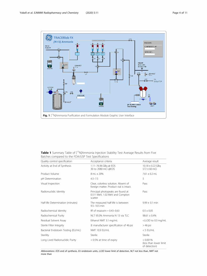
Fig. 1 [13N]Ammonia Purification and Formulation Module Graphic User Interface
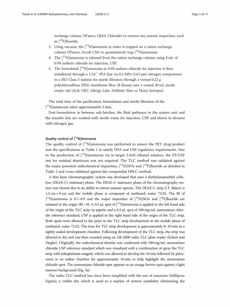
Table 1 Summary Table of [13N]Ammonia Injection Stability Test Average Results from FiveBatches compared to the FDA/USP Test Specifications
Quality control specification Acceptance criteria Average result
Activity at End of Synthesis 1.11–76.96 GBq @ EOS30 to 2080 mCi @EOS
10.18 ± 0.22 GBq572 ± 60 mCi
Product Volume 8mL ± 20% 7.61 ± 0.2 mL
pH Determination 4.5–7.5 5
Visual Inspection Clear, colorless solution. Absent offoreign matter. Product vial is intact.
Pass
Radionuclidic Identity Principal photopeaks are found at0.511 MeV, 1.02 MeV and Comptonscatter
Pass
Half-life Determination (minutes) The measured half-life is between9.5–10.5 min
9.99 ± 0.1 min
Radiochemical Identity Rf of resazurin = 0.43–0.63 0.5 ± 0.05
Radiochemical Purity NLT 95.0% Ammonia N 13 via TLC 98.61 ± 0.4%
Residual Solvent Assay Ethanol NMT 3.1 mg/mL <LLOD to 0.5 mg/mL
Sterile Filter Integrity ≥ manufacturer specification of 46 psi > 46 psi
Bacterial Endotoxin Testing (EU/mL) NMT 10.9 EU/mL < 5 EU/mL
Sterility Sterile Sterile
Long Lived Radionuclidic Purity < 0.5% at time of expiry < 0.001%(less than lower limitof detection)
Abbreviations: EOS end of synthesis, EU endotoxin units, LLOD lower limit of detection, NLT not less than, NMT notmore than
Yokell et al. EJNMMI Radiopharmacy and Chemistry (2020) 5:11 Page 4 of 11
exchange column (Waters, QMA Chloride) to remove any anionic impurities, such
as [18F]fluoride.
3. Using vacuum, the [13N]ammonia in water is trapped on a cation exchange
column (Waters, Accell CM) to quantitatively trap [13N]ammonia.
4. The [13N]ammonia is released from the cation exchange column using 8 mL of
0.9% sodium chloride for injection, USP.
5. The formulated [13N]ammonia in 0.9% sodium chloride for injection is then
transferred through a 1/16″ PFA line via 0.1 MPa (14.5 psi) nitrogen overpressure
to a ISO Class 5 isolator for sterile filtration through a vented 0.22 μ
polyethersulfone (PES) membrane filter (B Braun) into a vented 30 mL sterile
empty vial (ALK OKC Allergy Labs, Hollister Stier or Huayi Isotopes).
The total time of the purification, formulation and sterile filtration of the
[13N]ammonia takes approximately 5 min.
Post-formulation in between sub-batches, the fluid pathways in the system unit and
the transfer line are washed with sterile water for injection, USP and blown to dryness
with nitrogen gas.
Quality control of [13N]Ammonia
The quality control of [13N]ammonia was performed to ensure the PET drug product
met the specifications in Table 1 to satisfy FDA and USP regulatory requirements. Due
to the production of [13N]ammonia via in-target 5 mM ethanol solution, the EP/USP
test for residual aluminum was not required. The TLC method was validated against
the major potential radiochemical impurities, [13N]NOx and [18F]fluoride as detailed in
Table 2 and cross-validated against the compendial HPLC method.
A thin layer chromatographic system was developed that uses a diethylaminoethyl cellu-
lose (DEAE-C) stationary phase. The DEAE-C stationary phase of the chromatography sys-
tem was chosen due to its ability to attract anionic species. The DEAE-C strip (J.T. Baker) is
1.5 cm × 8 cm and the mobile phase is composed of methanol: water 75:25. The Rf of
[13N]ammonia is 0.7–0.9 and the major impurities of [13N]NOx and [18F]fluoride are
retained at the origin (Rf = 0). A 0.5 μL spot of [13N]ammonia is applied to the left hand side
of the origin of the TLC strip via pipette and a 0.5 μL spot of 100mg/mL ammonium chlor-
ide reference standard, USP is applied to the right hand side of the origin of the TLC strip.
Both spots were allowed to dry prior to the TLC strip development in the mobile phase of
methanol: water 75:25. The time for TLC strip development is approximately 8–10min in a
tightly sealed development chamber. Following development of the TLC strip, the strip was
allowed to dry and was then counted using an AR-2000 radio-TLC plate reader (Eckert and
Ziegler). Originally, the radiochemical identity was confirmed with 100mg/mL ammonium
chloride USP reference standard which was visualized with a combination of spray the TLC
strip with iodoplatinate reagent, which was allowed to develop for 10min followed by place-
ment in an iodine chamber for approximately 10min to help highlight the ammonium
chloride spot. The ammonium chloride spot appears as an orange brown spot against a light
maroon background (Fig. 3a).
The radio-TLC method has since been simplified with the use of resazurin (Millipore
Sigma), a visible dye which is used as a marker of system suitability eliminating the
Yokell et al. EJNMMI Radiopharmacy and Chemistry (2020) 5:11 Page 5 of 11
need to use ammonium chloride and the complicated development process of iodopla-
tinate reagent and iodine chamber to visualize the standard. Additionally, the visible
dye aids the operator by providing a visual pink-purple streak indicating proper TLC
development. A 0.5 μL spot of [13N]ammonia is applied to the left hand side of the ori-
gin of the TLC strip via pipette and a 0.5 μL spot of resazurin dye (1 mg/mL) is applied
to the right hand side of the origin of the TLC strip. Both spots were allowed to dry
prior to the TLC strip development in the mobile phase of methanol: water 75:25. The
other components of the TLC assay remain the same as described above. The Rf of
resazurin is 0.43–0.63 and an example TLC strip is shown in Fig. 3b. Table 2 below
contains the validation data of the [13N]ammonia TLC assay using resazurin as a sys-
tem suitability marker. For system suitability purposes, we require the front of the resa-
zurin peak to be 34–50mm.
Complete quality control testing and the [13N]ammonia test specifications for five
high activity stability batches is described in Table 1. For routine clinical production, a
quality control (QC) sub-batch is performed prior to manufacturing any sub-batches
for patient use. The routine clinical QC sub-batch QC process takes approximately 25
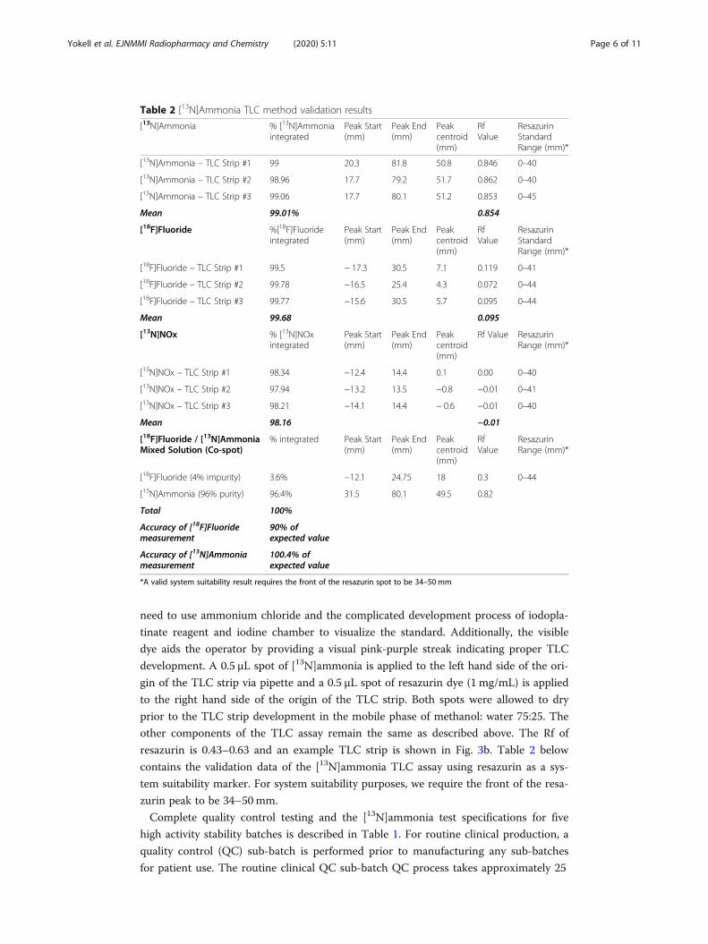
Table 2 [13N]Ammonia TLC method validation results
[13N]Ammonia % [13N]Ammoniaintegrated
Peak Start(mm)
Peak End(mm)
Peakcentroid(mm)
RfValue
ResazurinStandardRange (mm)*
[13N]Ammonia – TLC Strip #1 99 20.3 81.8 50.8 0.846 0–40
[13N]Ammonia – TLC Strip #2 98.96 17.7 79.2 51.7 0.862 0–40
[13N]Ammonia – TLC Strip #3 99.06 17.7 80.1 51.2 0.853 0–45
Mean 99.01% 0.854
[18F]Fluoride %[18F]Fluorideintegrated
Peak Start(mm)
Peak End(mm)
Peakcentroid(mm)
RfValue
ResazurinStandardRange (mm)*
[18F]Fluoride – TLC Strip #1 99.5 − 17.3 30.5 7.1 0.119 0–41
[18F]Fluoride – TLC Strip #2 99.78 −16.5 25.4 4.3 0.072 0–44
[18F]Fluoride – TLC Strip #3 99.77 −15.6 30.5 5.7 0.095 0–44
Mean 99.68 0.095
[13N]NOx % [13N]NOxintegrated
Peak Start(mm)
Peak End(mm)
Peakcentroid(mm)
Rf Value ResazurinRange (mm)*
[13N]NOx – TLC Strip #1 98.34 −12.4 14.4 0.1 0.00 0–40
[13N]NOx – TLC Strip #2 97.94 −13.2 13.5 −0.8 −0.01 0–41
[13N]NOx – TLC Strip #3 98.21 −14.1 14.4 − 0.6 −0.01 0–40
Mean 98.16 −0.01
[18F]Fluoride / [13N]AmmoniaMixed Solution (Co-spot)
% integrated Peak Start(mm)
Peak End(mm)
Peakcentroid(mm)
RfValue
ResazurinRange (mm)*
[18F]Fluoride (4% impurity) 3.6% −12.1 24.75 18 0.3 0–44
[13N]Ammonia (96% purity) 96.4% 31.5 80.1 49.5 0.82
Total 100%
Accuracy of [18F]Fluoridemeasurement
90% ofexpected value
Accuracy of [13N]Ammoniameasurement
100.4% ofexpected value
*A valid system suitability result requires the front of the resazurin spot to be 34–50 mm
Yokell et al. EJNMMI Radiopharmacy and Chemistry (2020) 5:11 Page 6 of 11
min. All of the QC tests are detailed below which are performed on the quality control
sub-batch, except for the periodic quality indicating tests (PQIT), which are performed
at their defined testing periods. For the patient sub-batches of [13N]ammonia, partial
quality control testing is performed, which is composed of final product vial visual in-
spection, product assay, and sterile filter integrity test.
Sterile filter integrity is performed using a manual bubble point test using a variable
pressure gas source with a calibrated pressure gauge according to filter manufacturing
directions. Visual inspection of the final product vial is performed by a qualified oper-
ator observing the vial through the lead glass window of the dispensing hot cell to
verify vial integrity as well as to ensure the solution is clear and particulate free. The
pH testing was performed using two pH strips (0–6, 2–9, EMD Millipore) and compar-
ing the result to pH strips spotted with the closest US NIST traceable pH reference
standards. Radio-TLC is performed to determine radiochemical purity and identity as
described above using the radio-TLC method. Sterility testing is performed within 30 h
of end of synthesis of the QC sub-batch using a validated modification of USP < 71>
direct inoculation method of 0.1–0.3 mL of [13N]ammonia into TSB and FTM hungate
10 mL sterility tubes. Bacterial Endotoxin testing is performed using the Endosafe
Nexgen PTS system (Charles River). Residual solvent testing for ethanol content (ICH
Class III solvent), is a periodic quality indicating test (PQIT) performed at least quar-
terly using a GC (Agilent 7890) with direct split injection (15:1) onto a USP G16 wax
column (Agilent DB Wax ETR, 30 m × 0.25 mm × 0.5 μm), using hydrogen as a carrier
gas (1.3 mL/min) and FID detector. GC oven at time of injection is 40 °C, and then
ramps 40 °C/min to 110 °C, where it holds for 1 min (3.25 min run time). Ethanol elutes
at approximately 2.7 min. Radionuclidic identity is performed as a PQIT at least annu-
ally using a high purity germanium detector system with Genie software (Mirion) which
automatically detects and identifies the 511 keV, 1022 keV, Compton scatter photo-
peaks as well as any unknown photopeaks. Radionuclidic purity is an annual PQIT
which is performed using the high purity germanium detector system using a two-hour
count to be able to quantitate 3.7 Bq (100pCi). The Genie software automatically calcu-
lates the amount of the known radionuclidic impurities feasible from the target body
and target windows as well as flags any identified unknown photopeaks for further ana-
lysis and identification.
Synthesis of [13N]Ammonia major radiochemical impurities, [13N]NOx and [18F]fluoride
[13N]NOx was produced by cyclotron bombardment of high purity water using identi-
cal irradiation conditions as described above for [13N]ammonia. [13N]NOx was used for
the methods validation without further purification. [18F]Fluoride was produced by
cyclotron bombardment via (p,n) reaction of ≥98% enriched [18O]water (Rotem or
Taiyo Nippon Sanso) using the GE Niobium [18F]fluoride target with variable beam
currents up to 65 μA. [18F]Fluoride was used for the methods validation after allowing
for decay of [13N]-species.
Results[13N]Ammonia was synthesized as described above. A summary of the 2018–2019
annual validation stability studies is detailed in Table 1 with the FDA approved
Yokell et al. EJNMMI Radiopharmacy and Chemistry (2020) 5:11 Page 7 of 11

[13N]ammonia product specifications and the average results for the five batches. All of
the batches met the FDA/USP product specifications. Long-lived radionuclidic analysis
revealed no detectable long-lived radionuclidic impurities from the ammonia cyclotron
target body or window. The validation of the radio-TLC method with resazurin as a
system suitability indicator for a valid test is detailed in Table 2. We demonstrated that
[13N]ammonia can be adequately separated from the known impurities, [18F]fluoride
and [13N]nitrous oxide (NOx). The two major impurities are retained on the radio-TLC
DEAE-C TLC strip at the origin while [13 N]ammonia migrates to the solvent front.
[13N]Ammonia and [18F]fluoride were mixed and co-spotted on the TLC strip and
counted post development. At time of measurement, the calculated percentages with
within 10% of the measured percentages for [18F]fluoride and [13N]ammonia. Table 2 also
contains the Rf data on the resazurin dye to validate it’s use as marker for system suitabil-
ity replacing the ammonium chloride reference standard. Figure 2 shows representative
radio-TLC chromatograms of [13N]ammonia, [18F]fluoride and [13N]NOx. Figure 3 show
representative radio-TLC strips developed with the ammonium chloride standard visual-
ized with iodoplatinate reagent/iodine vapor and resazurin indicating reagent.
DiscussionThe automated purification and formulation simplifies the operation of [13N]ammonia
produced via the in-target production method and eliminates the need for operators to
turn stopcocks or valves to purify and isolate [13N]ammonia for formulation. The puri-
fication process quantitatively removed all known radiochemical and radionuclidic im-
purities from the manufacturing process. The known radiochemical impurities which
can be theoretically made in the [13N]ammonia cyclotron irradiation include [18F]fluor-
ide, [15O]water and [13N]NO2− / [13N]NO3
− (NOx). [18F]fluoride is quantitatively
trapped on the QMA cartridge, which was documented on parts count via radioactive
decay analysis. [15O]water is not trapped on any of the SPEs and is sent to waste.
[13N]NOx species are quantitatively removed by the QMA cartridge. The long-lived
radionuclidic impurities produced from the silver body cyclotron target with HAVAR
window were identified as trapped on the QMA and CM cartridges upon further ana-
lysis. No long-lived radionuclidic impurities were identified as being carried into the
manufacturing process from the niobium body cyclotron target with HAVAR/niobium
double window, as niobium is the inner window.
The purification and formulation method described can be adapted with little to no
modifications for use on a variety of synthesis platforms, including cassette-based
systems such as the Ora Neptis, Trasis All-In-One, and GE FASTlab and MX systems.
The method for cassette can be further modified to have a number of purification
cartridges on the cassette to one cassette could be used for multiple runs without open-
ing the hot cell.
The radio-TLC method whose development is described in this publication allows for
[13N]ammonia to be simplified and eliminates the need to have a dedicated ion chro-
matography HPLC or add-on conductivity detector for a HPLC system. With the adop-
tion of the novel TLC method, a PET manufacturing site can perform [13N]ammonia
quality control using the same equipment required for [18F]fludeoxyglucose. The
method has been accepted and adopted by the USP as the new standard for radiochem-
ical identity and purity of [13N]ammonia with its publication in USP/NF 42–37 in
Yokell et al. EJNMMI Radiopharmacy and Chemistry (2020) 5:11 Page 8 of 11

Fig. 2 [13N]Ammonia Radio-TLC Chromatograms of [13N]ammonia (a), [13N]NOx (b), and [18F]fluoride (c)
Yokell et al. EJNMMI Radiopharmacy and Chemistry (2020) 5:11 Page 9 of 11

2019.([13N]Ammonia Monograph n.d.-c) We describe how the radio-TLC method has
been further simplified and improved with the replacement of the ammonium chloride
standard with the visible dye resazurin. The use the visible resazurin dye streamlines
the radio-TLC test method as it now allows the QC operator to see at time of strip
development if the TLC test is valid, saving valuable time with a short-lived isotope.
Additionally, it eliminates the need to use iodoplatinate spray reagent and an iodine
vapor chamber to visualize the ammonium chloride spot which can be cumbersome for
an operator to perform reproducibly. The visualization with iodoplatinate reagent and
iodine vapor chamber was identified as difficult to reproduce through email reports to
the authors from other PET sites.
We found that the radio-TLC analysis was best performed on a proportional count-
ing system, like the AR-2000, as the entire TLC strip is able to counted simultaneously,
as well as exhibit excellent sensitivity and resolution at low activity levels. We found
systems which scan the radio-TLC strip using a fixed or mobile NaI or similar detector
had unacceptably high noise and peak resolution due to low counting activity, as well
as scatter and decay during counting of the strip due to the 10-min half-life of [13N]am-
monia. Alternative counting methods using cut-strip method in a well counter were
not performed in our laboratory, but could theoretically be validated using the methods
outlined in this paper.
Both the automated purification and formulation method for [13N]ammonia produced
via the in-target production method and the novel radio-TLC radiochemical purity test
method have been accepted by the US FDA, including the updated radio-TLC method
with resazurin as a system suitability indicator for radiochemical purity. Additionally, the
radio-TLC radiochemical purity method has been adopted by the USP and has replaced
the radio-HPLC method which was difficult and time consuming to perform.
Fig. 3 Representative [13N]Ammonia TLC strips showing development with ammonium chloride standard(a) and with resazurin (b)
Yokell et al. EJNMMI Radiopharmacy and Chemistry (2020) 5:11 Page 10 of 11

ConclusionThe automated synthesis method for [13N]ammonia and the radio-TLC quality control
assay have been thoroughly validated and are ready to support the wider use of
[13N]ammonia globally for cardiac PET applications. The improved radio-TLC assay
described in this work is a simplification of the method described in the USP mono-
graph which improves the utility and ease of use of this assay in routine [13N]ammonia
quality control in a cGMP environment.
AbbreviationscGMP: current good manufacturing practices; DEAE-C: Diethylaminoethyl cellulose; EP: European Pharmacopeia;FDA: United States Food and Drug Administration; FDG: F-18 Fludeoxyglucose, 2-[18F]fluoro-2-deoxy-D-glucose;FTM: Fluid thioglycolate medium; HAVAR: UNS R30005, alloy of cobalt; NOx: Nitrous oxide; NIST: US National Instituteof Standards and Technology; PES: Polyethersulfone; PET: Position emission tomography; PFA: Perfluoroalkoxyfluoropolymer plastic; QC: Quality control; Rf: Retention factor; TLC: Thin layer chromatography; TSB: Trypticase soybroth; USP: United States Pharmacopeia
AcknowledgementsWe thank Tim Beaudoin, John A. Correia, David F. Lee Jr., Jessica Lee, Tiffany V. L’Heureux, Brian McAvoy, JacquelineNoel, Peter A. Rice, Hamid Sabet and Danielle Vesper of the Massachusetts General Hospital for isotope production,routine synthesis, quality control and technical support. We would also like to thank Reza Miraghaie from Trace-Abilityfor his technical support of the use of resazurin as a system suitability marker.
Authors’ contributionsDY is responsible for the overall design of the experiments, analysis and preparation of the manuscript. PR wasresponsible for experiment execution, co-developer of purification method and initial radio-TLC method developmentwith DY, as well as for experimental data review. RN was responsible for the development of the improved radio-TLCmethod as well as experimental execution. GEF was responsible for overall conduct of the experiments as well reviewand editing of the manuscript. The author(s) read and approved the final manuscript.
FundingThis work was partially funded through an unrestricted grant from Trace-Ability, Inc. to DY. Trace-Ability, Inc. had norole in the design of the studies described here, as well as data collection, analysis and manuscript preparation.
Availability of data and materialsAll data generated or analyzed during this study are included in this published article.
Ethics approval and consent to participateNot applicable
Consent for publicationNot applicable
Competing interestsAuthors DY, PR and Massachusetts General Hospital are patent holders of the purification method described in thispublication for [13N]Ammonia.
Received: 15 February 2020 Accepted: 4 May 2020
References[13N]Ammonia Monograph. (n.d.-a) US Pharmacopeia, Rockville. USP-NF 41–36. page 2955.[13N]Ammonia Monograph. (n.d.-b) European Pharmacopeia Ph. Eu 10, 965.[13N]Ammonia Monograph. (n.d.-c) US Pharmacopeia, Rockville. USP-NF 42–37. page 3150.Dilsizian V, Bacharach SL, Beanlands RS, et al. ASNC imaging guidelines/SNMMI procedure standard for positron emission
tomography (PET) nuclear cardiology procedures. J Nucl Cardiol. 2016;23:1187–226.Frank C, Winter G, Rensei F, et al. EJNMMI Radiopharm Chem. 2019;4:24 https://doi.org/10.1186/s41181-019-0077-0.Kumar R, Singh H, Jacob M, et al. Hell J Nucl Med. 2009;12:248–50.Pieper J, Patel VN, Escolero S, et al. J Nucl Cardiol. 2019; https://doi.org/10.1007/s12350-019-01886-7.Underwood SR, et al. Eur Heart J Cardiovasc Imaging. 2014;15:949–55.Wieland B, Bida G, Padgett H, et al. Appl Radiat Isot. 1991;42:1095–8.
Publisher’s NoteSpringer Nature remains neutral with regard to jurisdictional claims in published maps and institutional affiliations.
Yokell et al. EJNMMI Radiopharmacy and Chemistry (2020) 5:11 Page 11 of 11