Development of the Face, Tongue, Palate, Thyroid gland
54
Dr shittu LAJ
-
Upload
profgoodnewszion -
Category
Health & Medicine
-
view
241 -
download
5
Transcript of Development of the Face, Tongue, Palate, Thyroid gland

Dr shittu LAJ

Describe the development of the tongue and the thyroid gland.
Describe the development of the face, palate, and nasal cavity.
Describe the congenital anomalies associated with the development of the pharyngeal apparatus, face and palate.

The first endocrine gland to appear in embryonic development.
- 24 days after fertilization - Median endodermal thickening - Thyroid diverticulum - Thyroid glossal duct Becomes functional at about end of first trimester of
preg. With formation of follicular cells/follicles containing colloid for production of T3 and T4.
Also C-cells secreting cacitonin (ultimobrachial body-4th/5th arch)


is a diverticulum that originates at the level of the first pouch.
It arises from the floor of the pharynx and migrates caudally to a position ventral and inferior to the larynx.
This diverticulum forms a right and left lobe with an isthmus of thyroid tissue between.
During development, the thyroid gland continues to retain a connection with the pharyngeal lumen. This connection is known as the thyroglossal duct
Ordinarily, the thyroglossal duct closes off, leaving only an enlarged pit on the tongue (the foramen cecum) to mark its point of origin.

begins as a downward growth from the floor of the pharynx called the thyroid diverticulum.
As it descends down the neck to lie below the larynx and anterior to the trachea,
it remains connected to the tongue via the thyroglossal duct.
In the adult, a remnant of this duct persists in the tongue as the foramen cecum.

- foramen caecum - Isthmus - Pyramidal lobe


The endodermal derivatives of all the pharyngeal arches give rise to its formation in the region where the stomodeum and primitive pharynx meet.
It is seen initially as a proliferation of mesenchyme (mesoderm).
The stomodeum is lined by ectoderm NB: The lateral l ingual swell ings- which forms most
part of the body of the tongue, is lined by ectoderm.

Near the end of the fourth week. Median triangular elevation in the floor of the primitive
pharynx. Median tongue bud - Tuberculum Impar- First Arch Distal tongue bud Copula and Hypobranchial eminence - Third Arch

The tuberculum impar- at the caudal level of the first arch,
forms part of the body of the tongue and is covered by endoderm.
The root of the tongue develops from a primitive swelling - the hypo-branchial eminence (copula),
at the levels of the second, third and fourth pharyngeal arches.
At the level of the fourth arch, an epiglottic swelling arises which lies cephalic to the laryngotracheal groove.

A small nodule- the tuberculum impar, first appeared in the developing tongue in the floor of the pharynx.
This is later covered over by the lingual swellings, one on each side, derived from the first branchial arch.
They both fused in the midline to form the definitive anterior two-thirds of the tongue supplied by V and reinforced by chorda tympani (special sensory).
Posteriorly, this mass meets the copula (or hypobranchial eminence), a central swelling in the pharyngeal floor, which represents the 2nd, 3rd and 4th arches to form the posterior one-third of the tongue (nerve supply IX and X).
The tongue muscles are derived from the occipital myotomes, which migrate forward dragging with them their nerve supply (XII- the hypoglossal nerve).

◦ Contributions from all arches ◦ which changes with time ◦ begins as swelling rostral to foramen cecum ◦ median tongue bud - Tuberculum Impar
Arch 1 ◦ oral part of tongue (ant 2/3)
Arch 2 ◦ initial contribution to surface is lost
Arch 3 ◦ pharyngeal part of tongue (post 1/3)
Arch 4 ◦ epiglottis and adjacent regions
Salivary Glands ◦ epithelial buds in oral cavity (wk 6-7),◦ extend into mesenchyme ◦ parotid, submandibular, sublingual


4th week facial promordia appear around the stomodeum.
Under inductive influence - Proliferation of Neural crest cells.
Involvement of 1st pharyngeal arch and the whole process takes about 4-10 wks of development.
5 Facial Promordia develop Single Median Fronto nasal Prominence - V1
Paired Maxillary Prominence - V2
Paired Mandibular Prominence - V3


FNP Vesicles Nasal Placodes Nasal Pits Medial (MP) and Lateral Nasal Prominences (LNP)
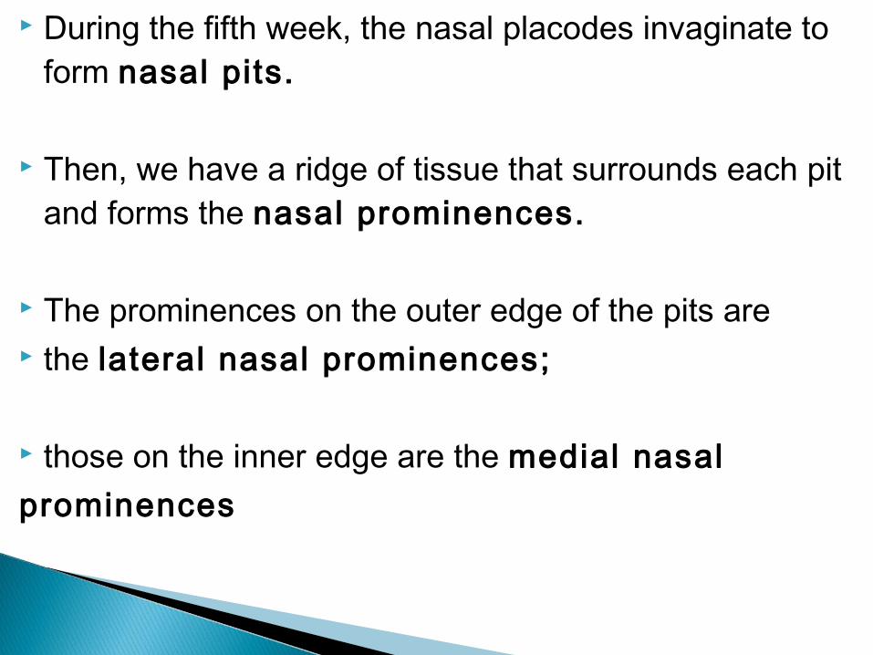
During the fifth week, the nasal placodes invaginate to form nasal pits.
Then, we have a ridge of tissue that surrounds each pit and forms the nasal prominences.
The prominences on the outer edge of the pits are the lateral nasal prominences;
those on the inner edge are the medial nasalprominences

1. Philtrum2. Premaxillary part of Maxilla (carries the 4
incissors)3. Primary Palate

the frontonasal process which projects down from the cranium.
Two olfactory pits develop in it and rupture into the pharynx to form the nostrils.
this process hence forms the nose, the nasal septum, nostril, the philtrum of the upper lip (the small midline depression) and the premaxilla
—the V-shaped anterior portion of the upper jaw which usually bears the four incisor teeth;

the maxillary processes on each side, which fuse with the frontonasal process and become:
the cheeks, upper lip (exclusive of the philtrum), Upper jaw and palate (apart from the premaxilla);

nasolacrimal groove nasolacrimal duct-canalized groove MP + LNP fusion, leads to the connection of the
nasolacrimal duct from the medial side of the eye to the inferior meatus of the nasal cavity.
Ear Auricles -◦ form from 6 auricular hillocks (week 5) ◦ 3 on each of arch 1 and 2

the mandibular processes, which meets in the midline form the lower jaw/mandible.
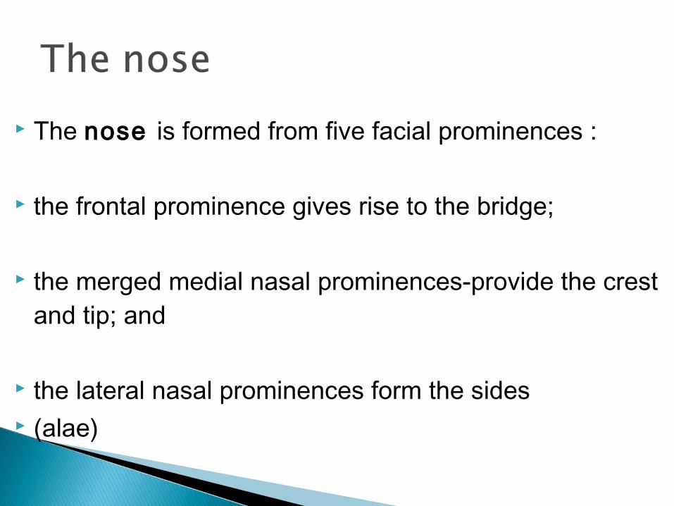
The nose is formed from five facial prominences : the frontal prominence gives rise to the bridge;
the merged medial nasal prominences-provide the crest and tip; and
the lateral nasal prominences form the sides (alae)


Intermaxillary segment of the maxilla
Premaxillary part of the maxilla

By wk 7-The floor of the nasal cavity at this stage is a posterior extension of the intermaxillary process known as the primary palate.
The medial walls of the maxillary swellings begin to produce a pair of thin medial extensions, 'palatine shelves', which grow inferiorly on either side of the tongue.
By wk 8-The tongue moves downward and the palatine shelves rapidly rotate upwards towards the midline, growing horizontally.
By wk 9- The palatine shelves begin to fuse ventrodorsally with each other, the primary palate and the inferior nasal septum.

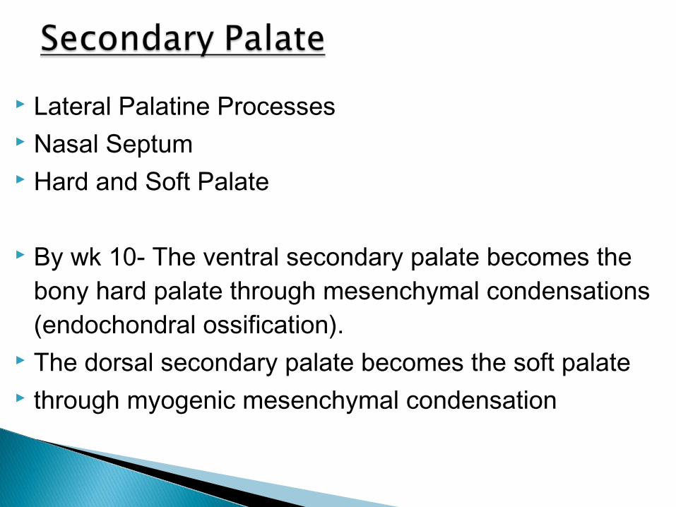
Lateral Palatine Processes Nasal Septum Hard and Soft Palate
By wk 10- The ventral secondary palate becomes the bony hard palate through mesenchymal condensations (endochondral ossification).
The dorsal secondary palate becomes the soft palate through myogenic mesenchymal condensation


Nasal placodes - become nasal pits Oronasal/bucconasal membrane The Conchae - Superior Middle Inferior-

The maxillary and medial nasal processes bulge forward together and the epithelium on their surfaces fuse to form a sheet, the nasal fin.
Subsequently this nasal fin breaks down such that both maxillary and medial nasal mesenchyme now intermingle.
The epithelial fins behind this fusion are stretched out laterally to form the bucconasal membranes and this also break down to form the nasal choanae that is connecting the nasal pits and the stomatodeum/stomodeum.


The tissues below the nasal pits now form the primary palate or intermaxillary segment.
The posterior part will form -the premaxillary part of the definitive palate,
the intermediate zone -premaxillary alveolar process and teeth,
the anterior zone- the medial portion of the upper lip.


5 facial prominences Bridge-frontal prominence Crest and tip- merged MNPs Side alar/labie-LNPs

During the sixth week, the nasal pits deepen considerably, partly due to growth of the surrounding nasal prominences and partly their penetration into the underlying mesenchyme.
At first the oronasal /bucco-nasal membrane separates the pits from the primitive oral cavity by way of the newly formed foramina, the primit ive choanae.
These choanae lie on each side of the midline and immediately behind the primary palate.
Later, with formation of the secondary palate and further development of the primitive nasal chambers,
the definit ive choanae lie at the junction of the nasal cavity and the pharynx.


Paranasal air sinuses develop as diverticula of the lateral nasal wall and
extend into the maxilla, ethmoid, frontal, and sphenoid bones.
They reach their maximum size during puberty and
contribute also to the definitive shape of the face.


First Arch Syndrome ◦ 2 major types, both result in extensive facial abnormalites ◦ Teacher Collins`s Syndrome ◦ Pierre Robin`s Syndrome
Cervical Fistulas and Cysts - Incomplete fusion or remnants of the walls of the cervical sinus (second branchial groove).
The internal openings of fistulas are at the sites of the pharyngeal membranes;
the external openings are along the anterior border of the sternocleidomastoid muscle.

Bif id Tongue - A midline split in the anterior two-thirds
of the tongue due to improper fusion of the lateral lingual swellings. Thyroglossal Duct Cyst - A cyst of remnants of all or
part of the thyroglossal duct. In removing this cyst, a portion of the body of the hyoid
bone is often removed, because the hyoid bone grows around the developmental path of this structure.
Retention and enlargement of that portion of the thyroglossal duct in contact with the thyroid results in the formation of a pyramidal lobe.

DiGeorge`s Syndrome ◦ absence of thymus and parathyroid glands ◦ 3rd and 4th pouch do not form ◦ disturbance of cervical neural crest migration.
Holoprosencephaly ◦ shh abnormality
NB: SHH genes◦ expressed in arches ◦ regulates midface formation
Pax-3 genes expressed in placode cells ◦ contribute to the CNV ◦ ophthalmic branch
Maternal Effects ◦ Retinoic Acid
present in skin ointments 1988 associated with facial developmental abnormalities Abnormalities from the Maternal Effects

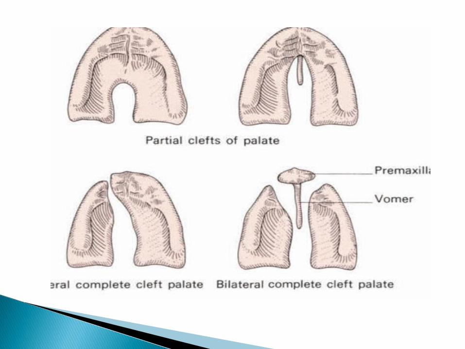
Cleft Lip and Palate ◦ 300+ different abnormalities ◦ different cleft forms and extent ◦ upper lip and ant. maxilla ◦ hard and soft palate
Etiology is again multifactorial, with the same teratogens as for cleft lip
an x-linked cleft palate syndrome has 1. Cleft lip◦ Unilateral◦ Bilateral
2. Median Cleft Lip-complete/incomplete 3. Cleft Palate 4. Facial Clefts -extremely rare

due to alcohol in early development (week 3+) facial and neurological abnormalities lowered ears, small face, mild retardation
Microcephaly - leads to small head circumference Short Palpebral fissure - opening of eye Epicanthal folds - fold of skin at inside of corner of eye Flat mid face Low nasal bridge Indistinct Philtrum - vertical grooves between nose and mouth Thin upper lip Micrognathia - small jaw
exposure of embryos in vitro to ethanol simulates premature differentiation of prechondrogenic mesenchyme of the facial primordial (1999)

these are pleuripotent cells that develop from the neural folds and migrate widely in the embryo to give rise to many nervous structures:
Spinal ganglia (dorsal root ganglia) Ganglia of the autonomic nervous system Ganglia of some cranial nerves Sheaths of peripheral nerves Meninges of brain and spinal cord Pigment cells Suprarenal medulla Skeletal and muscular components in the head

- Mandibulo-facial dysostosis results from a symmetrical loss of neural crest cells destined to migrate by the longer posterior route into the
face (Poswillo) There is hypoplasia of the zygomatic bone and a deficiency or absence of the arch. As a result there is an antimongoloid slant to the
palpebral fissure. There may be a coloboma of the lower eyelid with absence of lashes lateral
to the notch and hypoplasia of the mandible resulting in a lack of chin and an anterior open bite.
The ears are set low and the auricles and middle ear structures may be deficient in severe cases.
The abnormality tends to run in families.


![New {Anatomy/ Lec 6}————Thyroid gland / part 2Edited by : Rua’a … · 2020. 5. 9. · * Thyroid gland has to migrate from site of origin [ between future parts of tongue]](https://static.fdocuments.in/doc/165x107/605360ddde603a02de4f5d5e/new-anatomy-lec-6aaaathyroid-gland-part-2edited-by-ruaaa-2020.jpg)