Development of synapses between identified sensory neurones...
20
/. Embryol. exp. Morph. 86, 227-246 (1985) 227 Printed in Great Britain © The Company of Biologists Limited 1985 Development of synapses between identified sensory neurones and giant interneurones in the cockroach Periplaneta americana J. M. BLAGBURN, D. J. BEADLE Department of Biological Sciences, Thames Polytechnic, Wellington Street, Woolwich, London SE18, U.K. ANDD. B. SATTELLE A.F.R.C. Unit of Insect Neurophysiology and Pharmacology, Department of Zoology, University of Cambridge, Downing Street, Cambridge CB23EJ, U.K. SUMMARY The cereal afferent, giant interneurone pathway in Periplaneta americana was used as a model for synapse formation. The morphology of the two identified filiform hair sensory neurones (FHSNs) and of two giant interneurones (GI2 and GI3) was followed throughout embryogenesis by cobalt injection. The FHSN axons enter the CNS at the 45 % stage of embryogenesis, branch at 50 % and form complete arborizations by 70 %. The giant interneurones send out a primary dendrite at 45 %. Secondary branches form between 50 % and 60 % and elaboration of the branching pattern takes place until 80 % embryogenesis. At early stages the FHSN axons are within filopodial range of GI dendrites which may use these sensory processes as guidance cues. Synapse formation between the main FHSN axon shafts and GI dendrites was investigated by injection of the latter with HRP. From 55 % to 65 % the process is initiated by desmosome-like filopodial contacts, with subsequent vesicle clustering and formation of a small synaptic density. Numbers of contacts did not significantly increase after about 70 %, but the number of synapses doubled between 65 % and 75 %, with each GI process becoming postsynaptic to two FHSN synapses and the presynaptic densities lengthening to become bars. From 75 % embryogenesis to hatching there is a further small increase in synaptic bar length. In the first instar GI3 is postsynaptic to both FHSN axons, whereas GI2 forms very few synapses with the axon of the lateral FHSN (LFHSN). This imbalance of contacts is present throughout synaptogenesis, apart from some early filopodial contacts. GI 3 forms synapses with the lateral side of the LFHSN axon from 60 % embryogenesis but these are totally absent at hatching. The growth of glia along this side of the axon during the last 30 % of development appears to be associated with degeneration of synapses in this region. Thus, as the dendrites of the GIs grow to form a miniature version of the adult without loss of branches, there is little evidence of an initial overproduction of FHSN-GI synapses. Similarly there is no evidence that GI 2 forms 'incorrect' synapses with the axon of LFHSN. However, GI 3 contacts are removed from an inappropriate region of a correct synaptic partner, LFHSN. INTRODUCTION The problem of how neurones locate and form synapses with the correct targets is one of the central questions of neurobiology. This problem can be divided into Key words: Insect development, synaptogenesis, identified neurones, Periplaneta americana, interneurones.
Transcript of Development of synapses between identified sensory neurones...

/. Embryol. exp. Morph. 86, 227-246 (1985) 2 2 7Printed in Great Britain © The Company of Biologists Limited 1985
Development of synapses between identified sensoryneurones and giant interneurones in the cockroachPeriplaneta americana
J. M. BLAGBURN, D. J. BEADLE
Department of Biological Sciences, Thames Polytechnic, Wellington Street,Woolwich, London SE18, U.K.
A N D D . B. SATTELLE
A.F.R.C. Unit of Insect Neurophysiology and Pharmacology, Department ofZoology, University of Cambridge, Downing Street, Cambridge CB23EJ, U.K.
SUMMARY
The cereal afferent, giant interneurone pathway in Periplaneta americana was used as a modelfor synapse formation. The morphology of the two identified filiform hair sensory neurones(FHSNs) and of two giant interneurones (GI2 and GI3) was followed throughout embryogenesisby cobalt injection. The FHSN axons enter the CNS at the 45 % stage of embryogenesis, branchat 50 % and form complete arborizations by 70 %. The giant interneurones send out a primarydendrite at 45 %. Secondary branches form between 50 % and 60 % and elaboration of thebranching pattern takes place until 80 % embryogenesis. At early stages the FHSN axons arewithin filopodial range of GI dendrites which may use these sensory processes as guidance cues.
Synapse formation between the main FHSN axon shafts and GI dendrites was investigated byinjection of the latter with HRP. From 55 % to 65 % the process is initiated by desmosome-likefilopodial contacts, with subsequent vesicle clustering and formation of a small synaptic density.Numbers of contacts did not significantly increase after about 70 %, but the number of synapsesdoubled between 65 % and 75 %, with each GI process becoming postsynaptic to two FHSNsynapses and the presynaptic densities lengthening to become bars. From 75 % embryogenesisto hatching there is a further small increase in synaptic bar length.
In the first instar GI3 is postsynaptic to both FHSN axons, whereas GI2 forms very fewsynapses with the axon of the lateral FHSN (LFHSN). This imbalance of contacts is presentthroughout synaptogenesis, apart from some early filopodial contacts. GI 3 forms synapses withthe lateral side of the LFHSN axon from 60 % embryogenesis but these are totally absent athatching. The growth of glia along this side of the axon during the last 30 % of developmentappears to be associated with degeneration of synapses in this region.
Thus, as the dendrites of the GIs grow to form a miniature version of the adult without loss ofbranches, there is little evidence of an initial overproduction of FHSN-GI synapses. Similarlythere is no evidence that GI 2 forms 'incorrect' synapses with the axon of LFHSN. However, GI 3contacts are removed from an inappropriate region of a correct synaptic partner, LFHSN.
INTRODUCTION
The problem of how neurones locate and form synapses with the correct targetsis one of the central questions of neurobiology. This problem can be divided into
Key words: Insect development, synaptogenesis, identified neurones, Periplaneta americana,interneurones.

228 J. M. BLAGBURN, D. J. BEADLE AND D. B. SATTELLE
two aspects: the first concerns the means by which the growth cones of axons andtheir branches are able to navigate accurately to potential synaptic targets; thesecond concerns the way in which neurones establish synapses with the correct cellsand subsequently regulate the numbers of those contacts. Description of theembryonic development of simple model systems may help to elucidate thesemechanisms. In the present investigation we use the cockroach cereal afferent,giant interneurone pathway as a model for studying the morphogenesis of identifiedpresynaptic and postsynaptic neurones. In particular we describe the stages ofsynaptogenesis and investigate quantitatively the changes in synapse distributionbetween these structurally and physiologically identified cells which take placeduring embryogenesis.
Studies of the behaviour of growth cones in cell culture suggest that filopodia areregularly extended and retracted (Wessels et al., 1980). By a combination offilopodial adhesion to a particular substratum and the generation of tension, thegrowth cone moves towards the point of attachment (Bray, 1982). Adhesion offilopodia appears to guide the growth cones of grasshopper axons in vivo (Taghert,Bastiani, Ho & Goodman, 1982) and axons growing in the peripheral and centralnervous systems of Orthoptera follow specific guide-post cells and are themselvesused as 'labelled pathways' by later axons (Bate, 1976; Bentley & Keshishian, 1982;Raper, Bastiani, & Goodman, 1983a, b).
The growth patterns of neuronal branches vary, from 'initially directed growth'where no inappropriate branches are formed as in the grasshopper descendingcontralateral movement detector (Bentley & Toroian-Raymond, 1981), cerealsensory axons (Shankland, 1981a), medial giant interneurone (Shankland & Good-man, 1982) and the cockroach giant interneurone 2 (Blagburn, Beadle & Sattelle,1985) to cases in which transitory branches or axons are produced, such as grass-hopper stretch receptor (Heathcote, 1981), dorsal unpaired median extensor tibiae(Goodman & Spitzer, 1981) & H cell (Goodman, Bate & Spitzer, 1981). Little isknown about the cellular mechanisms which produce such growth patterns, al-though in the case of grasshopper medial giant interneurone (MGI), the presenceof cereal sensory afferent axons enhances the production of small dendriticbranches (Shankland, Bentley & Goodman, 1982). The spatial ordering of cricketcereal sensory axons within the terminal ganglion or within different ganglia isdetermined by the location of the cell bodies in the cereal array (Murphey, Bacon,Sakaguchi & Johnson, 1983; Murphey, Johnson & Sakaguchi, 1983).
The morphology of synaptogenesis has been widely studied. In vertebratesdesmosome-like contacts precede synaptic vesicle clustering and presynapticdensity formation (for example Rees, Bunge & Bunge, 1976; Hinds & Hinds, 1976)and similar morphological stages were seen in developing antennal lobes of the mothManduca sexta (Tolbert, Matsumoto & Hildebrand, 1983). The sequence of eventsinvolved in synaptogenesis can be studied more effectively using identified cells. Sucha model system could also provide information about the specificity of synapse forma-tion, and about changes in the numbers and size of synapses during development.

CODE JEM 15-3bJEM3488 page 15-3
Development of synapses between identified insect neurones 229Cereal sensory neurone, giant interneurone pathways are present in cockroaches
(Periplaneta americana) and aspects of their anatomy (Harrow, Hue, Pelhate &Sattelle, 1980; Daley, Vardi, Appignani & Camhi, 1981), pharmacology (Sattelle,1980; Sattelle et al. 1983) and behavioural physiology (Callec, Guillet, Pichon &Boistel, 1971; Westin, Langberg & Camhi, 1977; Camhi, Tom & Volman 1978;Ritzmann & Camhi, 1978) have been studied. For the purpose of developmentstudies, the cockroach cereal afferent, giant interneurone pathways offer a par-ticular advantage over the corresponding pathways in other Orthopteroid insects.In grasshoppers and crickets the sensory axons cannot be identified without the useof intracellular tracers, however first instar P. americana possess only two filiformhair sensilla on each cercus and each neurone gives rise to an axon which can beidentified in ultrathin sections by its characteristic size, position and morphology,without prior tracer injection (Blagburn & Beadle, 1982). In addition the neuronalcell bodies can be impaled with microelectrodes allowing dye injection andintracellular recording. Cobalt injection into the sensory neurones provides furtherevidence that the axonal morphology can be used to identify each sensory neurone(Blagburn, Beadle & Sattelle, 1984).
In previous studies the development of cholinergic sensitivity in the cell body anddendrites of giant interneurone 2 (GI2) has been described (Blagburn et al. 1985)and synapses between an identified filiform hair sensory neurone (FHSN) and GI 3have been located (Blagburn et al. 1984). In the present study we describe thecontributions of the two FHSNs to the synaptic input of GI 2 and GI 3 and thedevelopment of these synapses during embryogenesis.
MATERIALS AND METHODS
Oothecae projecting from female cockroaches were harvested daily and could be dated towithin 24 h. Each daily collection was stored at a temperature of 30 °C. The age of embryos wasexpressed as a percentage of the total time to hatching (normally 31 days). Detailed descriptionsof the embryos throughout embryogenesis are given elsewhere (Blagburn et al. 1985). Thedesignation A1-A6 is used for abdominal ganglia in the first instar; Ai-An is used to refer toganglionic rudiments present in the embryonic abdominal CNS. The terminal ganglion refers tothe postfusion ganglion (A6) formed from rudiments A7-A11.
Newly hatched first instar nymphs or embryos of the desired age were placed in saline of thefollowing composition: 150mM-NaCl, 3-lmM-KCl, 5-4mM-CaCl2, 5-0mM-HEPES buffer, pH7-4(based on Callec & Sattelle, 1973). The animals were dissected as described in an earlier paper(Blagburn et al. (1985) in order to remove the CNS which was then mounted on a microscopeslide. The A6 ganglion was secured, and the connective tissue sheath was softened by brief (15 s)exposure to saline containing l-0mg ml"1 protease (Type XIV, Sigma Chemical Co.).
Isolated preparations were viewed with a Zeiss X 40 water-immersion lens, using differentialinterference contrast (Nomarski) optics. Electrical isolation of the objective from the body of themicroscope was achieved with a Perspex insert. The somata of GI2 and GI3 were identified bytheir characteristic size, location and appearance. The procedure for viewing FHSNs has beendescribed previously (Blagburn et al. 1984).
For intracellular cobalt injection, neuronal cell bodies were impaled with 50-100 MQ micro-electrodes containing 6 % hexammine cobaltic chloride (Sigma Chemical Co.). Square, positivecurrent pulses, 0-5 s in duration at a frequency of 0-5 Hz and of 5nA amplitude, were passedthrough the electrode for 3-8 min. The preparation was left in saline for 10-15 min to allow for

230 J. M. BLAGBURN, D. J . BEADLE AND D. B. SATTELLE
distribution of cobalt, followed by precipitation with (NH^S and tissue fixation with alcoholicBouin's fixative for 30min. Cobalt-stained cells were silver-intensified using the whole-mountTimm's procedure of Bacon & Altman (1977), dehydrated in an alcohol series, cleared, andmounted in neutral Canada Balsam. Specimens were drawn with the aid of a Zeiss drawing tube.
Giant interneurones were also labelled intracellularly for electronmicroscopical examination.Cell bodies were impaled with microelectrodes containing 4 % horseradish peroxidase (HRPType VI, Sigma Chemical Co.) with 0-3M-KC1 and 0-2M-Tris buffer (pH7-4). Current pulses ofapproximately 3-4nA amplitude were passed through the electrode for 2-3 min. The preparationwas left in saline for 15min, fixed in 2-5 % glutaraldehyde in 0-lM-phosphate buffer (pH7-4)containing 0-2M-sucrose for 2h. The HRP reaction product was developed using the methoddescribed by Watson & Burrows (1981). Tissues were placed in 0-lM-Tris buffer pH7-4 for10min, then in Tris buffer containing 0-5 % cobalt chloride for 10min. After washing in Trisbuffer and phosphate buffer containing 0-2M-sucrose the ganglia were incubated in a mediumcontaining 10cm3 0-lM-phosphate buffer pH7-4, 4mg ammonium chloride, 20 mg /S-D-glucose,5 mg 3,3'-diaminobenzidine tetrahydrochloride (Sigma Isopac) and 20-30 units glucose oxidase(Type V, Sigma) for 20-40 min in the dark at 37 °C. After an overnight wash in 0-lM-cacodylatebuffer (pH7-4) containing 0-2M-sucrose, tissues were osmicated for l2h, dehydrated, embeddedand sectioned horizontally at 80 nm.
RESULTS
Morphology of filiform hair sensory neurones, GI2 and GI3 in the first ins tarThe two first instar filiform hair sensory neurones (FHSNs) from each cercus
form individually recognizable arborizations within the neuropile of the fusedganglion rudiments Ag-Au in the cockroach terminal abdominal ganglion, in aregion similar to the cereal glomerulus of first instar grasshoppers and crickets (cf.Shankland, 1981; Bacon & Murphey, 1981). As each axon enters the neuropilefrom cereal nerve XI (nomenclature of Roeder, Tozian & Weiant, 1960) it exhibitsone or two distinctive kinks and its diameter increases from approximately 2-3 jumto 5-8 /im. The axon arising from the lateral FHSN (Fig. 1A) is most lateral withinthe neuropile, and projects anteriorly along the ventral margin of the cerealglomerulus until it reaches the anterior boundary of the glomerulus which coincidesapproximately with the boundary between ganglion neuromeres A7 and A«. Atintervals the axon gives rise to four or five lateral branches which curve dorsally,branching extensively around the lateral margin of the glomerulus, and five or sixmedial branches which project across the ganglion and recurve dorsally around theglomerulus forming many small branches.
The medial FHSN (MFHSN) gives rise to an axon (Fig. IB) which runs medialand slightly dorsal to that of the lateral FHSN (LFHSN). It follows the latteranteriorly until the level of the A9 commissure when it deviates approximately 45 °in an anteromedial direction. The MFHSN axon also gives rise to four or five lateraland five or six medial branches whose arborization is similar to that of LFHSN.Both axons innervate similar regions of the cereal glomerulus with the exception ofthe anterolateral sector, from which MFHSN is largely absent as a result of thedeviation in its course. The branches of both axons bear numerous swellings(1-2 /im in diameter) which probably represent synaptic boutons.
In the first instar, GI2 and GI3 (Figs 1C and ID) display miniature versions of

Development of synapses between identified insect neurones 231the adult branching patterns described by Harrow et al. (1980) and Daley et al.(1981). The morphology of the first instar and embryonic GI2 has been describedpreviously (Blagburn et al. 1985). GI3 is similar in its overall morphology to GI2(Blagburn & Beadle, 1982) possessing a transverse neurite, anteriorly directed
Fig. 1. Drawings of silver-intensified cobalt fills of first instar filiform hair sensoryneurone (FHSN) axons and giant interneurones (GI2 and 3). (A) First instar lateralFHSN (LFHSN); (B) first instar medial FHSN (MFHSN); (C) first instar GI 3; (D) firstinstar GI2. Scale bar=50jum.

232 J. M. BLAGBURN, D. J. BEADLE AND D. B. SATTELLE
axon and dendrites in the contralateral cereal glomerulus and along the ipsilateralneurite. However, the soma of GI3 is somewhat larger than that of GI2(approximately 30 jum compared to 26 pan in diameter) and is located posterior tonerve VII instead of nerve VIII. The neurite of GI3 crosses the ganglionic midlinein the As commissure while that of GI2 runs in the A9 commissure. The majorcontralateral arborization fills the whole of the cereal glomerulus whereas that ofGI2 occupies only the posterior two-thirds. GI3 does not give rise to an axoncollateral branch in the A7 neuromere.
Growth of filiform hair sensory axonsThe FHSN axons were followed through embryogenesis using cobalt injection andelectronmicroscopy. The axons enter the CNS via nerve XI at about the 45 % stageof embryogenesis (Fig. 2A) and grow anteriorly. They do not fasciculate with themedial tracts of small cereal axons but take an independent course through thecentre of the neuropile (Figs 2B, E). During this period of growth the axons bearnumerous filopodia up to 20 fim in length. The growth cones of both axons tend tofollow temporarily the path of axons in nerve X, out of the ganglion (Fig. 2A). Longfilopodia are retained in this region after the growth cone has progressed anteriorly(Fig. 2E). LFHSN continues to grow anteriorly until about 60% embryogenesiswhen growth in this direction stops at the A7-A8 boundary. The MI*ISN axongrows alongside that of LFHSN until about 50-55 % when at the level of the A9commissure it deviates in an anteromedial direction. It is not certain what environ-mental cues dictate this change in direction. Branches of FHSN axons appear afterabout 50 % (Fig. 2C) and continue growing transversely, then dorsally, until65-70 % embryogenesis, when the aborizations are apparently complete (Fig. 2F).During the last 35 % of embryogenesis the cereal glomerulus shortens and widensand the unbranched posterior segments of the axons undergo a reduction in length(Figs 2D, G). The number of synaptic boutons increases during this period.
Growth of giant interneurones 2 and 3At 50 % embryogenesis both GI 2 and GI 3 have sent out transverse neurites
which extend to the contralateral longitudinal axon tract (Figs 3A,D). The neuriteof GI 3 runs through the anterior commissure of ganglion rudiment As, while thatof GI2 runs through the commissure in A9. Both axons turn anteriorly into thelongitudinal axon tract and by 50 % embryogenesis have left the terminal ganglion.At the turning points the axons give rise to posteriorly directed primary dendriteswhich form secondary branches between 50 % and 55 % embryogenesis. The small
Fig. 2. Drawings of silver-intensified cobalt fills of embryonic filiform hair sensoryneurone (FHSN) axons.
(A) 45% LFHSN; (B) 50% LFHSN; (C) 60% LFHSN; (D) 75% LFHSN; (E) 50%MFHSN; (F) 67% MFHSN; (G) 75% MFHSN. Percentages refer to the stage ofembryogenesis. Scale bar=50jum.
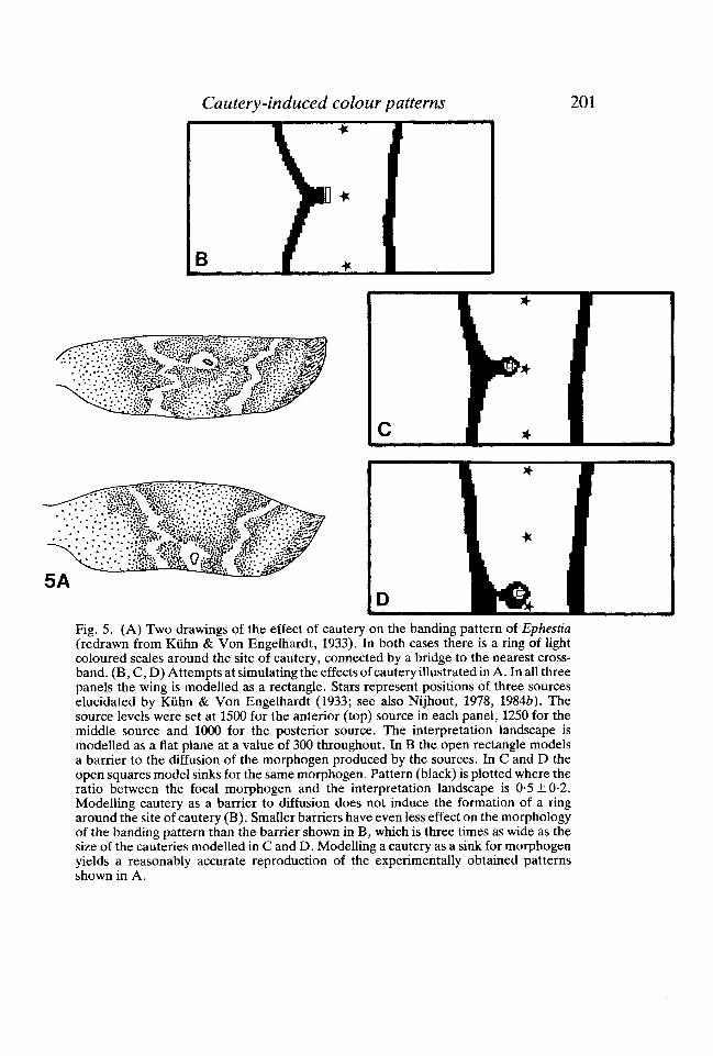
Cautery-induced colour patterns 201
Fig. 5. (A) Two drawings of the effect of cautery on the banding pattern of Ephestia (redrawn from Kiihn & Von Engelhardt, 1933). In both cases there is a ring of light coloured scales around the site of cautery, connected by a bridge to the nearest cross-band . (B, C, D) Attempts at simulating the effects of cautery illustrated in A. In all three panels the wing is modelled as a rectangle. Stars represent positions of three sources elucidated by Kiihn & Von Engelhardt (1933; see also Nijhout, 1978, 19846). The source levels were set at 1500 for the anterior (top) source in each panel, 1250 for the middle source and 1000 for the posterior source. The interpretation landscape is modelled as a flat plane at a value of 300 throughout. In B the open rectangle models a barrier to the diffusion of the morphogen produced by the sources. In C and D the open squares model sinks for the same morphogen. Pattern (black) is plotted where the ratio between the focal morphogen and the interpretation landscape is 0-5 ± 0-2. Modelling cautery as a barrier to diffusion does not induce the formation of a ring around the site of cautery (B). Smaller barriers have even less effect on the morphology of the banding pattern than the barrier shown in B, which is three times as wide as the size of the cauteries modelled in C and D. Modelling a cautery as a sink for morphogen yields a reasonably accurate reproduction of the experimentally obtained patterns shown in A.

234 J. M. BLAGBURN, D. J. BEADLE AND D. B. SATTELLE
Fig. 3

Development of synapses between identified insect neurones 235
\i •*
- 1 *
Fig. 4. The two FHSN axons can be identified in longitudinal ultrathin sections by theirsize, position and characteristic branching patterns. This figure contains examples ofelectronmicrographs of filiform hair sensory neurone, giant interneurone(LFHSN-GI3) synapses. (A) GI3 contact (asterisk) makes two dyadic synapses(arrows) with LFHSN axon. (B) High-power micrograph of synaptic bar in transversesection. The postsynaptic processes are not labelled with HRP. (C) GI3 branch(asterisk) forming output (closed arrow) and input (open arrow) synapses with unidenti-fied neurones. (D) Coated vesicles fusing with LFHSN membrane (arrow) close toLFHSN GI3 synapse. The GI3 process is labelled (asterisk). Scale bars=0-2jum.
dendritic branches in the neuropile ipsilateral to the cell body begin to develop inthis period as does the axon collateral of GI2 in the A7 ganglion rudiment. At 50,GI 3 also produces small collateral branches in this region but they disappear before60 % embryogenesis.
At this early stage the neuropile is proportionately narrower than it is afterconsolidation, with the result that the primary dendrites of the GIs are closer to theFHSN axons. Comparison of cobalt fills of afferents and interneurones suggests that
Fig. 3. Drawings of silver-intensified cobalt fills of embryonic giant interneurones (GI 2andGI3).
(A) 50% GI3; (B) 65% GI3; (C) 75% GI3; (D) 50% GI2; (E) 65% GI2; (F) 75%GI2. Percentages refer to the stage of embryogenesis. Scale bar=50jum.

236 J. M. BLAGBURN, D. J. BEADLE AND D. B. SATTELLE
all four neurones have the possibility of filopodial contact with each other since theyapproach to within 20/̂ m of each other and bear many filopodia which attain thislength. The arborizations of both GIs are elaborated by secondary and tertiarybranching between 50% and 80-85% (Figs 3B,C,E & F), after which timefilopodia disappear and the arborizations closely resemble those in the first instar.It is apparent that the branching patterns of the FHSNs are complete by 65-67 %whereas the GI dendrites bear filopodia until about 80 %, suggesting that they mayuse FHSN processes as guidance cues.
Synapses between the filiform hair sensory axons and GI2 and GI3 in the first instarIt has been shown previously that the axon of LFHSN makes numerous input
synapses onto dendrites of GI 3 in the first instar (Blagburn et al. 1984). The major-ity of the synapses between LFHSN, MFHSN and GI2 and GI3 exhibit a dyadicconfiguration with two postsynaptic processes, one of which belongs to the stainedcell (Fig. 4A). A presynaptic bar is located between the two postsynaptic profiles,this being triangular in cross section (Fig. 4B) and 200 ± 30 nm (n = 14) in length.On average there are two such bars at every FHSN-GI contact. Coated vesicles arecommonly seen fused to the presynaptic membrane in the vicinity of such contacts(Fig. 4D) and filamentous material is present in the synaptic cleft. Previous studiesof thoracic ganglionic neuropile have described similar presynaptic bars in uniden-tified neuronal processes (Wood, Pfenninger & Cohen 1977; Lane, Sattelle &Hufnagel, 1983) and in the latter study the fusion of coated vesicles to presynapticmembranes was thought to represent membrane retrieval.
Dendrites of GI 2 and GI 3 which receive synaptic input from the axon shafts ofLFHSN and MFHSN were reconstructed from serial or semiserial sections (Figs5A,B). The distribution of synapses shown in these reconstructions was found to berepresentative of all the other specimens examined. Dendrites of GI2 contact allsides of the MFHSN axon and approximately 66 MFHSN-GI2 synapses werecounted in this reconstruction. In contrast, despite the proximity of GI 2 dendritesto LFHSN, very few synaptic contacts were observed. In the reconstruction onesuch synapse was seen, but in most other specimens examined no synapses wereobserved between the axon shaft of LFHSN and dendrites of GI 2. Since the wholearborizations of the FHSN axons were not reconstructed, the possiblity remainsthat the relative contribution of LFHSN and MFHSN to the input of GI 2 may differwidely in other regions of the neuropile. GI3 dendrites form numerous synapticcontacts with both MFHSN and LFHSN (approximately 20 and 85 respectively inthis partial reconstruction). Although GI 3 dendrites were occasionally observed inthe neuropile on the lateral side of the LFHSN axon, in no case were synapsesformed between them. Much of the lateral side of this axon is sheathed in two orthree glial layers.
All of the synapses observed between the sensory axons and the interneuroneswere input synapses onto the latter. Although both GI2 and GI3 form outputsynapses in this region of neuropile (Fig. 4C) no such contacts were observed with

Development of synapses between identified insect neurones 237
A
Fig. 5. Reconstructions prepared using electronmicrographs of serial ultrathin sections,showing the distributions of synapses between the lateral (LFHSN) and medial(MFHSN) filiform hair receptor axons and GI2 and GI3 in the first instar. Small FHSNbranches have been omitted in order not to obscure GI dendrites. (A) Input synapsesfrom LFHSN and MFHSN onto dendritic branches of GI3. 85 LFHSN-GI 3 synapsesand 20 MFHSN-GI3 synapses were reconstructed in this area of the neuropile. GI 3forms more contacts with the FHSN axons which were not reconstructed. (B) Inputsynapses from the axons of the LFHSN and MFHSN onto branches of GI2. OneLFHSN-GI 2 synapse and 66 MFHSN-GI 2 synapses were seen. No other branches ofGI2 contact the main FHSN axons. Scale bar=

238 J. M. BLAGBURN, D. J. BEADLE AND D. B. SATTELLE
either MFHSN or LFHSN. However, GI-FHSN synapses may occur in otherregions of the neuropile.
Synapse formationThe ultrastructure and distribution of developing FHSN-GI synapses were
observed and the synapses formed with GI 3 by the medial side of the main LFHSNaxon were analysed quantitatively. Since small numbers of individuals weresampled the values should be taken only as estimates. By the 55 % stage of embryo-genesis both FHSN axons have established their main branches, whereas GI2 andGI 3 have extended a primary dendrite and some secondary branches. The earliestcontacts were seen at this stage, between filopodia of GI2 and GI3 and anteriorbranches of LFHSN and MFHSN. In later stages the GI dendrites grow outwardsfrom the centre of the cereal glomerulus and form contacts, first with the moreposterior FHSN branches (Fig. 6), then with the axon of MFHSN and finally withthe axon of LFHSN. In these stages an apparent sequence of contact types can beseen, ranging from preliminary filopodial contacts on the posterior regions of the
Fig. 6. Electronmicrograph of the neuropile at 65% embryogenesis, with anteriortowards top left. The FHSN axons can be recognised at this stage on the basis of size,position and branching pattern. Part of a medial LFHSN branch (LFHSN) is shown,near its origin on the main axon. An HRP-labelled GI3 dendrite (asterisk) is in closeproximity to the branch and makes filopodial contacts (arrows) with it and with othersmall, unidentified neuronal profiles. Scale bar=

Development of synapses between identified insect neurones 239FHSN axons, to synapses in the anterior regions and branches. By observation ofthis apparent sequence a tentative description of synapse formation can be made.
The first type of contact between FHSN and GIs was a simple filopodium-axoncontact (Fig. 7A). The cell membranes are closely apposed over an area of0-013 ± 0-002jUm2 (n = 8) and occasionally points of membrane contact are seen.Both membranes exhibit a slight increase in electron density with filamentousmaterial spanning the gap between them. This type of contact was not observedafter 75 % embryogenesis.
Later contacts are similar in appearance with the addition of a small cluster ofsynaptic vesicles. At the 65 % stage approximately 70% of this type of contactbetween the LFHSN axon and GI3 exhibit a small presynaptic dense body withdimensions of approximately 30x30x30nm (Fig. 7B) but in no case was adensity observed without a small vesicle cluster. At 65 % embryogenesis LFHSNand GI3 form approximately 0-5 ± 0-2 (n = 2) such contacts per jum2 of sensoryaxon membrane, although they are more numerous in the anterior portion of theaxon.
Maturation of synaptic contacts initially consists of an increase in the number ofsynaptic vesicles; evidence for two possible mechanisms was seen. At 55 % embryo-genesis, 45 nm diameter vesicles were observed, enclosed in larger membranousinclusions, within FHSN axons and at maturing synaptic contacts (Fig. 7C). At65 %, such inclusions were no longer seen but the presence of short lengths ofsmooth endoplasmic reticulum intermingled with synaptic vesicles suggests the insitu synthesis of vesicles (fig. 7D). Also present from this period were occasionaldense-core vesicles and coated vesicles, the latter often fused with the cell mem-brane.
At the 75 % stage the neuropile around the FHSN axons is densely filled withcellular processes and 80 % of the LFHSN-GI3 contacts possess a vesicle cluster,and 2 ± 0-3 (n = 7) presynaptic densities whose length has increased to 150 ± 20 nm(n = 14). The area of postsynaptic contact is also larger (0-1 ± 0-04 /an2, n = 9) andthe postsynaptic membrane density is more pronounced. The density of LFHSNaxon-GI3 synapses is 1-3 ± 0-1 pan'2 (n = 3).
Few further changes in the synapses take place up to 94 % embryogenesis, apartfrom a small increase in the mean length of presynaptic bars to 200 ± 30 nm (n = 14)and in the area of GI 3 contacts (0-13 ± 0-03 /an2, n = 10). By this stage 95 % of GI 3contacts with LFHSN are synaptic. In the first instar the mean density of LFHSN-GI 3 synapses (1-6 ± 0-2 jum"2, n = 3) is not significantly increased. From around60 % embryogenesis, when the first synapse-like contacts are formed between theaxon of LFHSN and dendrites of GI3, they represent approximately 20 % of thetotal number of synapses formed along the axon and this proportion does notchange significantly throughout embryogenesis.
In the first instar, synaptic contacts between the LFHSN axon and GI2 are veryrare. This situation exists throughout embryogenesis apart from the early stages(50-55 %) , when a small number of filopodial contacts are formed with the anterior

240 J. M. BLAGBURN, D. J. BEADLE AND D. B. SATTELLE
region of LFHSN. This observation suggests that GI2 dendrites selectively formsynaptic contacts with the axon of the MFHSN, not with that of LFHSN. Also, inthe first instar, there are no synapses between the lateral side of the LFHSN axon
1

Development of synapses between identified insect neurones 241and GI3 dendrites, despite the presence of some branches in that region ofneuropile. Access to that side of the axon is prevented by glial layers. However,LFHSN-GI3 synapses form on this side of the axon from 60 % embryogenesis andpersist until the 95 % stage, although as glia grow anteriorly along the axon somesynapses disappear. Synapses are not wholly absent in this region until after hatch-ing. At later stages of embryogenesis some vesicle clusters remain under a layer ofglia, sometimes associated with degenerating postsynaptic profiles (Fig. 7E). It isnot certain whether the glial growth results in this degeneration or whether thedeath of the postsynaptic process is necessary before the glia can grow over thesynapse.
DISCUSSION
The growth patterns of the lateral and medial FHSN together with GI2 and GI3within the terminal abdominal ganglion of Periplaneta americana resemble those ofthe cereal glomerular (G) afferents and medial giant interneurone (MGI) in thegrasshoppers Schistocerca nitens and Schistocerca americana (Shankland, 19816;Shankland & Goodman, 1982). The development timetables of GI2 and GI3 aresimilar to that of MGI, however in the grasshopper the cereal G afferents arrive inthe terminal ganglion at 55-60%, whereas in the cockroach the LFHSN andMFHSN arrive earlier, at approximately 45%. The arborizations of the FHSNaxons are established by 55-50 % whereas those of the GIs are complete by75-80 %. Thus, in the case of the main FHSN axons, synapses are formed as a resultof growth of the postsynaptic dendrites to the presynaptic cell. This is typical of thepatterns of growth observed in many other models of synaptogenesis, for example:vertebrate neuromuscular junction formation in vivo (Kullberg, Lentz & Cohen,1977) and in vitro (Nakajima, Kidokoro & Klier, 1980), vertebrate neurones in vivo(Landmesser & Pilar, 1972) and in vitro (Rees et al. 1976); moth neuromuscularjunctions (Stocker & Niiesch, 1975) & Mauthner cell innervation (Jacoby &Kimmel, 1982). However, in the optic lobe of Musca domestica photoreceptorsynapses are formed by growth of the postsynaptic cell to the synaptic sites (Froh-lich & Meinertzhagen, 1982). In the terminal abdominal ganglion of P. americanaboth types of growth may occur since postsynaptic GI dendrites grow to presynapticFHSN axons and synaptic endings are formed on the primary dendrites of GIs.
Fig. 7. Electronmicrographs of unidentified contacts with the LFHSN axon. (A) Initialfilopodial contacts (arrow) with LFHSN axon in a 55% stage embryo. Filamentous cleftmaterial and symmetrical membrane densities are present. (B) Early synapse-like con-tact (arrow) between LFHSN axon and unidentified filopodium. Small vesicle clusterand presynaptic dense body are visible in the 55% stage embryo. (C) LFHSN synapticending with vesicles transported in membrane (arrow), at 55% embryogenesis. (D)LFHSN synaptic ending at 65% with short lengths of smooth endoplasmic reticulum(arrow). (E) Degenerating synapse on lateral side of LFHSN. Myelin figures are visiblein postsynaptic processes, (arrow). A glial layer covers the synapse (asterisk). Scalebars=0-2jum.

242 J. M. BLAGBURN, D. J. BEADLE AND D. B. SATTELLE
Since the main branches of the sensory axons are established before the GI arboriza-tions are complete, deafferentation of embryonic cockroach GIs might be expectedto have a more radical effect upon the branching pattern than it does with grasshopperMGI where the sensory afferents grow in at a later stage (Shankland et al. 1982).
Injection of HRP into GI2 and GI3 enabled an analysis to be made of theirpatterns of connection with the two identifiable FHSN axons. GI3 was found toreceive synaptic input from both axons, in particular that of LFHSN, whereas GI 2apparently receives input almost exclusively from the MFHSN axon. Thedistribution of synapses seen in these restricted regions of the cells may or may notbe typical of the overall synapse distribution.
If the patterns of synapse distribution seen on the axons of the FHSNs is represen-tative of other parts of the arborizations then it can be inferred that GI 3 receivesexcitatory cholinergic synaptic input from both LFHSN and MFHSN while GI 2receives input mainly from MFHSN. It has been shown by Dagan & Volman (1982)that in the first instar cockroach the lateral FHSNs exhibit an increased spikingfrequency in response to wind from the front of the animal, while the medial FHSNsrespond when the wind direction is from the rear. If the distribution of morphologi-cal synapses is a reliable indicator of the strength of the physiological connection,then GI 3 will be excited by wind from all directions while GI 2 will be excited bywind mainly from the rear. However, in the adult, GI3 responds mainly to windfrom the front and the response of GI 2 shows no directionality (Westin et al. 1977).It may be that GI2 and GI3 undergo changes in directional sensitivity duringpostembryonic development, presumably as a result of changes in relative synapticinputs as large numbers of FHSN axons enter the ganglion in later instars.
Alternatively, although synapses between the LFHSN axon and GI2 are veryrare, they may occur in other regions of the cereal glomerulus. Since there are noapparent barriers preventing GI 2 dendrites from approaching the LFHSN axon,the most likely explanation of such an imbalance of contacts would be the presenceof different cell surface markers on the axon and branches of LFHSN, allowingadhesion of GI 2 filopodia to LFHSN branches but not to its axon. To answer thesequestions it will be necessary to record from first instar GIs while stimulating asingle FHSN.
An hypothetical sequence of synapse development was obtained by observationof FHSN-GI contacts throughout embryogenesis. This sequence is similar to thosedescribed in other studies of synaptogenesis, for example, in vertebrate neuronecultures (Rees et al. 1976); in Manduca antennal lobe (Tolbert et al. 1983). In allcases initial filopodial adhesion mediated by demosome-like contacts occursfollowed by accumulation of synaptic vesicles and formation of a presynaptic mem-brane density. It is not certain whether some initial filopodial adhesion sites persistand become modified into synapses, or whether a temporary adhesion simplyguides a dendritic growth cone to the site. The large number of immature synapse-like contacts formed by the FHSN axons with processes of small diameter that aresimilar to filopodia suggests that the former mechanism may be important. The

Development of synapses between identified insect neurones 243expansion in area of postsynaptic contacts seen by 75% embryogenesis may be aresult of dendritic growth in the direction of filopodia anchored by synapse-likejunctions. The mean number of LFHSN axon synapses at a single contact with GI3increases from one to two between 65% and 75% embryogenesis but there is littlefurther increase in the density of contacts. In addition the mean length of thepresynaptic densities increases during this period and the majority thereafter takethe form of synaptic bars.
Counts of a defined population of synapses (those between the axon of LFHSNand GI 3 dendrites) show that there is no initial overproduction then subsequentremoval of contacts, as is the case in the developing photoreceptor terminals inMusca (Frohlich & Meinertzhagen, 1983). The number of contacts rises until65-70% embryogenesis, and the number of synapses is approximately doubledbetween 65% and 75% by the expansion of postsynaptic processes and an increasein the number of synaptic bars per contact. These estimates refer to net numbersof synapses and it is possible that individual synapses may not survive for lengthyperiods of time.
Despite the possibility that regional differences may exist in the density of FHSN-GI synapses, the region of neuropile around the FHSN axons remains a valuablemodel for investigating the degree of specificity of synapse formation duringembryogenesis. There is no evidence of substantial synapse formation between theaxon of LFHSN and GI2 at any stage suggesting that, in this case, synapses areformed only between compatibly labelled cells or regions of cells. In contrast, inmany vertebrate systems relatively non-specific synapse formation occurs initially,followed by synapse elimination from some target cells (Purves & Lichtman, 1980).However, the lateral side of the LFHSN axon forms synapses with GI 3 and otherneurones which are later removed, either as a consequence of, or as a preliminaryto, glial growth. The simplest hypothesis would be that glial processes contact thepostsynaptic dendrites, somehow resulting in their degeneration and the gradualbreakdown of the presynaptic side of the synapse.
Spontaneous synaptic activity can be recorded from the cell bodies of GI2(Blagburn et al. 1985) and GI3 at 75% embryogenesis. This does not mean thatfunctional synapses are absent before this stage. For example, a certain numberof functional synaptic contacts may be necessary before spontaneous synapticactivity can be recorded in the soma. It has been reported that functional synaptictransmission precedes the appearance of anatomically recognizable synapticjunctions (Crain & Peterson, 1967; Landmesser & Pilar, 1972; Kullberg et al.1977) so it is possible that FHSN-GI synaptic transmission is present before70-75% embryogenesis. The doubling of the number of synapses seen between65% and 75% may result in an increase in the amplitude of compound epsps inGIs allowing them to be recorded from the cell body. Electrical stimulation ofFHSN axons and simultaneous recording from GIs will help to resolve thesequestions.
GI 2 becomes sensitive to carbamylcholine at about 40% when axonal outgrowth

244 J. M. BLAGBURN, D. J. BEADLE AND D. B. SATTELLE
commences, and the sensitivities of cell body and dendritic membranes diverge atapproximately 55% embryogenesis (Blagburn et al. 1985). GI2 filopodia formcontacts with other neurones as soon as the axon first sprouts, however it is not untilabout 50-55% embryogenesis when the dendritic tree begins to form, thatfilopodial contacts are established with FHSN axons. It is possible that contact withpotential synaptic partners triggers the divergence of chemosensitivity in the tworegions of the cell.
In the simple model system provided by cockroach filiform hair sensory neuronesand giant interneurones it appears that, at least in the axonal region of the pre-synaptic cells, synapse formation is a relatively specific process. At no stage duringits growth does GI2 form substantial numbers of 'incorrect' contacts with the axonof LFHSN. Similarly, no overproduction and subsequent loss of LFHSN-GI3synapses was observed, but a rapid increase in the net numbers of contacts was seen,followed by a doubling of the number of synapses per contact. However, synapsesbetween appropriate synaptic partners, LFHSN and GI3, which are formed in aninappropriate location, are removed towards the end of embryogenesis, possibly asa result of glial growth.
This study provides evidence that in the case of neurones whose dendrites appearto grow directly into a mature branching pattern, with no apparent loss of branchesas the pathway becomes behaviourally functional, there is a large degree of speci-ficity in the formation of synaptic contacts. Morphological studies of this type arean essential prerequisite for experiments in which manipulation of the synapticinputs onto developing neurones is attempted.
REFERENCESBACON, J. P. & ALTMAN, J. S. (1977) A silver intensification method for cobalt-filled neurones
in wholemount preparations. Brain Res. 138, 359-363.BACON, J. P. & MURPHEY, R. K. (1981) Cricket cereal filiform hairs have a somatotopic projection
in the terminal ganglion. Soc. Neurosci. Abstr. 7, 4.BATE, C. M. (1976) Pioneer neurons in an insect embryo Nature 260, 54-56.BENTLEY, D. & KESHISHIAN, H. (1982) Pathfinding by peripheral pioneer neurons in grass-
hoppers. Science 218, 1082-1088.BENTLEY, D. & TOROIAN-RAYMOND, A. (1981) Embryonic and post-embryonic morphogenesis of
a grasshopper interneuron. /. comp. Neurol. 210, 507-518.BLAGBURN, J. M. & BEADLE, D. J. (1982) Morphology of identified cereal afferents and
giant interneurones in the hatchling cockroach Periplaneta americana. J. exp. Biol. 97,421-426.
BLAGBURN, J. M., BEADLE, D. J. & SATTELLE, D. B. (1984) Synapses between an identified giantinterneurone and a filiform hair sensory neurone in the terminal ganglion of first instar cock-roaches {Periplaneta americana L.). /. exp. Biol. 113, 477-481.
BLAGBURN, J. M., BEADLE, D. J. & SATTELLE, D. B. (1985) Development of chemosensitivity ofan identified insect interneurone. /. Neurosci. Vol. 5, 1166-1174.
BRAY, D. (1982) Filopodial contraction and growth cone guidance. In Cell Behaviour (eds R.Bellairs, A. Curtis, & G. Dunn) London and New York: Cambridge Univ. Press.
CALLEC, J. J., GUILLET, J. C , PICHON, Y. & BOISTEL, J. (1971) Further studies on synaptictransmission in insects. II. Relations between sensory information and its synaptic integrationat the level of a single giant axon in the cockroach. J. exp. Biol. 55, 123-149.

Development of synapses between identified insect neurones 245CAMHI, J. M., TOM, W. & VOLMAN, S. (1978) The escape behaviour of the cockroach Periplaneta
americana. II. Detection of natural predators by air displacement. /. comp. Physiol. 128,203-212.
CRAIN, S. M. & PETERSON, E. R. (1967) Onset and development of functional interneuronal con-nections in explants of rat spinal cord ganglia during maturation in culture. Brain Res. 6,750-762.
DAGAN, D. & VOLMAN, S. (1982) Sensory basis for directional wind detection in first instarcockroaches, Periplaneta americana. J. comp. Physiol. 147, 471-478.
DALEY, D. L., VARDI, N., APPIGNANI, B. & CAMHI, J. M. (1981) Morphology of the giant inter-neurons and cereal nerve projections of the American cockroach. /. comp. Neurol. 196,41-52.
FROHLICH, A. & MEINERTZHAGEN, I. A. (1982) Synaptogenesis in the first optic neuropile of thefly's visual system. /. Neurocytol. 11, 159-180.
GOODMAN, C. S., BATE, C. M. & SPITZER, N. C. (1981) Embryonic development of identifiedneurones: origin and transformation of the H cell. /. Neurosci, 1, 94-102.
GOODMAN, C. S. & SPITZER, N. C. (1979) Embryonic development of identified neurones:differentiation from neuroblast to neurone. Nature 280, 208-214.
HARROW, I. D., HUE, B., PELHATE, M. & SATTELLE, D. B. (1980) Cockroach giant interneuronesstained by cobalt-backfilling of dissected axons. /. exp. Biol. 84, 341-343.
HEATHCOTE, R. D. (1981) Differentiation of an indentified sensory neuron (SR) and associatedstructures (CTO) in grasshopper embryos. / . comp. Neurol. 202,1-18.
HINDS, J. W. & HINDS, P. L. (1976) Synapse formation in the mouse olfactory bulb. II. Morpho-genesis. /. comp. Neurol. 169, 41-62.
JACOBY, J. & KIMMEL, C. B. (1982) Synaptogenesis and its relation to growth of the postsynapticcell: a quantitative study of the developing Mauthner neuron of the Axolotl. /. comp. Neurol.204, 364-376.
KULLBERG, R. W., LENTZ, T. L. & COHEN, M. W. (1977) Development of the myotomalneuromuscular junction in Xenopus laevis: an electrophysiological and fine structural study.Devi Biol. 60, 101-129.
LANDMESSER, L. & PILAR, G. (1972) The onset and development of transmission in the chickciliary ganglion. J. Physiol. Lond. 222, 691-713.
LANE, N. J., SATTELLE, D. B. & HUFNAGEL, L. A. (1983) Pre- and post-synaptic structures ininsect CNS: intramembranous features and sites of a-bungarotoxin binding. Tissue & Cell 15,921-937.
MURPHEY, R. K., BACON, J. P., SAKAGUCHI, D. S. & JOHNSON, S. E. (1983) Transplantationof cricket sensory neurons to ectopic locations: arborizations and synaptic connections.J. Neurosci. 3, 659-672.
MURPHEY, R. K., JOHNSON, S. E. & SAKAGUCHI, D. S. (1983) Anatomy and physiology ofsupernumerary cereal afferents in crickets: implications for pattern formation. /. Neurosci. 3,312-325.
NAKAJIMA, Y., KIDOKORO, Y. & KLIER, F. G. (1980) The development of functional neuro-muscular junctions in vitro: an ultrastructural and physiological study. Devi Biol 77, 52-72.
PURVES, D. & LICHTMAN, J. W. (1980) Elimination of synapses in the developing nervous system.Science 210, 153-157.
RAPER, J. A., BASTANI, M. J. & GOODMAN, C. S. (1983a) Pathfinding by neuronal growth conesin grasshopper embryos. I. Divergent choices made by the growth cones of sibling neurons./. Neurosci. 3, 20-30.
RAPER, J. A., BASTIANI, M. J. & GOODMAN, C. S. (19836). Pathfinding by neuronal growth conesin the grasshopper embryo. II. Selective fasciculation onto specific axonal pathways./. Neurosci. 3, 31-41.
REES, R. P. BUNGE, M. B. & BUNGE, R. P. (1976) Morpological changes in the neuritic growthcone and target neuron during synaptic junction development in culture. /. Cell Biol. 68,240-263.
RITZMANN, R. E. & CAMHI, J. M. (1978) Excitation of leg motorneurons by giant interneuronsin the cockroach Periplaneta americana. J. comp. Physiol. 125, 305-316.
ROEDER, K. D., TOZIAN, L. & WEIANT, E. A. (1960) Endogenous nerve activity and behaviourin the mantis and cockroach. /. Insect Physiol. 4, 45-62.
SATTELLE, D. B. (1980) Acetylcholine receptors of insects. Adv. Insect. Physiol. 15, 215-315.

246 J . M. BLAGBURN, D. J. BEADLE AND D. B. SATTELLE
SATTELLE, D. B., HARROW, I. D., HUE, B., PELHATE, M., GEPNER, J. I. & HALL, L. M. (1983)a-Bungarotoxin blocks excitatory synaptic transmission between cereal sensory neurones andgiant interneurone 2 of the cockroach Periplaneta americana. J. exp. Biol. 107, 473-489.
SHANKLAND, M. (1981ft) Embryonic development of a sensory afferent projection in the grass-hopper embryo. II. Growth and branching of peripheral sensory axons within the centralnervous system. /. Embryol. exp. Morph. 64, 187-209.
SHANKLAND, M., BENTLEY, D. & GOODMAN, C. S. (1982) Afferent innervation shapes the den-dritic branching pattern of the medial giant interneuron in grasshopper embryos raised inculture. Devi Biol. 92, 507-520.
SHANKLAND, M. & GOODMAN, C. S. (1982) Development of the dendritic branching pattern ofthe medial giant interneuron in the grasshopper embryo. Devi Biol. 92, 489-506.
STOCKER, R. F. & NUESCH, H. (1975) Ultrastructural studies on neuromuscular contacts and theformation of junctions in the flight muscle of Antherea polyphemus (Lep). I. Normal adultdevelopment. Cell Tissues Res. 159, 245-266.
TAGHERT, P. H., BASTIANI, M. J., Ho, R. K. & GOODMAN, C. S. (1982) Guidance of pioneergrowth cones: filopodial contacts and coupling revealed with an antibody to Lucifer Yellow.Devi Biol. 94, 391-399.
TOLBERT, L. P., MATSUMOTO, S. G. & HILDEBRAND, J. G. (1983) Development of synapses in theantennal lobes of the moth Manduca sexta during metamorphosis. /. Neurosci. 3, 1158-1175.
WATSON, A. H. D. & BURROWS, M. (1981) Input and output synapses on identified motorneurones of a locust revealed by the intracellular injection of horseradish peroxidase. CellTissue Res. 215, 326-332.
WESSELLS, N. K., LETOURNEAU, P. C , NUTTALL, R. P., LUDUENA-ANDERSON, M. & GUIDUSCHEK,J. M. (1980) Responses to cell contacts between growth cone neurites and ganglionic non-neuronal cells. J. Neurocytol. 9, 647-664.
WESTIN, J., LANGBERG, J. J. & CAMHI, J. M. (1977) Responses of giant interneurons of thecockroach Periplaneta americana to wind puffs of different directions and velocities. /. comp.Physiol. 121, 307-324.
WOOD, M. R., PFENNINGER, K. H. & COHEN, M. J. (1977) Two types of presynaptic configura-tions in insect central synapses: an ultrastructural analysis. Brain Res. 130, 25-45.
(Accepted 6 November, 1984)