Development of PCR-hybridization for the …budgetitc.dmsc.moph.go.th/research/pdf/201310.pdf1...
13
1 Development of PCR-hybridization for the identification of major Gram positive bacteria causing bacteremia. Athicha Mahayotha 1 , Rosarin Wongvilairat 1 , Surang Dejsirilert 2 , Tawatchai Sumpradit 1 , Anusak Kerdsin 2 . 1 Department of Microbiology and Parasitology, Faculty of Medical Science, Naresuan University, Phitsanulok, Thailand. 2 National Institute of Health, Department of Medical Sciences, Ministry of Public Health, Thailand. E-mail address: [email protected] Abstract Blood culture is considered to be a gold standard for diagnosis of bacterial septicemia. However, this method has some limitations. Molecular techniques for detection of DNA of bacteria causing septicemia such as PCR and hybridization are sensitive, specific and more rapid compared with blood culture. The aims of this project are to develop PCR – slot blot hybridization technique for detection and identification of Gram positive bacteria causing bacterimia. 16S rRNA gene of gram positive cocci were extracted and amplified by using universal primer. These fragments of each species were cloned in plasmids and preserved as clone libraries. Plasmid DNA was amplified by using M13 primer and PCR product used as DNA template to blot on a nylon membrane. V6 region in 16S rRNA gene of Streptococcus pneumoniae was amplified by PCR using digoxigenin-11-dUTP. PCR product was an initial experimental probe to optimize conditions of the hybridization with the reference DNA blotted on nylon membrane. Various anneal temperature and concentration of SSC buffer were tested to determine optimum hybridization conditions based on visual observation by using anti-DIG-AP conjugate, CDP-Star TM . This study found that the hybridize temperature at 65 o C exhibited more specific for probe of S. aureus, S. epidermidis and E. faecalis and unspecific for Streptococci. At 67 o C, hybridization exhibited more intense signal when used probe of all streptococcal species without any cross hybridization. The optimal condition of PCR-Slot blot Hybridization that was chosen as 67 o C of the hybridize temperature using for method validation. The lowest of pure DNA concentration of S. aureus ATCC 25923 which the PCR-Slot blot hybridization was given as positive results, was 0.7ng/μl. Sensitivity and specificity of PCR-Slot blot hybridization were highly value (100%). The results revealed that probe design and the optimized conditions were successful in the detection of gram positive septicemia-bacteria. Key words: Septicemia, Cultural method, PCR, Hybridization, 16S rRNA gene.
Transcript of Development of PCR-hybridization for the …budgetitc.dmsc.moph.go.th/research/pdf/201310.pdf1...

1
Development of PCR-hybridization for the identification of major Gram
positive bacteria causing bacteremia.
Athicha Mahayotha1, Rosarin Wongvilairat
1, Surang Dejsirilert
2, Tawatchai Sumpradit
1, Anusak Kerdsin
2.
1 Department of Microbiology and Parasitology, Faculty of Medical Science, Naresuan University, Phitsanulok, Thailand.
2 National Institute of Health, Department of Medical Sciences, Ministry of Public Health, Thailand.
E-mail address: [email protected]
Abstract
Blood culture is considered to be a gold standard for diagnosis of bacterial septicemia.
However, this method has some limitations. Molecular techniques for detection of DNA of
bacteria causing septicemia such as PCR and hybridization are sensitive, specific and more
rapid compared with blood culture. The aims of this project are to develop PCR – slot blot
hybridization technique for detection and identification of Gram positive bacteria causing
bacterimia.
16S rRNA gene of gram positive cocci were extracted and amplified by using
universal primer. These fragments of each species were cloned in plasmids and preserved as
clone libraries. Plasmid DNA was amplified by using M13 primer and PCR product used as
DNA template to blot on a nylon membrane. V6 region in 16S rRNA gene of Streptococcus
pneumoniae was amplified by PCR using digoxigenin-11-dUTP. PCR product was an initial
experimental probe to optimize conditions of the hybridization with the reference DNA
blotted on nylon membrane. Various anneal temperature and concentration of SSC buffer
were tested to determine optimum hybridization conditions based on visual observation by
using anti-DIG-AP conjugate, CDP-StarTM.
This study found that the hybridize temperature at 65oC exhibited more specific for
probe of S. aureus, S. epidermidis and E. faecalis and unspecific for Streptococci. At 67oC,
hybridization exhibited more intense signal when used probe of all streptococcal species
without any cross hybridization. The optimal condition of PCR-Slot blot Hybridization that
was chosen as 67oC of the hybridize temperature using for method validation. The lowest of
pure DNA concentration of S. aureus ATCC 25923 which the PCR-Slot blot hybridization
was given as positive results, was 0.7ng/µl. Sensitivity and specificity of PCR-Slot blot
hybridization were highly value (100%). The results revealed that probe design and the
optimized conditions were successful in the detection of gram positive septicemia-bacteria.
Key words: Septicemia, Cultural method, PCR, Hybridization, 16S rRNA gene.
2
Introduction
Septicemia is the presence of bacteria in blood ( bacteremia ) and is often
associated with severe disease. This disease is a serious; life- threatening infection that gets
worse very quickly. In case of severe sepsis is a serious condition that requires a hospital
stay and an intensive care unit (ICU) for admission (Amersfoort et al., 2003; Cunha, 2003;
Abraham, 1999; Ammerlaan et al., 2009). Early diagnosis of this disease is a key for
prevention of septic shock progression and associated with the correctly treatment of
clinicians. Physical examination and laboratory confirmation are used as a tool in septicemia
diagnosis. Especially laboratory tests play an important role in identifying the infectious
agent causing the infections (Agnihotri, 2004). Blood culture is considered to be the gold
standard for diagnosing bacterial septicemia (Baron et al., 2005; Chen et al., 2008). However,
this method has some limitations, including; a long time for growth of an organism(48 – 72
hrs), unacceptably low sensitivities in many clinical situations(40%) and inability to grow
by culturing or non-cultural bacteria (Bone, 1994; Keen et al., 2002; Thompson & Madeo,
1994; Craven, 2004 ). Molecular detection of septicemia bacteria based on DNA-based
detection methods including PCR and hybridization, both of which are sensitive and specific
and are rapid compared with blood culture (Kunakorn et al., 2000; Fu et al., 2002; Kim et al.,
2006; Hony et al., 2003; Mako et al., 2002). Here, we design one primer and one probe using
PCR-slot blot hybridization to recognize different strains within gram positive septicemia -
bacteria and will provide an rapid method for identification gram positive bacteria which
frequently in septicemia. And this should be applied in the hospital routine diagnostic
laboratory for early diagnosis gram positive bacteria.
Materials and Methods
Bacterial strains and culture. 11Reference septicemia bacteria (Table 1) were obtained from
the Culture Collection at the National Institute of Health, Department of Medical Sciences,
Ministry of Public Health (NIH, Nontaburi, Thailand). All strains were cultured on agars and
incubated 1-3 days depend on strains and species. Pure colonies are selected and sub-cultured
again for DNA extraction.
Extraction of bacterial DNA and DNA amplification. Bacterial cells of each strains were
harvested in sterile DW. DNA were extracted by using High pure PCR Template preparation
kit (Roche Diagnostic, USA) according to the manufacturer’s protocol. PCR amplification
of the 16S rRNA regions were conducted by PCR Thermal cycler (TC320, ASTEC, Japan)
using forward and reverse universal bacterial primers; 27F and 1492R (Table 2). PCR
reactions were performed in the final volume of 25 μl containing: 2.5 μl of 10X PCR reaction
buffer (100 mM Tris-HCl pH 8.3, 500 mM KCl, 20 mM MgCl2 and enhancer solution), 0.5 μl
of 10 mM dNTP mixtures, 1.25 μl of each of 10 pmol 27F and 1492R, 0.2 μl of 5 unit/μl of
Taq DNA polymerase (i-Taq), 1.0 μl of DNA template solution, and 18.3 μl of double
sterile distilled water.

3 Table 1: Reference strains of gram positive septicemia bacteria.
Bacterial species
Staphylococcus aureus ATCC 25923 DMST 8840,
S. epidermidis ATCC 12228 DMST 15505,
Sterptococus pyogenes DMST 17020,
S. agalactiae DMST 17129,
S. equi ATCC 33398T DMST 18774,
S. mitis DMST 8775,
S. bovis ATCC 33317T DMST 18769,
S. dysgalactiae DMST 173000,
S. pnuemoniae ATCC 49619 DMST 7945,
S. mutans ATCC 25175T DMST18777 Enterococcus faecalis ATCC 29212 DMST 4736
The PCR conditions were operated by pre-denaturation at 95oC for 3 min, followed by
35 cycles of denaturation at 95oC for 1 min, annealing at 58
oC for 45 sec, and extension at
72oC for 1 min, and final extension at 72
oC for 7 min. The PCR products were analyzed by 1
% agarose gel electrophoresis, and purified with Nucleospin®
Extract II (PCR Clean-up) Kit,
according to the manufacturer’s protocol.
Table 2: Sequences of primers
Name Sequences 5’- 3’ Expected size of product Universal primers:
primer 27F AGA GTT TGA TCM TGG CTC AG 1331 bp
primer 1492R GGT TAC CTT GTT ACG ACT T
vector primers :
M13F TGT AAA ACG ACG GCC AGT 1446 bp
M13R CAG GAA ACA GCT ATG ACC
V6 Primer
V6-F (961-978)* TCG ATG CAA CGC GAA GAA 125 bp
V6-R (1062-1085) ACA TTT CAC AAC ACG AGC TGA CGA
* = 16S ribosomal RNA gene position of E. coli (Chadravorty et al., 2007)
16S ribosomal RNA gene cloning. Purified 16S rDNA PCR products were cloned by using
Strataclone PCR Cloning kit (Stratagene, USA). In ligation reaction composed of 3.5 μl of
purified 16S fragments, 3.0 μl of ligation buffer and 1.0 μl of Strataclone vector. Then, the
mixtures were incubated at room temperature for 7 min after that kept the mixture on ice.
Transformation reactions were prepared, by adding 2.0 μl of ligation mixture to E. coli cells
(competent cells), incubated on ice for 20 min.
Transformations were performed at 42oC, 45 second and kept them on ice. 200 μl of
LB broth were added in competent cells and incubated at 37oC for 1 hr. After incubation, the
E. coli cells were spread on LB agar with X-gal and ampicillin, incubated at 37°C, overnight.

4 White colonies were selected to propagation of recombinant plasmid by streak on LB agar
with X-gal and ampicillin. Plasmid extractions were performed by using Plasmid DNA
Purification kit (Nucleospin®, Germany).
The recombinant plasmids were tested for inserted fragment by PCR using M13
primer. PCR reactions were performed in the final volume of 25 μl containing: 2.5 μl of 10X
PCR reaction buffer (100 mM Tris-HCl pH 8.3, 500 mM KCl, 20 mM MgCl2 and enhancer
solution), 0.5 μl of 10 mM dNTP mixtures, 1.25 μl of each of 10 pmol M13F and M13R, 0.2
μl of 5 unit/μl of Taq DNA polymerase (i-Taq), 1.0 μl of DNA template solution, and 18.3
μl of double sterile distilled water. The PCR conditions were operated similar in PCR
amplification of the 16S rRNA regions. The PCR products were analyzed by 1% agarose gel
electrophoresis. Each recombinant clones of all reference bacteria were stored at -70oC as
DNA templates to be applied hybridization.
Preparation of reference 16S rRNA gene for blotting on membrane. Each reference
bacterial genomic 16S rRNA gene from recombinant clones were amplified by using M13
primers. PCR reactions were performed in the final volume of 50 μl containing: 5.0 μl of 10X
PCR reaction buffer (100 mM Tris-HCl pH 8.3, 500 mM KCl, 20 mM MgCl2), 1.0 μl of 10
mM dNTP mixtures, 2.5 μl of each of 10 pmol of M13 forward and reverse primers, 0.4 μl of
5 unit/μl of Taq DNA polymerase (i-Taq), 2.0 μl of DNA template solution, and 36.6 μl of
double sterile distilled water. The PCR conditions were operated similar in PCR
amplification of the 16S rRNA regions. DNA concentrations were determined by UV-
Spectrophotometer. 50 ng/ml of each target DNA was prepared and boiled at 100oC for 10
min before apply on nylon membrane by slot blot machine (Bio-RAD, USA). Then
membranes were fixed by UV-Transluminator for 30 sec and were stored at -20oC.
Probe labeling. This study, probe was the PCR product of V6 of 16S rRNA gene from
bacteria (Chadravorty et al., 2007). PCR reaction was performed in a final volume of 50 μl
containing: 5.0 μl of 10X PCR reaction buffer (100 mM Tris-HCl pH 8.3, 500 mM KCl, 20
mM MgCl2 , 5.0 μl of 10 mM dNTP mixtures with digoxigenin-11-dUTP, 2.5 μl of each of
10 pmol of forward and reverse primer for V6 region of 16S rRNA, 0.4 μl of 5 unit/μl of Taq
DNA polymerase (i-Taq), 2.0 μl of DNA template solution, and 32.6 μl of double sterile
distilled water. The PCR condition was operated by pre-denaturation at 95oC for 3 min,
followed by 35 cycles of denaturation at 95oC for 1 min, annealing at 58
oC for 30 sec, and
extension at 72oC for 1 min, and final extension at 72
oC for 7 min. Probe concentration was
determined by UV-Spectrophotometer.
Probe hybridization setting and optimized method. The nylon membrane was placed on
filter paper and let it to room temperature. After membrane was cut and labeled, inserted it to
rolling bottle and added 5 ml of DIG Easy Hybridization (Roche Diagnostic, USA). Pre-
hybridize by placing rolling bottle rotated at 65oC for 1 hour in hybridization incubator. The
specific probe of S. pneumoniae ATCC 49619 was an initial experimental probe to optimize
condition of the hybridization. 2 μl of 50 ng/ml of labeled probe was added in 500 μl of DIG
Easy Hybridization and denatured at 100oC for 2 min. Add 2 μl of denatured probe to rolling
bottle, the hybridization temperature will be variable from 58 - 70oC, overnight.

5
Washing of the membrane by variable concentration of standard saline-citrate buffer,
SSC (Roche Diagnostic, USA) at 68oC for 15 min from 2X, 0.5X and 0.1X, respectively.
Membrane was soaked in plastic box with maleic acid buffer and drained. After that added 40
ml of blocking reagent to membrane, mixed gently and shake at room temperature for 1 hour.
Prepared conjugate by mixing of 2.5 μl of Anti-DIG-AP conjugate and 10 ml of blocking
reagent gently and poured to membrane in plastic box, shake gently at room temperature for
30 min. Membrane washing by wash buffer at room temperature for 20 min, 3 times and
maleic acid buffer for 5 min, twice. In the visualization step, rinsed membrane briefly in 20
ml of detection buffer. Placed membrane on plastic sheet and applied 150 μl of 1: 100 diluted
of CDP-StarTM
(Roche Diagnostic, USA). Immediately covered the membrane with the
second sheet to spread the substrate evenly and without air bubbles over the membrane,
incubate for 1 min. Exposed to X-ray film for 30 sec, placed X-ray film in development
buffer for 3 min. Stop reaction on X-ray film by soaking film in water for 1 min and then film
was fixed in fixing buffer for 5 min and dried.
The testing of the optimized conditions of hybridization. The other probes of gram
positive bacteria were tested with these conditions, including S. aureus, S. epidermidis, S.
pyogenes, S. agalactiae, S. equi, S. mitis, S. bovis, S. dysgalactiae, S. mutans and E. faecalis.
Method validation
Method validation: limit of detection, sensitivity, specificity are determined by using
positive blood culture in clinical specimen under the optimal PCR-Slot blot Hybridization
conditions in comparison with conventional blood culture results (Alonzo & Pepe 1999).
Limit of Detection. The detection limit of PCR-Slot blot Hybridization had been
determined by using pure DNA 12- diluted series of S. aureus ATCC 25923. DNA
concentrations were detected and had been started with 43.6, 22.8, 7.7, 0.7 ng/µl and below
limit of detection (not detectable).
Sensitivity and specificity of the PCR-Slot blot hybridization. The sensitivity and
specificity of the PCR-Slot blot hybridization was evaluated by testing with 13 blood culture
samples that had positive results within Gram positive septicemia bacteria such as S. aureus,
E. faecalis, S. pneumonia, S. mutans and S. agalactiae and 30 blood culture samples that had
negative results within Gram positive septicemia bacteria. The other groups of blood culture
samples that had positive results in the other gram positive bacteria, included 12 samples of
coagulase-negative Staphylococci, 2 samples of E. faecium, a sample of S. suis and 5 samples
of Bacillus spp.) were also determined its sensitivity and specificity with this method.
Results
In the step of DNA template preparation comprised of reference bacterial cultures,
extraction of bacterial nucleic acids, DNA target amplification by using universal primer and
DNA target cloning. 1,446 bps- DNA target-template of all gram bacteria blotted on
membrane were synthesized by using M13 primers via PCR, shown in Fig. 1.
6
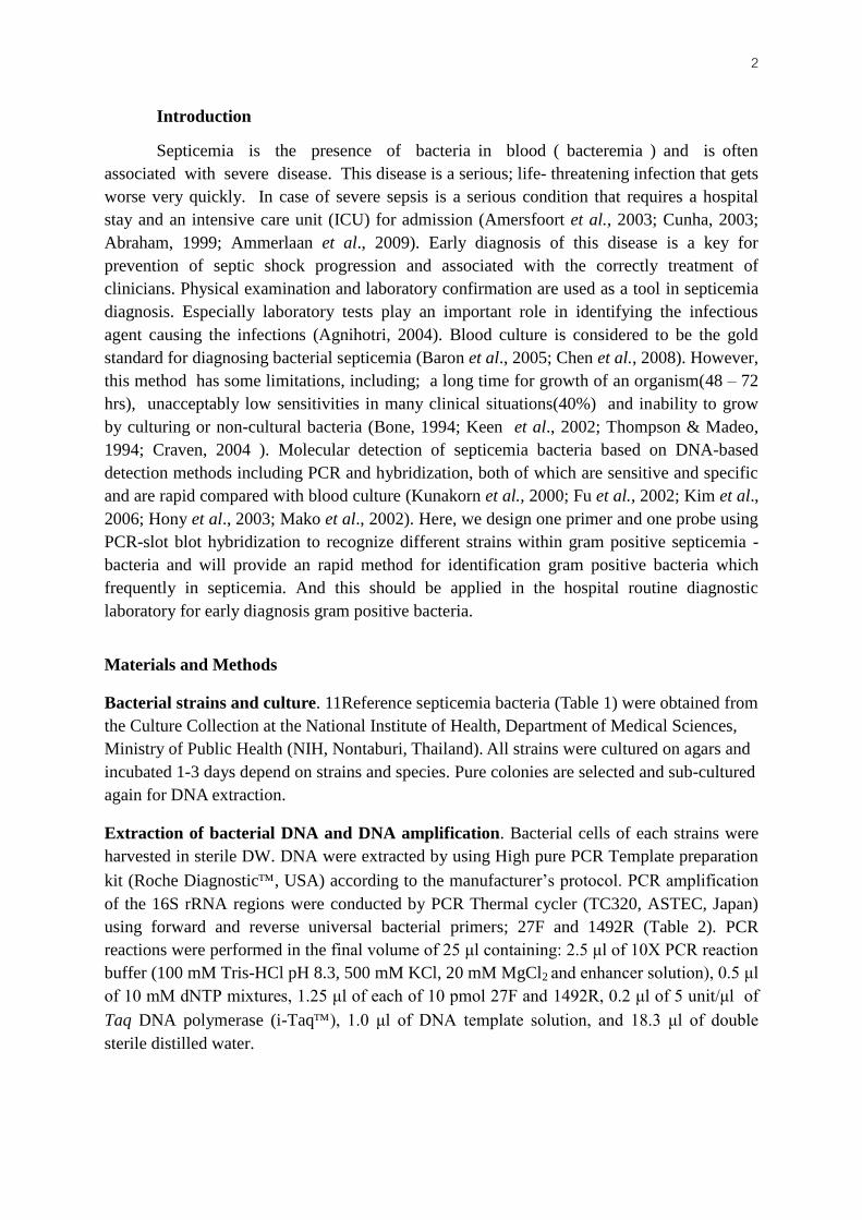
Polynucleotide probe-labeled sized 125 bps was synthesized by using a primer pair (V6F,
V6R) and DIG-11-dUTPs via PCR, shown in Fig. 2.
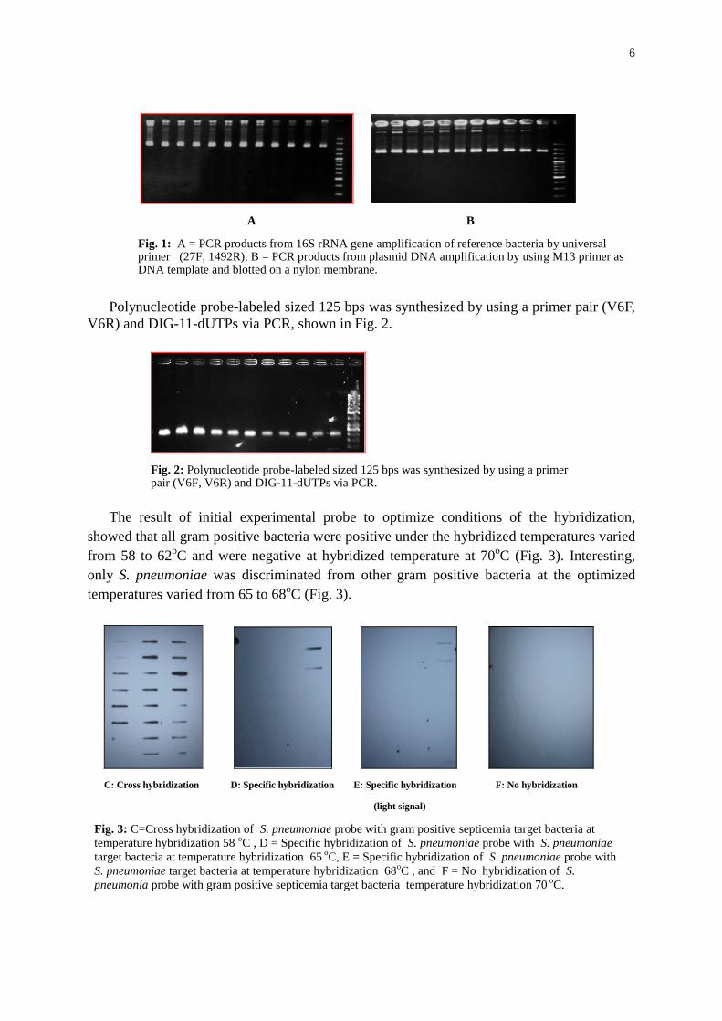
The result of initial experimental probe to optimize conditions of the hybridization,
showed that all gram positive bacteria were positive under the hybridized temperatures varied
from 58 to 62oC and were negative at hybridized temperature at 70
oC (Fig. 3). Interesting,
only S. pneumoniae was discriminated from other gram positive bacteria at the optimized
temperatures varied from 65 to 68oC (Fig. 3).
Fig. 2: Polynucleotide probe-labeled sized 125 bps was synthesized by using a primer pair (V6F, V6R) and DIG-11-dUTPs via PCR.
A B
Fig. 1: A = PCR products from 16S rRNA gene amplification of reference bacteria by universal primer (27F, 1492R), B = PCR products from plasmid DNA amplification by using M13 primer as DNA template and blotted on a nylon membrane.
C: Cross hybridization D: Specific hybridization E: Specific hybridization F: No hybridization
(light signal)
Fig. 3: C=Cross hybridization of S. pneumoniae probe with gram positive septicemia target bacteria at
temperature hybridization 58 oC , D = Specific hybridization of S. pneumoniae probe with S. pneumoniae
target bacteria at temperature hybridization 65 oC, E = Specific hybridization of S. pneumoniae probe with
S. pneumoniae target bacteria at temperature hybridization 68oC , and F = No hybridization of S.
pneumonia probe with gram positive septicemia target bacteria temperature hybridization 70 oC.
7
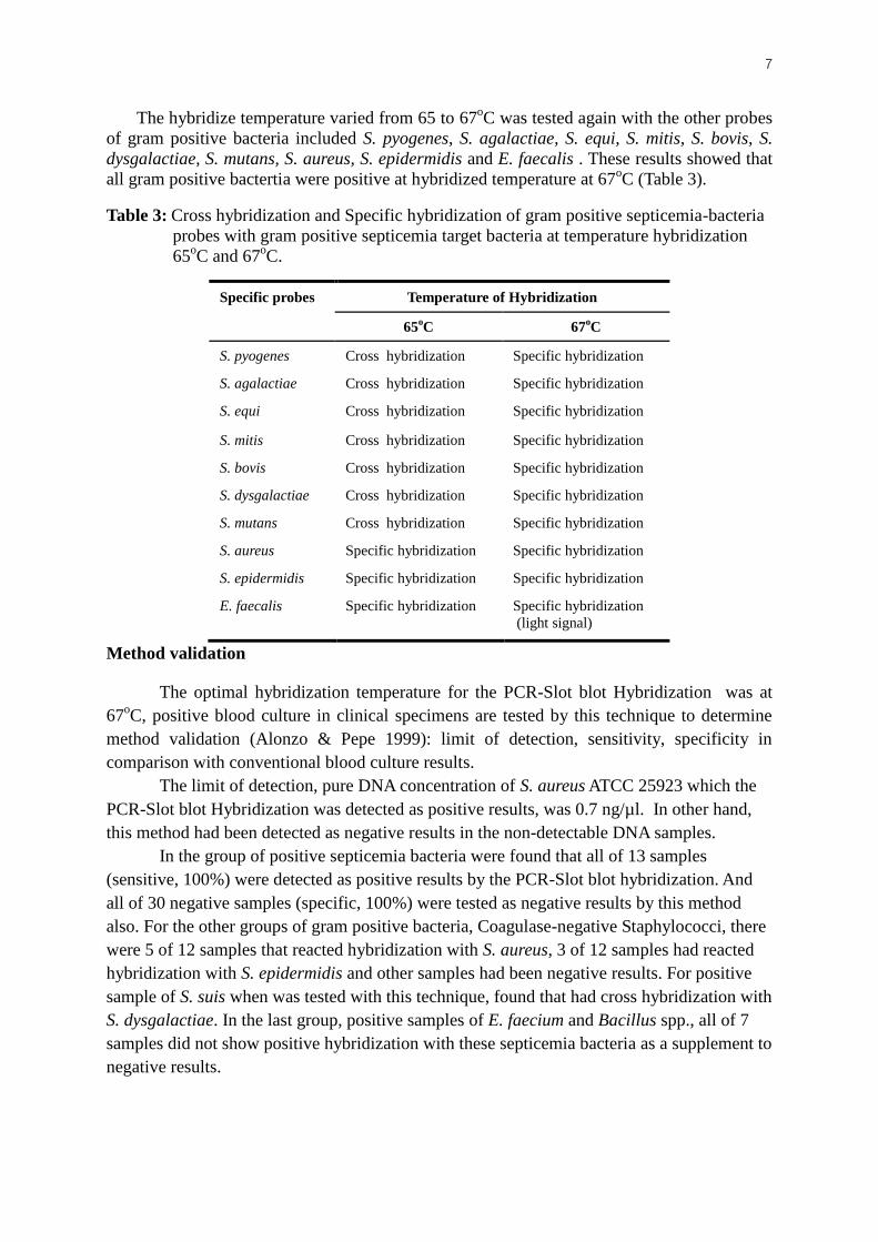
The hybridize temperature varied from 65 to 67oC was tested again with the other probes
of gram positive bacteria included S. pyogenes, S. agalactiae, S. equi, S. mitis, S. bovis, S.
dysgalactiae, S. mutans, S. aureus, S. epidermidis and E. faecalis . These results showed that
all gram positive bactertia were positive at hybridized temperature at 67oC (Table 3).
Table 3: Cross hybridization and Specific hybridization of gram positive septicemia-bacteria
probes with gram positive septicemia target bacteria at temperature hybridization
65oC and 67
oC.
Specific probes Temperature of Hybridization
65oC 67
oC
S. pyogenes Cross hybridization Specific hybridization
S. agalactiae Cross hybridization Specific hybridization
S. equi Cross hybridization Specific hybridization
S. mitis Cross hybridization Specific hybridization
S. bovis Cross hybridization Specific hybridization
S. dysgalactiae Cross hybridization Specific hybridization
S. mutans Cross hybridization Specific hybridization
S. aureus Specific hybridization Specific hybridization
S. epidermidis Specific hybridization Specific hybridization
E. faecalis Specific hybridization Specific hybridization
(light signal)
Method validation
The optimal hybridization temperature for the PCR-Slot blot Hybridization was at
67oC, positive blood culture in clinical specimens are tested by this technique to determine
method validation (Alonzo & Pepe 1999): limit of detection, sensitivity, specificity in
comparison with conventional blood culture results.
The limit of detection, pure DNA concentration of S. aureus ATCC 25923 which the
PCR-Slot blot Hybridization was detected as positive results, was 0.7 ng/µl. In other hand,
this method had been detected as negative results in the non-detectable DNA samples.
In the group of positive septicemia bacteria were found that all of 13 samples
(sensitive, 100%) were detected as positive results by the PCR-Slot blot hybridization. And
all of 30 negative samples (specific, 100%) were tested as negative results by this method
also. For the other groups of gram positive bacteria, Coagulase-negative Staphylococci, there
were 5 of 12 samples that reacted hybridization with S. aureus, 3 of 12 samples had reacted
hybridization with S. epidermidis and other samples had been negative results. For positive
sample of S. suis when was tested with this technique, found that had cross hybridization with
S. dysgalactiae. In the last group, positive samples of E. faecium and Bacillus spp., all of 7
samples did not show positive hybridization with these septicemia bacteria as a supplement to
negative results.
8 Discussion
The results of this study showed that gram positive bacteria causing frequently
septicemia were positive under the hybridize temperature varied from 58 to 62oC and were
negative at hybridize temperature at 70oC. Interesting, only S. pneumoniae was discriminated
from other gram positive septicemia-bacteria at the optimized temperature varied from 65 to
68oC. But the detection signal of hybridization at 68
oC was light signal as a consequence that
this temperature was higher than the melting temperature (Table 4) and may have substantial
effects on weaker hydrogen bonding between probe-target sequences (DIG Application
Manual, Roche applied science, 2009). This polynucleotide probe will benefit for the
identification S. pneumoniae in heamo-cultures samples by using the specific optimized
conditions (Davis & Fuller 1991; Zhang et al., 1995; Chadravorty et al., 2007).
The hybridize temperature varied from 65 to 67oC was tested again with the other
specific probes of gram positive septicemia-bacteria included S. pyogenes, S. agalactiae, S.
equi, S. mitis, S. bovis, S. dysgalactiae, S. mutans, S. aureus, S. epidermidis and E. faecalis.
The hybridize temperature at 65oC exhibited more specific for S. aureus, S. epidermidis and
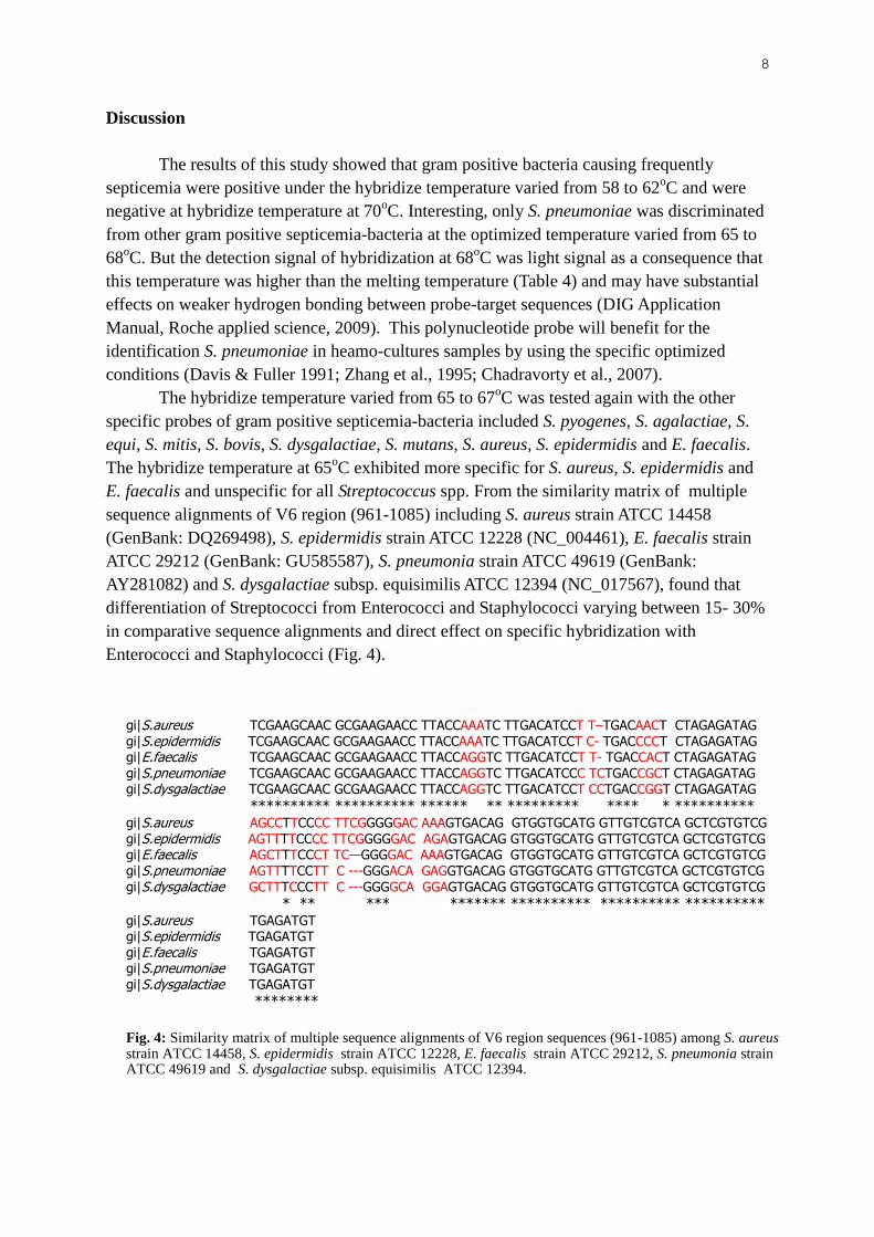
E. faecalis and unspecific for all Streptococcus spp. From the similarity matrix of multiple
sequence alignments of V6 region (961-1085) including S. aureus strain ATCC 14458
(GenBank: DQ269498), S. epidermidis strain ATCC 12228 (NC_004461), E. faecalis strain
ATCC 29212 (GenBank: GU585587), S. pneumonia strain ATCC 49619 (GenBank:
AY281082) and S. dysgalactiae subsp. equisimilis ATCC 12394 (NC_017567), found that
differentiation of Streptococci from Enterococci and Staphylococci varying between 15- 30%
in comparative sequence alignments and direct effect on specific hybridization with
Enterococci and Staphylococci (Fig. 4).
gi|S.aureus TCGAAGCAAC GCGAAGAACC TTACCAAATC TTGACATCCT T–TGACAACT CTAGAGATAG gi|S.epidermidis TCGAAGCAAC GCGAAGAACC TTACCAAATC TTGACATCCT C- TGACCCCT CTAGAGATAG gi|E.faecalis TCGAAGCAAC GCGAAGAACC TTACCAGGTC TTGACATCCT T- TGACCACT CTAGAGATAG gi|S.pneumoniae TCGAAGCAAC GCGAAGAACC TTACCAGGTC TTGACATCCC TCTGACCGCT CTAGAGATAG gi|S.dysgalactiae TCGAAGCAAC GCGAAGAACC TTACCAGGTC TTGACATCCT CCTGACCGGT CTAGAGATAG
********** ********** ****** ** ********* **** * ********** gi|S.aureus AGCCTTCCCC TTCGGGGGAC AAAGTGACAG GTGGTGCATG GTTGTCGTCA GCTCGTGTCG gi|S.epidermidis AGTTTTCCCC TTCGGGGGAC AGAGTGACAG GTGGTGCATG GTTGTCGTCA GCTCGTGTCG gi|E.faecalis AGCTTTCCCT TC—GGGGAC AAAGTGACAG GTGGTGCATG GTTGTCGTCA GCTCGTGTCG gi|S.pneumoniae AGTTTTCCTT C ---GGGACA GAGGTGACAG GTGGTGCATG GTTGTCGTCA GCTCGTGTCG
gi|S.dysgalactiae GCTTTCCCTT C ---GGGGCA GGAGTGACAG GTGGTGCATG GTTGTCGTCA GCTCGTGTCG
* ** *** ******* ********** ********** ********** gi|S.aureus TGAGATGT gi|S.epidermidis TGAGATGT gi|E.faecalis TGAGATGT gi|S.pneumoniae TGAGATGT gi|S.dysgalactiae TGAGATGT
********
Fig. 4: Similarity matrix of multiple sequence alignments of V6 region sequences (961-1085) among S. aureus strain ATCC 14458, S. epidermidis strain ATCC 12228, E. faecalis strain ATCC 29212, S. pneumonia strain ATCC 49619 and S. dysgalactiae subsp. equisimilis ATCC 12394.
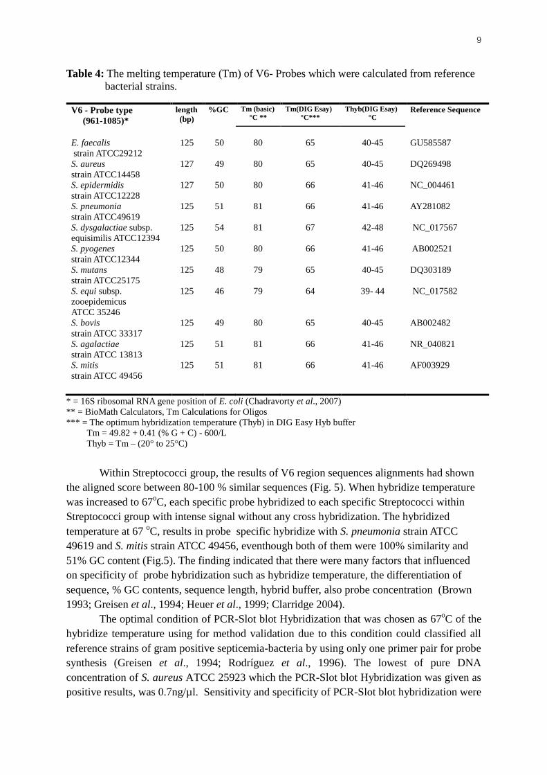
9 Table 4: The melting temperature (Tm) of V6- Probes which were calculated from reference
bacterial strains.
V6 - Probe type
(961-1085)*
length
(bp) %GC
Tm (basic)
°C ** Tm(DIG Esay)
°C***
Thyb(DIG Esay)
°C Reference Sequence
E. faecalis
strain ATCC29212
S. aureus
strain ATCC14458
S. epidermidis
strain ATCC12228
S. pneumonia
strain ATCC49619
S. dysgalactiae subsp.
equisimilis ATCC12394
S. pyogenes
strain ATCC12344
S. mutans
strain ATCC25175
S. equi subsp.
zooepidemicus
ATCC 35246
S. bovis
strain ATCC 33317
S. agalactiae
strain ATCC 13813
S. mitis
strain ATCC 49456
125
127
127
125
125
125
125
125
125
125
125
50
49
50
51
54
50
48
46
49
51
51
80
80
80
81
81
80
79
79
80
81
81
65
65
66
66
67
66
65
64
65
66
66
40-45
40-45
41-46
41-46
42-48
41-46
40-45
39- 44
40-45
41-46
41-46
GU585587
DQ269498
NC_004461
AY281082
NC_017567
AB002521
DQ303189
NC_017582
AB002482
NR_040821
AF003929
* = 16S ribosomal RNA gene position of E. coli (Chadravorty et al., 2007)
** = BioMath Calculators, Tm Calculations for Oligos
*** = The optimum hybridization temperature (Thyb) in DIG Easy Hyb buffer
Tm = 49.82 + 0.41 (% G + C) - 600/L
Thyb = Tm – (20° to 25°C)
Within Streptococci group, the results of V6 region sequences alignments had shown
the aligned score between 80-100 % similar sequences (Fig. 5). When hybridize temperature
was increased to 67oC, each specific probe hybridized to each specific Streptococci within
Streptococci group with intense signal without any cross hybridization. The hybridized
temperature at 67 oC, results in probe specific hybridize with S. pneumonia strain ATCC
49619 and S. mitis strain ATCC 49456, eventhough both of them were 100% similarity and
51% GC content (Fig.5). The finding indicated that there were many factors that influenced
on specificity of probe hybridization such as hybridize temperature, the differentiation of
sequence, % GC contents, sequence length, hybrid buffer, also probe concentration (Brown
1993; Greisen et al., 1994; Heuer et al., 1999; Clarridge 2004).
The optimal condition of PCR-Slot blot Hybridization that was chosen as 67oC of the
hybridize temperature using for method validation due to this condition could classified all
reference strains of gram positive septicemia-bacteria by using only one primer pair for probe
synthesis (Greisen et al., 1994; Rodríguez et al., 1996). The lowest of pure DNA
concentration of S. aureus ATCC 25923 which the PCR-Slot blot Hybridization was given as
positive results, was 0.7ng/µl. Sensitivity and specificity of PCR-Slot blot hybridization were

10 highly value, 100% (Alonzo & Pepe 1999); Zhang et al., 1995; Borchardt et al., 2004). In the
cases of Coagulase-negative Staphylococci were not confirmed to bacterial species, these
cases may be identified to many species of Staphylococci. Few samples hybridized with S.
epidermidis which may truly be S. epidermidis, a more detailed identification test could be
experimental to confirm this hybridization by using biochemical tests as in analytical profile
index tests methods. For some samples of cross hybridization with S. aureus are therefore
critically important in false reports in some species. A false negative may be perceived as the
samples are not allowed time to cool for 30 minutes at room temperature or 10 minutes in the
freezer, given the fact that the serum will be sufficient for melting (Sperber & Tatini, 1975;
Essers and Radebold, 1980; Dickson & Marples, 1986). Cross hybridization between S. suis
and S. dysgalactiae revealed the closest similarity between theV6-16SrRNA region of these
two bacteria, increasing the hybridize temperature would be increase the discrimination
power in order to separate S. suis and S. dysgalactiae (Fig.6). For the last group, E. faecium
and Bacillus spp., all of 7 samples, results found that there were no cross hybridization with
these septicemia bacteria that supporting the negative results.
gi|S.dysgalactiae TCGAAGCAAC GCGAAGAACC TTACCAGGTC TTGACATCCT CCTGACCGGT CTAGAGATAG gi|S.agalactiae TCGAAGCAAC GCGAAGAACC TTACCAGGTC TTGACATCCT TCTGACCGGC CTAGAGATAG gi|S.pneumoniae TCGAAGCAAC GCGAAGAACC TTACCAGGTC TTGACATCCC TCTGACCGCT CTAGAGATAG gi|S.mitis TCGAAGCAAC GCGAAGAACC TTACCAGGTC TTGACATCCC TCTGACCGCT CTAGAGATAG gi|S.pyogenes TCGAAGCAAC GCGAAGAACC TTACCAGGTC TTGACATCCC GATGCCCGCT CTAGAGATAG gi|S.mutans TCGATGCAAC GCGAAGAACC TTACCAGGTC TTGACATCCC GATGCTATTC TTAGAGATAG gi|S.equi TCGAAGCAAC GCGAAGAACC TTACCAGGTC TTGACATCCC GATGCTATTC TTAGAGATAA gi|S.bovis TCGATGCAAC CGCAAGAACC TTACCAGGTC TTGACATCCC GATGCTATTC TTAGAGATAG
**** ***** ********* ********** ********* ** *********
gi|S.dysgalactiae GCTTTCCCTT CGGGGCAGGA GTGACAGGTG GTGCATGGTT GTCGTCAGCT CGTGTCGTGA gi|S.agalactiae GCTTTCTCTT CGGAGCAGAA GTGACAGGTG GTGCATGGTT GTCGTCAGCT CGTGTCGTGA gi|S.pneumoniae AGTTTTCCTT CGGGACAGAG GTGACAGGTG GTGCATGGTT GTCGTCAGCT CGTGTCGTGA gi|S.mitis AGTTTTCCTT CGGGACAGAG GTGACAGGTG GTGCATGGTT GTCGTCAGCT CGTGTCGTGA gi|S.pyogenes AGTTTTACTT CGGTACATCG GTGACAGGTG GTGCATGGTT GTCGTCAGCT CGTGTCGTGA gi|S.mutans GAAGTTACTT CGGTACATCG GAGACAGGTG GTGCATGGTT GTCGTCAGCT CGTGTCGTGA gi|S.equi GAAGTTACTT CGGTACATTG GAGACAGGTG GTGCATGGTT GTCGTCAGCT CGTGTCGTGA gi|S.bovis GGTTTCTCTT CGGAACATCG GTGACAGGTG GTGCATGGTT GTCGTCAGCT CGTGTTGTGA
* *** *** ** * ******** ********** ********** ***** **** gi|S.dysgalactiae GATGT gi|S.agalactiae GATGT gi|S.pneumoniae GATGT gi|S.mitis GATGT gi|S.pyogenes GATGT gi|S.mutans GATGT gi|S.equi GATGT gi|S.bovis GATGT
*****
Fig. 5: Similarity matrix of multiple sequence alignments of V6 region sequences (961-1085) within Streptococci composed S. pneumonia strain ATCC 49619, S. dysgalactiae subsp. equisimilis ATCC 12394, S. pyogenes strain ATCC 12344, S. mutans strain ATCC 25175, S. equi subsp. zooepidemicus ATCC 35246, S. bovis strain ATCC 33317, S. agalactiae strain ATCC 13813 and S. mitis strain ATCC 49456.

11
Conclusion
The results revealed that probe design and the optimized conditions were successful in
the detection of gram positive septicemia-bacteria. From these results, polynucleotide probes
targeting a region of the V6 of 16S rRNA gene probe, and the specific optimized
hybridization conditions at 67oC proved to be more specific for the identification each
septicemia- gram positive bacteria. This finding is a major progress; however further
investigation of the gram negative septicemia-bacteria would be required for a broader
identification of septicemia bacteria.
Acknowledgments
We would like to thank you the department of Medical Sciences, Ministry of Public
Health, Thailand to support the finance of this project and really thank you the faculty of
Medical Science, Naresuan University to give us about introduction, equipment for all of the
project processes. And thank you of officer of the Khon Kaen hospital and Regional Medical
Science Center, Khon Kaen to help me to collect blood culture samples and sample
preparation.
References
Amersfoort, E. S. V., Berkel, T. J. C. V. & Kuiper, J. (2003). Receptors, Mediators, and
Mechanisms Involved in Bacterial Sepsis and Septic Shock. Clin Microbiol Rev 16, 379–414.
Cunha, B. A. (1996). Fever in the intensive care unit. Intensive Care Med 25, 648-651.
Abraham, E. (1999). Why immunomodulatary therapies have not worked in sepsis.
Intensive Care Med 25, 556-566.
Ammerlaan, H., Seifert, H., Harbarth, S., Brun-Buisson, C., Torres, A., Antonelli, M. &
other authors. (2009). Adequacy of antimicrobial treatment and outcome of Staphylococcus
aureus bacteremia in 9 Western European countries. Clin Infect Dis 49, 997-1005.
Agnihotri, N., Kaistha, N. & Gupta, V. (2004). Antimicrobial susceptibility of isolates
from neonatal septicemia. Jpn J Infect Dis 57, 273-275.
gi|S.dysgalactiae TCGAAGCAAC GCGAAGAACC TTACCAGGTC TTGACATCCT CCTGACCGGT CTAGAGATAG gi|S.suis TCGATGCAAC GCGAAGAACC TTACCAGGTC TTGACATCCC GATGACCGCC CTAGAGATAG
**** ***** ********** ********** ********* ****** ********** gi|S.dysgalactiae GCTTTCCCTT CGGGGCAGGA GTGACAGGTG GTGCATGGTT GTCGTCAGCT CGTGTCGTGA gi|S.suis GGTTTCTCTT CGGAGCATCG GTGACAGGTG GTGCATGGTT GTCGTCAGCT CGTGTCGTGA
* **** *** *** *** ********** ********** ********** ********** gi|S.dysgalactiae GATGT gi|S.suis GATGT
*****
Fig. 6: Similarity matrix created of sequence alignments of V6 region sequences (961-1085) between S. dysgalactiae subsp. equisimilis ATCC 12394 and S. suis strain ATCC 43765.

12 Baron, E. J., Scott, J. D. & Tompkins, L. S. (2005). Prolonged incubation and extensive
sub-culturing do not increase recovery of clinically significant microorganisms from standard
automated blood cultures. J Clin Infect Dis 41, 1677-1680.
Chen, J. R., Lee, S. Y., Yang, B. H. & Lu, J. S. (2008). Rapid identification and
Susceptibility testing using the VITEK2 System using culture fluids from positive
BacT/ALERT blood cultures. J Microbiol Immunol Infect 41, 259-264.
Bone, R.C. (1994). Sepsis and its complications: the clinical problem. Crit Care Med 22, S8-
S11.
Keen, A., Knoblock, L., Edelman, L. & Saffle, J. (2002). Effective limitation of blood
culture use in the burn unit. J Burn Care Rehabil 23, 183-189.
Thompson, F. & Madeo, M. (1994). Blood cultures: towards zero false positives. J Clin
Pathol 47, 796-798.
Craven, D.A. (2004). Blood Cultures for Community-Acquired Pneumonia. Am J Res Crit
Care Med 169, 327-328.
Kunakorn, M., Raksakait, K., Sethaudom, C., Sermswan, R. W. & Dharakul, T. (2000).
Comparison of three PCR primer set for diagnosis of septicemic melioidosis. Acta Tropical
74, 247-251.
Fu, J-f., Yu, H-Y., Shang, S-Q., Hong, W-L., Lu, M-Q. & Li, J-P. (2002). A molecular
biological study on identification of common septicemia bacteria using 16S-23S rRNA gene
spacer regions. J Zhejiang University (SCIENCE) 3, 237-242.
Kim, S., Frye, J. G., Hu, J., Fedorka-Cray, P. J., Gautom, R. & Boyle, D.S. (2006).
Multiplex PCR-Based Method for identification of common clinical serotypes of Salmonella
enterica subsp. enterica. J Clin Microbiol 44, 3608-3615.
Hony, Y., Berrang, M. E., Liv, T., Hafacre, C.L., Sanchez, S., Wang, L. & other authors
(2003). Rapid detection of Campylobacter coli, C. jejuni, and Salmonella enterica, on poultry
carcasses by using PCR-Enzyme-Linked Immunosorbent Assay. Appl Environ Microbiol 69,
3492-3499.
Mako, K., Eiichi, M., Hisashi, K., Nobuyasu, Y., Katsuji, T. & Masao, N. (2002). 16S
Ribosomal DNA-Based Analysis of Bacterial Diversity in Purified Water Used in
Pharmaceutical Manufacturing Processes by PCR and Denaturing Gradient Gel
Electrophoresis. Appl Environ Microbiol 68, 699-704.
Chadravorty, S., Helb, D., Burday, M., Connell, N. & Alland, D. (2007). A detailed
analysis of 16S ribosomal RNA gene segments for the diagnosis of pathogenic bacteria. J
Microbiol Methods 69, 330-339.
Alonzo, T. A. & Pepe, M. S. (1999). Assessing accuracy of new diagnostic test. Stat Med
18, 2987-3003.
Procedures for Nonradioactive Labeling and Detection. (2009). DIG Application Manual
for Filter Hybridization, Roche Applied Science, Retrieved December 29, 2012, from
http://www.roche-applied-science.com/PROD_INF/MANUALS/DIG_MAN/dig_toc.htm.
Davis, T. E. & Fuller, D. D. (1991). Direct identification of bacterial isolates in blood
cultures by using a DNA probe. J Clin Microbiol 29, 2193-96.
13 Zhang, Y., Isaacman, D. J., Waduwsky, R. M., Rydquist-White, J., Post, J. C. &
Ehrlich, G. D. (1995). Detection of Streptococcus pneumonia in whole blood by PCR. J Clin
Microbiol 33, 596-601.
Wang, Y. & Qian, P.Y. (2009.) Conservative fragments in bacterial 16S rRNA genes and
primer design for 16S ribosomal DNA amplicons in metagenomic studies. PLoS ONE 4:
e7401.
Brown, T. A. (1993). Nucleic acid blotting and hybridization. In Molecular Biology Lab
Fax: Gene Analysis, pp. 1-19. Edited by T. A. Brown: Academic Press.
Greisen, K., Loeffelholz, M., Purohit, A. & Leong, D. (1994). PCR primers and probes for
the 16S rRNA gene of most species of pathogenic bacteria, including bacteria found in
cerebrospinal fluid. J Clin Microbiol 32, 335-51.
Heuer, H., Hartung, K., Wieland, G., Kramer, I. & Smalla, K. (1999). Polynucleotide
probes that target a hypervariable region of 16S rRNA genes to identify bacterial isolates
corresponding to bands of community fingerprints. Appl Environ Microbiol 65, 1045-49.
Clarridge, J. E. (2004). Impact of 16S rRNA gene sequence analysis for identification of
bacteria on clinical microbiology and infectious diseases. J Clin Microbiol Rev 17, 840-862.
Rodríguez, M., Núñez, F., Córdoba, J. J., Bermúdez, E. & Asensio, M A. (1996). Gram-
positive, catalase-positive cocci from dry cured Iberian ham and their enterotoxigenic
potential. Appl Environ Microbiol 62, 1897-1902.
Borchardt, S. M., Foxman, B., Chaffin, D.O., Rubens, C. E., Tallman, P. A. & Manning,
S. D. (2004). Comparison of DNA Dot Blot Hybridization and Lancefield Capillary
Precipitin Methods for Group B Streptococcal Capsular Typing. J Clin Microbiol 42, 146-
150.
Sperber, W. H. & Tatini, S. R. (1975). Interpretation of the Tube Coagulase Test for
Identification of Staphylococcus aureus. Appl Microbiol 29, 502-5.
Essers, L. & Radebold, K. (1980). Rapid and reliable identification of Staphylococcus
aureus by a latex agglutination test. J Clin Microbiol 12, 641-643.
Dickson, J. I. & Marples, R. R. (1986). Coagulase production by strains of Staphylococcus
aureus of differing resistance characters: a comparison of two traditional methods with a
latex agglutination system detecting both clumping factor and protein A. J Clin Pathol 39,
371-375.