AES2013 Harnett plenary talk: Electrodes for microfluidic applications

Neuroscience Center and Division of Neuroscience and Physiology
Department of Biosciences Faculty of Biological and Environmental Sciences
Integrative Life Science Doctoral Program University of Helsinki
Development of Microfluidic Applications to Study the Role of Kainate Receptors in Synaptogenesis
Prasanna Sakha
ACADEMIC DISSERTATION
To be presented for public examination, with the permission of the Faculty of Biological and Environmental Sciences of the University of Helsinki, in lecture room 2402,
Viikki Biocenter 3, on 22nd April 2016, 12 o’ clock noon.
Helsinki 2016

ii
Supervised by Docent Sari Lauri, PhD Neuroscience Center and Department of Biosciences University of Helsinki, Helsinki, Finland Docent Henri Huttunen, PhD Neuroscience Center University of Helsinki, Helsinki, Finland Thesis Committee Professor Sami Franssila, PhD Department of Materials Science and Engineering Aalto University, Espoo, Finland Docent Pirta Hotulainen, PhD Minerva Institute for Medical Research University of Helsinki, Helsinki, Finland Reviewed by Docent Irma Holopainen, MD, PhD Department of Pharmacology University of Turku, Turku, Finland Tommi Möykkynen, PhD Department of Biosciences University of Helsinki, Helsinki, Finland Opponent Docent Susanna Narkilahti, PhD NeuroGroup, Institute of Biomedical Technology University of Tampere, Tampere, Finland Custos Professor Juha Voipio, PhD Department of Biosciences University of Helsinki, Helsinki, Finland ISSN 2342-3161 (Print) ISSN 2342-317X (Online) ISBN 978-951-51-2031-1 (Paperback) ISBN 978-951-51-2032-8 (PDF) Unigrafia, Helsinki 2016
iii
Table of Contents List of Original Publications ................................................................................................. vii
Abbreviations...................................................................................................................... viii
Abstract ................................................................................................................................. 1
1 Literature Review ............................................................................................................... 2
1.1 Kainate Receptors ........................................................................................................ 2
1.1.1 Subunit composition and structure of Kainate Receptors ................................... 3
1.1.2 RNA editing of the KAR subunits .......................................................................... 4
1.1.3 Post translational modifications ........................................................................... 6
1.2 Subcellular distribution of Kainate Receptors and Trafficking .................................... 6
1.2.1 Exocytosis from ER to the surface ........................................................................ 7
1.2.1.1 Synaptic targeting .............................................................................................. 9
1.2.2 Kainate Receptor endocytosis and degradation ................................................ 11
1.3 KAR Signaling Pathways ............................................................................................. 12
1.3.1 Ionotropic signaling ............................................................................................ 12
1.3.2 G protein-coupled signaling................................................................................ 13
1.4 Physiological Functions .............................................................................................. 14
1.4.1 Presynaptic KARs: Modulation of neurotransmitter release ............................. 15
1.4.2 Postsynaptic Kainate Receptors ......................................................................... 16
1.4.3 Role of KARs in synaptic plasticity ...................................................................... 17
1.5 Synaptogenesis .......................................................................................................... 18
1.5.1 Synapse progenesis ............................................................................................ 18
1.5.2 Pre and postsynaptic differentiation .................................................................. 19
1.5.2.1 Presynaptic maturation ................................................................................... 19
1.5.2.2 Synaptic vesicles and markers of presynaptic differentiation ......................... 20
1.5.2.3 Postsynaptic maturation ................................................................................. 22
1.5.2.4 Functional maturation of synapse ................................................................... 22
1.6 Morphological development and Regulation of Axonal mobility by KARs ................ 23
1.6.1 KARs in neurite outgrowth and morphological maturation of neurons ............ 23
iv
1.6.2 Physiological regulation of filopodial motility by KAR activation ....................... 25
1.6.3 Kainate Receptors in functional maturation of synapses ................................... 26
1.7 Microfluidics neuronal culture and optogenetic application in Neuroscience ......... 27
1.7.1 Microfluidic neuronal cultures to study neuronal processes ............................. 27
1.7.2 Advantages of microfluidic application in neurobiology .................................... 28
1.7.3 Optogenetic tools in neuroscience ..................................................................... 29
2. Aims of the Study............................................................................................................. 31
3. Material and Methods ..................................................................................................... 32
3.1 Cell culture ................................................................................................................. 32
3.1.1 Neuronal cultures ............................................................................................... 32
3.1.1.1 Isolation of embryonic rat hippocampal neurons ........................................... 32
3.1.1.2 Microfluidic cultures ........................................................................................ 32
3.1.2 Dispersed neuronal cultures ............................................................................... 34
3.1.3 Hippocampal slice preparations ......................................................................... 34
3.2 Production of lentiviral vectors and viral transfection .............................................. 34
3.2.1 List of viruses used.............................................................................................. 35
3.3 Molecular-Biochemical techniques ........................................................................... 35
3.3.1 Immunostaining .................................................................................................. 35
3.3.2 List of antibodies ................................................................................................ 36
3.3.2 In-Situ hybridization ........................................................................................... 36
3.3.3 RT-PCR ................................................................................................................ 36
3.3.4 Western Blot ....................................................................................................... 37
3.4 Confocal Imaging ....................................................................................................... 37
3.5 Image Analysis ........................................................................................................... 37
3.5.1 Receptor classification at proximal and distal dendrites ................................... 37
3.5.2 Filopodial analysis ............................................................................................... 38
3.5.3 Synaptic vesicle, PSD-95 and colocalization density .......................................... 38
3.5.4 Dendritic spines analysis..................................................................................... 38
3.5.5 Axonal localization of GluK1c with GluK4/5 ....................................................... 39

v
3.6 Electron Microscopy .................................................................................................. 39
3.7 Electrophysiology ...................................................................................................... 39
3.7.1 Microfluidic culture electrophysiology and optogenetics .................................. 39
3.7.2 Hippocampal slice electrophysiology ................................................................. 40
3.8 Pharmacological Tools ............................................................................................... 40
3.9 Statistical Analysis ..................................................................................................... 40
4. Results ............................................................................................................................. 41
4.1 Development of microfluidic chamber for rat hippocampal neurons ...................... 41
4.1.1 Characterization of axonal growth and functional viability of the neurons in the microfluidic chamber ................................................................................................... 42
4.1.2 Asymmetric fluidic isolation of neurons ............................................................. 43
4.1.3 Optogenetic activation of microfluidically isolated axons ................................. 44
4.2 Subcellular localization of GluK1 subunit containing KARs ....................................... 45
4.2.1 Heteromerization of GluK1c with high-affinity KARs promotes distal targeting of GluK1c in dendritic processes ...................................................................................... 45
4.3 KARs promote formation of axonal and dendritic protrusions ................................. 47
4.3.1 Expression profile of KAR subunits in microfluidic cultures ............................... 47
4.3.2 KARs promote formation of axonal protrusions ................................................ 48
4.3.2.1 Enrichment of axonal filopodia protrusion is independent of KAR subunit type .............................................................................................................................. 48
4.3.3. GluK2 KARs promote maturation of dendritic spines ....................................... 51
4.3.4. GluK1 editing and channel inhibition does not affect outgrowth of filopodial protrusions .................................................................................................................. 55
4.4 KARs regulate clustering of synaptic vesicles ............................................................ 55
4.4.1 Low-affinity KARs enhance synaptic vesicle clustering ...................................... 55
4.4.2 High-affinity KARs circumvent synaptic vesicle clustering ................................. 57
4.4.3 Heteromerization of high and low affinity KARs decreases synaptic vesicle clusters......................................................................................................................... 58
4.5 Regulation of filopodia and synaptic vesicle clusters via KARs follow distinct but converging pathway ........................................................................................................ 58

vi
4.6 Regulation of glutamatergic transmission by presynaptic KARs ............................... 60
4.6.1 Presynaptic calcium permeable low-affinity KARs enhance success rate of synaptic vesicle release ............................................................................................... 60
4.6.2 Heteromeric KARs mimic dominant features of high affinity KARs in suppressing glutamatergic transmission ......................................................................................... 61
4.6. 3. Low-affinity KARs affect synaptic ultrastructure ............................................. 61
5 Discussion ......................................................................................................................... 65
5.1 Asymmetrical neuronal growth in the microfluidic chamber ................................... 65
5.2 Axonal targeting of KAR subunits .............................................................................. 67
5.3 Synaptogenesis – An interplay of KARs, filopodiogenesis and clustering of synaptic vesicles ............................................................................................................................. 68
5.4 Postsynaptic roles of KARs in synaptic differentiation .............................................. 70
5.5 Synaptic maturation by KARs .................................................................................... 71
5.6 Significance in brain dysfunction ............................................................................... 73
6 Conclusions ....................................................................................................................... 75
7 Acknowledgements .......................................................................................................... 76
8 References ........................................................................................................................ 77
vii
List of Original Publications
I. A microfluidic chip for axonal isolation and electrophysiological measurements. Ville Jokinen*, Prasanna Sakha*, Pia Suvanto, Claudio Rivera, Sami Franssila, Sari
E.Lauri & Henri J. Huttunen (2013). Journal of Neuroscience Methods 212: 276-282 *equal contribution
II. Expression of GluK1c underlies the developmental switch in presynaptic kainate
receptor function. Aino Vesikansa, Prasanna Sakha, Juha KUja-Panula, Svetlana Molchanova, Claudio
Rivera, Henri J. Huttunen, Heikki Rauvala, Tomi Taira & Sari E. Lauri (2012). Scientific Reports 2:310 III. Axonal Kainate receptors regulate the strength of efferent connectivity by promoting
presynaptic differentiation. Prasanna Sakha, Aino Vesikansa, Ester Orav, Joonas Heikkinen, Ville Jokinen, Tiina-
Kaisa Kukku-Lukjanov, Alexandra Shintyapina, Sami Franssila, Henri J. Huttunen & Sari E. Lauri (2016).Frontiers in Cellular Neuroscience 10:3
The candidate has done all the cell biological and electrophysiological experiments in I (Figures 2-4) and the experiments addressing axonal targeting of various kainate receptor subunits in II (Figure 5 and partly Figure 4). Most of the experimental work in III, except for the data presented in the Figures 1B and 1C has been done by the candidate.
The candidate has also actively participated in design and development of the microfluidic tools, optimization of the culture conditions and planning of the experiments in I, II and III, as well as in analyzing the data and writing the manuscripts.

viii
Abbreviations aa amino acid ACSF artificial cerebrospinal fluid AMPA α-amino-3-hydroxy-5-methyl-4-isoxazolepropionic acid AMPAR AMPA receptor AZ active zone BAR Bin-Amphiphysin-Rvs) BDNF brain derived neurotropic factor BSA bovine serum albumin Ca2+ calcium ion CA1 Cornu ammonis region 1 CA3 Cornu ammonis region 3 CAMs cell adhesion molecules CAMKII calcium/calmodulin-dependent protein kinase-II ChR1 Channelrhodopsin-1 (ChR1) ChR2 Channelrhodopsin-2 (ChR2) Cl- chloride ion CNS Central nervous system COPI coatamer protein complex-1 CRMP2 collapsin response mediator protein 2 DIV days in vitro DRG dorsal root ganglia DsRED Discosoma sp. red fluorescent protein EPSC excitatory post synaptic Current ER endoplasmic reticulum FGF fibroblast growth factor GABA γ-Aminobutyric acid GDNF glia cell-derived growth factor GFP green fluorescent protein GRIP glutamate receptor interacting protein HEK293T human embryonic kidney 293-T antigen cells HR halorhodposins ICC immunocytochemistry IPSC inhibitory postsynaptic current ITO indium-tin iodide IsAHP slow Ca2+ activated K+ current IAHP Ca2+ dependent K+ channel mediated after hyperpolarization current JNK c-Jun N-terminal kinase KA kainic acid (kainate) KAR kainate receptor (kainic acid receptor)

ix
KCC2 K+-Cl- cotransporter LTD long-term depression LTP long-term potentiation mIPSCs miniature IPSCs MEA micro electrode array MSB multiple synapse bouton NB neurobasal Na+ sodium ion NB neurobasal NETO neuropilin and tolloid-like NMDA N-Methyl-D-aspartic acid or N-Methyl-D-aspartate NMDAR NMDA receptor NMJ neuromuscular junction NTD N-terminal domain PBS phosphate buffered saline PDMS polydimethylsiloxane PDZ post synaptic density protein 95 (PSD95), Drosophila disk large tumor
suppressor (Dlg1), and zonula occludens-1 protein (ZO-1) PFA paraformaldehyde PKA protein Kinase A PKC protein Kinase C PLL poly-L-lysine PM plasma membrane PNS peripheral nervous system PPF paired-pulse facilitation Pr release probability PSD postsynaptic density PSD95 postsynaptic density-95 RHN rat hippocampal neuron RIM Rab3 interacting proteins RRP readily releasable pool sAHP postspike slow after-hyperpolarization potential SNAP25 synaptosomal-associated protein 25 SNARE SNAP (Soluble SNF Attachment Protein) REceptor SSB single synapse bouton SUMO-1 small ubiquitin-related modifier 1 SV synaptic vesicle SVs synaptic vesicles SynCAM synaptic cell adhesive molecule TM transmembrane uIPSCs unitary-IPSCs

1
Abstract Neurons have polar morphology with distinctive subcellular features comprising of cell soma, axons and dendrites. Axons are very thin in diameter in comparison to the dendritic processes. Studying axons has been traditionally difficult due to the lack of functional tools. Consequently, the molecular mechanisms that regulate morphological differentiation and polarized assembly of presynaptic structures are not well understood. Primary objective of this study was to develop a novel microfluidic device for spatial isolation of axons from the somatodendritic compartment of cultured hippocampal neurons. A new method was developed for asymmetrical genetic manipulation improving specificity in studies of how individual proteins affect axonal morphology, presynaptic development and function. Subsequently, the microfluidic culture system was used to study the signaling events involved in synaptogenesis, focusing on the roles of kainate type of glutamate receptors (KARs). Functional studies have shown that KARs are present in axons and may regulate presynaptic function. However, the molecular composition and detailed subcellular localization of axonal KARs as well as their roles in presynaptic differentiation are largely unknown.
The results show that different subunits of KARs are involved invariantly in early stages of synaptogenesis. Expression of low (GluK1-3) and high affinity (GluK4-5) KAR subunits promoted filopodiogenesis irrespective of its channel function at the isolated axons. In addition, axonal low affinity subunits enhanced clustering of synaptic vesicles and transmission efficacy at nascent glutamatergic synapses, an effect which was associated with widening of presynaptic active zone. Likewise, low affinity subunit GluK2 affected generation of dendritic spines. On the other hand, high affinity KAR subunits had no effect on synaptic vesicle clustering, nor presynaptic transmission efficacy. However their heteromerization with low affinity subunits completely prevented the synapse promoting effects and instead lead to strong inhibition of presynaptic transmission efficacy. The presynaptic effects of GluK1-3 on synaptic vesicle clustering involved both PKA and PKC pathways. Moreover, heteromerization of GluK1 with individual high-affinity KAR subunits affected its subcellular targeting in the neurons. GluK1 expression was developmentally regulated in neonatal and juvenile hippocampus and heteromeric combination of GluK1c with high affinity subunits suppressed glutamatergic synaptic transmission.
KARs are linked to various neurological and neuropsychiatric disorders. Our observations and previous findings strongly suggest that KARs are involved in morphological maturation of neurons and in refinement of neuronal circuitry in the brain. The present results provide novel insights into the involvement of different types of KAR subunits in synaptic development and morphological differentiation. Hence, they are potential therapeutic targets in various developmentally originating neurological disorders.

2
1 Literature Review
1.1 Kainate Receptors Forewords: Generation of synapses and refinement of synaptic connectivity is essential for the establishment of functional circuitry during brain development. Synapses can be heterogeneous both structurally and functionally, but still particular in transmitting specific yet diverse signals between distant communicating partners. The overall operation of such biomechanical organ is responsible for coordinating and sensing basic biological functions and in guiding cognitive consciousness. This is where memories are processed, stored and generated. Yet, deciphering how brains exactly work has been a daunting challenge for the neurobiologist.
Synaptic signals are transmitted by neurotransmitters via chemical synapses. Glutamate primarily transmits excitatory signals in the central nervous system (CNS) (e.g. review Pinheiro & Mulle, 2006; Lerma 2003; Contractor et al. 2011). Glutamate acts on three different types of ligand-gated ionotropic receptors, namely N-Methyl-D-aspartic acid receptors (NMDAR), kainate receptors (KARs) and α-amino-3-hydroxy-5-methyl-4-isoxazolepropionic acid receptors (AMPAR) located diversely in the brain. The nomenclature of these receptors was pharmacologically defined in respect to the exogenous activation by their agonist. Most of the current knowledge about these receptors comes from different animal models, and particularly from the rodent hippocampus. AMPAR and NMDAR are found primarily in the postsynaptic region whilst KARs are present also presynaptically to regulate excitatory and inhibitory synaptic transmission. Additionally, KARs are unique as they also couple with unconventional G-protein signaling pathways (e.g. review Lauri & Taira 2012; Carta et al. 2014, Sihra et al. 2013; Rodríguez-Moreno & Sihra 2007).
There are five different subunits of KARs, namely GluK1, GluK2, GluK3, GluK4 and GluK5 (previous nomenclature GluK5, GluK6, GluK7, KA1 and KA2, respectively), transcribed from GRIK1, GRIK2, GRIK3, GRIK4 and GRIK5 genes respectively (review Lerma 2003; Coussen 2009; González-González et al. 2012; Contractor et al. 2011). The length of amino acid (aa) chain varies depending on the subunit. Predicted polypeptide length of GluK1: 854-871 aa, GluK2: 869-889 aa, GluK3: 910-919 aa, GluK4: 956, and GluK5: 979 aa long (review Lerma 2003; NP_036704.1 n.d.; NP_113696.1 n.d.). Recent studies ascribe integral roles of KARs at the physiological regulation of neuronal networks in the hippocampus and other brain regions. However, little information exists on how these findings translate at the
3
behavioral level and to link to brain disorders (Carta et al. 2014; Lerma & Marques 2013). Moreover, increasing evidence suggests that apart from modulating synaptic transmission and plasticity, KARs may be involved in the development of neuronal network. However, the exact roles of KARs and in particular, presynaptic KARs in circuit development are largely unclear.
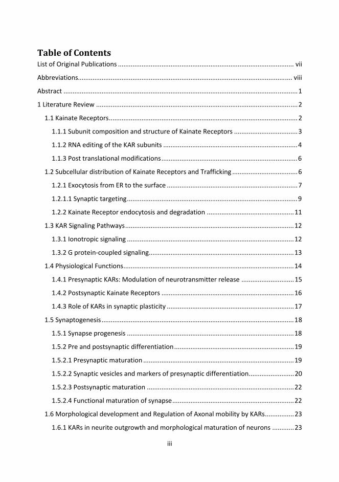
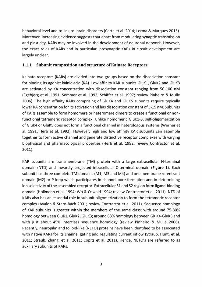
1.1.1 Subunit composition and structure of Kainate Receptors Kainate receptors (KARs) are divided into two groups based on the dissociation constant for binding its agonist kainic acid (KA). Low affinity KAR subunits GluK1, GluK2 and GluK3 are activated by KA concentration with dissociation constant ranging from 50-100 nM (Egebjerg et al. 1991; Sommer et al. 1992; Schiffer et al. 1997; review Pinheiro & Mulle 2006). The high affinity KARs comprising of GluK4 and GluK5 subunits require typically lower KA concentration for its activation and has dissociation constant of 5-15 nM. Subunits of KARs assemble to form homomere or heteromere dimers to create a functional or non-functional tetrameric receptor complex. Unlike homomeric GluK1-3, self-oligomerization of GluK4 or GluK5 does not form a functional channel in heterologous systems (Werner et al. 1991; Herb et al. 1992). However, high and low affinity KAR subunits can assemble together to form active channel and generate distinctive receptor complexes with varying biophysical and pharmacological properties (Herb et al. 1992; review Contractor et al. 2011). KAR subunits are transmembrane (TM) protein with a large extracellular N-terminal domain (NTD) and inwardly projected intracellular C-terminal domain (Figure 1). Each subunit has three complete TM domains (M1, M3 and M4) and one membrane re-entrant domain (M2) or P-loop which participates in channel pore formation and in determining ion selectivity of the assembled receptor. Extracellular S1 and S2 region form ligand-binding domain (Hollmann et al. 1994; Wo & Oswald 1994; review Contractor et al. 2011). NTD of KARs also has an essential role in subunit oligomerization to form the tetrameric receptor complex (Ayalon & Stern-Bach 2001; review Contractor et al. 2011). Sequence homology of KAR subunits is greater within the members of the same class; with around 75-80% homology between GluK1, GluK2, GluK3; around 68% homology between GluK4-GluK5 and with just about 45% interclass sequence homology (review Pinheiro & Mulle 2006). Recently, neuropilin and tolloid-like (NETO) proteins have been identified to be associated with native KARs for its channel gating and regulating current inflow (Straub, Hunt, et al. 2011; Straub, Zhang, et al. 2011; Copits et al. 2011). Hence, NETO’s are referred to as auxiliary subunits of KARs.
4
Figure 1: Topographic representation of KAR subunit with its ligand binding domain (S1, S2) and four membrane (M1-M4) associated hydrophobic domains of which M1, M3 and M4 spans the membrane completely. M2 forms re-entrant loop and its hydrophilic region forms the channel poor and contains Q/R editing sites. (Adapted from González-González et al. 2012)
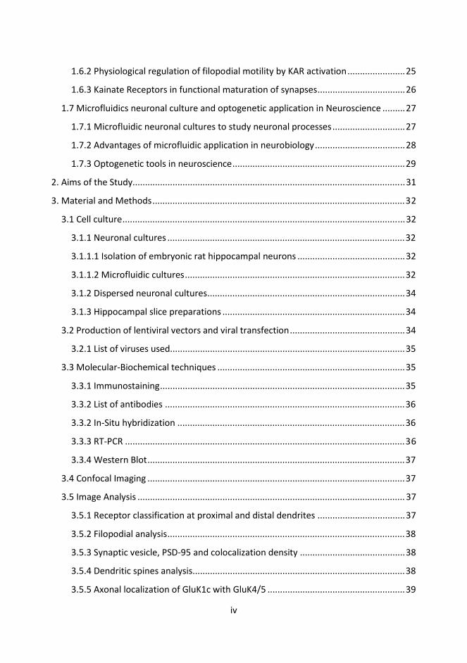
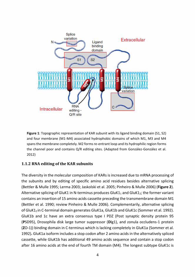
1.1.2 RNA editing of the KAR subunits The diversity in the molecular composition of KARs is increased due to mRNA processing of the subunits and by editing of specific amino acid residues besides alternative splicing (Bettler & Mulle 1995; Lerma 2003; Jaskolski et al. 2005; Pinheiro & Mulle 2006) (Figure 2). Alternative splicing of GluK1 in N-terminus produces GluK11 and GluK12; the former variant contains an insertion of 15 amino acids cassette preceding the transmembrane domain M1 (Bettler et al. 1990; review Pinheiro & Mulle 2006). Complementarily, alternative splicing of GluK12 in C-terminal domain generates GluK1a, GluK1b and GluK1c (Sommer et al. 1992). GluK1b and 1c have an extra consensus type I PDZ (Post synaptic density protein 95 (PSD95), Drosophila disk large tumor suppressor (Dlg1), and zonula occludens-1 protein (ZO-1)) binding domain in C-terminus which is lacking completely in GluK1a (Sommer et al. 1992). GluK1a isoform includes a stop codon after 2 amino acids in the alternatively spliced cassette, while GluK1b has additional 49 amino acids sequence and contain a stop codon after 16 amino acids at the end of fourth TM domain (M4). The longest subtype GluK1c is

5
formed by insertion of extra 29 amino acids into the C-terminal sequence of GluK1b (Sommer et al. 1992). GluK2 has two main C-terminal splice variants, GluK2a and shorter GluK2b marked by two distinctive amino acid sequences consisting of 54 and 15 residues, respectively, at the C-terminal end (Gregor et al. 1993). Apart from these, individual human specific splice variants GluK1d (Barbon et al. 2001) and GluK2c (Barbon et al. 2001) have also been reported. There are two known C-terminus splice variants for GluK3 (GluK3a and GluK3b) with non-homologous amino acid sequences of 55 residues in C-terminal tail (Schiffer et al. 1997). GluK4 and GluK5 do not have any known splice variants. For all of the C terminal splice variants of GluK1-3 there is a conserved sequence of 15 amino acid sequences just after the last TM domain (Pinheiro & Mulle 2006).
Figure 2: Illustration of different subtypes and splice variants of KAR subunits. The region in blue marks the membrane domains and different m-RNA editing sites are also indicated. The colored boxes at the C-terminus represent alternative splice variant regions. (Adapted from: González-González et al. 2012)
Physiological diversity of KARs further depends on editing of mRNA coding for few amino acids lining its channel pore. Q/R editing takes place in both GluK1 and GluK2 at second membrane domain (Figure 2). Replacement of glutamine with more positively charged or basic amino acid arginine residue makes KAR less permeable to Ca2+ ions and affects channel rectification: thus edited KARs display a characteristic linear current/voltage relationship while inwardly rectifying current/voltage curve is typical for unedited KARs (Burnashev et al. 1995; Bähring et al. 1997). In addition, GluK2 undergoes two separate mRNA editing in its first TM domain by substituting valine for isoleucine (I/V) and cysteine for tyrosine (Y/C) (Köhler et al. 1993). Conversely, GluK3-5 subunits do not undergo similar
6
subtype editing as with GluK1 and GluK2 (Pinheiro & Mulle 2006; Contractor et al. 2011; González-González et al. 2012).
1.1.3 Post translational modifications In addition to subunit diversity and subtype variability, post-translation modifications also participate in defining distribution and physiological functions of KARs. GluK2 is phosphorylated by protein kinase C (PKC) at Ser-846 and Ser-868 residues (Nasu-Nishimura et al. 2010). Protein kinase A (PKA) phosphorylates GluK2a at Ser-825 and Ser-837 residues (Raymond et al. 1993; Traynelis & Wahl 1997; Wong & Mayer 1993; Kornreich et al. 2007). Likewise GluK1b is phosphorylated by PKC at Ser-880 and Ser886 residues (Hirbec et al. 2003). GluK5 is recently also shown to be phosphorylated by calcium/calmodulin-dependent protein kinase-II (CaMKII) (Carta et al. 2013). Palmitoylation of GluK2 at its two C-terminal cysteine residues can reduce the susceptibility to PKC phosphorylation (Pickering et al. 1995). SUMOylation is another post translational modification on KARs where SUMO-1 (small ubiquitin-like modifier 1) a macromolecular (97 amino acid) protein is covalently attached to specific lysine residues (such as in Lys-886 in GluK2) (Martin et al. 2007; Wilkinson & Henley 2010). Post translational modifications on KARs might derive contrasting physiological functions in intermediating KARs trafficking and surface recycling (see Chapter 1.2).
1.2 Subcellular distribution of Kainate Receptors and Trafficking Kainate receptor subunits are located in various neuronal subdomains, the localized pools comprising of receptors with diverse molecular composition and functional properties. Different KAR subunits interact with diverse range of proteins that are involved in forward trafficking, endocytosis, recycling and receptor degradation. These protein partners assist KARs in defining the functional complexity and result in a distinctive pre and post synaptic compartmental distribution (reviews González-González et al. 2012; Coussen 2009; Pinheiro & Mulle 2006). Likewise, such interactions may critically influence downstream signaling of KARs. Localization of KAR subunits has been studied using in situ hybridization to detect mRNA expression pattern, restricted profiling by receptor overexpression, colocalization with different cellular markers and physiological characterization by electrophysiological recordings. The study of KARs diversity is subdued by non-specificity of available antibodies and pharmacological tools, hence rendering precise understanding on location and composition of KARs at various synapses incompletely verifiable (reviews González-

7
González et al. 2012; Pinheiro & Mulle 2006). Furthermore, most of these studies were conducted in different heterologous cell lines and sparsely in neurons, so the exact localization of various KAR subtypes in different areas of the CNS remains largely unresolved.
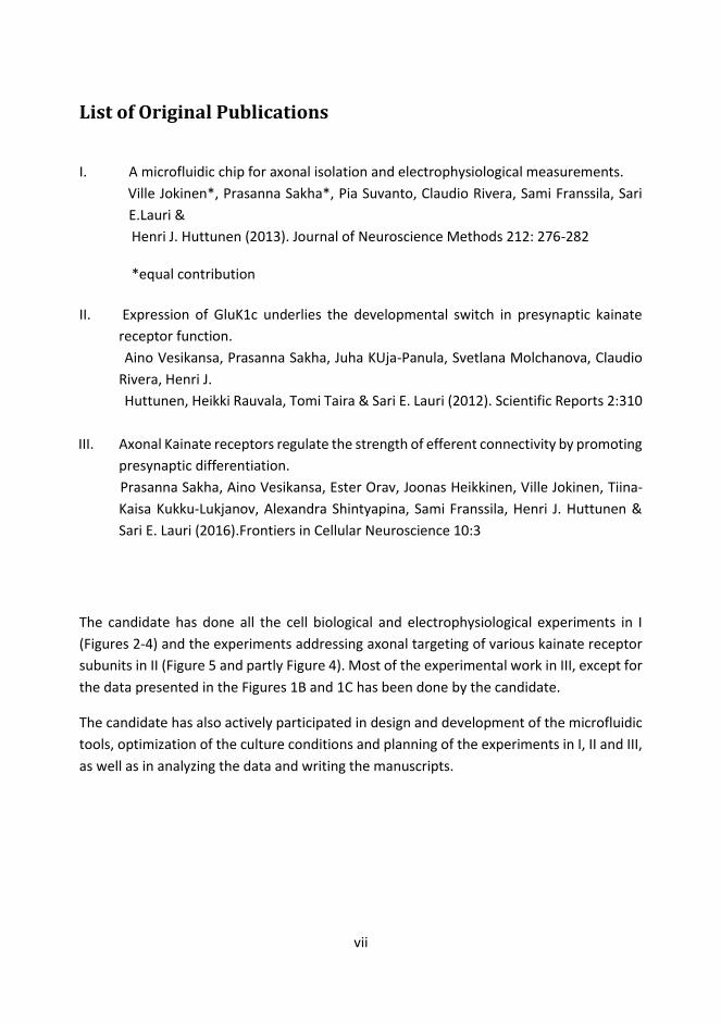
1.2.1 Exocytosis from ER to the surface Surface expression of KAR complex depends on the subunit composition and on presence of C-terminal splice variants. Homomeric GluK2a and GluK3a are highly expressed in the plasma membrane (PM) as compared to the GluK1a, GluK1b, GluK2b and GluK3b, which tend to be retained in the endoplasmic reticulum (ER) (Jaskolski et al. 2004; Jaskolski, Normand, et al. 2005) (Figure 3). Apart from being extensively present in the PM due to the presence of forward trafficking sequence (CQRRLKHK motif) at their C-terminal, both GluK2a and GluK3a can promote surface expression of other subunits that contain ER retention motifs (Jaskolski et al. 2004; Jaskolski, Normand, et al. 2005; Jaskolski, Coussen, et al. 2005). This is achieved by rendering steric hindrance against the intracellular retention motifs (Ren et al. 2003; Pinheiro & Mulle 2006). Certain subunits can still display differential pattern of surface expression in different subcellular domains (Ren, Riley, Needleman, et al. 2003; Jaskolski et al. 2004). For instance, in heterologous cell lines, GluK1c is entirely retained in the ER due to the presence of RXR motif, while the surface expression is sparse though not completely lacking in cultured hippocampal neurons (Jaskolski et al. 2004) . The complete picture on secretory pathway for the KARs from the ER to the cell surface is still unclear. Based on the proteomic studies, GluK2a interacts with different sets of cytosolic proteins such as Spectrin, F3-contactin, dynamin1, 14-3-3δ and calmodulin to regulate surface trafficking mechanisms, while GluK2b interacts with VILIP-1, neurocalcinin- δ, calcineurin, calmodulin and profiling-IIa (Coussen et al. 2005; Kjarland et al. 2006). Few studies have revealed involvement of coatomer protein complex I (COPI) and 14-3-3 proteins in bidirectional modulation of GluK5 subunit from cellular periphery. Arginine rich ER retention motif in GluK5 is bound to COPI and ER retained (Vivithanaporn et al. 2006).Therefore, the high affinity KARs such as GluK5 are retained in the ER in absence of low affinity KAR partners (Gallyas et al. 2003; Hayes et al. 2003; Ren, Nathan J Riley, et al. 2003). Heteromeric combination with low affinity KARs sterically shield the ER retention/retrieval signal of GluK5 to allow surface delivery by generation functional receptors (Ren, Nathan J Riley, et al. 2003). Constitutively, assembly of GluK2 and GluK5 enhances interaction of 14-3-3Ϛ with GluK5, resulting in superfluous surface expression of GluK5 (Vivithanaporn et al. 2006).

8
Figure 3: Trafficking of KARs from endoplasmic reticulum to the plasma membrane depends on the molecular composition of its receptor complex formed after diverse combination of different KAR subunits. The figure exemplifies relative distribution of different homomeric and heteromeric KAR complexes. (Adapted from: González-González et al. 2012)
Forward trafficking of both homomeric and heteromeric KARs from the ER depend on formation of an intact glutamate binding site with conformational precision in the multimeric receptor complex (Mah et al. 2005; Valluru et al. 2005). In GluK5 for example, mutation of ligand binding domain (Thr-675) leads to reduced affinity for binding glutamate and Kainate. This mutated GluK5 can still assemble with GluK2 but is degraded without further subsequent forward trafficking to the PM (Valluru et al. 2005). In addition, linker region between TM domain 3 and S2 domain of GluK2 is crucial in receptor complex biogenesis during dimeric and tetrameric assembly and also for post assembly trafficking from the ER. Additionally, the same region is compliant for energy transduction during channel opening mediated by its ligand binding (Vivithanaporn et al. 2007). NTD of KARs also has an important part to play in KAR surface expression, as a quality control checkpoint per se and may prevent cross-assembly with other ionotropic glutamatergic receptors (review Hansen et al. 2010).
Flexible oligomerization of different KAR subunits promotes surface expression of diversified receptor complexes. However, many KAR subtypes are retained in ER due to the retention/retrieval motifs present in its c-terminus. This scenario appears baffling and raises questions on possible channel independent intracellular roles by these receptors.
Phosphorylation by PKA and PKC have been extensively studied in forward trafficking of various KARs, albeit significance of basal KARs phosphorylation is not well illustrated. PKA phosphorylation of GluK2a (Ser-825 and Ser-837) potentiates KAR currents by increasing

9
channel opening probability (Raymond et al. 1993; Traynelis & Wahl 1997; Wong & Mayer 1993; Kornreich et al. 2007). Likewise, phosphorylation of GluK2 (Ser-846 and Ser-868) by PKC up-regulates surface expression by dynamically modifying receptor endocytosis (Nasu-Nishimura et al. 2010). On the contrary, PKC inhibition was previously found to prevent both NMDA and kainate-evoked GluK2 endocytosis (Martin & Henley 2004). This apparent mutually exclusive activity of PKC in GluK2 phosphorylation is not clearly understood. However, it could be speculated that phosphorylation can bidirectionally conjugate KARs with proteins involved in protein cargo trafficking and endocytosis from the cell surface (González-González et al. 2012). Hence, PKC phosphorylation may have concurrent but independent roles in forward trafficking of GluK2 and in endocytosis.
PKC inhibition reduces KAR EPSC amplitude in hippocampal CA3 synapses (Hirbec et al. 2003). Group 1 mGlu-receptor activation also enhances GluK1 phosphorylation by PKC and increase KAR mediated excitatory postsynaptic current (EPSC) (Cho et al. 2003). On other hand, in DRG (dorsal root ganglia) neurons expressing mostly GluK1 and GluK5, PKC activation has been found to reduce surface expression of KARs (Rivera et al. 2007). These contrasting results may be explained by cell type dependent indirect / direct consequences of PKA activity and KAR phosphorylation. More insights of these endogenous signaling pathways involved in KAR trafficking require defining the downstream consequences of KAR phosphorylation by PKA and PKC (González-González et al. 2012).
1.2.1.1 Synaptic targeting GluK1b, GluK1c and GluK2a contain c-terminal PDZ binding motif (comprising of 80-90 amino acids) (Coussen 2009). Interactions with PDZ domain containing proteins are involved in stabilizing KARs in postsynaptic structures (Hirbec et al. 2003). For example, PSD95 (with its three PDZ domains) interacts with GluK2 containing KARs to promote receptor clustering (Garcia et al. 1998; Mehta et al. 2001). SAP-90 and SAP-102 are present at close proximity or co-localized with GluK5 and GluK2 subunit at the dendrites (Garcia et al. 1998). Postsynaptic density (PSD) proteins such as spectrin can tether KARs to the actin cytoskeleton (Coussen et al. 2005). Another actin binding protein profillin-II inhibits clathrin-mediated endocytosis of KARs via interfering with dynamin-1 activity (Gareus et al. 2006) but also unconventionally down regulates KAR surface expression (Gareus et al. 2006; Mondin et al. 2010). Disruption of PSD95-GluK2 complex has been linked to reduced excitotoxicity plus protection against KA evoked ischemic neuronal death (Pei et al. 2006). Otherwise, PSD95-GluK2 interaction leads to the activation of JNK kinase and subsequent neuronal

10
hyperactivity (Savinainen et al. 2001). GluK1b, GluK1c and GluK2a can also associate with PICK1, GRIP and syntenin via PDZ ligation (Hirbec et al. 2003). Disruption of KAR interaction with either PICK1 or GRIP leads to loss of synaptic KAR , with reciprocal increase in AMPA receptor activity suggesting that PDZ domain containing proteins can have divergent regulatory mechanisms for Kainate and AMPA receptors (Hirbec et al. 2003; Park et al. 2006). On the similar note, interaction of PDZ binding proteins regulate NMDARs trafficking from the ER to the plasma membrane (Standley et al. 2000; Scott et al. 2001), while this is not the case for KARs.
Apart from the direct PDZ domain containing interactors, the NETO auxiliary subunits might influence targeting of KARs into the synapses. In hippocampal cultures, exogenous NETO2 can direct a fraction of exogenous GluK1 into the synapses (Copits et al. 2011); and similarly GluK2 in cerebellar granule cells (Zhang et al. 2009). Interestingly, however both GluK1 and GluK2 can be targeted in the synapses in absence of these auxiliary subunits (Tang et al. 2011; review Lerma & Marques 2013; see Sheng et al., 2015). More recently, NETO2 was found to also interact with neuronal KCC2 ( K+-Cl- cotransporter) (Ivakine et al. 2013); and initially NETO1 was identified as an interacting partner for NMDARs responsible for its synaptic localization (Ng et al. 2009). Overall, NETO1/2 may have essential novel functions via its cross-talk mechanisms amongst different receptors and ion transporter, and its influential roles exclusive to KARs may require further understanding on selectivity of certain receptor population restrictive to the NETOs (review Lerma & Marques 2013).
Very little is known on the mechanisms guiding axonal and presynaptic targeting of various KAR subunits. Presynaptic functions of KAR subunits may be directly influenced by its interaction with presynaptic components. These may eventually lead to stabilization of KARs in the presynaptic terminals. Most of immunoprecipitation and proteome based affinity studies from the rodent brain or heterologous cell lysates have displayed that KAR subunits varyingly interact with different presynaptic proteins. GluK2a interacts indirectly with β-catenin and binds directly to cadherin and p120 proteins (Coussen et al. 2002). Likewise, this subunit also interacts with Velis/LIN-7 and CASK/LIN-2 complex which might essentially locate KARs in the synapses (Coussen et al. 2002). Both GluK2a/b subtypes interact with calmodulin and may have downstream influence in regulating intracellular Ca2+ at presynaptic terminals. Likewise GluK2 also interacts with cytoskeletal proteins such as spectrin and profilin-IIa (see Coussen et al. 2005 for additional protein partners). GluK1b interacts with β-catenin and synapsin-II, and these interactions may have significant presynaptic roles during the synapse formation (Rutkowska-Wlodarczyk et al. 2015).
11
1.2.2 Kainate Receptor endocytosis and degradation Another important mechanism regulating abundance of surface KARs is receptor endocytosis to early endosomes followed by recycling or degradation of these receptors depending on its internalization stimulus (Martin & Henley 2004). For instance, NMDAR activation induces Ca2+, PKA and PKC dependent endocytosis of GluK2 into early endosomes which are then rapidly recycled back to the PM. While, kainate mediated activation leads to inflow of Ca2+, followed by PKC dependent but PKA independent endocytosis of GluK2 for lysosomal degradation instead of surface recycling (Martin & Henley 2004). Another study shows that removal of GluK5 containing KARs from the synapses involves interaction with synaptosomal-associated protein 25 (SNAP25) (Selak et al. 2009). This is also a mutual protein partner of PICK1-GRIP-GluK5 interaction complex. Antagonism of SNAP25 leads to GluK5 dependent increase in synaptic KARs (Selak et al. 2009). Palmitoylation and phosphorylation of GluK2 containing KARs also results in receptor internalization (Pickering et al. 1995; Huang & El-Husseini 2005; Martin & Henley 2004; Martin et al. 2008). SUMOylation of GluK2 facilitates rapid removal of receptors from the PM, observed as reduced KAR mediated excitatory postsynaptic currents (KAR EPSCs)(Martin et al. 2007; Wilkinson & Henley 2010). Coherence between phosphorylation and SUMOylation may have an important implication for long-term depression (LTD) of KAR EPSCs in Mossy fiber-CA3 synapses where phosphorylation by PKC (Ser-868) results in subsequent SUMOylation (Lys-886) and endocytosis of KARs (Chamberlain et al. 2012). It is worth mentioning that with relatively few studies presenting how KARs could undergo endocytosis, more could be known regarding its retrograde trafficking. Also the fact that GluK2 can reshuffle by lateral diffusion between the dendritic shafts and the spines (Martin et al. 2008) open a wider horizon on the diligence of KAR arrangements in synaptic zones and perhaps even within the intracellular compartments.
Fate of internalized KARs depends on ubiquitination followed by proteasomal or lysosomal degradation. The proteasomal degradation involves removal of misfolded and unassembled receptors while the lysosomal degradation disposes excess receptors from the PM (González-González et al. 2012; Mabb & Ehlers 2010). For example, prolonged KA activation leads to internalization of KARs for lysosomal degradation (Martin et al. 2008). Direct ubiquitination and proteasomal degradation of GluK1 and GluK2 is mediated by binding to Kelch domain of actifilin to its c-terminus and targeting them for recruiting Cullin 3 ubiquitin ligase complex (Salinas et al. 2006; González-González et al. 2012). This mechanism is unique for GluK1 and GluK2a, but not for GluK5 and nor for AMPAR (Salinas

12
et al. 2006). As described above, SUMOylation of KARs in response to pharmacological activation can facilitate receptor endocytosis (Martin et al. 2007). However, it is unclear if these internalized receptors would undergo degradation, despite SUMOylated GluK2a are detected in intracellular compartments.
1.3 KAR Signaling Pathways Accumulating studies enunciate the ability of KARs to signal via two distinct mechanisms: the ionotropic signaling typical for all members of the receptor family (AMPA, NMDA and KAR receptors) and non-canonical G-protein dependent signaling. How exactly these two modes of action interact to define the topical roles of KARs in synaptic modulation is still largely unclear (also see Chapter 1.4).
1.3.1 Ionotropic signaling Ionotropic signaling is mediated by ligand-gated ion channels. These are TM proteins consisting of channel pore and extracellular ligand binding domain. Ligand binding to KARs leads to conformational change in its ion channel pore which is permeable to Na+, K+ and in some cases also to Ca2+ ions. Ionotropic signaling is critically influenced by the presence of subunit and subtype specific variants in KAR complex. The unedited variants of GluK1 and GluK2 at Q/R site are calcium permeable and predominant at immature brain (Bernard et al. 1999; Lee et al. 2001). A striking feature that differentiates KARs from AMPAR is deactivation and desensitization (Bowie 2010). Gating properties of AMPARs are not effected by external ion concentration, while KARs display faster rate of deactivation and desensitization in extracellular environment with low ionic strength (Bowie 2002; Bowie & Lange 2002). These features have been proposed to be due to KARs behaving as monomers to tetramers in low and high ionic environments, respectively (Bowie 2010). KAR complex has discrete anion (Cl-) and cation (2 Na+) binding pockets which are allosterically and structurally coupled (Plested & Mayer 2007; Wong et al. 2007). The exact physiological functions of such channel gating by extracellular ions are not yet known. It has been speculated that under an intense neuronal activation, a sudden drop in extracellular monovalent ions may limit functioning of KARs, which may become unresponsive to the neurotransmitter if the receptors are in an unbound states by these extracellular ions (Lerma & Marques 2013; Plested et al. 2008). Native KAR-EPSCs are typically slower and have lower amplitude as compared to AMPAR mediated EPSCs (Castillo et al. 1997; Vignes et al. 1998). This unique feature of native KAR-EPSCs is in sharp contrast to recombinant KAR-EPSCs which instead features rapid

13
activation, deactivation and desensitization in the range of just few milliseconds (Herb et al. 1992; Cui & Mayer 1999). Most of the differences in channel properties between native and recombinant KARs have been recently clarified with the identification of NETO auxiliary subunits for KARs. They are responsible for modulating agonist binding affinity and kinetics of KARs (Straub, Hunt, et al. 2011; Straub, Zhang, et al. 2011). This results in higher agonist binding affinity and slower deactivation kinetics of KARs. In addition, association of KARs with NETOs greatly reduces inward rectification of KAR currents without effecting extracellular Ca2+ permeability (Frerking & Ohliger-Frerking 2002; review Lerma & Marques 2013).
1.3.2 G protein-coupled signaling G protein-coupled KAR signaling was first described in the CA1 region of hippocampus where KAR mediated regulation of GABA release was shown to be sensitive to pertussis toxin and PKC inhibitors but independent on its ion channel activity (Rodríguez-Moreno & Lerma 1998). Similarly, tonic inhibition of glutamate release by GluK1 containing KARs in immature hippocampus is also obstructed by pertussis toxin suggesting involvement of G-protein dependent signaling (Lauri et al. 2006; Sallert et al. 2009). It has been postulated that activation of G protein-coupled KARs might regulate voltage dependent Ca2+ channels thus explaining the effects on transmitter release ( Frerking et al., 2001; Kamiya and Ozawa, 1998; Rozas et al. 2003). Another important G-protein dependent function of KARs is seen during inhibition of postspike slow after-hyperpolarization potential (sAHP) in hippocampal pyramidal neurons which results in enhanced neuronal excitability (Melyan et al. 2002). KARs do not have a conventional binding motifs for directly coupling to G-proteins at its C-terminus domain. Instead, it has been suggested that intermediator protein partners can act as scaffolds or as a transducer in interceding metabotropic action via KAR activation (Frerking et al. 1998; Chergui et al. 2000; Coussen 2009). One study involving GluK5 knockout has essentially displayed the direct interaction of GluK5 with Gαq/11 protein which modulates slow Ca2+ activated K+ current (IsAHP) (Ruiz et al. 2005). However, kainate receptor-mediated inhibition of the IsAHP was intact in GluK4/GluK5 knockout mice (Fernandes et al. 2009). Recently, proteomic and functional analysis have displayed direct interaction of GluK1 and Gαo in mediating IAHP (Ca2+ dependent K+ channel mediated after hyperpolarization current) in DRG neurons (Rutkowska-Wlodarczyk et al. 2015). However, these biochemical interactions remain controversial and to be further confirmed.
Downstream signaling of G protein-coupled KAR may involve PKC (Rodríguez-Moreno & Lerma 1998), which however is not the sole downstream target. Thus, presynaptic effect

14
of G protein-coupled KAR in CA3-CA1 synapse can be completely inhibited by PKC antagonism in immature brain but not in juvenile CA3-CA1 synapses (Lauri et al. 2005; Sallert et al. 2007). Likewise, G protein-coupled KAR mediated modulation of sAHP can be blocked by PKC inhibition in pyramidal neurons (Melyan et al. 2002) while in interneurons this modulation is still prevalent despite inhibiting PKC activity (Segerstråle et al. 2010). In addition, despite the lack of accurate mechanism explaining G protein-coupled KAR signaling at pre or postsynaptic regions, there are studies which suggest coordinated action between both ionotropic and G protein-coupled KARs in physiological context. For example, in neonatal CA3, GluK1 subunit containing KARs regulate glutamate release in G-protein dependent manner while the high affinity KARs (lacking GluK1) act to increase axonal excitability (Lauri et al. 2005; Juuri et al. 2010), together controlling the excitability of the network to allow the physiological type activity patterns.
1.4 Physiological Functions Amongst other glutamatergic receptors, KARs have distinctive activities in CNS with unique synaptic and extra-synaptic functions. It has become evident that KARs modulate both glutamatergic and GABAergic transmission to regulate and maintain excitability of neuronal circuitry in addition to their crucial but refined roles in maintaining long and short term synaptic plasticity (review Lerma 2003; Pinheiro & Mulle 2006; Contractor et al. 2011; González-González et al. 2012; Lauri & Taira 2012). Apart from these synaptic functions, somatodendritic KARs regulate neuronal excitability , via direct ionotropic action or via regulating the after-hyperpolarizing currents via G-protein coupled mechanisms (e.g. Melyan et al. 2002; Segerstråle et al., 2010) (see chapter 1.3.2). Accumulating evidence suggest that KARs may regulate presynaptic function and hence synaptic transmission via Ca2+ influx through its ion channel (e.g. Pinheiro et al. 2007; Lauri et al. 2003) or via metabotropic signaling particularly at immature synapses (Lauri et al. 2005; Lauri et al. 2006; Rodríguez-Moreno & Lerma 1998; Melyan et al. 2002). These physiological functions of KARs have been revealed since the discovery of 2,3-benzodiazepines such as GYKI 53655 (LY300268) which can specifically block AMPA receptors and enabled pharmacological distinction between AMPA and KARs (Paternain et al. 1995; Wilding & Huettner 1995). In fact, in hippocampal pyramidal synapses even low agonist concentration of 1 μM KA can activate both of these non-NMDA receptors (Mulle et al. 1998; Bureau et al. 1999). The broader prospect of KARs functions have subsequently been studied by the development of additional drugs, some with subunit specificity, and by the expansion of rodent lines deficient in different KAR subunits (review Lerma 2003; Pinheiro & Mulle 2006; Lauri & Taira 2012).

15
1.4.1 Presynaptic KARs: Modulation of neurotransmitter release Early evidence of presynaptic KARs involved 3H KA binding studies in CA3 pyramidal neurons where selective destruction of afferent mossy fiber lead to substantial reduction of agonist binding in the proximal dendritic region (Agrawal & Evans 1986; Represa et al. 1987). Following this, numerous studies have surfaced where presynaptic KARs are shown to modulate excitatory and inhibitory synaptic transmission, by facilitating or inhibiting neurotransmitter release depending on the synapse type (review Lerma 2003; Pinheiro & Mulle 2006; Lauri & Taira 2012). In mossy fibres with relatively large presynaptic terminals, KAR activation facilitates synaptic release during high-frequency transmission via membrane depolarization and Ca2+ influx (Kamiya et al. 2002; Schmitz et al. 2001; Lauri et al. 2001). However, the KAR conductance may also shunt the membrane and inactivate voltage sensitive ion channels, hence causing suppression of presynaptic function (Kamiya et al. 2002; Schmitz et al. 2001). This dual action of KARs, manifest as presynaptic facilitation and depression of transmission might be due to the presence of different populations of KARs in hippocampal mossy fibre-CA3 circuitry. Similar physiology of presynaptic KARs is also seen in neocortex-thalamocortical circuitry (Jouhanneau et al. 2011). Instead, in smaller structures such as filopodial protrusions presynaptic depolarization shunts the membrane even with small conductance to inhibit vesicle release (Kidd et al. 2002). This supports presynaptic ionotropic KARs to be an on-off synaptic switch, regulating vesicle release bidirectionally based on the structural features and intrinsic electrical properties of the presynaptic terminal.
Thus, activation of presynaptic KARs can tune up the release probability (as seen in calyx of Held: Awatramani et al. 2005) via direct Ca2+ influx or by the activation of other voltage gated Na+ channels (Engel & Jonas 2005). In addition, at mossy fiber-CA3 synapse, Ca2+
released from intracellular stores is implicated in presynaptic actions of KARs (Lauri et al. 2003; Pinheiro et al. 2005). Via this action at the mossy fibre synapses, KARs act as auto-receptors to sense glutamate release and then subsequently facilitate further release of glutamate. This effect depends on the stimulus frequency used; presynaptic KARs are activated with either repetitive low (0.1- 3 Hz) or short trains of high (20 -100 Hz) frequency activity (Salin et al. 1996). This KARs mediated synaptic facilitation is impaired in GluK2-/- mice (Contractor et al. 2001), while involvement of GluK1 subunit have been reported based on use of its selective antagonist (LY382884) (Lauri et al. 2001). However, studies in GluK1-/- mice and additional study in GluK5-/- mice contradicted involvement of GluK1 (Contractor et al. 2001; Contractor et al. 2003). Such discrepancies may be due to

16
generation of unforeseen and unknown physiological compensation in the knock-out lines. Furthermore, given the complex rules regulating KAR subcellular targeting, subunit knockout may also cause inexplicit receptor organization, subcellular localization and finally impede its endogenous signaling mechanisms. Finally, the molecular composition of facilitatory KARs may vary during development and between cell types. For example in CA3-CA3 pyramidal neuron collaterals, robust increase in network excitability is displayed by low agonist concentration suggesting presence of the high affinity GluK4/5 (Juuri et al. 2010). On the other hand, in barrel cortex, thalamocortical inputs, presynaptic facilitatory activity require high agonist concentration thus suggesting absence of GluK4/5 (Jouhanneau et al. 2011).
In the developing hippocampus, presynaptic GluK1 subunit containing KARs in CA3-CA1 tonically inhibits synaptic transmission in a G-protein dependent manner (Lauri et al. 2006). This mechanism participates in regulating synaptic response to short bursts of high frequency activity, typical for the developing hippocampus. Presynaptic inhibitory KARs are also suggested to be present in the hippocampal interneurons where KAR agonists inhibit evoked inhibitory postsynaptic currents (IPSCs) , reduce frequency of miniature IPSCs (mIPSCs) and increase failure rate of transmission between cell-pairs in acute hippocampal slices (Clarke et al. 1997; Rodríguez-Moreno & Lerma 1998; Castillo et al. 1997; Min et al. 1999; but see Frerking et al. 1998). It is still debated whether presynaptic inhibitory KARs are present in GABAergic interneuron terminals, or are the effects due to receptors located in the axons or at the somatodendritic region. KA application induces rapid interneuronal firing due to activation of somatodendritic KARs in the interneurons (DeVries & Schwartz 1999; Kidd & Isaac 1999) which might indirectly cause depression of IPSCs (Frerking et al. 1998).
Unitary-IPSCs (uIPSCs) recording between CA1 pyramidal neuron and stratum radiatum interneuron cell pairs suggested that low concentration of KAR agonist (300 nM KA), ambient glutamate or stimulation of stratum radiatum can facilitate GABAergic synapses by activating interneuronal presynaptic or axonal KARs (Jiang et al. 2001). Similarly, the frequency of spontaneous IPSCs is enhanced by low concentration of KA (250 nM) in the interneurons (Semyanov & Kullmann 2001), thus suggesting heterogeneous roles of presynaptic KARs in regulating GABAergic synapses (Cossart et al. 2001; Ali et al. 2001; review Lerma 2003).
1.4.2 Postsynaptic Kainate Receptors Early evidence of postsynaptic KARs was observed in CA3 pyramidal cells which displayed slow EPSC to the stimulation of mossy fibres in presence of AMPA and NMDA antagonist,
17
that was sensitive to antagonism by AMPA/KAR blocker CNQX (Castillo et al. 1997; Vignes & Collingridge 1997). This current was absent in GluK2-/- mice suggesting a critical role of GluK2 subunits (Mulle et al. 1998). KAR mediated EPSCs have also been reported in other parts of the brain including GABAergic interneurons in CA1 region (Cossart et al. 1998; Frerking et al. 1998), cerebellar Golgi cells (Bureau et al. 2000), Purkinje cells (Huang et al. 2004), neocortical pyramidal and interneurons (Kidd & Isaac 1999; Ali 2003; Eder et al. 2003; Wu et al. 2005) and in basolateral amygdala (Li & Rogawski 1998). A compelling function of post synaptic KARs is the ability to encode temporal information; for example, the slow KAR currents in CA1 interneurons may cause tonic depolarization of these interneurons even during slight presynaptic activity (Frerking et al. 1998; Frerking & Ohliger-Frerking 2002). Hence, KARs can have an integrative role prompting sustained depolarization during repetitive firing, henceforth influencing the network physiology. This unique property of KARs differs from AMPA-EPSCs which mediate phasic and time locked excitation (Frerking & Ohliger-Frerking 2002).
1.4.3 Role of KARs in synaptic plasticity KARs are involved in regulation of long-term potentiation (LTP) in the hippocampal mossy fibre pathway (Bortolotto et al. 1999; Contractor et al. 2001). Remarkably, this mossy fiber LTP is still inducible in presence of CNQX which is supposed to completely block the activity of AMPA and Kainate receptors (Weisskopf & Nicoll 1995), indicating that KAR are not necessary for LTP induction in this pathway but rather play a regulatory role. Presynaptic KARs may boost LTP induction via facilitating glutamate release during the LTP inducing stimuli (Lauri et al. 2001). In addition, postsynaptic KARs are shown to influence associative LTP in area CA3 by amplifying spike transmission (Sachidhanandam et al. 2009). At interneurons, activation of postsynaptic KARs during physiologically relevant activity patterns influence LTP induction via regulation of feed-forward inhibition (Clarke et al. 2012). In addition to contributing to plasticity induction, KARs are regulated in response to activity-dependent plasticity. Relationship between synaptic plasticity and KAR function is well studied in the immature hippocampal CA3-CA1 circuitry, where pairing induced LTP causes down regulation of tonic inhibitory activity of GluK1 KARs to increase probability of release (Lauri et al. 2006). On the other hand, upregulation of GluK1 activity can contribute to LTD (Sallert et al. 2007; Clarke et al. 2014). The endogenous KAR activity as well as its plasticity in CA3-CA1 circuitry is lost during development (Lauri et al. 2006; Sallert et al. 2007) which functionally coincides to the developmental maturation of hippocampi,

18
henceforth paralleling KARs switching from immature to the mature form (review Lauri & Taira 2012).
1.5 Synaptogenesis Central nervous system (CNS) synaptogenesis is a complex associational process initiated by asymmetrical cell-cell adhesion between two nerve cells and designated to ultimately mediate rapid and efficient chemical transmission between the neurons (review Kelsch et al. 2010; Waites et al. 2005; Garner et al. 2002). This process in vertebrates begins during the period of embryonic neurogenesis, is refined in early postnatal life and continues throughout the adulthood to imprint learning, memory, cognition and consciousness. The research in this field has established many molecules that act locally or distantly for the formation of appropriate contact or induce signals which cascade into formation of these highly specialized structures transmitting information between neurons.
1.5.1 Synapse progenesis During the establishment of neuronal circuitry, axons navigate extensive distances both in CNS and in PNS (peripheral nervous system) to find their targets, retracting from unspecific synaptic contacts prior to reaching the synaptic destination. This early patterning follows guidelines set by regulatory genes encoding guidance proteins. Synapse induction requires both intrinsic cues, consisting of unidentified priming molecules that turn neurons competent to undergo this process and subsequently, target-derived factors or inducing molecules which trigger synaptogenesis (review Waites et al. 2005). Synaptogenic molecules belonging to Wnt and fibroblast growth factor (FGF) families can instigate axonal arborisation and accumulation of recycling synaptic vesicles of afferent innervating axons (Scheiffele 2003). For example, Mossy fiber axons expressing FGF2 receptor show enhanced active zone formation due to increased responsiveness to FGF22 that is secreted from granule cell neurons (Umemori et al. 2004). Likewise, secretion of Wnt-7 from granule cells lead to clustering of synaptic vesicle associated protein synapsin-1 in innervating mossy fibre terminals (Hall et al. 2000). Neurotrophins such as brain derived neurotropic factor (BDNF) (Alsina et al. 2001) and non-neuronal priming molecules like glial-derived factors (Nägler et al. 2001; Ullian et al. 2001) can regulate the density of synaptic innervations and facilitate maturation of synaptic connections. Additional target derived molecules such as netrins and semaphorins (Bagri & Tessier-Lavigne 2002; Pascual et al. 2004) act diffusely from the local sources in promoting synapse development.
19
Initial stage of CNS synaptogenesis is marked by contact formation at axo-dendritic, axon-somatic or axo-axonal sites by axonal growth cones and dendritic filopodial protrusions. The contact formation is determined by several classes of cell-adhesion molecules (CAMs) (Vaughn 1989; Marrs et al. 2001; Okabe et al. 2001; Wong & Wong 2001; Ziv & Garner 2001). These include members of calcium dependent CAMs such as cadherins and protocadherin (Shapiro & Colman 1999; Takai et al. 2003). CNS has over 20 different types of cadherins involved in early stages of synaptogenesis for target specification and stabilization during initial contact formation (Yagi & Takeichi 2000).
Synaptic differentiation is propagated by two sets of secreted proteins: Narp and Ephrin capable of clustering subsets of post synaptic proteins ( O’Brien et al. 1999; Scheiffele et al. 2000). Narp binds extracellular domain of AMPAR and promotes its clustering (O’Brien et al. 1999). Narp also influences clustering of NMDAR in specific classes of interneurons (Mi et al. 2002) although NMDA clustering is primarily initiated by EphrinB family members that directly bind extracellular domain of NR1 subunit (Dalva et al. 2000). Clustering of EphB receptors via EphrinB leads to dendritic spine development (Murai et al. 2003) and spine maturation (Penzes et al. 2003). On the presynaptic side, clustering of neuroligins with β-neurexin (Dean et al. 2003) and homophilic/hetrophilic association of SynCAM on the either region of synapse lead to the formation of active zone (AZ) (Biederer et al. 2002; Shingai et al. 2003). Synapse specialization is dependent on dynamic interplay of these interacting proteins. For example, neuroligin initiate presynaptic differentiation after contacting an axon, while post synaptic differentiation via its binding partner β-neurexin induces local clustering of PSD-95 and NMDARs (Graf et al. 2004; Waites et al. 2005).
1.5.2 Pre and postsynaptic differentiation 1.5.2.1 Presynaptic maturation Based on a deep etch freeze fracture studies the presynaptic active zone (AZ) is visible as electron-dense presynaptic cytoskeletal matrix (Landis 1988; Zhai et al. 2001). AZ contains essential presynaptic components such as Piccolo, Bassoon and Rab3 interacting proteins (RIM) (Lee et al. 2003; Ohtsuka et al. 2002; Shapira et al. 2003) and also the proteins involved in synaptic vesicle (SV) release machinery including syntaxin, SNAP25 and N-type voltage gated Ca2+ channels (Shapira et al. 2003; Zhai et al. 2001). Assembly of presynaptic boutons is accompanied by the appearance of pleomorphic vesicle clusters all closely associated to microtubules in the axons and in growth cones (Ahmari et al. 2000; Zhai et al. 2001). These supposedly are the precursors of synaptic vesicles (SVs)

20
and contain numerous multidomain scaffold proteins of the AZ. These tubulovesicular structures can also carry cytosolic pools of post-golgi membrane cargos resembling endosomal intermediates onto the site of synapse initiation. It is not yet clear if these vesicular structures are in the process of sequential cargo trafficking to the site of synapse formation, or whether they can be aligned to the synapse initiation sites in the PM following induction of AZ by neuroligin and SynCAM (Garner et al. 2002; Waites et al. 2005). However, the subsequent fusion of these 80 nm dense core vesicles with the PM is believed to initiate rapid establishment of functional SV docking and fusion sites followed by consequent delivery of additional presynaptic proteins and SV precursors which presumably lead to biogenesis and clustering of mature SVs in the presynaptic zone (Ziv & Garner 2004).
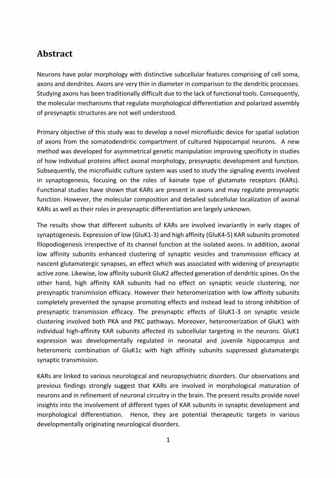
1.5.2.2 Synaptic vesicles and markers of presynaptic differentiation Synaptic vesicles (SVs) are aligned in an orderly fashion to the proximity of the release side in the AZ at the synapse, and are broadly classified into three groups, i) readily releasable pools (RRP) containing SVs primed and docked to the AZ for release up on stimulation, ii) recycling pool contain vesicles scattered throughout the nerve terminal and recycled upon moderate stimulation, and iii) reserve pool contain most of the SV clusters and released only upon a very strong stimulation (Rizzoli & Betz 2005). The livelihood of different synaptic vesicle pools depends on recycling of the released vesicles following diverse recycling pathways (Figure 4).
There are certain essential proteins common to all the SVs, such as synaptobrevin ( 7̴0 copies), synaptotagmin ( 1̴5 copies), synaptophysin ( 3̴0 copies) and synapsin ( ̴8 copies), while others like endosomal SNARE (SNAP (Soluble SNF Attachment Protein) REceptor) fusion proteins are sparsely present (Takamori et al. 2006). The specific vesicle pool cannot be defined by these specific proteins. Finding specific pool tags would require spatial segregation of SVs, complicated by the fact that SVs are dynamically mobile in presynaptic terminals (Westphal et al. 2008; Shtrahman et al. 2005; Yeung et al. 2007; review Denker & Rizzoli 2010; Jähne et al. 2015; Saheki & De Camilli 2012). In addition, upon exocytosis or during recycling these tag proteins might be exchanged with the PM and then arbitrarily distributed to the newly generated SVs (Zhu et al. 2009, but see Opazo et al. 2010). Apart from these eminent proteins, there are other interacting proteins involved in vesicle recycling, clathrin assembly/disassembly, BAR (Bin-Amphiphysin-Rvs) domain proteins for sensing and generating bilayer curvature along with other scaffolding proteins and

21
numerous decisive protein metabolites (for comprehensive list of proteins: Saheki & De Camilli 2012) involved in regulating the life cycle of SVs in the presynaptic terminal.
Figure 4: Representation of different synaptic vesicle (SV) recycling modules. 1) Complete fusion of SV to the membrane followed by clathrin-mediated endocytosis. 2) Bulk endocytosis of presynaptic membrane followed by clathrin-mediated endocytosis. 3) Ultrafast endocytosis characterized by incomplete fusion of the SV to the presynaptic membrane. And, 4) Kiss and run release leading to incomplete fusion of the SV and presynaptic membranes. The exact recycling pathways of SVs after endocytosis are unclear. Endosomes have been linked to most of these recycling pathways except for kiss and run release. However, endosomes-like structures are observed during strong stimulation of the nerve terminals, formed by large infoldings of the plasma membrane (Clayton et al. 2007) during ultrafast endocytosis (Watanabe et al. 2013). Likewise, bulk endocytosis has also been associated with excessive stimulation (Clayton et al. 2007). (Adapted and modified from: Jähne et al. 2015)
Early phase of synaptic differentiation coincides with expression of bassoon along the SV recycling sites (Tom Dieck et al. 1998; Zhai et al. 2000). Golgi derived Piccolo-Bassoon transport vesicles along with RIMα and ELKS2/CAST bring essential components for AZ scaffolding at the axons (Zhai et al. 2001; Shapira et al. 2003). Munc-13 exits Golgi

22
independently, and may assist Bassoon-ELK2 complexes to scaffold (Maas et al. 2012; Wang & Schwarz 2009). The detailed stepwise-procedures involved in the formation of AZ are yet unclear. However, above mentioned components have self-assembly property to initiate synaptogenesis, and are highly dependent on the vitality of the molecular motors regulating microtubules in the axons (e.g. Chia et al. 2013). Another cytoskeletal protein F-actin stabilizes young pre- and postsynaptic structures (Zhang & Benson 2001; Hotulainen & Hoogenraad 2010) and associates with presynaptic complexes such as Neurabin/NAB-1 (Chia et al. 2013) and β-Pix (Sun & Bamji 2011) during synapse formation.
1.5.2.3 Postsynaptic maturation Assembly of postsynaptic structures consist of gradual accumulation of molecules initiated by the recruitment of scaffolding protein belonging to PSD-95 family after axon-dendritic contact formation (Bresler et al. 2001). This is followed by accumulation of NMDA type glutamatergic receptors in the nascent synapse (Washbourne et al. 2002; Bresler et al. 2004). Insertion of AMPA receptors occurs independently from NMDA receptor recruitment (Passafaro et al. 2001; Borgdorff & Choquet 2002). Different AMPAR subunits follow distinct and activity-dependent rules for synaptic insertion, and interact with various sets of PDZ domain containing protein, chaperones, endocytosis adaptors and cytoskeletal proteins which effect their synaptic delivery (see Waites et al. 2005; Garner et al. 2002). Apart from these, accumulation of other proteins such as CAMKII, Homer and Shank takes place in the cytosolic pool of post synaptic structures (Shen & Meyer 1999; Bresler et al. 2004; Okabe et al. 2001).
1.5.2.4 Functional maturation of synapse The unique feature of synapse development is its prolonged maturation phase characterized by the enlargement in presynaptic terminal volume due to accumulation of increasing number of SVs and with corresponding structural maturation of postsynaptic density (review Waites et al. 2005; Denker & Rizzoli 2010). Structural maturation of excitatory synapses involves formation of dendritic spine (Fischer et al. 1998) with varying morphological forms such as mushroom, branched, thin or stubby projections (e.g. review Waites et al. 2005). Synaptic maturation is reflected as changes in synaptic function such as changes in transmitter release probability and the size of reserve pool of vesicles (Bolshakov & Siegelbaum 1995). Synapse maturation can be dynamically modulated by neuronal activity; however synapse assembling can take place in conditions where neuronal activity is prevented or strongly reduced (Verhage et al. 2000; Varoqueaux et al. 2002; Rao & Craig 1997). This infer that synaptogenesis is driven by imprinted neuronal

23
properties, while the activity-dependent processes are essential to refine the connectivity into its adult functions. Synaptic pruning refers to elimination of nonsensical synaptic connections during circuit refinement. It is generally observed that there are more synapses formed in the developing brain, although later on as the neurons mature, the number of active synapses are fewer than initially established thus suggesting that synapse elimination must be a crucial part of brain development (Hashimoto & Kano 2003; Huttenlocher et al. 1982). Activity-dependent fine tuning of synapses by LTP or LTD may either stabilize or eliminate them, respectively. LTP induction enhances postsynaptic AMPAR responses and may result in dendritic structural plasticity (Harvey & Svoboda 2007; Matsuzaki et al. 2004; Bourne & Harris 2011; review Nicoll & Roche 2013). LTD may cause internalization of both AMPAR and NMDARs from the synapses resulting in reduced sensitivity to glutamate and eventually leading to synapse elimination (see review Collingridge et al. 2010). Array of different downstream signaling mechanisms and intricate protein interactions with scaffolding proteins underlie such activity-dependent processes guiding maturation of the synaptic connectivity.
1.6 Morphological development and Regulation of Axonal mobility by KARs Several studies have implicated both glutamatergic and serotonergic neurotransmitter receptors in morphological maturation of neurons, via direct or synergistic signaling with different growth factors including BDNF, bFGF, IGF1, NGF and GDNF (review Mattson 2008; Ponimaskin et al. 2007). This interplay between diffusible neurotransmitters and neurotropic signaling have an impending role in neuronal life cycle by influencing different stages of neurogenesis, maturation, plasticity and ultimately in programmed cell death (or apoptosis) (Ponimaskin et al. 2007). KAR subunits are highly expressed in the developing brain, with distinct cell type specific and developmentally regulated dynamic expression pattern (Bahn et al. 1994; Ritter et al. 2002) (Table 1). This implicates advisory roles for KARs in formation of the synaptic connectivity , best characterized in the hippocampus (review Lauri & Taira 2011; Lauri & Taira 2012). This chapter will focus on the sparsely available studies that provide direct evidence on how KARs are involved in neuronal development and maturation.
1.6.1 KARs in neurite outgrowth and morphological maturation of neurons Glutamate release from the growth cones can alter neurite growth to influence synaptogenesis. Early studies in hippocampal neuronal cultures indicated that exogenous glutamate application inhibits dendritic but not axonal growth cones (Mattson, Dou, et al. 1988; Mattson, Lee, et al. 1988). Similarly, in axons, sustained electrical stimulation can

24
cease axon elongation due to growth cone collapse (Cohan & Kater 1986; Diefenbach et al. 2000). These effects were attributed to glutamate induced Ca2+ influx which can alter dynamics of cytoskeletal proteins, by inhibiting polymerization or by depolymerisation of microfilaments and microtubules thus causing cessation of neurite outgrowth (Mattson 1992; Mattson 2008).
Table 1: Kainate receptor expression in developing rat hippocampus
Table 1. Dynamic expression pattern of Kainate receptor (KAR) subunits at different regions of hippocampus during different stages of brain development. KAR subunit expression was detected by in situ hybridization and the level of expression was scored either by qualitative observation (Bahn et al. 1994; Kask et al. 2000) or by quantitative (Ritter et al. 2002) analysis. Different subunits of KARs are expressed variably at different developmental stages (E19 = embryonic day 19; P0 = postnatal day or at birth; P1 = postnatal day 1 or a day after birth, and so on till P35). Scoring of expression level was synthesized as: (+) very weakly detectable, + weakly detectable, ++ moderately detectable, +++ detectable and, ++++ abundantly detectable (ND = not determined). The green highlighted boxes represent transient increase in the expression level at different subfields of the hippocampus. Likewise, the grey highlights at the end emphasize overall regulation of these subunits at P35 from it expression levels at birth. GluK1 expression peaks during early postnatal time and its level is downregulated during the development. GluK2 and GluK5 have highest expression in all sub regions and their level is not dramatically downregulated. GluK3 expression is strongest in DG and is downregulated. GluK4 is highest in the CA3 and is strongly downregulated during the development.

25
More recent studies have identified a critical regulatory role for glutamate receptors and in particular, Kainate receptors in axon growth. Exogenous KA (20 μM) produced axonal growth cone stalling via activation of low affinity KAR (GluK2) (Ibarretxe et al. 2007). Equivalently, action potential generated (around 7 - 10 Hz) activation of somatodendritic KARs also stalled the growth cones due to fractional shift in intracellular Ca2+ levels.
The roles of KARs in neurite outgrowth and maturation have been best described in the DRG neurons, where low KA concentration (300nM) enhanced overall neurite extensions and delayed neuronal maturation while high concentration of KA (3–10 μM) resulted in suppression of neurite growth and promoted maturation (Marques et al. 2013). The neurite outgrowth stimulant and maturation suppressant property by KAR activation is dependent on Gi/o-protein coupled pathway, involves PKC dependent phosphorylation of GSK3β and modulation of neuronal development associated protein CRMP2 (Collapsin Response mediator Protein 2). Resulting phosphorylation of CRMP2 at T555 reduces Ca2+ channel activity (Marques et al. 2013). In another study, permeability of Ca2+ ions through unedited low affinity KARs and release of Ca2+ from intracellular micro-domains induced branching of primary neurites in DRG neurons, whereas KAR antagonism enhanced the neurite length (Joseph et al. 2011). These variable effects on neurite elongation by different KARs in these two studies could be influenced by different neuronal culture paradigms (i.e. with or without glial co-culture and growth factors) and analysis parameters (total neurites outgrowth vs. primary neurite) (Marques et al. 2013; Joseph et al. 2011). For example, growth factors in the culture medium might infer with neuronal glutamate metabolism, calcium influx or with tyrosine kinase activity (example review Mattson 2008) and hence KAR downstream signaling mechanisms responsible for neurite outgrowth.
1.6.2 Physiological regulation of filopodial motility by KAR activation Axonal filopodia reside along the entire length of axonal shaft. In the growth cone tips filopodia and lamillipodia function typically relates to navigation, while the filopodial protrusions along the shaft are thought to be responsible for initiating synapses as they also hold SVs (Kraszewski et al. 1995). The motility of filopodia has been suggested to promote synaptogenesis by enhancing the probability of contact formation (Tashiro et al. 2003). In cultured hippocampal neurons (6 DIV) exogenous glutamate has an inhibitory effect on axonal filopodial mobility prompted by a very local activation of AMPA/Kainate (but disengaged NMDA) receptors activating voltage dependent Ca2+ channels (Chang & De Camilli 2001). Hippocampal slice cultures (from postnatal day 1 to 2-3 weeks) show extensive mobility of filopodial extensions in terminals of mossy fibers at second week cultures, indistinguishable from filopodia mobility seen in the acute slices of corresponding age (Tashiro et al. 2003).

26
This mobility was drastically reduced in 3rd week of culture suggesting that mobility of filopodia is inversely correlated to contact formation onto postsynaptic partner. Filopodial mobility was dependent on activation of KARs: 10 μM KA repressed motility while 1 μM KA caused a two fold increase in filopodial mobility. This unique bidirectional regulation of mossy fiber filopodial motility together with the effects on growth cone mobility observed in dispersed neuronal cultures infers distinct roles of KARs in modulation filopodia movement at different axonal microstructures (Tashiro et al. 2003; but see Ibarretxe et al. 2007).
Implication of these different studies should be cautiously recognized in the context of developmental stage, agonist type and concentrations, culture conditions and analysis parameters used. However it is evident that KAR present in the axonal terminals can modulate filopodial mobility characteristically during the time when synaptic connectivity is being formed, but possibly also later on in brain development.
1.6.3 Kainate Receptors in functional maturation of synapses The immature networks display spontaneous activity that guides the activity-dependent fine-tuning of the synaptic circuitry, involving stabilization of certain connections and elimination of others. Most of this spontaneous activity in developing hippocampus is seen as high frequency bursts of synchronous activity (Lamsa et al. 2000). Physiological activation of high-affinity KARs participate in regulating this activity (Lauri et al. 2005; Juuri et al. 2010), which might indirectly influence circuit development (Lauri & Taira 2011; Lauri & Taira 2012). Strikingly, high-affinity GluK1 subunit containing KARs are physiologically activated only in the immature hippocampus, during the time synaptic connectivity is forming, and this activity is gradually lost during second postnatal week (Lauri et al. 2005; Lauri et al. 2006; Sallert et al. 2007; Segerstråle et al. 2010; Lauri & Taira 2012). LTP induction can turn off the immature type KAR activity (Lauri et al. 2006). Together these data strongly suggests involvement of KARs in activity-dependent maturation of the synaptic circuitry. Accordingly, activation of endogenous GluK1 subunit containing KARs leads to pronounced increase in the number of functional glutamatergic synapses in CA1 (Vesikansa et al. 2007). Interestingly, GluK2-/- mice display delayed functional development of the mossy fiber-CA3 synapses with presynaptic structural dysmorphosis of mossy terminals (Lanore et al. 2012). This implies that in addition to the indirect regulation of network maturation via activity-dependent regulation, KARs might have more direct signaling roles in synaptogenesis.

27
1.7 Microfluidics neuronal culture and optogenetic application in Neuroscience
1.7.1 Microfluidic neuronal cultures to study neuronal processes Neuronal growth has been patterned in vitro by direct coating and contact printing of cellular matrix proteins to mimic cues as growth facilitators (Hammarback et al. 1988; Wu et al. 2010). More precise spatial isolation of axons and dendrites can be achieved by growing neurons in microfluidic devices made of polymers containing spatially separated cell culture reservoirs connected with narrow tunnels, allowing growth of axons while the cell soma and dendrites have structural restrictions in crossing the micro-tunnels (e.g. Campenot 1977; Taylor et al. 2005). Most of these devices can be assembled or sealed on top of a smooth glass surface by forming water-tight seal. Likewise, neurons can be functionally connected by directed or superficially generated pathways using photo thermal itching in low melting agarose gel microchamber (Suzuki et al. 2004; Suzuki et al. 2005) (Figure 5).
Figure 5: Illustration of different techniques used for spatial isolation of axons from somatodendritic region. A) Campenot chamber consist of Teflon ring sealed on a regular cell culture plate or on top of clean glass surface with silicon. DRG neurons are plated on the central cell reservoirs and axons are attracted by NGF to reach the peripheral reservoirs. B) Microfluidic devices are commonly made of polydimethylsiloxane (PDMS) as they can form water-tight seal on glass surface. Cell reservoirs are connected by the microgrooves or tunnels. These narrow tunnels (width: 3 - 10 μm; length: variable, but > 450 μm) are designed to allow axonal entry, whereas dendrites do not travel very far into the tunnels as they growth relatively shorter than axons. Neuronal soma are also restricted to the cell reservoir(s) due to their larger size. C) Low melting agarose etching technique can be used to plate individual neurons in agarose micro chamber (AMC) to allow a directed or patterned growth and form artificial neuronal networks (Suzuki et al. 2004) . Glass surface is layered with indium-tin oxide (ITO) and agarose, and infrared laser beam is used to melt agarose and guide neurons to send neurites to form artificial network with neighboring neurons. (Adapted from Brunello et al. 2013)

28
In addition to dispersed neurons, hippocampal slices have been successfully cultured in microfluidic platforms (Berdichevsky et al. 2009). Epigenetic cues were mimicked by growing hippocampal slices with/without poly-d-lysine coated glass surface which was transferred to custom made microelectrode array (MEA) with polydimethylsiloxane (PDMS) chip containing microtunnels. Uncoated glass surface resulted into shrinking of brain slice volume and had lesser proliferation of neurites; while, poly-d-lysine coated glass surface caused circumferential spreading of organotypic slice with `halo´ appearance formed by axonal sprouting and neurites outgrowth (even as early as 3 DIV). Similar neurite outgrowth was previously observed from organotypic slice grown on a nanoporous membrane coated with collage or laminin (Molnár & Blakemore 1999). This excessive growth and spatial isolation of axons both in organotypic slice and in dissociated neuronal microfluidic cultures has helped study specific biochemical, developmental and pathophysiological properties of axons (see following Chapter 1.7.2).
1.7.2 Advantages of microfluidic application in neurobiology Microfluidic devices have proved to be an immense advantage for studying neuronal biology because of the polar morphology of neurons. Compartmentalized microfluidic isolation of axons can have biggest advantage in separating axons from the somatodendritic mass. Spatially isolated axons are suitable to extract axonal RNA and proteins to study axonal composition (Wu et al. 2010). Likewise, neuron-glial microfluidic co-culture has been used to study neuron-oligodendrocyte signaling during axonal myelination (Taylor et al. 2005; Park et al. 2012). Constant electrical stimulation in neuron-glia co-cultures revealed enhanced myelination of axons (Yang et al. 2012). Tracing axons have opened the possibility to study trafficking of intracellular molecules or cargoes in axons. For instance, this technique has been used to study retrograde trafficking of NGF (K. Zhang et al. 2010). Axons play a central role in neurodegenerative diseases and microfluidic provide reliable tools to examine metabolic dysfunctions in axons. Using such techniques, misfolded protein such as α-synuclein fibrils was found to be propagated between neurons through axonal transport (Freundt et al. 2012). Spatially isolated axons can be also useful to study CNS injury. Axotomy can be mimicked using blunt forces, chemical substrate or powerful laser to damage axons locally (e.g. Kim et al. 2012; Dollé et al. 2013; review Brunello et al. 2013). Future studies may shed novel insights into crucial steps involving axonal degeneration and lead to the study of possible axon regeneration mechanisms. For this, microfluidic devices will still be a pivotal research platform.
29
Spatial neuronal compartmentalization has also been extremely useful in studying synaptic biology (Taylor et al. 2010). PDMS chip with precisely engineered junctions capable of simultaneous patch clamp of multiple cells have been developed (Ionescu-Zanetti et al. 2005; Martina et al. 2011). Controlled pharmacological treatment of neurons can be introduced by integrating mosaic system of pressurized pumps and valves to the microfluidic devices (see Brunello et al. 2013). Microelectrode array (MEA) devices allow noninvasive stimulation and extracellular electrophysiological measurement of neuronal activity in long-term microfluidic cultures. Integration of MEA with microfluidic platforms also have an added benefit of enabling independent stimulation and recording of different cell populations (e.g. Takeuchi et al. 2011). Likewise MEA can be integrated with agarose gel based microchamber to create controlled topographical networks by using photo thermal itching process (Suzuki et al. 2005; Suzuki et al. 2005)(Figure 5 C). Neurons are seeded on MEA electrode and as neuron grow they are guided to form connection with another neuron. Neurons can be assessed for changes in activity before and after the formation of patterned connectivity. These advancements in neuronal microfluidics tools open an interlude of opportunities for the neuroscientists to interrogate dynamic neuronal behaviors to achieve a breakthrough in solving baffling pursuits.
1.7.3 Optogenetic tools in neuroscience Light responsive recombinant type-I microbial opsins are 7-TM helix channels or pumps that have innovative functions in neuroscience (review Zhang et al. 2011; Fenno et al. 2011). Use of such optogenetic tools in neuronal system have never been so vivid than recently, as it was found that expression of these proteins in mammalian neurons produces light-sensitive ion channels (Boyden et al. 2005; Zhang et al. 2006; Fenno et al. 2011). Channelrhodopsin-1 (ChR1) and Channelrhodopsin-2 (ChR2) from Chlamydomonas reinhardtii are activated by blue-light and permeate mono and divalent cations non-specifically to induce neuronal excitation (Nagel et al. 2002; Nagel et al. 2003). Halorhodopsins (HR and NpHR) are derived from halobacterium Natronomonas pharaonis and are yellow light driven electrogenic chloride pump reducing neuronal excitability (Zhang et al. 2007; Gradinaru et al. 2008; Fenno et al. 2011). One of the single most achievements of optogenetic tools is its success in generating regulated spatiotemporal switch to enhance and subdue neuronal activity. In addition to taking advantage of different wavelength to precisely regulate neuronal excitability, these optogenetic proteins they can be subcloned into viral expression vectors with selective promoters for various cell-type specific expression. Selective mutations of these channels have been generated to control the properties of the light-evoked current (e.g. kinetics and

30
conductance) (Berndt et al. 2009; Zhang et al. 2010; Yizhar, L. E. Fenno, Davidson, et al. 2011; Zhang et al. 2011). Moreover, generation of enhanced step function opsins mutation (SFO) in ChR2 for instance can alter wavelength sensitivity of these channels thus displaying a bistable function. This can cause prolonged channel opening in response to light stimulation that can be switched-off only by stimulating with another light pulse of different wavelength, doubtlessly suited for animal behavioral experiments (Fenno et al. 2011). Optogenetic tools in far red wavelengths have proven to be versatile also in studying functions of deep brain structures in rodents since these wavelengths have a better tissue penetrance (Yizhar, L. Fenno, et al. 2011). Alternatively, in both dispersed and in vivo cultures, optogenetic tools operating in separate spectra of wavelength can be used to simultaneously stimulate identical or individual population of neurons per se, thus neuronal inhibition and excitation can be spatiotemporally controlled simultaneously by using either step functions ChRs or independently by using channelrhodopsin and halorhodopsin (Deisseroth et al. 2006; Deisseroth 2011; Zhang et al. 2011). Importantly, different neurological and neuropsychiatric disease models have been subserved by application of optogenetic tools (e.g. epileptiform study: Tønnesen et al. 2009; Depression model: Covington et al. 2010; Parkinson’s Circuitry: Gradinaru et al. 2009; social behavior: Yizhar, L. E. Fenno, Prigge, et al. 2011). Some other studies involve exploring brain circuitry of fear stimulation (e.g. Ciocchi et al. 2010) and dissecting circuitry integration in cortical area (Cardin et al. 2009). Most of these studies are conceptually progressive, and, have already assisted neuroscience research in uncovering both physiological and behavioral parameters of brain functions.

31
2. Aims of the Study The overall aim of this study was to develop novel microfluidic cell culture platform for compartmentalized isolation of neuronal structures and its versatile application for electrophysiological studies. The microfluidic device consisted of axonal tunnels spatially isolated between two neuronal culture reservoirs. This methodology was then utilized to understand in detail the role of various types of KARs and in particular, axonal KARs in synapse development and maturation. I addition, acute hippocampal slices and its organotypic cultures were used to study localization and physiological roles of GluK1 subunit containing KARs. Specifically the aims were:
1. Design and develop a novel microfluidic chamber allowing axonal isolation, asymmetric genetic manipulation, patch clamp electrophysiology and optogenetic manipulation to study axonal and presynaptic functions.
2. Define exact localization of various homomeric and heteromeric KARs containing the GluK1 subunit in dispersed neuronal cultures and hippocampal slices, focusing on axonal and presynaptic compartment using microfluidic cultures.
3. Study the effect of KAR subunits on axonal and dendritic morphology and on synaptic ultrastructure.
4. Dissect the effect of axonal KARs on clustering of synaptic vesicles and characterize the underlying signaling mechanisms.
5. Examine how glutamatergic synaptic transmission is regulated by different types of presynaptic KARs.
32
3. Material and Methods The listed methods briefly describe experiments conducted by the author and provides an overview of other supporting methods used in the original studies. The detailed description of the methodologies can be found in the original publications. All experiments have the ethical approval from the `Ethics Committee for Animal research´ of the University of Helsinki.
3.1 Cell culture
3.1.1 Neuronal cultures 3.1.1.1 Isolation of embryonic rat hippocampal neurons Dissociated rat hippocampal neurons (RHN) were provided by Neuronal Culture Unit of Neuroscience Center, University of Helsinki, Helsinki, Finland. Hippocampi were dissected from 17/18 day old rat embryos, chemically dissociated by treating with papain (500 μg/ ml), followed by mechanically trituration to obtain single cell suspension. This suspension was centrifuged to obtain cell pellet that was suspended in the neurobasal (NB) media before plating.
3.1.1.2 Microfluidic cultures Modular PDMS (polydimethyl-siloxane) microfluidic chips were fabricated by the co-authors in the Department of Materials Science and Engineering, Aalto University, Espoo, Finland. These chips contained three resealable layers and had two cell reservoirs. Detailed description on chip fabrication is in the original publication (article I). Microfluidics chips were rinsed in 96% ethanol overnight and dried for at least 2 hours before assembling them on a poly-L-lysine (PLL) coated glass coverslips. The thin bottom layer containing tunnel grooves for the axonal passage were placed (tunnel faced down) on the coverslips and the other two layers were gradually added. The circular media reservoirs of different chip layers were symmetrically aligned, and the assembled chamber was gently pressed by the forceps without applying immense pressure to the middle part containing axonal tunnels. Apart for these custom made chambers, commercial microfluidic chamber containing axonal tunnels (xonamicrofluidics, Taylor et al. 2003;
33
Taylor et al. 2005) with a single PDMS layer and 4 cell culture reservoirs was used in some experiments. The assembled chamber was filled with full NB (neurobasal, supplemented by 2% B27, 0.5 mM L-Glutamine and 1% v/v penicillin-streptomycin). They were pre-incubated in cell culture incubator (5% CO2 and water vapor saturated, + 37 oC) before seeding neurons. RHN was seeded either unilaterally or bilaterally in the density ranging from 10,000 to 25,000 per reservoir. The single layered microfluidic chamber (from xonamicrofluidics) required 150,000 to 200,000 RHN, and it was critical that neurons were sufficiently dispensed into the internal passage (Taylor et al. 2003; Taylor et al. 2005; J. W. Park et al. 2006). Around 180 -250 μl of full NB was used in these chambers. However, asymmetrical viral transduction was optimized in our tri-layered microfluidic chip. ¾th of media was replaced by equivalently (but slightly excess volume of 10 - 15 μl) amount of fresh-warm media every 3rd or 4th day during the culture periods.
For asymmetrical viral transduction, media difference was introduced in the cell reservoir with the third layer by adding excess of 70 μl media 3 DIV onwards. Viral transduction was carried out in the cell reservoir with smaller media volume.
Leaky chambers were discarded from any experiments. Obvious media leakage from the chambers was definitely spotted; however, internal media leakages between two cell reservoirs were also carefully checked during media changes. For this, after removing calculated volume of media from both cell reservoirs, either one of the reservoirs was replenished by fresh media and checked for any possible media run down into the opposite reservoir. These were the signs of assembled chambers being leaky internally and were discarded. Likewise, all fixed chambers were assed for clumpy neurons or ones with a lot of neuronal fasciculation, as these morphological distinctions would signify unhealthy neuronal cultures. Mobility of axons was monitored on the fixed samples. Any bilaterally plated neuronal chambers with axons displaying restricted crossing into the tunnels were discarded. Likewise, from unilateral neuronal plating, at least 20% tunnels with axons crossing into the other reservoirs, and between 60-100 % of tunnels with axons reaching the half length of the tunnels was required. Only these fixed microfluidic RHN samples were processed for the immunostaining experiments. Moreover, depending on the experimental conditions, fixed cultures were strictly quality controlled by only including samples with affluent inflow of axons into the tunnels, or/and by discarding cultures with a lot of neurites fasciculation in the somatodendritic compartments.

34
3.1.2 Dispersed neuronal cultures
Dispersed neuronal preparation were made in 48 well plates, on a PLL coated 9 mm coverslips. Around 40,000 neurons were plated in 250 μl of media. 3/4th of old culture media was replaced every 3rd or 4th day.
3.1.3 Hippocampal slice preparations Hippocampal organotypic cultures and acute slices from rat pups were used for the electrophysiological experiments. These were conducted by the co-authors. Detailed descriptions are in the original paper (article II) (also see Vesikansa et al. 2007) (also see following Chapter 3.2).
3.2 Production of lentiviral vectors and viral transfection HEK 293T cells were seeded at the density of 3X10⁶ on 10 cm2 plates. On the following day, these cells were transfected with mixtures of Fugene6 and plasmids containing 0.75 μg envelop-coding plasmid pMD.G, 2.25 μg packaging plasmid psPAX2 and 3 μg of transfer vector. Media suspensions containing viral particles were collected 48 hrs post transfection. Media was cleared of debris and virus particles were concentrated either by ultra-centrifugation or by adding PEG-itTM to the media for subsequent centrifugation. The viral particles were diluted with DMEM in 1/100 of the original volume and stored in - 80 oC freezer. The titer of the stock lentiviral particles were typically between 1X10⁷-1X10⁸ transducing unit/ml which were determined by ELISA assay. Lentiviral infections in the dispersed cultures were done in 3 or 7 DIV. Likewise microfluidic cultures were asymmetrically infected at 3 DIV after maintaining media volume difference in custom made tri-layered chambers. 0.5 to 2 μl of virus suspensions were used for different experiments. Viral transduction efficiency was kept between 40 to 100 % for different experiments. For in vivo infection, 0-2 day old rat pups were anaesthetized, their skull exposed and a hole was made with the dental drill. Lentivirus particle was injected into the CA3 region, then the wound was sutured, treated with antiseptics and pup was left to recover. Likewise acute hippocampal slices were made at P7 - 8 and lentivirus were injected into the CA3 region for organotypic cultures. These experiments were carried out by the coauthors. Descriptions in detail are present in the original publication (article II) (also see Lauri et al. 2006).
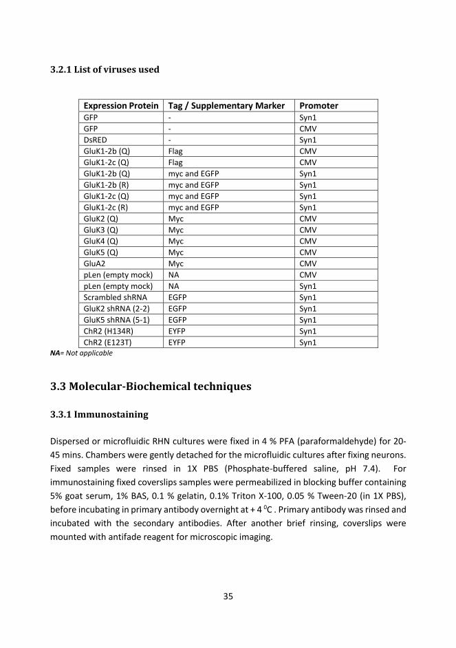
35
3.2.1 List of viruses used
Expression Protein Tag / Supplementary Marker Promoter GFP - Syn1 GFP - CMV DsRED - Syn1 GluK1-2b (Q) Flag CMV GluK1-2c (Q) Flag CMV GluK1-2b (Q) myc and EGFP Syn1 GluK1-2b (R) myc and EGFP Syn1 GluK1-2c (Q) myc and EGFP Syn1 GluK1-2c (R) myc and EGFP Syn1 GluK2 (Q) Myc CMV GluK3 (Q) Myc CMV GluK4 (Q) Myc CMV GluK5 (Q) Myc CMV GluA2 Myc CMV pLen (empty mock) NA CMV pLen (empty mock) NA Syn1 Scrambled shRNA EGFP Syn1 GluK2 shRNA (2-2) EGFP Syn1 GluK5 shRNA (5-1) EGFP Syn1 ChR2 (H134R) EYFP Syn1 ChR2 (E123T) EYFP Syn1
NA= Not applicable
3.3 Molecular-Biochemical techniques 3.3.1 Immunostaining Dispersed or microfluidic RHN cultures were fixed in 4 % PFA (paraformaldehyde) for 20-45 mins. Chambers were gently detached for the microfluidic cultures after fixing neurons. Fixed samples were rinsed in 1X PBS (Phosphate-buffered saline, pH 7.4). For immunostaining fixed coverslips samples were permeabilized in blocking buffer containing 5% goat serum, 1% BAS, 0.1 % gelatin, 0.1% Triton X-100, 0.05 % Tween-20 (in 1X PBS), before incubating in primary antibody overnight at + 4 0C . Primary antibody was rinsed and incubated with the secondary antibodies. After another brief rinsing, coverslips were mounted with antifade reagent for microscopic imaging.

36
3.3.2 List of antibodies
Primary Antibody Application Dilution Source
Myc-anti rabbit ICC 1:1500 Upstate
Flag-anti-mouse ICC 1:1500 Sigma Aldrich
Synapsin1-anti mouse ICC 1:1000 Synaptic Systems
Synaptophysin1-anti guinea pig ICC 1:1000 Synaptic Systems
Tau-anti mouse ICC 1:1000 Cell Signaling Technology
Phospho-Tau (Ser 396)-anti mouse ICC 1:1000 Cell Signaling Technology
MAP2- anti chicken and -anti rabbit ICC 1:8000 Synaptic Systems; Millipore
PSD-95 ICC 1:500 BD Transduction Laboratories
ICC = Immunocytochemistry 3.3.2 In-Situ hybridization For double in situ hybridization 7 μm thick brain slices from P3 and P15 rats were fixed in 4 % PFA and paraffinated. The procedure was similar as in (Huberfeld et al. 2007). These experiments were carried out by the co-authors. The cloning details, procedure for the generation of labeled sense and anti-sense strands, hybridization and imaging methods are mentioned in the original publication (article II).
3.3.3 RT-PCR RNA was extracted from hippocampi of P3 and P15 age rats or from 7 DIV and 14 DIV dispersed cultures using RNeasy Mini Kit. cDNA was synthesized with 1 μg of total RNA with oligo-dT primers using RevertAId First Strand cDNA synthesis Kit (Theromo Scientific) or RETROscript® (ambion) reverse transcription (RT) kit. Absolute quantification PCR or aqPCR was carried out in a CFX96 Real-Time PCR detection system (Bio-Rad) using SYBR Green qPCR Master Mix (Thermo Scientific). These experiments were carried out by the co-authors. The detailed procedure and the lists of forward and reverse primers are listed in the original publications (article II and III).

37
3.3.4 Western Blot HEK 293T cells or RHN cultures expressing different vectors or viral constructs were lysed in laemmili buffer. Standard BSA (bovine serum albumin) protein quantification method was used to quantify protein concentration before resolving the protein homogenates by SDS-PAGE. Gel separated proteins were transferred to nitrocellulose filters, blocked and stained with different primary antibodies. Protein bands were detected on X-ray films after staining the filters with HRP (horseradish peroxidase) conjugated secondary antibodies. This method was used to test different lenti-viruses expression in dispersed cultures. The experiment in the original publication (article III) was carried out by the co-author.
3.4 Confocal Imaging Following Confocal microscopes were used for image acquisition
Name of the Microscope Objective Used Zeiss LSM 5 Pascal Axioplan 2 Microscope C-Apochromat 40X N.A 1.2 water objective MP Leica TCS SP5 confocal Microscope HPX PL APO 63X 1.30 Objective LSM Zeiss700 Microscope I LCI Plan-Neofluar 63X/1.3 Imm Korr DIC M27
Objective LSM Zeiss 710 Confocal Microscope alpha Plan-Apochromat 63X/1.46 Oil Korr M27
Objective Imagining parameters were optimized using control samples and kept constant for imaging KAR expressing samples. Fluorescent images along z-axis were obtained with 0.1 μm – 1 μm stack series for different experiments. Images were compiled by maximum intensity projection for further analysis. Leica LAS AF lite, ZEN Blue, ZEN Black, Zeiss LSM Browser, NeuronStudio, Imaris and adobe Photoshop Software were used for different image analysis and post analysis image representation. Detailed staining parameters are listed in the table 3.3.2 (List of antibodies) and detailed descriptions of the analysis are mentioned in the original publication.
3.5 Image Analysis 3.5.1 Receptor classification at proximal and distal dendrites Determination of dendritic distal localization of GluK1 with GluK4/5 was based on the detection of the receptors being present 50 μm away from the soma (also refer Kayadjanian et al. 2007)
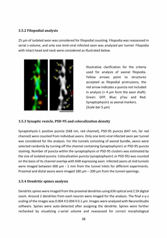
38
3.5.2 Filopodial analysis 25 μm of isolated axon was considered for filopodial counting. Filopodia was reassessed in serial z-volume, and only one lenti-viral infected axon was analyzed per tunnel. Filopodia with intact head and neck were considered as illustrated below.
3.5.3 Synaptic vesicle, PSD-95 and colocalization density Synaptohysin-1 positive puncta (568 nm, red channel), PSD-95 puncta (647 nm, far red channel) were counted from individual axons. Only one lenti-viral infected axon per tunnel was considered for the analysis. For the tunnels consisting of axonal bundle, axons were selected randomly by turning off the channel containing Synaptophysin1 or PSD-95 puncta staining. Number of puncta within the synaptophysin or PSD-95 clusters was estimated by the size of isolated puncta. Colocalization puncta (synaptophysin1 or PSD-95) was counted on the basis of its channel overlap with KAR expressing axon. Infected axons at mid tunnels were imaged between 600 μm - 1 mm from the tunnel inlets for different experiments. Proximal and distal axons were imaged 180 μm – 200 μm from the tunnel openings.
3.5.4 Dendritic spines analysis Dendritic spines were imaged from the proximal dendrites using 63X optical and 2.5X digital zoom. Around 2 dendrites from each neuron were imaged for the analysis. The final x-y-z scaling of the images was 0.004 X 0.004 X 0.1 μm. Images were analyzed with NeuronStudio software. Spines were auto-detected after assigning the dendrite. Spines were further rechecked by visualizing z-serial volume and reassessed for correct morphological
Illustrative clarification for the criteria used for analysis of axonal filopodia. Yellow arrows point to structures accepted as filopodial protrusions, the red arrow indicates a puncta not included in analysis (> 4 μm form the axon shaft). Green: GFP, Blue: pTau and Red: Synaptophysin1 as axonal markers. (Scale bar 5 μm)
39
detection by the software. Statistical analysis of spine dimensions was carried out from the raw data by pooling morphological dimensions of individual spines from 4 independent cultures. 3.5.5 Axonal localization of GluK1c with GluK4/5 Isolated axons were imaged 350 μm - 550 μm from the tunnel inlets. Total number of axons per tunnel was determined by bright field imaging.
3.6 Electron Microscopy RHN cultures were fixed in 2% glutaraldehyde + 2% PFA, and subsequently blocked, dehydrated and embedded in epon capsules. Embedded samples were sectioned, transferred into the grids and post-stained with 0.5 % uranyl acetate. The sample processing was carried out by the personnel of the Electron Microscopy Unit of University of Helsinki. Joel 1400 transmission electron microscope was used to image intact symmetrical synapses with the visible active zones with 4000-6000 X magnification. At least 3 serial grids were imaged per sample (an average of 35 ± 14 pictures per serial section). ImageJ software was used to measure the length of active zones.
3.7 Electrophysiology
3.7.1 Microfluidic culture electrophysiology and optogenetics Whole cell patch clamp recording of RHN cultures were performed between 15 to 24 DIV. Top two layers of microfluidic chambers were gently removed, and the remaining bottom layer on a coverslip containing neurons were placed in a submerged recording chamber mounted on Olympus BX51 fluorescence microscope. The chambers were perfused with artificial cerebrospinal fluid (ACSF). Glass pipette electrode (resistance 4-7 MΩ) was used to gain whole cell access of patched neurons. Blue light source (470 nm) from the OptoLED was custom fitted to the objective of top-down microscope in the recording setup to stimulate ChR2 during optogenetic experiments. The electrophysiological data was analyzed using pClamp software. Detailed experimental protocol is mentioned in Jokinen et al. 2013 and Sakha et al. 2013.

40
3.7.2 Hippocampal Slice Electrophysiology
Slices with clear lentiviral expression (fluorescence) in the hippocampal CA3 region but not in CA1 were considered for the study. Slices were placed in the submerged recording chamber and constantly perfused with ACSF. Glass electrode (resistance 4-7 MΩ) was used to gain whole cell access. These experiments were conducted by the coauthors, and the detailed procedure is mentioned in the original publication (article II).
3.8 Pharmacological Tools List of drugs used
Drug Concentration Pharmacology (9R,10S,12S)-2,3,9,10,11,12-Hexahydro-10-hydroxy-9-methyl-1-oxo-9,12-epoxy-1H-diindolo[1,2,3-fg:3',2',1'-kl]pyrrolo[3,4-i][1,6]benzodiazocine-10-carboxylic acid, hexyl ester (KT5720)
1 μM Protein Kinase A inhibitor
(S)-1-(2-Amino-2-carboxyethyl)-3-(2-carboxy-5-phenylthiophene-3-yl-methyl)-5-methylpyrimidine-2,4-dione (ACET)
200 nM Competitive antagonist for GluK1
6-cyano-7-nitroquinoxaline-2,3-dione (CNQX)
10 μM AMPAR and KAR antagonist
Picrotoxin 100 μM GABAA receptor antagonist
bisindolylmaleimide VII acetate (BIS) 0.5 μM Protein Kinase C inhibitor
penicillin-streptomycin1 1% (v/v) antibiotics2 1= used in cell culture medium; 2= combined action against gram positive and gram negative bacteria. Penicillin inhibits cell wall synthesis and streptomycin inhibits protein synthesis
3.9 Statistical Analysis Following Statistical Analysis was performed: 1) Student’s two-tailed t-test, or, 2) ANOVA and post hoc analysis by Dunnett’s or Turkey’s correction procedure. (Statistical significances and N values are mentioned in the text here or/and in the original publications.)

41
4. Results
4.1 Development of microfluidic chamber for rat hippocampal neurons Microfluidic PDMS chamber containing two spatially isolated neuronal reservoirs separated by microtunnels for axonal passage was designed and optimized for experiments involving both morphological and functional (patch clamp electrophysiology) characterization of neurons (article I). The chamber consisted of three layers (Figure 6). The first layer was 700 μm thick and comprised of axonal tunnels measuring 2 mm X 7.5 μm X 73 μm. A total of 34 tunnels were designed to connect two primary cell culture reservoirs of 7 mm X 7 mm dimension. The second layer was 5 mm thick and comprised two circular cavities which could be aligned vertically on top of the cell culture reservoirs of the first layer chip. Finally, the third chip or the topmost layer was 2mm thick, used for accommodating excess volume of media to just one side of the reservoir (refer Chapter 4.1.2). Due to the elastic property of PDMS it can stick to clear surfaces even with a gentle pressure thus forming water-tight seal and can be reversely detached simply by a moderate physical force (McDonald & Whitesides 2002). This property enabled 3 distinctive PDMS layers to seal during chamber assembly. Furthermore, the top two layers of the chamber could be removed without perturbing the viability of the neurons, to allow positioning on the stage of inverted microscope for electrophysiological recordings. With the shallow PDMF layer the patch electrode had sufficient access to the neurons grown on the coverslip.
Figure 6: Top panel: Image of assembled 3 layered PDMS chamber for neuronal culture filled with full NB media (left hand side). Pictures illustrating the chamber with only the bottom layer in an electrophysiological setup (right hand side). The access of the electrode was optimal for patching neurons. Bottom panel: Graphical illustration of the top panel images. 3 layers of PDMS chips are labelled numerically. (Figure modified from article I)

42
4.1.1 Characterization of axonal growth and functional viability of the neurons in the microfluidic chamber PDMS chambers were assembled on poly-l-lysine (PLL) coated dry glass coverslips and rat hippocampal neurons (RHN) were seeded on one side only (unilaterally) or on both sides. With this arrangement, most of the assembled chambers formed water-tight seal without any media leakage. From day 3 onwards periodical media changes were conducted on both sides of the neuronal reservoirs every 3-4 days by replacing a bit over ¾ of the remaining media volume. Under these conditions, the axons crossed half length of the tunnels during the first week of culturing and took at least 10 days to cross the 2 mm distance to enter the other reservoir of the chamber when plated unilaterally (Figure 2 A, article I). As expected and revealed by MAP immunostaining, dendrites did not travel far and grew roughly 100 - 250 μm into the tunnels even after second week of culturing. We also tested commercially available neuronal PDMS microfluidic chambers (xonamicrofluidics). These chambers had 4 external neuronal reservoirs and internal main channels with somatic and axonal compartments separated by tunnels (Taylor et al. 2003; Taylor et al. 2005; J. W. Park et al. 2006). These were of two kinds with variable tunnel length (450 μm and 900 μm). This chamber was primarily designed to plate neurons unilaterally so that axons were sent from somatic side to the axonal side. However, it was still possible to plate neurons bilaterally and it required seeding neurons in all four external reservoirs (150,000-200,000 RHN per reservoir). It was rather challenging to get neurons into the somatic compartment through the main channel, so neurons had to be pipetted in-and-out few times during seeding. This occasionally resulted in the formation of air bubbles in the main channels. Hence, the internal neuronal compartment did not always have optimal neuronal density and sometimes neurons did not reach the somatic compartment(s) as neuronal passage got blocked by air bubbles. The previous design also did not allow efficient viral transduction in these internal somatic compartment as neurons were inaccessible once assembled on the glass coverslip. These technical issues inspired us to design simplified chamber with only two accessible neuronal culture reservoirs connected by 2mm tunnels. Inclusion of top third chip in one of the reservoirs also permitted us to maintain media volume difference between two neuronal reservoirs for asymmetrical viral transduction (see Chapter 4.1.2). Direct pharmacological treatment on the neurons was also possible in this design. Our design required less neurons (10,000 to 25,000 per reservoir). One of the biggest advantage over commercially available chambers was the possibility of patching neurons directly from the neuronal reservoirs. This was due to the versatile engineering of 3 separate layers so that the bottom 1st layer
43
containing tunnels and primary neuronal reservoir provided a) inter-compartmental axonal entry by neurons, and b) sufficient angular access for the electrode to patch neurons without the need of detaching the PDMS chamber completely from the glass coverslip. In addition, this 2mm tunnel length provided sufficient spatial separation for axons from the dendrites at its middle part. Speculation around the general health of neurons in microvolume of media during prolonged time period (2-3 weeks) was understandable. The functional viability of neurons was assessed by testing the active membrane properties in current clamp mode. Three week old neurons displayed consistent action potential frequency of 15.55 ± 1.13 Hz and 19.69 ± 0.63 Hz (N = 8) in response to 60 pA and 80 pA step depolarization, respectively, with spike adaptation pattern typical for hippocampal pyramidal neurons (Figure 2 C, article I). Furthermore, to check the state of neuronal connectivity within the culture reservoir, spontaneous activity was pharmacologically characterized. The cultures displayed prominent spontaneous synaptic activity which was mostly glutamatergic and sensitive to global AMPA/KA receptor antagonist (CNQX). The remaining synaptic activity was blocked by picrotoxin (a noncompetitive GABAA receptor antagonist) (Figure 2 C, article I). This confirmed that cultures were synaptically connected in the dispersed network with in the cell culture reservoir primarily by glutamatergic connection and with some GABAergic synapses.
4.1.2 Asymmetric fluidic isolation of neurons Asymmetrical media volume was maintained by including excess media volume of 70 μl in one of the reservoirs accommodating the extra third chip (Figure 3 A, article I). This extra dimension served two very essential functions a) primarily to avoid cross spillage of media as the radial distance between two reservoirs was small, and b) to prevent excessive media evaporation during prolonged cultures. Sufficient micro volume of culture media had to be accommodated in these reservoirs which was only achieved by maintaining the media curvature convex against the resting surface. The fluidic isolation between the two reservoirs was confirmed by infecting the two cell culture reservoirs with separate lentiviruses coding for green fluorescent protein (GFP) and double-stranded red (DsRED) at 3 DIV (Days in vitro) (Figure 3 B and 3 C, article I). This resulted in an asymmetrical neuronal transduction, as fixed neurons (DIV 14) exhibited uneven viral transduction pattern between these two reservoirs. Minor cross contamination on the opposite reservoir was only seen if transduction was done in reservoir with excess media. Furthermore, despite of these two reservoirs being connected

44
by the tunnels, the volume difference between the two reservoirs was maintained and media did not get displaced from higher to lower volume during prolonged culturing. With asymmetrical fluidic isolation, some viral cross-transduction was only observed around the periphery of the tunnel outlet of reservoir with smaller media volume. Thus, even if the volume difference between the reservoirs was maintained, the media connecting the tunnels may have diffused constituents from the larger media volume reservoir. The nature of molecules diffusing into these tunnels may however display an exclusion factor, depending relatively on its molecular dimensions. Apart from the pressures holding the fluid asymmetrically between these two reservoirs, additional viscous stress tensor force may also be present. These may result in a corrective module between the physical counteractive forces generated by i) the internal and hydrostatic pressure, ii) along with the diffusion of media into the tunnels, and iii) encountering biomechanical forces generated by the motion of cytoskeletal structures responsible for axonal movement and growth. This results in relatively fewer average number of axons to enter the tunnels from the cell reservoir containing bigger media volume than from the opposite reservoir with smaller volume (2.3 ± 0.3 average axons versus 3.3 ± 0.5, p = 0.03, respectively, Student’s t-test, S.D. N = 4 chambers, from 3 independent cultures), provided that RHN were equally plated (20,000) in both reservoirs initially (unpublished results). However surprisingly, despite unequal number of axonal entry from either sides, the numbers of axons exiting the tunnels into opposite reservoirs was equal (1.5 ± 0.5 average axons versus 1.2 ± 0.4, N=4 chambers). Likewise, for control cultures without any media difference relatively same number of axons entered the tunnels from both the reservoirs (2.4 ± 0.6 and 2.5 ± 0.7, N = 3 chambers, from corresponding sister cultures) and fewer exited (1.3 ± 0.3 and 1.2 ± 0.3, N=3). Together, these results indicate that the third layer to accommodate adequate volume of media was highly useful, preventing a) media cross contamination, b) external media leakage and c) peculiarly excessive rate of media evaporation, critical for asymmetric manipulation and prolonged neuronal cultivation.
4.1.3 Optogenetic activation of microfluidically isolated axons With optimization of asymmetrical fluidic isolation for viral transduction, we next wanted to check if axons crossing the tunnels can make functional connections to neurons at the other reservoir. To this end, we used optogenetics and electrophysiological recordings. For optogenetic excitation, two separate lentiviral constructs containing ChR2 with H134R mutation or ChR2 with E123T (ChETA) were tested. The former, ChR2(H134R) can generate

45
larger photo current compared to the wild-type ChR2 but has slower channel kinetics (Nagel et al. 2005). The ChETA version exhibits relatively smaller photo current but with an ultrafast precision in its channel kinetics (Gunaydin et al. 2010). Voltage clamping the neurons expressing these ChRs confirmed that the photo current in neurons expressing H134R was roughly 3-5 time larger in ChETA expressing cells, although the rate of viral infection was indifferent between these constructs (data not shown). To reliably excite the axons, we chose ChR2 (H134R) for the rest of our studies. The density of axons crossing the tunnel was highest near the tunnel outlets and relatively abundant just around 1 mm peripheral distance from the tunnel openings (Figure 4 B, article I). Limited number of the neurons near the tunnel opening (13 % out of N = 23) responded with temporally precise monosynaptic EPSC to blue light stimulation. This confirmed the presence of a direct connectivity between ChR2 expressing axons crossing the tunnel and postsynaptic wild type neurons. The success rate of light evoked transmission was relatively low suggestive of a monosynaptic connection. Furthermore, these currents were sensitive to AMPAR/KAR antagonist (CNQX) thus confirming them to be glutamatergic synaptic responses. This novel method allows introducing protein of interest with/without ChR2 to this system to analyze its effect on presynaptic function in high-resolution.
4.2 Subcellular localization of GluK1 subunit containing KARs GluK1 subunit is highly expressed in the developing hippocampus and has prominent effects on synaptic transmission and network excitability (Lauri et al. 2006; Vesikansa et al. 2007; Sallert et al. 2007; Lauri et al. 2005). However, the exact molecular composition of the receptors responsible for this functional modulation at immature synapses is unclear. This led us to investigate the expression pattern and subcellular localization of various different types of GluK1 subunit containing receptors in hippocampal neurons, in order to correlate its molecular structure to physiological function.
4.2.1 Heteromerization of GluK1c with high-affinity KARs promotes distal targeting of GluK1c in dendritic processes In situ hybridization revealed that GluK1 subunit was highly expressed in pyramidal neurons of CA1, CA3 and granule cells of dentate gyrus (here in principal neurons) of neonate brain (P3) while its level was strongly downregulated during development at P15 (Figure 1 C, article II). Double in situ hybridization of GluK1 with GluK4 revealed strong co-expression in the principal neurons of CA1, CA3 and dentate gyrus at P3, but not in the juvenile brain
46
(P15) (Figure 2 C, article II). GluK5 was also co-expressed with GluK1 in these hippocampal regions at P3, and the expression was developmentally downregulated (Figure 2 D, article II). This reduction in co-expression is mainly due to the loss of GluK1 from the pyramidal neurons. GluK1 splice variants displayed a neuron type specific expression pattern in P3 and P15 hippocampus (Figure 3 D, article II). GluK1a was confirmed not to be detectable in the hippocampus (as in Jaskolski et al. 2004)(Figure 3 A, article II). GluK1c splice variants were primarily present in the principal cells of hippocampus including CA3 and CA1 pyramidal neurons and granule cells of the dentate gyrus. GluK1b subtype was equivalently detected in the interneurons at P3 (Figure 3 C and 3 E, article II). GluK1c expression was strongly downregulated in parallel with development and maturation of the circuit (Figure 3 D, article II). The subcellular expression pattern of GluK1c was studied using lentiviral vectors to express recombinant GluK1c (flag-tagged) with or without the high affinity recombinant subunits (myc-tagged GluK4 or GluK5) in dispersed hippocampal cultures (15 DIV) (Figure 4 B, article II). Recombinant GluK1c was mainly detected in the neuronal soma and proximal dendrites ( ̴ 50 μm post soma). Inclusion of either GluK4 or GluK5 visibly promoted targeting of GluK1c into distal dendritic processes (> 50 μm from soma) (Figure 4 C, article II). Co-expression of GluK1b with GluK4/5 had dissimilar pattern than GluK1c (unpublished data). In most of the neurons GluK1b was localized in the soma (79%, N = 361). GluK4 expression had no effect in dendritic dispersion of GluK1b, while GluK5 co-expression promoted targeting of GluK1b to the distal dendritic compartments. In dispersed cultures, GluK1c was observed also in few axons, identified with absence of MAP2 staining. Heteromerization of GluK1c with GluK4 but not with GluK5 enhanced its localization to axons. In order to avoid coincidental identification of axons in the dispersed cultures and not to oversee extremely slender dimensions of the axons amongst the dense dendritic processes we further conducted microfluidic experiments to spatially isolate the axons from the somatic dendritic compartments (article I). Isolated axons displayed abundant expression of GluK1c and this was not affected by co-expression of GluK4. In contrast, the frequency of axonal GluK1c expression was reduced as a consequence of GluK5 co-expression. Vice versa, axonal expression of GluK4 but not GluK5 was enhanced by including GluK1c (Figure 5 D and 5 E, article II). This suggested a role of GluK4 rather than GluK5 for GluK1c distal targeting in the axons. High resolution imaging revealed that heteromerization with the high-affinity subunits affected GluK1c expression pattern in the axonal shaft and in filopodial processes (Figure 5 F, article II). When expressed alone, GluK1c, GluK4 and GluK5 subunits were equally
47
present in axonal shafts and in the filopodial protrusions (Figure 5 B and 5 C, article II). When co-expressed, GluK1c had preferential localization in the axonal shaft while GluK4 and in particular GluK5 were localized in filopodial protrusions in the mid part of the tunnel. As the mid part of tunnels does not contain any post synaptic partners, this peripheral shift of GluK4 or GluK5 may function to sense post synaptic targets. Contrariwise, these high affinity subunits may function to retain GluK1c in the axonal shafts in the absence of functional post synaptic partners. Interestingly, at the distal axons that made connections to dendrites, GluK1c was equally present in axo-dendritic contact zones. Qualitative assessment suggested GluK1c to co-localize more with GluK4 versus GluK5 after dendritic contact formation, although colocalization with both was detected. Together, these data support GluK1c+GluK4 heteromers to be responsible for endogenous presynaptic GluK1 function in the immature hippocampus.
4.3 KARs promote formation of axonal and dendritic protrusions
4.3.1 Expression profile of KAR subunits in microfluidic cultures First, we wanted to check expression of different endogenous KAR subunits in RHN dispersed cultures at 7 and 14 DIV. To this end, total RNA extracts from the cultures were processed for qPCR, aimed at two purposes. First, to determine copy number of individual subunits, and to check relative expression levels of these subunits between first and second week old dispersed cultures. Comparing Ct values of the samples with the standard curve plot gave us the initial copy number of individual subunits (Figure 1 B, article III). Neurons at both time points expressed all KAR subunits, although GluK1 and in particular GluK3 were less abundant. Gene expression differences between these week old apart neurons were calculated by 2-
ddct method against GAPDH and Rpl19 reference genes. The expression patterns seemed to be relatively stable during these culture periods for most of the subunits, except for GluK3. However, this difference may be due to random variability as the copy number of GluK3 was very low. Establishing endogenous expression pattern of these subunits justified the control of their endogenous expression levels using shRNA sequences against rat GluK2 (GRIK2, 889-912) and rat GluK5 (GRIK5, 752-776). These shRNA target sequence constructs were selected based on their ability to inhibit expression of recombinant and endogenous GluK2 and GluK5 in HEK 293T cells and hippocampal cultured neurons, respectively (Figure 1 C, article

48
III). The selected shRNAs resulted in specific knockdown of target subunit without cross reactivity against other homologous subunits.
4.3.2 KARs promote formation of axonal protrusions Presynaptic Kainate receptors regulate filopodial mobility in immature and premature neurons (Chang & De Camilli 2001; Tashiro et al. 2003). However, whether distinct KAR subunits influence filopodial morphogenesis is not known. Here, we used microfluidic chambers to cultivate RHN expressing epitope tagged KARs subunits asymmetrically in spatially isolated axons in order to analyze their effects on axonal morphology.
4.3.2.1 Enrichment of axonal filopodia protrusion is independent of KAR subunit type In the mid part of the tunnels within the microfluidic chamber, axons grow in complete isolation from the somatodendritic compartments and glial connections. Since the width of these microgrooves was only 7.5 μm, axons tended to bundle up conspicuously but not entirely along the length. Axonal marker pTau was used to identify axons (explained below in Figure 8) while transduced axons were distinguished from the uninfected ones based on the epitope tag. At 15-16 DIV, spatially isolated axons expressing recombinant unedited KAR subunits developed extensive filopodial protrusions from the axonal shafts (unpublished data) in comparison to GFP expressing filopodial processes (0.45 ± 0.03 /μm) (Figure 7 B). Co-expression of high+low affinity subunits also resulted in significant increase in filopodial density as compared to GFP (GluK1+GluK5 = 193 ± 10 %, GluK2+GluK5 = 169 ± 17 % of GFP, N = 45, 52 respectively, GFP N = 112, p < 0.005 at both cases, based on immunolabelling of high affinity KAR subunit, see Chapter 4.2.1). To make sure these increments in axonal filopodial density was because of transduced KAR subunits we assessed GluA2 AMPA subunit as they seemed to be equally targeted to the filopodial processes here as for the myc-KAR expressing filopodia. AMPARs in general do not cross-assemble with any KAR subunits (review Pinheiro & Mulle 2006; Hansen et al. 2010) and GluA2 overexpression did not display visible changes in filopodial numbers (0.45 ± 0.03 /μm, N = 85; 91 ± 9 % of GFP). Conversely, knockdown of endogenous GluK2 or GluK5 with EGFP containing shRNA constructs led to reciprocal decrease in filopodial number against mock construct infected axons (Figure 7 C, unpublished data). Neurons with early knockdown (3 DIV) of these subunits did not survive till 15 DIV (hence fixed at 11 DIV). However late knockdown at 7 DIV led to the survival of neuronal cultures possibly due to the formation of stable neurite
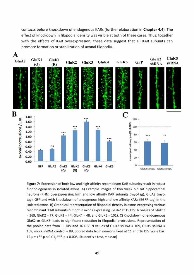
49
contacts before knockdown of endogenous KARs (further elaboration in Chapter 4.4). The effect of knockdown in filopodial density was visible at both of these cases. Thus, together with the effects of KAR overexpression, these data suggest that all KAR subunits can promote formation or stabilization of axonal filopodia.
Figure 7: Expression of both low and high-affinity recombinant KAR subunits result in robust filopodiogenesis in isolated axons. A) Example images of two week old rat hippocampal neurons (RHN) overexpressing high and low affinity KAR subunits (myc-tag), GluA2 (myc-tag), GFP and with knockdown of endogenous high and low affinity KARs (EGFP-tag) in the isolated axons. B) Graphical representation of filopodial density in axons expressing various recombinant KAR subunits but not in axons expressing GluA2 at 15 DIV. N values of GluK1c = 169, GluK2 = 77, GluK3 = 44, GluK4 = 48, and GluK5 = 101). C) Knockdown of endogenous GluK2 or GluK5 leads to significant reduction in filopodial protrusions. Representation of the pooled data from 11 DIV and 16 DIV. N values of GluK2 shRNA = 109, GluK5 shRNA = 109, mock shRNA control = 89, pooled data from neurons fixed at 11 and 16 DIV.Scale bar: 12 μm (** p < 0.01, *** p < 0.005, Student’s t-test, ± s.e.m)

50
Figure 8: Immuno-labeling rat hippocampal neurons (RHN) with Tau and phospho- Tau (Ser 396) ( p-Tau) antibody at different culture ages. A-B) Wild-type dispersed RHN fixed at 6 and 10 DIV labeled with Tau and p-tau antibodies, respectively. At both instances, both Tau and p-Tau staining (green channel) was specific to the axons, while MAP-2 differentiated the dendritic labeling. C) RHN expressing GluK4 KARs at the isolated tunnels of the microfluidic chamber at 15 DIV. p-Tau antibody had better specificity for detecting axonal shafts at these differently aged cultures both with and without KAR subunit expression. Scale bar: A, B = 70 μm and C = 50 μm.
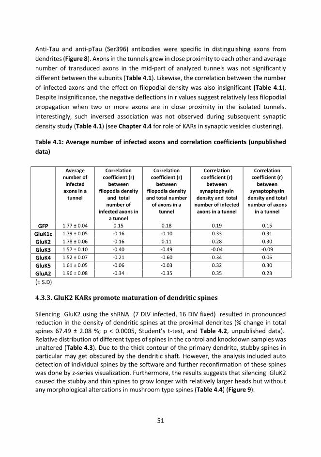
51
Anti-Tau and anti-pTau (Ser396) antibodies were specific in distinguishing axons from dendrites (Figure 8). Axons in the tunnels grew in close proximity to each other and average number of transduced axons in the mid-part of analyzed tunnels was not significantly different between the subunits (Table 4.1). Likewise, the correlation between the number of infected axons and the effect on filopodial density was also insignificant (Table 4.1). Despite insignificance, the negative deflections in r values suggest relatively less filopodial propagation when two or more axons are in close proximity in the isolated tunnels. Interestingly, such inversed association was not observed during subsequent synaptic density study (Table 4.1) (see Chapter 4.4 for role of KARs in synaptic vesicles clustering).
Table 4.1: Average number of infected axons and correlation coefficients (unpublished data) Average
number of infected
axons in a tunnel
Correlation coefficient (r)
between filopodia density
and total number of
infected axons in a tunnel
Correlation coefficient (r)
between filopodia density and total number
of axons in a tunnel
Correlation coefficient (r)
between synaptophysin
density and total number of infected
axons in a tunnel
Correlation coefficient (r)
between synaptophysin
density and total number of axons
in a tunnel
GFP 1.77 ± 0.04 0.15 0.18 0.19 0.15 GluK1c 1.79 ± 0.05 -0.16 -0.10 0.33 0.31 GluK2 1.78 ± 0.06 -0.16 0.11 0.28 0.30 GluK3 1.57 ± 0.10 -0.40 -0.49 -0.04 -0.09 GluK4 1.52 ± 0.07 -0.21 -0.60 0.34 0.06 GluK5 1.61 ± 0.05 -0.06 -0.03 0.32 0.30 GluA2 1.96 ± 0.08 -0.34 -0.35 0.35 0.23
(± S.D)
4.3.3. GluK2 KARs promote maturation of dendritic spines Silencing GluK2 using the shRNA (7 DIV infected, 16 DIV fixed) resulted in pronounced reduction in the density of dendritic spines at the proximal dendrites (% change in total spines 67.49 ± 2.08 %; p < 0.0005, Student’s t-test, and Table 4.2, unpublished data). Relative distribution of different types of spines in the control and knockdown samples was unaltered (Table 4.3). Due to the thick contour of the primary dendrite, stubby spines in particular may get obscured by the dendritic shaft. However, the analysis included auto detection of individual spines by the software and further reconfirmation of these spines was done by z-series visualization. Furthermore, the results suggests that silencing GluK2 caused the stubby and thin spines to grow longer with relatively larger heads but without any morphological altercations in mushroom type spines (Table 4.4) (Figure 9).

52
Correspondingly, number of dendritic spines at the proximal dendrites increased with recombinant GluK2 expression (% change in total spines 207.7 ± 6.12 %; p < 0.0005, Student’s t-test; and Table 4. 2, unpublished data). With GluK2 overexpression there were relatively more mushroom and branched type spines as compared to control (Table 4.3). Interestingly, stubby and thin spines had relatively smaller head diameter, but the morphology of the mushroom spines and the length of overall spines were not drastically altered (Table 4.4). However, despite optimizing imagining parameters, the fluorophore intensity between different fluorophores namely myc-tagged GluK2 receptors and GFP (controls and GluK2shRNA) in the spines may effect these morphological measurements to certain degree. Despite this, there was a definite and very selective pattern observed in the morphological paradigms of the spines between the controls and receptor overexpressed or knockdown samples (Figure 9).
Table 4.2: Change in spine density and analysis parameters
Spine type % change in spine density with respect to control
Significant with respective control
GluK2-shRNA GluK2 GluK2-shRNA GluK2 Stubby 64.61 ± 2.81 %, 173.2 ± 7.1%, *** *** Thin 72.5 ± 3.4 % 178.27 ± 7 % *** *** Mushroom 70.32 ± 4.31%, 197.34 ± 14.6 % *** *** Other branched
86.51 ± 10.83 %, 615.2 ± 36.6 % ns ***
Mock control GluK2-shRNA GFP-control GluK2 Average dendritic length from soma
44.17 ± 0.8 μm 46.55 ± 0.98 μm 45 ± 0.94 μm 52.8 ± 1.21 μm
Average spine density
0.64 ± 0.02 / μm 0.43 ± 0.01 / μm 0.55 ± 0.02 / μm 1.12 ± 0.03 / μm
Number of branches analyzed
142 143 135 137
*** p < 0.0005, ns= not significant; Student’s t-test; (± s.e.m), and (± S.D) for number of branches. N values for different spine type in Table 4.3
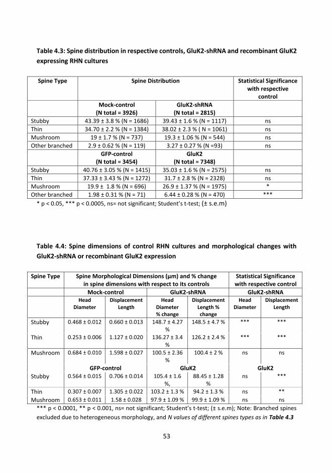
53
Table 4.3: Spine distribution in respective controls, GluK2-shRNA and recombinant GluK2 expressing RHN cultures Spine Type Spine Distribution Statistical Significance
with respective control
Mock-control (N total = 3926)
GluK2-shRNA (N total = 2815)
Stubby 43.39 ± 3.8 % (N = 1686) 39.43 ± 1.6 % (N = 1117) ns Thin 34.70 ± 2.2 % (N = 1384) 38.02 ± 2.3 % ( N = 1061) ns Mushroom 19 ± 1.7 % (N = 737) 19.3 ± 1.06 % (N = 544) ns Other branched 2.9 ± 0.62 % (N = 119) 3.27 ± 0.27 % (N =93) ns GFP-control
(N total = 3454) GluK2
(N total = 7348)
Stubby 40.76 ± 3.05 % (N = 1415) 35.03 ± 1.6 % (N = 2575) ns Thin 37.33 ± 3.43 % (N = 1272) 31.7 ± 2.8 % (N = 2328) ns Mushroom 19.9 ± 1.8 % (N = 696) 26.9 ± 1.37 % (N = 1975) * Other branched 1.98 ± 0.31 % (N = 71) 6.44 ± 0.28 % (N = 470) ***
* p < 0.05, *** p < 0.0005, ns= not significant; Student’s t-test; (± s.e.m)
Table 4.4: Spine dimensions of control RHN cultures and morphological changes with GluK2-shRNA or recombinant GluK2 expression
Spine Type Spine Morphological Dimensions (μm) and % change in spine dimensions with respect to its controls
Statistical Significance with respective control
Mock-control GluK2-shRNA GluK2-shRNA Head
Diameter Displacement
Length Head
Diameter % change
Displacement Length % change
Head Diameter
Displacement Length
Stubby 0.468 ± 0.012 0.660 ± 0.013 148.7 ± 4.27 %
148.5 ± 4.7 % *** ***
Thin 0.253 ± 0.006 1.127 ± 0.020 136.27 ± 3.4 %
126.2 ± 2.4 % *** ***
Mushroom 0.684 ± 0.010 1.598 ± 0.027 100.5 ± 2.36 %
100.4 ± 2 % ns ns
GFP-control GluK2 GluK2 Stubby 0.564 ± 0.015 0.706 ± 0.014 105.4 ± 1.6
%, 88.45 ± 1.28
% ns ***
Thin 0.307 ± 0.007 1.305 ± 0.022 103.2 ± 1.3 % 94.2 ± 1.3 % ns ** Mushroom 0.653 ± 0.011 1.58 ± 0.028 97.9 ± 1.09 % 99.9 ± 1.09 % ns ns
*** p < 0.0001, ** p < 0.001, ns= not significant; Student’s t-test; (± s.e.m); Note: Branched spines excluded due to heterogeneous morphology, and N values of different spines types as in Table 4.3

54
Figure 9: Representative images of 2 weeks old rat hippocampal neurons with spines at the proximal dendrites. Left panel figures: GluK2 shRNA infected dendrites; Middle panel figures: GFP expressing dendrites; and Right panel figures: GluK2 subunit expressing dendrites. Scale bar: 14 μm
55
4.3.4. GluK1 editing and channel inhibition does not affect outgrowth of filopodial protrusions Influx of Ca2+ ions in response to the activation of glutamate receptors including KARs has been suggested to have a dynamic role in filopodial morphogenesis (Mattson 2008; Chang & De Camilli 2001; Ibarretxe et al. 2007). GluK1 and GluK2 subunits are basically unedited and permeable to Ca2+ ions during birth, and are later replaced by its edited variants as brain develops (refer Chapter 1.1). This led us to check the role of Q/R editing in filopodial morphogenesis, using Q and R variants of the GluK1c subtype. Remarkably, equal filopodial density was observed in the isolated axon, irrespective to Ca2+ permeability of the recombinant subunit expressed (GluK1c (Q) 0.91 ± 0.03 /μm, N = 127 and GluK1c(R) 0.91 ± 0.06 /μm, N = 67; (unpublished data; Figure 7 A). Furthermore, calcium permeable GluK1c (Q) still displayed filopodial enrichment in the presence of a selective antagonist, ACET, but to the lesser extent than for the untreated receptors (165 ± 6.9 % of GFP control, p < 0.05; or; 79.2 ± 3.1 % of untreated GluK1c(Q), p < 0.05, Student’s t-test, GFP + ACET N = 119, 120; GluK1 + ACET N = 117 and 128, 2 weeks old RHN, unpublished data). Likewise, in these growth permissive isolated tunnels of microfluidic chambers, both unedited and edited GluK1b splice variants resulted in pronounced increase in the density of axonal filopodia (GluK1b (Q) = 0.98 ± 0.04; N = 36, GluK1b (R) 1.05 ± 0.05; N = 64) (unpublished data).Together this proved that KARs mediated filopodial enhancement is independent from channel activity and its permeability for Ca2+ ions.
4.4 KARs regulate clustering of synaptic vesicles
4.4.1 Low-affinity KARs enhance synaptic vesicle clustering To study the direct involvement of axonal KARs in presynaptic differentiation we checked the prevalence of synaptic vesicle (SVs) clusters in the axons expressing different recombinant KAR subunits using synaptophysin staining. All recombinant KAR subunits co-localized with synaptophysin puncta at the isolated axons (Pearson’s coefficient for colocalization of KARs and Synaptophysin: GluK1 0.63; GluK2 0.61; GluK3 0.56; GluK4 0.47 and GluK5 0.63). GluK2 and GluK3 were more frequently detected with synaptophysin puncta than GluK1, GluK4 or GluK5 (Figure 2 A, article III). This colocalization for recombinant receptors was not altered between isolated axons and axons with dendritic contacts at the distal tunnels. This suggested that localization of recombinant receptors to axonal release site was not altered with the dendritic contacts. At the distal tunnels PSD-
56
95 colocalization revealed that 61 ± 2 % of KAR positive synaptophysin puncta had dendritic contact without any significant difference between the subunits (Figure 2 B, article III). Expression of the low affinity subunits GluK1, GluK2 and GluK3 resulted in pronounced increase in the density of synaptophysin positive puncta in isolated axons as compared to GFP controls (Figure 3 A, article III). To check if this presynaptic differentiation was specific to KARs, we also tested the effect of GluA2 and it did not alter the density of synaptophysin puncta. Knockdown of endogenous GluK2 subunit led to reduction in synaptophysin density in the isolated axons (Figure 3 A, article III, pooled data from 11 and 16 DIV fixed cultures). The role of the calcium permeability of GluK1c was tested using the Q/R editing variants. Expression of both edited and unedited variants of GluK1c displayed enhanced synaptophysin puncta as compared to GFP, while the effect of edited GluK1c (R) was significantly smaller as compared to that of unedited Q subtype (Figure 4 A, article III). However, unlike for GluK1c synaptophysin density was only increased by Ca2+ permeable GluK1b in the isolated axons (GluK1b (Q) 122.3 ± 6.5 %, p < 0.005; GluK1b (R) 104.3 ± 5.3 %, of control GFP, N = 38, 64 respectively, Student’s t-test, unpublished data). Likewise, GluK1c (Q) splice variant had slightly stronger effect in SV clustering than GluK1b (Q) splice variant (GluK1c (Q) 132.6 ± 5.7 %; p < 0.0005 GluK1c (R) 118.6 ± 6.2 %, p < 0.05, % of control GFP, N = 91, 76, respectively, Student’s t-test)(unpublished data). Furthermore, inhibition of endogenous GluK1 receptors by ACET resulted in reduction of synaptophysin density as compared to control conditions, and ACET fully blocked the effect of GluK1 overexpression (Figure 4 A, article III). This suggests that the channel activation and calcium permeability contribute to clustering of SVs in KAR expressing axons.
The axons make synaptic contacts to somatodendritic compartments before entering the tunnels. Additionally, these axons also contact wild type neurons in uninfected reservoir at distal tunnels. Synaptophysin density at the distal end was robustly increased upon expression of calcium permeable low-affinity KARs, whereas GluA2 had no effect (Figure 3 B, article III). Interestingly, GluK2 shRNA had no effect on the number of synaptophysin puncta at the distal tunnels (Figure 3 B, article III, pooled data from 11 and 16 DIV fixed cultures). However, at the proximal part, early knockdown of endogenous GluK2 significantly reduced SVs density (11 DIV fixed = 48 ± 6 %, N = 54, p < 0.0005; verses 16 DIV fixed = 108 ± 10 %, N = 42, % change of control, Mock control N = 37 and 38, respectively, pooled data illustrated in Figure 3 C, article III). This suggests that endogenous GluK2 subunit affects presynaptic differentiation in the early phase of synaptogenesis. However, once the neurite networks have been formed (by 7 DIV), knockdown of endogenous GluK2 is no longer efficient in reducing synaptophysin clusters at the proximal tunnels. On the other hand, the density of synaptophysin puncta was enhanced at these proximal tunnels
57
with overexpression of low-affinity recombinant KAR subunits (GluK1 = 137 ± 5.7 %, p < 0.005; N = 165, GluK2 = 141 ± 5.2 %, p < 0.005, N = 79, GluK3 = 120 ± 8.5 %, p < 0.005, N = 44, % change of control GFP; GFP = 0.56 ± 0.02 /μm, N = 238, unpublished data) and there was no effect with GluA2 overexpression (109.8 ± 5% of GFP, N = 87). However, this effect was relatively smaller than at the distal axons containing only presynaptic subunits. This data suggests that the postsynaptic KARs might also influence synaptic differentiation, although the data here is not sufficient to make definite conclusions on their exact roles.
4.4.2 High-affinity KARs circumvent synaptic vesicle clustering Unlike the low affinity subunits, expression of high affinity GluK4 and GluK5 subunits did not influence synaptophysin clusters in the isolated part of the tunnels (Figure 3 A, article III) even though these subunits were equally effective in enhancing the density of axonal filopodia. This implies that SVs clustering and filopodial propagation by KARs follow a distinct pathway, and may have specific but firmly related functions during synaptogenesis. Expression of GluK4 or GluK5 had no effect on synaptophysin puncta at the distal end of tunnel, (Figure 3 B, article III) but surprisingly reduced the density of synaptophysin puncta at proximal tunnels (GluK4 = 79.2 ± 6.4 %,N = 47, p = not significant; GluK5 = 71.4 ± 4.9 %, N = 41, p < 0.005, % change of control, Student’s t-test; unpublished data). This suggests that overexpression of high affinity subunits suppress synaptic differentiation under certain conditions. However, silencing expression of endogenous GluK5 also led to the reduction of synaptophysin puncta in axons at mid (pooled data from 11 and 16 DIV illustrated in Figure 3 A, article III) and proximal tunnels (proximal axons : 11 DIV fixed = 35 ± 4 %, N = 64; 16 DIV fixed = 62 ± 7 %, N = 40, % change of mock control, p < 0.005, pooled data illustrated in Figure 3 C, article III), but not at the distal tunnels (pooled data from 11 and 16 DIV, Figure 3 B, article III). The late knockdown effect of GluK5 implies temporal difference in the presynaptic effects of GluK2 and GluK5; GluK5 having a prolonged role in synaptogenesis or synapse stabilization as compared to GluK2 (as in Chapter 4.4.1). The finding that both overexpression and knockdown of GluK5 reduced synaptophysin puncta at proximal tunnel suggests that GluK5 might influence synaptic differentiation via several mechanisms, possibly involving indirect and/or postsynaptic effects.

58
4.4.3 Heteromerization of high and low affinity KARs decreases synaptic vesicle clusters Given the distinct effects of the low- and high affinity subunits on synaptophysin puncta, it was of interest to see the consequence of their co-expression, expectedly resulting in expression of heteromeric receptors. Interestingly, co-transduction of GluK1+GluK5 and GluK2+GluK5 led to pronounced reduction of synaptophysin density at the isolated and distal axons (isolated: distal GluK1 + GluK5 87 ± 10.2 %, 73.2 ± 8.6 %; GluK2 + GluK5 = 72.8 ± 8.3 %, 72.3 ± 8.8 %, of control GFP, respectively; and p < 0.005 against low affinity subunits from sister cultures; N = 45, 63, 45 and 59,respectively, GFP N = 114, 76, unpublished data). Similar phenotype was also observed for GluK1+GluK5 at proximal part of tunnels while GluK2+GluK5 had not effect in this region (GluK1+GluK5 = 72.45 ± 7.5 %, GluK2+GluK5 = 91.28 ± 8.4 % of GFP, p < 0.005 against sister low affinity subunits; N = 48 and 77, respectively, GFP N = 118, unpublished data). These data suggest that high affinity KARs have a dominant role in heteromeric receptor, suppressing the synaptophysin cluster-promoting effect of the low-affinity subunits.
4.5 Regulation of filopodia and synaptic vesicle clusters via KARs follow distinct but converging pathway After checking individual and combined effect of high and low affinity KAR subunits in synaptic differentiation, we wanted to gain insights on the downstream signaling mechanisms involved. We were particularly interested in the possible roles of PKA and PKC, identified in previous studies to be involved in metabotropic KARs signaling (review Rodríguez-Moreno & Sihra 2007; Lauri & Taira 2012). Axonal filopodial protrusion and synaptophysin puncta densities were analyzed after incubating RHN (4 DIV onwards) in presence of inhibitors KT5720 (1 μM) and bisindolylmaleimide VII acetate (BIS, 0.5 μM) for PKA and PKC respectively. For simplification, effects of inhibitors were only studied in isolated presynaptic axons in unilaterally plated RHN. Neither PKA nor PKC inhibition changed basal synaptophysin density in control GFP expressing cultures (Figure 4 B, article III). However, filopodial protrusions were enhanced significantly (Figure 10, unpublished data). Hence, filopodia and synaptophysin densities of control and drug treated KAR subunits were normalized to respective controls (DMSO or corresponding drug treated). Statistics were also performed similarly by cross checking with similar drug treated control samples of GFP or within sister controls expressing KAR subunits The effect of GluK1, GluK2 and GluK5 on filopodial density was significantly reduced in the presence of PKA and PKC inhibitors (Figure 10, unpublished data). PKA inhibition was more

59
efficient in decreasing filopodial protrusions in axons expressing low affinity subunits GluK1 and GluK2 than for GluK5 which still resulted in slight increase in filopodial number despite PKA inhibition. On the other hand, PKC inhibition reduced effects of GluK1 and GluK2 on filopodial density, and completely blocked the effect of GluK5. (Unpublished data: effect of PKA and PKC inhibition on axonal filopodial protrusions).
Figure 10: Pooled data on axonal filopodial density in the isolated tunnels under various experimental conditions. The data for PKA and PKC inhibitors, KT5720 and BIS respectively for GFP are represented as the % change level of DMSO treated GFP controls within the same culture batches. Likewise, KAR expressing axonal pharmacological data is normalized to the level of GFP expressing sister cultures with corresponding drug treatments. * p < 0.5 , ** p < 0.005, *** p< 0.005 as compared to corresponding GFP treatments; # p < 0.05 in comparison to respective KARs with control DMSO treatment. N values of GFP + DMSO = 85, GFP + KT5720 = 89 and GFP + BIS = 98; GluK1 + DMSO = 100, GluK1 + KT5720 = 84 and, GluK1 + BIS = 83; GluK2 + DMSO = 91, GluK2 + KT5720 = 94 and GluK2 + BIS = 88; GluK5 + DMSO = 88, GluK5 + KT5720 = 85, and GluK5 + BIS = 85)
PKA inhibition reversed the effect of GluK1 and GluK2 on the density of synaptophysin puncta, leading to reduced SV density as compared to drug treated GFP. On the other hand, PKC inhibition significantly prevented or reversed the effect of GluK2 and GluK1 on synaptophysin density respectively (Figure 4 B, article III). GluK5 had no effect on synaptophysin density under control conditions or in the presence of PKA inhibitor (98.7 ±

60
6.4 %, N = 88 and 89.2 ± 8.5 %, N = 85, respectively of corresponding GFP controls, unpublished data), but in the presence of PKC inhibitor a significant reduction in synaptophysin puncta was observed (58.2 ± 6.3 % of drug treated GFP control; N = 85, p < 0.005, unpublished data). These divergences support a role for both PKA and PKC signaling in the effects of KAR subunits on axonal filopodia and on synaptic vesicle clustering.
4.6 Regulation of glutamatergic transmission by presynaptic KARs Presynaptic KARs affected synaptic differentiation by regulating axonal filopodia and clustering of synaptic vesicles. These unique effects are expected to also influence synaptic function at KAR expressing axons. Light activated ChR2 (H134R) (Boyden et al. 2005) was co-expressed with KAR subunits asymmetrically in the microfluidic chamber to investigate the effect of various recombinant KARs on presynaptic function (article I, also refer Chapter 4.1.3). Light induced excitatory post synaptic currents (EPSCs) were recorded from the wild type neurons at 14-18 DIV. The average success rate of EPSCs in GFP/ChR2(H134R) expressing synapses was 34 ± 6 %, with average amplitude of 11 ± 3 pA and average potency of 28 ± 5 pA (Figure 5 B, article III). Paired light stimulation with inter pulse interval of 100 ms was applied to check the prevalence of short term plasticity represented as paired-pulse facilitation (PPF). However, PPF was not observed (second / first pulse current amplitude, 0.96 ± 0.24) in control samples, thus this property was not further investigated. 4.6.1 Presynaptic calcium permeable low-affinity KARs enhance success rate of synaptic vesicle release Synaptic responses at KAR/ChR2(H134R) expressing cultures were normalized to the corresponding responses of GFP controls from the sister batches to compensate for variation between cultures. Light evoked stimulation of presynaptic axons expressing calcium permeable (Q) variants of the low affinity subunits GluK1-3 led to significant increase in success rate of EPSCs recorded from postsynaptic wild type neurons without significant changes in the potency (Figure 5 C and 5 D, article III). These effects depended on the Ca2+ permeability of the low affinity subunits since Ca2+ impermeable GluK1 (R) had no effect on transmission. This data suggests that presynaptic expression of Ca2+ permeable low affinity subunits leads to increase in glutamate release probability (Pr).
Presynaptic expression of high affinity GluK4 and GluK5 subunits resulted in reduction in success rate. However, only GluK5 subunit displayed significant reduction in Pr (Figure 5 C, article III). Likewise EPSC amplitude was reduced by presynaptic expression of GluK5
61
(Figure 5 C, article III). These data suggest that expression pattern of KAR subunits in the presynaptic neuron may define enhancement or reduction in the Pr.
4.6.2 Heteromeric KARs mimic dominant features of high affinity KARs in suppressing glutamatergic transmission After observing unique effects of the low and high affinity presynaptic KAR subunits on synaptic transmission we further checked consequences of subunit co-expression. Surprisingly, the dominant feature of GluK5 in repressing success rate was prevalent in heteromeric receptors (GluK1c+GluK5 52.38 ± 12.21 %, GluK2+GluK5 24.77 ± 5.4 %, of GFP, N = 5 and 8, respectively, unpublished data). However, co-expression of high and low affinity KARs did not change the current amplitude (potency change: GluK1c+GluK5 115.15 ± 29.31 %, GluK2+GluK5 111.5 ± 39.5 %, of GFP). These data suggest that the high-affinity KARs subunits may alter the signaling properties of the presynaptic KARs, and this is a dominant feature in heteromeric receptors. We had also observed that GluK1c is highly expressed together with GulK4/5 in the pyramidal neurons of CA3 and CA1 of the neonatal hippocampus (P3). Similarly, targeting of GluK1c in the axons was mostly due to co-expression with GluK4 subunit (Chapter 4.2.1, article II). In the organotypic slice cultures presynaptic expression of GluK1c in CA3-CA1 synapses inhibited glutamate release and enhanced paired pulse ratio (Figure 6 C, article II) . Likewise, in vivo expression of GluK1c in juvenile CA3 pyramidal neurons resulted in inhibition of glutamate release probability at CA3-CA1 synapses, (Figure 7 C, 7 D and 7E, article II).
Together these data imply that in the neonatal CA3-CA1 synapses, immature type presynaptic KAR activity is mediated by GluK1c in heteromeric combination with the high affinity subunits in axons, and most probably with GluK4 because of the stronger association in co-expression pattern. This also reflects the dominant role played by high affinity KAR subunits during heteromerization with low affinity subunits as observed above in the microfluidic recordings.
4.6. 3. Low-affinity KARs affect synaptic ultrastructure Direct correlation between Pr and synaptic active zone (AZ) morphology has been suggested (e.g. Pierce & Lewin 1994; Schikorski & Stevens 1997). To start to check if different KAR subunits affected synaptic ultrastructure we characterized their effects on AZ structure in dispersed two week old RHN cultures. Electron micrographs were obtained from the symmetrical synapses with distinct integral pre and postsynaptic structures and

62
with visibly intact electron dense synaptic cleft, and the length of the AZ was measured (Schikorski & Stevens 1997). Only symmetrical synapses with distinctive post and presynaptic structure containing SVs but without round or flattened pleomorphic vesicles in the presynaptic periphery were included to exclude GABAergic synapses (Harris & Landis 1986). Due to heterogeneity (e.g. Shepherd & Harris 1998; Schikorski & Stevens 1997), after acquisition of electron micrographs synapses were classified in three groups based on pre and post synaptic pairing: i) presynaptic terminal-post synaptic dendritic spines (group I), ii) between axon-dendritic shafts (group II), and iii) axons-somatodendritic structures (group III). Majority of the analyzed synapses belonged to first two groups without drastic differences in inter group distribution after recombinant KAR subunit expression (Table 4.5; batch average N = 146 ± 25). The median distribution of synapses between these three groups in GFP control and KAR subunits were 43.43 % (avg dev = 3.88 %), 40.76 % (avg dev = 3.36 %) and 15.82 % (avg dev = 0.52 %), respectively. The length of AZ (at 15 DIV) was enhanced in cultures expressing low affinity KAR, while high affinity GluK5 had no significant effect (Figure 4 C, article III). Interestingly expression of calcium impermeable GluK1 (R) equally resulted in elongation of synaptic AZ (126 ± 4.75 %, of GFP, N = 185, unpublished data).
We observed that not all the synapses had single synapse bouton (SSB) and multiple active zones with independent post synaptic structures were also observed in a single presynaptic varicosity both in the shafts and axonal terminals (around 2-3 AZ) (e.g. Shepherd & Harris 1998). Pooled data represented length of all AZ from these multiple synapse bouton (MSB) as it was not practical to selectively pick only one among others from the same synaptic varicosity. So we further wanted to check if these drastic increments in the length of synaptic AZ, at least with low affinity subunits was due to selective inclusion of synapses belong to either SSB or MSB. Synapse morphology was separated into these two groups and there was no significant differences between the groups (mean distribution SSB = 53.33 ± 5.27%, group deviation 5.42 %; MSB = 46.77 ± 5.27 %, group deviation 5.42%).
Furthermore, the presence of mitochondria in the synapses was also analyzed because of their role in regulation of cellular calcium ions and ATP metabolism (Südhof 1995; Morris & Hollenbeck 1993; Overly et al. 1996). Proximity of Mitochondria to the AZ was measured to obtain edge-to-edge displacement length. There was no significant difference in mitochondrial association of control and KAR subunits expressing synapses (GluK1 101.2 ± 8.67 %, GluK2 91.89 ± 9.63 %, and GluK5 114.5 ± 14.25 % of control GFP, GFP 0.47 ± 0.04 μm). However, not all the synapses had mitochondria in its proximity. In general, around
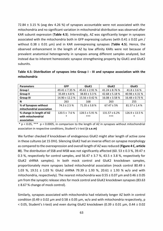
63
72.84 ± 3.15 % (avg dev 4.26 %) of synapses accountable were not associated with the mitochondria and no significant variation in mitochondrial distribution was observed after KAR subunit expression (Table 4.5). Interestingly, AZ was significantly longer in synapses associated with the mitochondria both in GFP expressing cultures (with 0.41 ± 0.02 μm / without 0.38 ± 0.01 μm) and in KAR overexpressing synapses (Table 4.5). Hence, the observed enhancement in the length of AZ by low affinity KARs were not because of prevalent anatomical heterogeneity in synapses among different samples analyzed, but instead due to inherent homeostatic synapse strengthening property by GluK1 and GluK2 subunits.
Table 4.5: Distribution of synapses into Group I - III and synapse association with the mitochondria
Parameters GFP GluK1 GluK2 GluK5 Group I 49.41 ± 7.35 % 45.61 ± 2.91 % 41.24 ± 8.76 % 41.8 ± 3.6 % Group II 35.69 ± 4.8 % 38.83 ± 3.3 % 42.68 ± 5.18 % 40.98 ± 4.31 % Group III 14.90 ± 12.2 % 15.56 ± 0.42 % 16.08 ± 3.58 % 16.48 ± 0.73 % N 263 168 263 255 % of Synapses without mitochondrial association
74.3 ± 2.5 % 71.35 ± 3.8 % 67.47 ± 5% 81.57 ± 1.4 %
% change in length of AZ with mitochondrial association
120.5 ± 7.6 % *
128.5 ± 5.91 % ***
131.57 ± 6.2% ***
126.9 ± 13.5 % *
* p < 0.05, *** p < 0.0005, in comparison to the length of AZ in synapses without mitochondrial association in respective conditions, Student’s t-test (± s.e.m)
We further checked if knockdown of endogenous GluK2 might alter length of active zone in these cultures (at 15 DIV). Silencing GluK2 had an inverse effect on synapse morphology as compared to the overexpression and overall length of AZ was reduced (Figure 4 C, article III). The distribution of SSB and MSB was not significantly affected (60. 53 ± 0.3 %, 39.47 ± 0.3 %, respectively for control samples, and 56.47 ± 3.7 %, 43.5 ± 3.8 %, respectively for GluK2 shRNA samples). In both mock control and GluK2 knockdown samples, proportionately more synapses lacked mitochondrial association (mock control 80.49 ± 1.03 %, 19.51 ± 1.03 %: Gluk2 shRNA 79.39 ± 1.93 %, 20.61 ± 1.93 % w/o and with mitochondria, respectively). The nearest mitochondria was 0.55 ± 0.07 μm and 0.46 ± 0.05 μm from the synaptic release sites for mock control and GluK2 knockdown synapses (83.63 ± 8.67 % change of mock control). Similarly, synapses associated with mitochondria had relatively longer AZ both in control condition (0.49 ± 0.02 μm and 0.58 ± 0.05 μm, w/o and with mitochondria respectively, p < 0.05, Student’s t-test) and even during GluK2 knockdown (0.39 ± 0.01 μm, 0.44 ± 0.02
64
μm, w/o and with mitochondria respectively, p < 0.05, Student’s t-test). Consequently, relative decrease in the AZ length with GluK2 knock down was visible in both synapses with or without mitochondrial association (78.96 ± 2.38 %, p < 0.001 and 76.25 ± 4.27 %, p < 0.01 of mock control, respectively, Student’s t-test, ± s.e.m). Both control and GluK2 shRNA expressing synapses had relative distribution of synapses into Group I, II and III based on pre and post synaptic partnering (39.10%, avg dev 0.28 %; 50.47 %, avg dev 1.86 % and 10.43%, avg dev 2.14 %, Group I, II and III, respectively). Hence, the reduction in the length of AZ with GluK2 shRNA expression is unaccountable to the procedural disproportionality during synapse identification, but undeniably represents impaired synapse maturation caused by the lack of endogenous GluK2 subunit.

65
5 Discussion
5.1 Asymmetrical neuronal growth in the microfluidic chamber
A microfluidic device for spatially isolating axons between the two neuronal somatodendritic compartments was successfully developed. This chamber allowed effortless electrophysiological recordings as well as asymmetrical viral transduction, due to novel three layered design with two reservoirs connected by axonal tunnels. Compared to commercially available chamber our device had more reliable application for a) plating neurons directly in the cell reservoirs apposing axonal tunnel inlets, b) direct pharmacological treatments of neurons and c) chamber specific viral transduction by introducing media volume difference between two cell reservoirs. The only commercially available device has four separate external reservoirs and main internal channels, primarily meant for single sided neuronal plating (Taylor et al. 2003; Taylor et al. 2005; J. W. Park et al. 2006) with limited usages in bilateral neuronal plating. In addition, these internal channels and compartments were not accessible once the chamber had been assembled on a glass coverslips. Reliable viral transduction and direct pharmacological treatment of neurons in the internal somatic compartment was hence a limiting factor. On the contrary, our versatile design was suitable for bilateral neuronal plating and for the optimization of asymmetrical neuronal transduction. Our multi-purpose design allowed removing top two layers and the remaining intact bottom first layer (sealed onto PLL coated glass coverslip with long-term cultivated neurons) allowed sufficient electrode access for the electrophysiological experiments. In the commercially purchased chambers the electrode access to cultured neurons was hindered by the thicker PDMS body. So removing the PDMS chip for patch electrode access would have damaged the delicate neuronal connections. Another benefit of our device was it required relatively fewer number of cells (by around 1/10th per reservoir).
During culture, the layered structure supports maintenance of media volume difference between the culture reservoirs to promoting fluidic isolation. The explanation behind this asymmetrical fluidic isolation may be closely related to microfluidic passive pumping phenomenon. In microfluidic passive pumping micro-volume or a drop of media is replaced due to the pressure differences between the inlet port containing smaller volume and the outlet port containing larger media volume in a unitary microtunnel (Berthier & Beebe 2007; Resto et al. 2010). During passive pumping, the internal pressure in smaller drop is greater due to its smaller internal radius compared to the large drop with bigger internal radius. This is founded on the Laplace’s law, which accounts for the relationship of surface tension at the liquid-air interface with that of the pressure difference between two uneven

66
micro volumes of media separated in the microchannel. However, our system had a multitude of 34 microtunnels combined for connecting two relatively larger reservoirs. In this case, hydrostatic pressure of media holding bigger volume (Taylor et al. 2005) may also oppose most of the internal pressure of smaller volume. This may have curiously resulted in the fractional equilibrium of these two counteracting pressures hence creating asymmetrical force to prevent selective cross-contamination of lentiviral particles. The outcome, based on experimental data, clearly indicates that inclusion of lentiviral vectors at the reservoir accommodating lesser media volume produced asymmetric genetic manipulation with no cross contamination in the other side.
Another interesting phenomenon of neuron growth in these two reservoirs is the regulation of axonal mobility perhaps by viscous stress tensor in the tunnels. Without any definitive mathematical model to absolutely represent fluidic resistance and counterbalancing forces between these two cell reservoirs and in the tunnels at this point, it can only be assumed that axons need to overcome the stress forces at the entry points of tunnels in the reservoir. The reservoir with lesser media with excessive passive internal pressure can assist in free passage of axons into the tunnels without hinder mobility. Likewise, since there was no significant difference in average number of axons exiting the tunnels at either end, the fluidic viscous stress might be uniformly distributed along the length of the tunnels, with more resistance on the top and less at the floor of the tunnel. So despite unequal axonal entry initially, equal number of axons from either direction may be able to crawl though the PLL coated coverslips to reach postsynaptic somatodendritic partner through the tunnel outlets. On top of that, axons in the tunnels tend to grow in close proximity to each other, or they are in occasional contact, and this might have additional effects in facilitating growth.
Another general observation during neuronal growth in these chambers was an affluent axonal entry into the tunnels when rate of culture media evaporation was not drastic or if the chambers were fluidically sealed without any leakages. The average time for any of the axons to cross 2 mm tunnel in this microfluidic chamber was 10 days. For instance tunnel length of 450 μm might have facilitated axons to cross the tunnel quicker. However, this tunnel dimension would have been too short to spatially isolate axons at the mid part of the tunnel because of bidirectional dendritic entry in bilaterally plated neuronal cultures. In such cases, both axonal and dendritic markers would have been required for their identification, creating technical limitations in our immunostaining studies for virally transduced neuronal cultures. 2mm length of our chamber provided reassurance that axons at mid part of the tunnels did not have any dendritic contacts even when neurons
67
were plated bilaterally. This was also confirmed with MAP and anti-PSD-95 immunostaining which was absent at the mid part of the tunnels (article I, II, III).
5.2 Axonal targeting of KAR subunits
Ambient glutamate activates presynaptic KARs to inhibiting glutamate release probability in neonatal CA1 and CA3 (Lauri et al. 2005). These tonically activated immature KARs are replaced by adult type receptors which are non-responsive to ambient glutamate (e.g. Lauri et al. 2006; Clarke & Collingridge 2002). In situ hybridization data portrayed GluK1c mRNA to be abundantly co-expressed with GluK4 in the principal cells of the immature hippocampus, and detectable association with GluK5 was also observed in these principal cells (article II). Developmental maturation coincided with profound reduction of GluK1c mRNA expression and selective loss of its co-expression with GluK4 and GluK5 in CA3 and CA1 regions (article II). Based on these data, the immature type presynaptic KARs suppressing transmission at CA3-CA1 synapses most likely contain GluK1c and GluK4 subunits (Chapter 4.6.2). While frequently implicated in regulation of presynaptic transmission, very little is understood on the mechanisms responsible for axonal targeting of various KAR subunits. In the microfluidic chamber, axons at the mid tunnel grew isolated in the absence of local post synaptic contacts. Axons were unbranched at the isolated tunnels and at > 7 DIV their growth cones were further away in the distal part (also see Shepherd & Harris 1998; Molnár & Blakemore 1999). This represented an ideal environment for us to investigate in detail the subcellular localization of KAR subunits in the axonal compartment, and further, the developmental roles of different types of KAR subunits in axons. The effects observed in the isolated conditions could not have been influenced by surrounding tissue mass or by postsynaptic interactions. In these cultures, we observed that both GluK4 and GluK5 can equally target GluK1c to distal dendrites but axonal targeting of GluK1c is exceptionally supported by co-expression of GluK4 (Chapter 4.2.1). All recombinant KAR subunits were detected at axonal shaft and at the filopodial protrusions in the isolated axons. In contrast, inclusion of high affinity KAR subunits changed the localization of GluK1c to be mostly observed at the axonal shafts. Upon dendritic contact at distal tunnels GluK1c was localized at the synaptic contact zones along with the co-expressed high affinity subunit. Similarly, all recombinant subunits colocalized with synaptophysin puncta, both at mid and distal tunnels. Dendritic contact at the distal tunnels did not significantly change the relative localization of different recombinant KAR subunits at the axonal release sites. Concurrently, in immature brain

68
GluK1c may be prominently targeted to the axons due to co-expression of GluK4 subunits. Eventually, later on in development in the juvenile brain the strong down regulation in co-expression of GluK1c and GluK4 might explain the loss of high agonist affinity of the presynaptic KARs and the switch from immature to adult-type KAR function at the CA3-CA1 synapses.
5.3 Synaptogenesis – An interplay of KARs, filopodiogenesis and clustering of synaptic vesicles
Active filopodial motion is typically observed during `critical phase´ of vigorous search for synaptic partners, or before synapse establishment (2nd week) (Chang & De Camilli 2001; Tashiro et al. 2003). After stabilization of synapses, filopodial mobility is no longer required (Tashiro et al. 2003). Entry of Ca2+ ions following the activation of AMPA/KAR inhibits filopodial mobility in hippocampal cultures (Chang & De Camilli 2001). However, glutamate (100 μm) and KA (1 μM, but not 10 μM) promoted filopodial motility in two week old postnatal slices, whilst repressing mobility at third week (Tashiro et al. 2003). The molecular identity of the KARs responsible for regulation of filopodial dynamics is not known. Here for the first time, we observed a robust increase in the density of axonal filopodia in hippocampal neurons in response to expression of recombinant KAR subunits. Strikingly, all KAR subunits were capable of increasing the density of axonal filopodia. This effect was independent on channel function or calcium ion permeability, and thus distinct from previous studies that included topical activation of KAR complexes by exogenous agonists. Rather than regulating the motility, the increase in filopodial density observed here suggest that KAR expression may directly or indirectly influence actin polymerization through yet unidentified signaling pathways.
The clustering of SVs was significantly promoted by expression of low affinity in KARs axons lacking post synaptic partners, an effect which may or may not be related to the observed increase in the density of filopodial protrusions. Silencing of endogenous GluK2 or GluK5 using shRNA led to decrease in both, synaptophysin puncta and filopodial density, confirming a role for endogenous KARs. Similarly, density of thorny excrescences , filopodial like structural elements in mossy fibres synaptic boutons, was impaired in 2-3 week old GluK2-/- mice in comparison to wild type GluK2+/+ (Lanore et al. 2012), speaking for a role of GluK2 in presynaptic maturation in vivo.
The finding that GluK5 silencing reduced synaptophysin puncta was somewhat surprising as GluK5 overexpression had no effect. The reason for the reduction in SV density at proximal and isolated tunnels with knockdown of endogenous GluK5 is unclear. One explanation could be that GluK5 knockdown may interrupt the functions of GluK2 subunits

69
(Fernandes et al. 2009; Fisher & Housley 2013; Palacios-Filardo et al. 2014). Hence GluK5 knockdown may suppress presynaptic differentiation in axons indirectly via loss of function of endogenous GluK2.
It is yet not clear if the synaptophysin positive vesicles in isolated axons are in constant phase of recycling from the filopodial terminals, or whether they represent active release sites. SV pools can move bidirectional between presynaptic boutons or become very mobile in the axons even in the absence of dendritic contacts (Krueger et al. 2003; Darcy et al. 2006; Westphal et al. 2008). Stabilization of such early vesicle clusters is thought to dependent on postsynaptic contact and consequent signaling initiated by cell adhesion molecules. Mid part of tunnels are deprived of dendritic contacts although axo-axonic contact are feasible. Such contacts can support homomeric adhesion that might influence synapse induction (Biederer et al. 2002; Coussen et al. 2002; Chen et al. 2012). How exactly KARs contribute to this process is currently unknown. However, ACET inhibition of GluK1c showed that receptor activation is required for the regulation of SVs puncta in the isolated axons, while the downstream signaling apparently involved both PKA and PKC (Chapter 4.4 and 4.5).
Alternatively, SVs clustering in the isolated axons expressing low affinity KARs could be a result of extensively generated SVs from the synaptic contacts residing at proximal and distal part of the tunnels near the somatodendritic compartments. Indeed, if neurons were bilaterally plated, the increase in the density of synaptophysin puncta in KAR expressing axons was apparently higher than in unilaterally plated neurons (e.g. GluK2 = 210 ± 12.6 % and 150.1 ± 9.2 %, of control GFP from bilaterally and unilaterally plated cultures; N = 77 and 99, respectively; but GluK1c(Q) = 147.1 ± 8 % and 140 ± 7.9 %, of control GFP from bilaterally and unilaterally plated cultures; N = 177 and 100, respectively, unpublished results). Earlier studies in hippocampal cultures show that low affinity KAR activation by exogenous KA application (ranging from 200 nM-100 μM) can promote mobility of SVs in the growth cones (Gelsomino et al. 2013). However, with the limited information generated with immunostaining method used here, it cannot be ascertained if these vesicles are driven in anterograde or retrograde direction in the KAR expressing axons. Understanding the dynamics of SVs mobility influenced by KAR subunits in the axons and presynaptic terminals would be an important future step towards understanding the mechanisms behind the observed effects.
Our study suggests that filopodial growth may project towards SV clustering, provided that active signaling via Ca2+ dependent low-affinity KAR is present. In contrast, in the presence of high-affinity subunits (most likely expressed as heteromeric combinations of high/low
70
subunits), KAR cannot perform this function. Thus, high affinity KARs dominated the effect of low affinity KARs during receptor heteromerization. The mechanism underlying this functional switch is unknown, but could possibly involve altered signaling or localization of the receptors upon heteromerization.
Our observations prove that both PKA and PKC pathways are involved in regulation of filopodial growth. Prolonged drug treatments during critical phase of synaptogenesis (4 DIV onwards till 16 DIV) inhibited the effects of low and high affinity KAR subunits on filopodial density, suggesting involvement of metabotropic signaling via KARs. Based on our subsequent findings, the effects of low-affinity KARs on synaptophysin puncta also required PKA and PKC signaling cascades. Together with divergent and converging intracellular signaling pathways KARs may influence accumulation of presynaptic components as the synapses differentiate.
5.4 Postsynaptic roles of KARs in synaptic differentiation
Interestingly, recombinant or endogenous KARs in the somatodendritic region might also have an important function in synaptic differentiation. GluK2 inhibition led to significant reduction in the spine density, with associated changes in the spine morphology towards morphologically enlarged stubby and thin spines. With GluK2 overexpression, spines were more abundant as compared to GFP expressing neurons with relatively smaller head diameter for stubby and thin spines. The differences observed in the length of the spines were based on the maximal displacement length of the spines from the dendritic shafts (auto detected by the software). However, the angle of deflection of the spines to the shaft may influence its length to certain level and was unaccountable for the analysis but should be acknowledged. Likewise, numerous spines formed with GluK2 overexpression may dilute any peculiar effects seen in lengthwise maturation from the pooled data. In case of GluK2 knock down, there were very few spines but with distinctive morphology that stood out from the control conditions.
Dendritic spines are structurally very dynamic and are responsive to the synaptoplastic events (e.g. Colgan & Yasuda 2014). The role of KARs in spine dynamics has not been studied. However, the data presented here as well as the recently characterized interaction of GluK2 with KCC2, implicated in regulation of spine morphology (Mahadevan et al. 2014) suggests that similarly to axonal filopodia, KARs might have critical roles in regulation of spine dynamics.
Another piece of evidence to suggest a role for postsynaptic KARs in synaptogenesis involves comparison of the effects of KAR subunits in the proximal vs distal parts of the

71
tunnels. In the latter, recombinant KARs are only expressed presynaptically, while in the proximal parts they are also expressed at the dendrites. Curiously, expression of high-affinity subunits GluK4 and GluK5 suppressed synaptophysin puncta at the proximal tunnels, but had no effect at the distal parts, consistent with an effect mediated via postsynaptic KARs. Growth arrest and maturation promoting effect of KARs has been previously suggested in the context of neurite outgrowth. KAR activation by low KA concentration have been found to promote neurite extension and delay maturation of DRG neurons (Marques et al. 2013). In contrast, high concentration of KA promoted neuronal maturation by suppressing neurite extension. This growth promoting effect caused by high affinity KAR activation resulted in enhanced activity of CRMP2 (Marques et al. 2013). So postsynaptically these KAR subunits may promote neurite maturation while presynaptically they may induce axonal and filopodial growth. How exactly such interplay between distinct KAR populations could influence synaptogenesis is not known, but would be expected to involve trans-synaptic or retrograde signals.
5.5 Synaptic maturation by KARs
Expression of low affinity KARs resulted in maturation of the synapses, evidenced by morphological (increase in AZ length) and functional (higher Pr) criteria, while high affinity subunits did not. Complementarily; knockdown of endogenous GluK2 reduced the size of AZ. These effects may be related to the observed effects of KAR subunits on synapse differentiation (i.e. synaptophysin puncta) (Chapter 4.3, 4.4 and 4.6). For instance, presynaptic expression of GluK1 and GluK2 lead to enhanced Pr which might be explained by longer AZ containing larger amount readily releasable docked vesicles or by perforated AZ with several release sites. However, both GluK1c (R) and GluK1 (Q) enhanced the length of AZ, but only the calcium permeable Q variant enhanced Pr. Moreover, expression of GluK5 did not alter AZ length, but when expressed presynaptically, inhibited synaptic release. These data indicate that apart from the morphological development, KAR regulate presynaptic function also via acute effects. For example, even though both GluK1c (Q) and GluK1c (R) had proportionate features in the presynaptic differentiation (Chapter 4.4); the release of SV was eventually dependent on the channel permeability for Ca2+ ions, as shown before on physiological experiments (e.g. Lauri et al. 2003). Furthermore, most of the subunits did not display changes in potency of EPSCs thus referring that quantal release of neurotransmitter was not altered, except for GluK5 which caused reduction in potency. Presynaptically, this suggests that GluK5 might promote incomplete fusion of SV into the membrane during synaptic release, which would result in less glutamate to be released manifest as smaller EPSC. This intriguing possibility might be involved in the inhibitory effects of KARs on transmitter release in both glutamatergic and GABAergic synapses in

72
various areas of the brain (reviewed Lerma & Marques 2013; Pinheiro & Mulle 2006) (summary, Figure 11).
Figure 11: All axonal Kainate receptor subunits promote filopodial growth. Knockdown of endogenous KAR subunits lead to reduction in filopodia density, whereas its expression of recombinant KAR subunits results in an increase in filopodial density. This enhancement is independent of channel function but depends on PKA and PKC activity. Presynaptic, low affinity KAR subunits enhance clustering of synaptic vesicles (synaptophysin puncta). At the synapse, low affinity KAR subunits promotes synaptic maturation by enhancing the length of synaptic active zone (AZ). These subunits also facilitate glutamatergic transmission and enhance release probability (Pr), depending on its channel function. Expression of high affinity recombinant GluK4 had no effect on synaptic transmission. Whereas recombinant GluK5 subunit reduces the probability of synaptic transmission and the potency of the transmitted current. Heteromerization of low and high affinity subunits is marked by the dominant feature of high affinity subunit, decreasing the probability of transmitter release. (ND = not determine; -- no effect)

73
5.6 Significance in brain dysfunction
Research on KARs in just over two decades has elucidated various physiological roles but also linked these receptors to different neurological and neuropsychiatric disorders, such as in epilepsy, autism, mood disorders, and schizophrenia (refer Lerma & Marques 2013 for comprehensive review). The basic question of physiological functions of KARs in the brain however still awaits more comprehensive answer (Lerma & Marques 2013). In addition, determination of the translational roles of KARs during neuro-pathophysiology suffer from the lack of correct animal models representing the disease phenotypes. Exceptionally though, a few behavioral studies in mood disorders including depression, schizophrenia and bipolar disorders or in pain perception and learning disability have been directly and indirectly linked to the KARs (Lerma & Marques 2013;Pinheiro & Mulle 2006). Mutations in KAR subunit encoding genes have been linked to neurological disorders such as epilepsy, autism, schizophrenia and mood disorders (see Pinheiro & Mulle 2006; Lerma & Marques 2013). However, these observations require careful biochemical validation for clear understanding the ascertained roles of KARs in the disease mechanisms.
Gradual, but still significant novel roles of KARs have emerged in the recent times. Mostly these studies have examined the involvement of KARs in the neuronal morphological maturation, axonal growth cone mobility and SV turn over in the axonal growth cones (Ibarretxe et al. 2007; Joseph et al. 2011; Marques et al. 2013; Chang & De Camilli 2001; Tashiro et al. 2003; Gelsomino et al. 2013) as well as described critical roles in regulation of synaptic plasticity in various areas of the brain (e.g. Clarke et al. 2014; Shin et al. 2013 etc.). Nevertheless, these new finds encouragingly suggests that besides neurotransmission, KARs have an important multiple roles in network maturation and neuronal development.
Based on our morphological data, different KARs modulated axonal filopodiogenesis and spinogenesis. These would have direct effects in the synaptogenesis, via both ionotropic and G protein-coupled signaling. These suggest KARs have growth permissive roles during the vigorous search for the local synaptic partners. Interestingly, KARs have been recently shown to mediate axonal sprouting in the granule cells of the hippocampus during pathological conditions , contributing to as the pathogenesis of temporal lobe epilepsy (Epsztein et al. 2005; Artinian et al. 2011). Moreover, human epileptic tissue sample from the hippocampus indicated up-regulation of GluK1 subunits (Li et al. 2010), thus suggesting that these receptors may have more adult type specific roles that may be differently related than its inclined roles in early brain development.
74
Even if the cellular phenotype such as the formation of excessive synaptic structures may look similar, the functions of KARs during the development and under pathophysiological conditions may be different. Neuronal sprouting in epilepsy may be due to the coping mechanism of brain cells. Yet, the distinction around the cause and the effect of such morphological delineation would require further attention in such induced pathophysiological conditions. For instance, during neuronal development, isolated axons had more filopodiogenesis and corresponded to accommodating more SV in the axons which are mediated by low affinity KAR subunits. However, this scenario is not the case for the high affinity subunits, although abundant filopodiogenesis was observed. During pathophysiological condition, such conserved or defined roles of these subunits may be dynamically altered. Since KARs are diversely located in the different parts of the brain with dynamic expression pattern (Table 1) and may function both by canonical and non-canonical pathways, strenuous analysis would be required to dissect the function of these independent pathways in different neuro-circuitry.
75
6 Conclusions
1. Modular design of the multi-functional microfluidic chamber was developed and validated to be optimal for asymmetrical viral transduction of neurons. Neurons cultured in the novel chamber proved resourceful for investigation of different stages of synaptogenesis and in particular, for studying secluded presynaptic functions of afferent axons.
2. GluK1 subunit was highly expressed in the neonatal hippocampus and its expression was developmentally downregulated. GluK1c subtype was localized at the distal dendrites in the presence of high affinity KARs. GluK1b displayed similar dispersion to lesser extent but in association with GluK5. GluK1c was more likely to heteromerize with GluK4 at the distal axons but significant association with GluK5 was also observed.
3. Both low and high affinity KARs enhanced the density of axonal filopodia in the absence of dendritic contact. This feature was independent of channel function suggesting that filopodial propagation is an inherent competitive feature of all KAR subunits.
4. Low affinity KARs enhanced clustering synaptic vesicles, which was suppressed by inclusion of the high affinity subunits in the receptor complex.
5. Low affinity calcium permeable KARs promoted presynaptic maturation by enhancing probability of glutamate release and the width of the active zone
Altogether, these data confirm target derived temporal roles of KARs in different stages of synaptogenesis. Calcium permeable low-affinity subunits GluK1-3 had overall purpose in promoting efferent connectivity, by enhancing synaptic contact formation and transmitter release. High affinity subunits (GluK4-5) on the other hand, restricted the effects of the low-affinity subunits and inhibited transmitter release in the hippocampal microfluidic cultures and in slices. These robust effects on synaptic development and differentiation suggest that the primary function of KARs may be linked to circuit development and implies a role for KAR dysfunction in developmentally originating brain disorders.
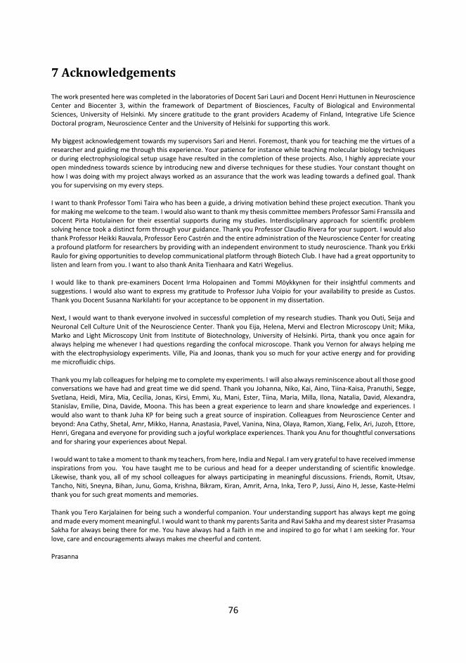
76
7 Acknowledgements The work presented here was completed in the laboratories of Docent Sari Lauri and Docent Henri Huttunen in Neuroscience Center and Biocenter 3, within the framework of Department of Biosciences, Faculty of Biological and Environmental Sciences, University of Helsinki. My sincere gratitude to the grant providers Academy of Finland, Integrative Life Science Doctoral program, Neuroscience Center and the University of Helsinki for supporting this work. My biggest acknowledgement towards my supervisors Sari and Henri. Foremost, thank you for teaching me the virtues of a researcher and guiding me through this experience. Your patience for instance while teaching molecular biology techniques or during electrophysiological setup usage have resulted in the completion of these projects. Also, I highly appreciate your open mindedness towards science by introducing new and diverse techniques for these studies. Your constant thought on how I was doing with my project always worked as an assurance that the work was leading towards a defined goal. Thank you for supervising on my every steps. I want to thank Professor Tomi Taira who has been a guide, a driving motivation behind these project execution. Thank you for making me welcome to the team. I would also want to thank my thesis committee members Professor Sami Franssila and Docent Pirta Hotulainen for their essential supports during my studies. Interdisciplinary approach for scientific problem solving hence took a distinct form through your guidance. Thank you Professor Claudio Rivera for your support. I would also thank Professor Heikki Rauvala, Professor Eero Castrén and the entire administration of the Neuroscience Center for creating a profound platform for researchers by providing with an independent environment to study neuroscience. Thank you Erkki Raulo for giving opportunities to develop communicational platform through Biotech Club. I have had a great opportunity to listen and learn from you. I want to also thank Anita Tienhaara and Katri Wegelius. I would like to thank pre-examiners Docent Irma Holopainen and Tommi Möykkynen for their insightful comments and suggestions. I would also want to express my gratitude to Professor Juha Voipio for your availability to preside as Custos. Thank you Docent Susanna Narkilahti for your acceptance to be opponent in my dissertation. Next, I would want to thank everyone involved in successful completion of my research studies. Thank you Outi, Seija and Neuronal Cell Culture Unit of the Neuroscience Center. Thank you Eija, Helena, Mervi and Electron Microscopy Unit; Mika, Marko and Light Microscopy Unit from Institute of Biotechnology, University of Helsinki. Pirta, thank you once again for always helping me whenever I had questions regarding the confocal microscope. Thank you Vernon for always helping me with the electrophysiology experiments. Ville, Pia and Joonas, thank you so much for your active energy and for providing me microfluidic chips. Thank you my lab colleagues for helping me to complete my experiments. I will also always reminiscence about all those good conversations we have had and great time we did spend. Thank you Johanna, Niko, Kai, Aino, Tiina-Kaisa, Pranuthi, Segge, Svetlana, Heidi, Mira, Mia, Cecilia, Jonas, Kirsi, Emmi, Xu, Mani, Ester, Tiina, Maria, Milla, Ilona, Natalia, David, Alexandra, Stanislav, Emilie, Dina, Davide, Moona. This has been a great experience to learn and share knowledge and experiences. I would also want to thank Juha KP for being such a great source of inspiration. Colleagues from Neuroscience Center and beyond: Ana Cathy, Shetal, Amr, Mikko, Hanna, Anastasia, Pavel, Vanina, Nina, Olaya, Ramon, Xiang, Felix, Ari, Juzoh, Ettore, Henri, Gregana and everyone for providing such a joyful workplace experiences. Thank you Anu for thoughtful conversations and for sharing your experiences about Nepal. I would want to take a moment to thank my teachers, from here, India and Nepal. I am very grateful to have received immense inspirations from you. You have taught me to be curious and head for a deeper understanding of scientific knowledge. Likewise, thank you, all of my school colleagues for always participating in meaningful discussions. Friends, Romit, Utsav, Tancho, Niti, Sneyna, Bihan, Junu, Goma, Krishna, Bikram, Kiran, Amrit, Arna, Inka, Tero P, Jussi, Aino H, Jesse, Kaste-Helmi thank you for such great moments and memories. Thank you Tero Karjalainen for being such a wonderful companion. Your understanding support has always kept me going and made every moment meaningful. I would want to thank my parents Sarita and Ravi Sakha and my dearest sister Prasamsa Sakha for always being there for me. You have always had a faith in me and inspired to go for what I am seeking for. Your love, care and encouragements always makes me cheerful and content. Prasanna

77
8 References Agrawal, S.G. & Evans, R.H., 1986. The primary afferent depolarizing action of kainate in the rat.
British journal of pharmacology, 87(2), pp.345–55. Ahmari, S.E., Buchanan, J. & Smith, S.J., 2000. Assembly of presynaptic active zones from cytoplasmic
transport packets. Nature neuroscience, 3(5), pp.445–51. Ali, A.B., 2003. Involvement of post-synaptic kainate receptors during synaptic transmission
between unitary connections in rat neocortex. The European journal of neuroscience, 17(11), pp.2344–50.
Ali, A.B. et al., 2001. Kainate receptors regulate unitary IPSCs elicited in pyramidal cells by fast-spiking interneurons in the neocortex. The Journal of neuroscience , 21(9), pp.2992–9.
Alsina, B., Vu, T. & Cohen-Cory, S., 2001. Visualizing synapse formation in arborizing optic axons in vivo: dynamics and modulation by BDNF. Nature neuroscience, 4(11), pp.1093–101.
Artinian, J. et al., 2011. Synaptic kainate receptors in interplay with INaP shift the sparse firing of dentate granule cells to a sustained rhythmic mode in temporal lobe epilepsy. The Journal of neuroscience , 31(30), pp.10811–8.
Awatramani, G.B., Price, G.D. & Trussell, L.O., 2005. Modulation of transmitter release by presynaptic resting potential and background calcium levels. Neuron, 48(1), pp.109–21.
Ayalon, G. & Stern-Bach, Y., 2001. Functional assembly of AMPA and kainate receptors is mediated by several discrete protein-protein interactions. Neuron, 31(1), pp.103–13.
Bagri, A. & Tessier-Lavigne, M., 2002. Neuropilins as Semaphorin receptors: in vivo functions in neuronal cell migration and axon guidance. Advances in experimental medicine and biology, 515, pp.13–31.
Bahn, S., Volk, B. & Wisden, W., 1994. Kainate receptor gene expression in the developing rat brain. The Journal of neuroscience, 14(9), pp.5525–47.
Barbon, A., Vallini, I. & Barlati, S., 2001. Genomic organization of the human GRIK2 gene and evidence for multiple splicing variants. Gene, 274(1-2), pp.187–97.
Berdichevsky, Y. et al., 2009. Microfluidics and multielectrode array-compatible organotypic slice culture method. Journal of neuroscience methods, 178(1), pp.59–64.
Bernard, A. et al., 1999. Q/R editing of the rat GluR5 and GluR6 kainate receptors in vivo and in vitro: evidence for independent developmental, pathological and cellular regulation. The European journal of neuroscience, 11(2), pp.604–16.
Berndt, A. et al., 2009. Bi-stable neural state switches. Nature neuroscience, 12(2), pp.229–34. Berthier, E. & Beebe, D.J., 2007. Flow rate analysis of a surface tension driven passive micropump.
Lab on a chip, 7(11), pp.1475–8. Bettler, B. et al., 1990. Cloning of a novel glutamate receptor subunit, GluR5: expression in the
nervous system during development. Neuron, 5(5), pp.583–95. Bettler, B. & Mulle, C., 1995. Review: neurotransmitter receptors. II. AMPA and kainate receptors.
Neuropharmacology, 34(2), pp.123–39. Biederer, T. et al., 2002. SynCAM, a synaptic adhesion molecule that drives synapse assembly.

78
Science (New York, N.Y.), 297(5586), pp.1525–31. Bolshakov, V.Y. & Siegelbaum, S.A., 1995. Regulation of hippocampal transmitter release during
development and long-term potentiation. Science (New York, N.Y.), 269(5231), pp.1730–4. Borgdorff, A.J. & Choquet, D., 2002. Regulation of AMPA receptor lateral movements. Nature,
417(6889), pp.649–53. Bortolotto, Z.A. et al., 1999. Kainate receptors are involved in synaptic plasticity. Nature, 402(6759),
pp.297–301. Bourne, J.N. & Harris, K.M., 2011. Coordination of size and number of excitatory and inhibitory
synapses results in a balanced structural plasticity along mature hippocampal CA1 dendrites during LTP. Hippocampus, 21(4), pp.354–73.
Bowie, D., 2002. External anions and cations distinguish between AMPA and kainate receptor gating mechanisms. The Journal of physiology, 539(Pt 3), pp.725–33.
Bowie, D., 2010. Ion-dependent gating of kainate receptors. The Journal of physiology, 588(Pt 1), pp.67–81.
Bowie, D. & Lange, G.D., 2002. Functional stoichiometry of glutamate receptor desensitization. The Journal of neuroscience, 22(9), pp.3392–403.
Boyden, E.S. et al., 2005. Millisecond-timescale, genetically targeted optical control of neural activity. Nature neuroscience, 8(9), pp.1263–8.
Bresler, T. et al., 2004. Postsynaptic density assembly is fundamentally different from presynaptic active zone assembly. The Journal of neuroscience, 24(6), pp.1507–20.
Bresler, T. et al., 2001. The dynamics of SAP90/PSD-95 recruitment to new synaptic junctions. Molecular and cellular neurosciences, 18(2), pp.149–67.
Brunello, C. a et al., 2013. Microtechnologies to fuel neurobiological research with nanometer precision. Journal of nanobiotechnology, 11, p.11.
Bureau, I. et al., 1999. Kainate receptor-mediated responses in the CA1 field of wild-type and GluR6-deficient mice. The Journal of neuroscience, 19(2), pp.653–63.
Bureau, I. et al., 2000. Kainate receptor-mediated synaptic currents in cerebellar Golgi cells are not shaped by diffusion of glutamate. Proceedings of the National Academy of Sciences of the United States of America, 97(12), pp.6838–43.
Burnashev, N. et al., 1995. Fractional calcium currents through recombinant GluR channels of the NMDA, AMPA and kainate receptor subtypes. The Journal of physiology, 485 ( Pt 2, pp.403–18.
Bähring, R. et al., 1997. Permeation and block of rat GluR6 glutamate receptor channels by internal and external polyamines. The Journal of physiology, 502 ( Pt 3, pp.575–89.
Campenot, R.B., 1977. Local control of neurite development by nerve growth factor. Proceedings of the National Academy of Sciences of the United States of America, 74(10), pp.4516–9.
Cardin, J.A. et al., 2009. Driving fast-spiking cells induces gamma rhythm and controls sensory responses. Nature, 459(7247), pp.663–7.
Carta, M. et al., 2013. CaMKII-dependent phosphorylation of GluK5 mediates plasticity of kainate receptors. The EMBO journal, 32(4), pp.496–510.
79
Carta, M. et al., 2014. Kainate receptors in the hippocampus. The European journal of neuroscience, 39(11), pp.1835–44.
Castillo, P.E., Malenka, R.C. & Nicoll, R.A., 1997. Kainate receptors mediate a slow postsynaptic current in hippocampal CA3 neurons. Nature, 388(6638), pp.182–6.
Chamberlain, S.E.L. et al., 2012. SUMOylation and phosphorylation of GluK2 regulate kainate receptor trafficking and synaptic plasticity. Nature neuroscience, 15(6), pp.845–52.
Chang, S. & De Camilli, P., 2001. Glutamate regulates actin-based motility in axonal filopodia. Nature neuroscience, 4(8), pp.787–793.
Chen, C. et al., 2012. The function of p120 catenin in filopodial growth and synaptic vesicle clustering in neurons. Molecular biology of the cell, 23(14), pp.2680–91.
Chergui, K. et al., 2000. Functional GluR6 kainate receptors in the striatum: indirect downregulation of synaptic transmission. The Journal of neuroscience, 20(6), pp.2175–82.
Chia, P.H. et al., 2013. Intramolecular regulation of presynaptic scaffold protein SYD-2/liprin-α. Molecular and cellular neurosciences, 56, pp.76–84.
Cho, K. et al., 2003. Regulation of kainate receptors by protein kinase C and metabotropic glutamate receptors. The Journal of physiology, 548(Pt 3), pp.723–30.
Ciocchi, S. et al., 2010. Encoding of conditioned fear in central amygdala inhibitory circuits. Nature, 468(7321), pp.277–82.
Clarke, V.R. et al., 1997. A hippocampal GluR5 kainate receptor regulating inhibitory synaptic transmission. Nature, 389(6651), pp.599–603.
Clarke, V.R.J. et al., 2014. Activity-dependent upregulation of presynaptic kainate receptors at immature CA3-CA1 synapses. The Journal of neuroscience, 34(50), pp.16902–16.
Clarke, V.R.J. et al., 2012. Synaptic kainate receptors in CA1 interneurons gate the threshold of theta-frequency-induced long-term potentiation. The Journal of neuroscience, 32(50), pp.18215–26.
Clarke, V.R.J. & Collingridge, G.L., 2002. Characterisation of the effects of ATPA, a GLU(K5) receptor selective agonist, on excitatory synaptic transmission in area CA1 of rat hippocampal slices. Neuropharmacology, 42(7), pp.889–902.
Clayton, E.L., Evans, G.J.O. & Cousin, M.A., 2007. Activity-dependent control of bulk endocytosis by protein dephosphorylation in central nerve terminals. The Journal of physiology, 585(Pt 3), pp.687–91.
Cohan, C.S. & Kater, S.B., 1986. Suppression of neurite elongation and growth cone motility by electrical activity. Science (New York, N.Y.), 232(4758), pp.1638–40.
Colgan, L.A. & Yasuda, R., 2014. Plasticity of dendritic spines: subcompartmentalization of signaling. Annual review of physiology, 76, pp.365–85.
Collingridge, G.L. et al., 2010. Long-term depression in the CNS. Nature reviews. Neuroscience, 11(7), pp.459–73.
Contractor, A. et al., 2003. Loss of kainate receptor-mediated heterosynaptic facilitation of mossy-fiber synapses in KA2-/- mice. The Journal of neuroscience, 23(2), pp.422–9.
Contractor, A., Mulle, C. & Swanson, G.T., 2011. Kainate receptors coming of age: Milestones of two decades of research. Trends in Neurosciences, 34(3), pp.154–163.

80
Contractor, A., Swanson, G. & Heinemann, S.F., 2001. Kainate receptors are involved in short- and long-term plasticity at mossy fiber synapses in the hippocampus. Neuron, 29(1), pp.209–16.
Copits, B.A. et al., 2011. Synaptic targeting and functional modulation of GluK1 kainate receptors by the auxiliary neuropilin and tolloid-like (NETO) proteins. The Journal of neuroscience, 31(20), pp.7334–40.
Cossart, R. et al., 1998. GluR5 kainate receptor activation in interneurons increases tonic inhibition of pyramidal cells. Nature neuroscience, 1(6), pp.470–8.
Cossart, R. et al., 2001. Presynaptic kainate receptors that enhance the release of GABA on CA1 hippocampal interneurons. Neuron, 29(2), pp.497–508.
Coussen, F. et al., 2005. Coassembly of two GluR6 kainate receptor splice variants within a functional protein complex. Neuron, 47(4), pp.555–566.
Coussen, F., 2009. Molecular determinants of kainate receptor trafficking. Neuroscience, 158(1), pp.25–35.
Coussen, F. et al., 2002. Recruitment of the kainate receptor subunit glutamate receptor 6 by cadherin/catenin complexes. The Journal of neuroscience, 22(15), pp.6426–36.
Covington, H.E. et al., 2010. Antidepressant effect of optogenetic stimulation of the medial prefrontal cortex. The Journal of neuroscience, 30(48), pp.16082–90.
Cui, C. & Mayer, M.L., 1999. Heteromeric kainate receptors formed by the coassembly of GluR5, GluR6, and GluR7. The Journal of neuroscience, 19(19), pp.8281–91.
Dalva, M.B. et al., 2000. EphB receptors interact with NMDA receptors and regulate excitatory synapse formation. Cell, 103(6), pp.945–56.
Darcy, K.J. et al., 2006. Constitutive sharing of recycling synaptic vesicles between presynaptic boutons. Nature neuroscience, 9(3), pp.315–21.
Dean, C. et al., 2003. Neurexin mediates the assembly of presynaptic terminals. Nature neuroscience, 6(7), pp.708–16.
Deisseroth, K. et al., 2006. Next-Generation Optical Technologies for Illuminating Genetically Targeted Brain Circuits. The Journal of Neuroscience, 26(41), pp.10380–10386.
Deisseroth, K., 2011. Optogenetics. Nature methods, 8(1), pp.26–9. Denker, A. & Rizzoli, S.O., 2010. Synaptic vesicle pools: an update. Frontiers in synaptic neuroscience,
2, p.135. DeVries, S.H. & Schwartz, E.A., 1999. Kainate receptors mediate synaptic transmission between
cones and “Off” bipolar cells in a mammalian retina. Nature, 397(6715), pp.157–60. Diefenbach, T.J., Guthrie, P.B. & Kater, S.B., 2000. Stimulus history alters behavioral responses of
neuronal growth cones. The Journal of neuroscience, 20(4), pp.1484–94. Dollé, J.-P. et al., 2013. An organotypic uniaxial strain model using microfluidics. Lab on a chip, 13(3),
pp.432–42. Eder, M. et al., 2003. Distribution and properties of functional postsynaptic kainate receptors on
neocortical layer V pyramidal neurons. The Journal of neuroscience, 23(16), pp.6660–70. Egebjerg, J. et al., 1991. Cloning of a cDNA for a glutamate receptor subunit activated by kainate but
not AMPA. Nature, 351(6329), pp.745–8.

81
Engel, D. & Jonas, P., 2005. Presynaptic action potential amplification by voltage-gated Na+ channels in hippocampal mossy fiber boutons. Neuron, 45(3), pp.405–17.
Epsztein, J. et al., 2005. Recurrent mossy fibers establish aberrant kainate receptor-operated synapses on granule cells from epileptic rats. The Journal of neuroscience, 25(36), pp.8229–39.
Fenno, L., Yizhar, O. & Deisseroth, K., 2011. The development and application of optogenetics. Annual review of neuroscience, 34, pp.389–412.
Fernandes, H.B. et al., 2009. High-affinity kainate receptor subunits are necessary for ionotropic but not metabotropic signaling. Neuron, 63(6), pp.818–29.
Fischer, M. et al., 1998. Rapid actin-based plasticity in dendritic spines. Neuron, 20(5), pp.847–54. Fisher, J.L. & Housley, P.R., 2013. Agonist binding to the GluK5 subunit is sufficient for functional
surface expression of heteromeric GluK2/GluK5 kainate receptors. Cellular and molecular neurobiology, 33(8), pp.1099–108.
Frerking, M., Malenka, R.C. & Nicoll, R.A., 1998. Synaptic activation of kainate receptors on hippocampal interneurons. Nature neuroscience, 1(6), pp.479–86.
Frerking, M. & Ohliger-Frerking, P., 2002. AMPA receptors and kainate receptors encode different features of afferent activity. The Journal of neuroscience, 22(17), pp.7434–43.
Freundt, E.C. et al., 2012. Neuron-to-neuron transmission of α-synuclein fibrils through axonal transport. Annals of neurology, 72(4), pp.517–24.
Gallyas, F., Ball, S.M. & Molnar, E., 2003. Assembly and cell surface expression of KA-2 subunit-containing kainate receptors. Journal of neurochemistry, 86(6), pp.1414–27.
Garcia, E.P. et al., 1998. SAP90 binds and clusters kainate receptors causing incomplete desensitization. Neuron, 21(4), pp.727–39.
Gareus, R. et al., 2006. Mouse profilin 2 regulates endocytosis and competes with SH3 ligand binding to dynamin 1. The Journal of biological chemistry, 281(5), pp.2803–11.
Garner, C.C. et al., 2002. Molecular mechanisms of CNS synaptogenesis. Trends in neurosciences, 25(5), pp.243–51.
Gelsomino, G. et al., 2013. Kainate induces mobilization of synaptic vesicles at the growth cone through the activation of protein kinase A. Cerebral cortex (New York, N.Y. : 1991), 23(3), pp.531–41.
González-González, I.M. et al., 2012. Kainate receptor trafficking. Wiley Interdisciplinary Reviews: Membrane Transport and Signaling, 1(1), pp.31–44.
Gradinaru, V. et al., 2009. Optical deconstruction of parkinsonian neural circuitry. Science (New York, N.Y.), 324(5925), pp.354–9.
Gradinaru, V., Thompson, K.R. & Deisseroth, K., 2008. eNpHR: a Natronomonas halorhodopsin enhanced for optogenetic applications. Brain cell biology, 36(1-4), pp.129–39.
Graf, E.R. et al., 2004. Neurexins induce differentiation of GABA and glutamate postsynaptic specializations via neuroligins. Cell, 119(7), pp.1013–26.
Gregor, P. et al., 1993. Expression and novel subunit isoforms of glutamate receptor genes GluR5 and GluR6. Neuroreport, 4(12), pp.1343–6.

82
Gunaydin, L.A. et al., 2010. Ultrafast optogenetic control. Nature Neuroscience, 13(3), pp.387–392. Hall, A.C., Lucas, F.R. & Salinas, P.C., 2000. Axonal remodeling and synaptic differentiation in the
cerebellum is regulated by WNT-7a signaling. Cell, 100(5), pp.525–35. Hammarback, J.A. et al., 1988. Growth cone guidance by substrate-bound laminin pathways is
correlated with neuron-to-pathway adhesivity. Developmental biology, 126(1), pp.29–39. Hansen, K.B., Furukawa, H. & Traynelis, S.F., 2010. Control of assembly and function of glutamate
receptors by the amino-terminal domain. Molecular pharmacology, 78(4), pp.535–49. Harris, K.M. & Landis, D.M., 1986. Membrane structure at synaptic junctions in area CA1 of the rat
hippocampus. Neuroscience, 19(3), pp.857–72. Harvey, C.D. & Svoboda, K., 2007. Locally dynamic synaptic learning rules in pyramidal neuron
dendrites. Nature, 450(7173), pp.1195–200. Hashimoto, K. & Kano, M., 2003. Functional differentiation of multiple climbing fiber inputs during
synapse elimination in the developing cerebellum. Neuron, 38(5), pp.785–96. Hayes, D.M. et al., 2003. Trafficking and surface expression of the glutamate receptor subunit, KA2.
Biochemical and biophysical research communications, 310(1), pp.8–13. Herb, A. et al., 1992. The KA-2 subunit of excitatory amino acid receptors shows widespread
expression in brain and forms ion channels with distantly related subunits. Neuron, 8(4), pp.775–85.
Hirbec, H. et al., 2003. Rapid and differential regulation of AMPA and kainate receptors at hippocampal mossy fibre synapses by PICK1 and GRIP. Neuron, 37(4), pp.625–638.
Hollmann, M., Maron, C. & Heinemann, S., 1994. N-glycosylation site tagging suggests a three transmembrane domain topology for the glutamate receptor GluR1. Neuron, 13(6), pp.1331–43.
Hotulainen, P. & Hoogenraad, C.C., 2010. Actin in dendritic spines: connecting dynamics to function. The Journal of cell biology, 189(4), pp.619–29.
Huang, K. & El-Husseini, A., 2005. Modulation of neuronal protein trafficking and function by palmitoylation. Current opinion in neurobiology, 15(5), pp.527–35.
Huang, Y.H. et al., 2004. Climbing fiber activation of EAAT4 transporters and kainate receptors in cerebellar Purkinje cells. The Journal of neuroscience, 24(1), pp.103–11.
Huberfeld, G. et al., 2007. Perturbed chloride homeostasis and GABAergic signaling in human temporal lobe epilepsy. The Journal of neuroscience, 27(37), pp.9866–73.
Huttenlocher, P.R. et al., 1982. Synaptogenesis in human visual cortex--evidence for synapse elimination during normal development. Neuroscience letters, 33(3), pp.247–52.
Ibarretxe, G. et al., 2007. Fast regulation of axonal growth cone motility by electrical activity. The Journal of neuroscience, 27(29), pp.7684–7695.
Ionescu-Zanetti, C. et al., 2005. Mammalian electrophysiology on a microfluidic platform. Proceedings of the National Academy of Sciences of the United States of America, 102(26), pp.9112–7.
Ivakine, E.A. et al., 2013. Neto2 is a KCC2 interacting protein required for neuronal Cl- regulation in hippocampal neurons. Proceedings of the National Academy of Sciences of the United States

83
of America, 110(9), pp.3561–6. Jaskolski, F., Normand, E., et al., 2005. Differential trafficking of GluR7 kainate receptor subunit
splice variants. The Journal of biological chemistry, 280(24), pp.22968–76. Jaskolski, F. et al., 2004. Subunit composition and alternative splicing regulate membrane delivery
of kainate receptors. The Journal of neuroscience, 24(10), pp.2506–2515. Jaskolski, F., Coussen, F. & Mulle, C., 2005. Subcellular localization and trafficking of kainate
receptors. Trends in pharmacological sciences, 26(1), pp.20–6. Jiang, L. et al., 2001. A kainate receptor increases the efficacy of GABAergic synapses. Neuron, 30(2),
pp.503–13. Jokinen, V. et al., 2013. A microfluidic chip for axonal isolation and electrophysiological
measurements. Journal of Neuroscience Methods, 212(2), pp.276–282. Joseph, D.J., Williams, D.J. & MacDermott, A.B., 2011. Modulation of neurite outgrowth by activation
of calcium-permeable kainate receptors expressed by rat nociceptive-like dorsal root ganglion neurons. Developmental Neurobiology, 71(10), pp.818–835.
Jouhanneau, J.-S. et al., 2011. Mechanisms of bi-directional modulation of thalamocortical transmission in barrel cortex by presynaptic kainate receptors. Neuropharmacology, 60(6), pp.832–41.
Juuri, J. et al., 2010. Kainate receptor-induced ectopic spiking of CA3 pyramidal neurons initiates network bursts in neonatal hippocampus. Journal of neurophysiology, 104(3), pp.1696–706.
Jähne, S., Rizzoli, S.O. & Helm, M.S., 2015. The structure and function of presynaptic endosomes. Experimental cell research, 335(2), pp.172–9.
Kamiya, H. et al., 2002. Presynaptic Ca2+ entry is unchanged during hippocampal mossy fiber long-term potentiation. The Journal of neuroscience, 22(24), pp.10524–8.
Kamiya, H. & Ozawa, S., 1998. Kainate receptor-mediated inhibition of presynaptic Ca2+ influx and EPSP in area CA1 of the rat hippocampus. The Journal of physiology, 509 ( Pt 3, pp.833–45.
Kask, K. et al., 2000. Developmental profile of kainate receptor subunit KA1 revealed by Cre expression in YAC transgenic mice. Brain research, 876(1-2), pp.55–61.
Kayadjanian, N. et al., 2007. Localization of glutamate receptors to distal dendrites depends on subunit composition and the kinesin motor protein KIF17. Molecular and cellular neurosciences, 34(2), pp.219–30.
Kelsch, W., Sim, S. & Lois, C., 2010. Watching synaptogenesis in the adult brain. Annual review of neuroscience, 33, pp.131–49.
Kidd, F.L. et al., 2002. A presynaptic kainate receptor is involved in regulating the dynamic properties of thalamocortical synapses during development. Neuron, 34(4), pp.635–46.
Kidd, F.L. & Isaac, J.T., 1999. Developmental and activity-dependent regulation of kainate receptors at thalamocortical synapses. Nature, 400(6744), pp.569–73.
Kim, H.J. et al., 2012. Integrated microfluidics platforms for investigating injury and regeneration of CNS axons. Annals of biomedical engineering, 40(6), pp.1268–76.
Kjarland, E., Keen, T.J. & Kleppe, R., 2006. Does isoform diversity explain functional differences in the 14-3-3 protein family? Current pharmaceutical biotechnology, 7(3), pp.217–23.
84
Kornreich, B.G. et al., 2007. Identification of C-terminal domain residues involved in protein kinase A-mediated potentiation of kainate receptor subtype 6. Neuroscience, 146(3), pp.1158–68.
Kraszewski, K. et al., 1995. Synaptic vesicle dynamics in living cultured hippocampal neurons visualized with CY3-conjugated antibodies directed against the lumenal domain of synaptotagmin. The Journal of neuroscience, 15(6), pp.4328–42.
Krueger, S.R., Kolar, A. & Fitzsimonds, R.M., 2003. The presynaptic release apparatus is functional in the absence of dendritic contact and highly mobile within isolated axons. Neuron, 40(5), pp.945–57.
Köhler, M. et al., 1993. Determinants of Ca2+ permeability in both TM1 and TM2 of high affinity kainate receptor channels: diversity by RNA editing. Neuron, 10(3), pp.491–500.
Lamsa, K. et al., 2000. Synaptic GABA(A) activation inhibits AMPA-kainate receptor-mediated bursting in the newborn (P0-P2) rat hippocampus. Journal of neurophysiology, 83(1), pp.359–66.
Landis, D.M., 1988. Membrane and cytoplasmic structure at synaptic junctions in the mammalian central nervous system. Journal of electron microscopy technique, 10(2), pp.129–51.
Lanore, F. et al., 2012. Deficits in morphofunctional maturation of hippocampal mossy fiber synapses in a mouse model of intellectual disability. The Journal of neuroscience, 32(49), pp.17882–93.
Lauri, S. & Taira, T., 2012. Kainate receptors in developing presynaptic terminals. Wiley Interdisciplinary Reviews: Membrane Transport and Signaling, 1(1), pp.45–55.
Lauri, S.E. et al., 2001. A critical role of a facilitatory presynaptic kainate receptor in mossy fiber LTP. Neuron, 32(4), pp.697–709.
Lauri, S.E. et al., 2003. A role for Ca2+ stores in kainate receptor-dependent synaptic facilitation and LTP at mossy fiber synapses in the hippocampus. Neuron, 39(2), pp.327–41.
Lauri, S.E. et al., 2005. Endogenous activation of kainate receptors regulates glutamate release and network activity in the developing hippocampus. The Journal of neuroscience, 25(18), pp.4473–84.
Lauri, S.E. et al., 2006. Functional Maturation of CA1 Synapses Involves Activity-Dependent Loss of Tonic Kainate Receptor-Mediated Inhibition of Glutamate Release. Neuron, 50(3), pp.415–429.
Lauri, S.E. & Taira, T., 2011. Role of kainate receptors in network activity during development. Advances in Experimental Medicine and Biology, 717, pp.81–91.
Lee, C.J. et al., 2001. Kainate receptors expressed by a subpopulation of developing nociceptors rapidly switch from high to low Ca2+ permeability. The Journal of neuroscience, 21(13), pp.4572–81.
Lee, R.C. et al., 2003. The protocadherin Flamingo is required for axon target selection in the Drosophila visual system. Nature neuroscience, 6(6), pp.557–63.
Lerma, J., 2003. Roles and rules of kainate receptors in synaptic transmission. Nature reviews. Neuroscience, 4(6), pp.481–95.
Lerma, J. & Marques, J.M., 2013. Kainate receptors in health and disease. Neuron, 80(2), pp.292–311.

85
Li, H. & Rogawski, M.A., 1998. GluR5 kainate receptor mediated synaptic transmission in rat basolateral amygdala in vitro. Neuropharmacology, 37(10-11), pp.1279–86.
Li, J.-M. et al., 2010. Aberrant glutamate receptor 5 expression in temporal lobe epilepsy lesions. Brain research, 1311, pp.166–74.
Maas, C. et al., 2012. Formation of Golgi-derived active zone precursor vesicles. The Journal of neuroscience, 32(32), pp.11095–108.
Mabb, A.M. & Ehlers, M.D., 2010. Ubiquitination in postsynaptic function and plasticity. Annual review of cell and developmental biology, 26, pp.179–210.
Mah, S.J. et al., 2005. Glutamate receptor trafficking: endoplasmic reticulum quality control involves ligand binding and receptor function. The Journal of neuroscience, 25(9), pp.2215–25.
Mahadevan, V. et al., 2014. Kainate Receptors Coexist in a Functional Complex with KCC2 and Regulate Chloride Homeostasis in Hippocampal Neurons. Cell Reports, 7(6), pp.1762–1770.
Marques, J.M. et al., 2013. CRMP2 Tethers Kainate Receptor Activity to Cytoskeleton Dynamics during Neuronal Maturation. Journal of Neuroscience, 33(46), pp.18298–18310.
Marrs, G.S., Green, S.H. & Dailey, M.E., 2001. Rapid formation and remodeling of postsynaptic densities in developing dendrites. Nature neuroscience, 4(10), pp.1006–13.
Martin, S. et al., 2008. Bidirectional regulation of kainate receptor surface expression in hippocampal neurons. The Journal of biological chemistry, 283(52), pp.36435–40.
Martin, S. et al., 2007. SUMOylation regulates kainate-receptor-mediated synaptic transmission. Nature, 447(7142), pp.321–5.
Martin, S. & Henley, J.M., 2004. Activity-dependent endocytic sorting of kainate receptors to recycling or degradation pathways. The EMBO journal, 23(24), pp.4749–59.
Martina, M. et al., 2011. Recordings of cultured neurons and synaptic activity using patch-clamp chips. Journal of neural engineering, 8(3), p.034002.
Matsuzaki, M. et al., 2004. Structural basis of long-term potentiation in single dendritic spines. Nature, 429(6993), pp.761–6.
Mattson, M.P., 1992. Calcium as sculptor and destroyer of neural circuitry. Experimental gerontology, 27(1), pp.29–49.
Mattson, M.P., 2008. Glutamate and Neurotrophic Factors in Neuronal Plasticity and Disease. Annals of the New York Academy of Sciences, 1144(1), pp.97–112.
Mattson, M.P., Lee, R.E., et al., 1988. Interactions between entorhinal axons and target hippocampal neurons: a role for glutamate in the development of hippocampal circuitry. Neuron, 1(9), pp.865–76.
Mattson, M.P., Dou, P. & Kater, S.B., 1988. Outgrowth-regulating actions of glutamate in isolated hippocampal pyramidal neurons. The Journal of neuroscience, 8(6), pp.2087–100.
McDonald, J.C. & Whitesides, G.M., 2002. Poly(dimethylsiloxane) as a material for fabricating microfluidic devices. Accounts of chemical research, 35(7), pp.491–9.
Mehta, S. et al., 2001. Molecular mechanisms regulating the differential association of kainate receptor subunits with SAP90/PSD-95 and SAP97. The Journal of biological chemistry, 276(19), pp.16092–9.

86
Melyan, Z., Wheal, H. V & Lancaster, B., 2002. Metabotropic-mediated kainate receptor regulation of IsAHP and excitability in pyramidal cells. Neuron, 34(1), pp.107–14.
Mi, R. et al., 2002. Differing mechanisms for glutamate receptor aggregation on dendritic spines and shafts in cultured hippocampal neurons. The Journal of neuroscience, 22(17), pp.7606–16.
Min, M.Y., Melyan, Z. & Kullmann, D.M., 1999. Synaptically released glutamate reduces gamma-aminobutyric acid (GABA)ergic inhibition in the hippocampus via kainate receptors. Proceedings of the National Academy of Sciences of the United States of America, 96(17), pp.9932–7.
Molnár, Z. & Blakemore, C., 1999. Development of signals influencing the growth and termination of thalamocortical axons in organotypic culture. Experimental neurology, 156(2), pp.363–93.
Mondin, M. et al., 2010. Profilin II regulates the exocytosis of kainate glutamate receptors. The Journal of biological chemistry, 285(51), pp.40060–71.
Morris, R.L. & Hollenbeck, P.J., 1993. The regulation of bidirectional mitochondrial transport is coordinated with axonal outgrowth. Journal of cell science, 104 ( Pt 3, pp.917–27.
Mulle, C. et al., 1998. Altered synaptic physiology and reduced susceptibility to kainate-induced seizures in GluR6-deficient mice. Nature, 392(6676), pp.601–5.
Murai, K.K. et al., 2003. Control of hippocampal dendritic spine morphology through ephrin-A3/EphA4 signaling. Nature neuroscience, 6(2), pp.153–60.
Nagel, G. et al., 2002. Channelrhodopsin-1: a light-gated proton channel in green algae. Science (New York, N.Y.), 296(5577), pp.2395–8.
Nagel, G. et al., 2003. Channelrhodopsin-2, a directly light-gated cation-selective membrane channel. Proceedings of the National Academy of Sciences of the United States of America, 100(24), pp.13940–5.
Nagel, G. et al., 2005. Light activation of channelrhodopsin-2 in excitable cells of Caenorhabditis elegans triggers rapid behavioral responses. Current biology : CB, 15(24), pp.2279–84.
Nasu-Nishimura, Y. et al., 2010. Differential regulation of kainate receptor trafficking by phosphorylation of distinct sites on GluR6. The Journal of biological chemistry, 285(4), pp.2847–56.
Ng, D. et al., 2009. Neto1 is a novel CUB-domain NMDA receptor-interacting protein required for synaptic plasticity and learning. PLoS biology, 7(2), p.e41.
Nicoll, R.A. & Roche, K.W., 2013. Long-term potentiation: peeling the onion. Neuropharmacology, 74, pp.18–22.
NP_036704.1, N.R.S., glutamate receptor ionotropic, kainate 4 precursor [Rattus norvegicus] - Protein - NCBI.
NP_113696.1, N.R.S., glutamate receptor ionotropic, kainate 5 precursor [Rattus norvegicus] - Protein - NCBI.
Nägler, K., Mauch, D.H. & Pfrieger, F.W., 2001. Glia-derived signals induce synapse formation in neurones of the rat central nervous system. The Journal of physiology, 533(Pt 3), pp.665–79.
O’Brien, R.J. et al., 1999. Synaptic clustering of AMPA receptors by the extracellular immediate-early gene product Narp. Neuron, 23(2), pp.309–23.

87
Ohtsuka, T. et al., 2002. Cast: a novel protein of the cytomatrix at the active zone of synapses that forms a ternary complex with RIM1 and munc13-1. The Journal of cell biology, 158(3), pp.577–90.
Okabe, S. et al., 2001. Rapid redistribution of the postsynaptic density protein PSD-Zip45 (Homer 1c) and its differential regulation by NMDA receptors and calcium channels. The Journal of neuroscience, 21(24), pp.9561–71.
Opazo, F. et al., 2010. Limited intermixing of synaptic vesicle components upon vesicle recycling. Traffic (Copenhagen, Denmark), 11(6), pp.800–12.
Overly, C.C., Rieff, H.I. & Hollenbeck, P.J., 1996. Organelle motility and metabolism in axons vs dendrites of cultured hippocampal neurons. Journal of cell science, 109 ( Pt 5, pp.971–80.
Palacios-Filardo, J., Aller, M.I. & Lerma, J., 2014. Synaptic Targeting of Kainate Receptors. Cerebral cortex (New York, N.Y. : 1991).
Park, J. et al., 2012. Multi-compartment neuron-glia co-culture platform for localized CNS axon-glia interaction study. Lab on a chip, 12(18), pp.3296–304.
Park, J.W. et al., 2006. Microfluidic culture platform for neuroscience research. Nature protocols, 1(4), pp.2128–36.
Park, Y. et al., 2006. Long-term depression of kainate receptor-mediated synaptic transmission. Neuron, 49(1), pp.95–106.
Pascual, M. et al., 2004. Coordinated functions of Netrin-1 and Class 3 secreted Semaphorins in the guidance of reciprocal septohippocampal connections. Molecular and cellular neurosciences, 26(1), pp.24–33.
Passafaro, M., Piëch, V. & Sheng, M., 2001. Subunit-specific temporal and spatial patterns of AMPA receptor exocytosis in hippocampal neurons. Nature neuroscience, 4(9), pp.917–26.
Paternain, A. V, Morales, M. & Lerma, J., 1995. Selective antagonism of AMPA receptors unmasks kainate receptor-mediated responses in hippocampal neurons. Neuron, 14(1), pp.185–9.
Pei, D.-S. et al., 2006. Neuroprotection against ischaemic brain injury by a GluR6-9c peptide containing the TAT protein transduction sequence. Brain : a journal of neurology, 129(Pt 2), pp.465–79.
Penzes, P. et al., 2003. Rapid induction of dendritic spine morphogenesis by trans-synaptic ephrinB-EphB receptor activation of the Rho-GEF kalirin. Neuron, 37(2), pp.263–74.
Pickering, D.S. et al., 1995. Palmitoylation of the GluR6 kainate receptor. Proceedings of the National Academy of Sciences of the United States of America, 92(26), pp.12090–4.
Pierce, J.P. & Lewin, G.R., 1994. An ultrastructural size principle. Neuroscience, 58(3), pp.441–6. Pinheiro, P. & Mulle, C., 2006. Kainate receptors. Cell and Tissue Research, 326(2), pp.457–482. Pinheiro, P.S. et al., 2007. GluR7 is an essential subunit of presynaptic kainate autoreceptors at
hippocampal mossy fiber synapses. Proceedings of the National Academy of Sciences of the United States of America, 104(29), pp.12181–6.
Pinheiro, P.S. et al., 2005. Presynaptic kainate receptors are localized close to release sites in rat hippocampal synapses. Neurochemistry international, 47(5), pp.309–16.
Plested, A.J.R. et al., 2008. Molecular basis of kainate receptor modulation by sodium. Neuron, 58(5),

88
pp.720–35. Plested, A.J.R. & Mayer, M.L., 2007. Structure and mechanism of kainate receptor modulation by
anions. Neuron, 53(6), pp.829–41. Ponimaskin, E. et al., 2007. Morphogenic signaling in neurons via neurotransmitter receptors and
small GTPases. Molecular neurobiology, 35(3), pp.278–87. Rao, A. & Craig, A.M., 1997. Activity regulates the synaptic localization of the NMDA receptor in
hippocampal neurons. Neuron, 19(4), pp.801–12. Raymond, L.A., Blackstone, C.D. & Huganir, R.L., 1993. Phosphorylation and modulation of
recombinant GluR6 glutamate receptors by cAMP-dependent protein kinase. Nature, 361(6413), pp.637–41.
Ren, Z., Riley, N.J., et al., 2003. Cell Surface Expression of GluR5 Kainate Receptors is Regulated by an Endoplasmic Reticulum Retention Signal. Journal of Biological Chemistry, 278(52), pp.52700–52709.
Ren, Z., Riley, N.J., et al., 2003. Multiple trafficking signals regulate kainate receptor KA2 subunit surface expression. The Journal of neuroscience, 23(16), pp.6608–6616.
Represa, A., Tremblay, E. & Ben-Ari, Y., 1987. Kainate binding sites in the hippocampal mossy fibers: localization and plasticity. Neuroscience, 20(3), pp.739–48.
Resto, P.J. et al., 2010. An automated microdroplet passive pumping platform for high-speed and packeted microfluidic flow applications. Lab on a chip, 10(1), pp.23–6.
Ritter, L.M., Vazquez, D.M. & Meador-Woodruff, J.H., 2002. Ontogeny of ionotropic glutamate receptor subunit expression in the rat hippocampus. Brain research. Developmental brain research, 139(2), pp.227–36.
Rivera, R., Rozas, J.L. & Lerma, J., 2007. PKC-dependent autoregulation of membrane kainate receptors. The EMBO journal, 26(20), pp.4359–67.
Rizzoli, S.O. & Betz, W.J., 2005. Synaptic vesicle pools. Nature reviews. Neuroscience, 6(1), pp.57–69.
Rodríguez-Moreno, A. & Lerma, J., 1998. Kainate receptor modulation of GABA release involves a metabotropic function. Neuron, 20(6), pp.1211–8.
Rodríguez-Moreno, A. & Sihra, T.S., 2007. Kainate receptors with a metabotropic modus operandi. Trends in neurosciences, 30(12), pp.630–7.
Rozas, J.L., Paternain, A. V & Lerma, J., 2003. Noncanonical signaling by ionotropic kainate receptors. Neuron, 39(3), pp.543–53.
Ruiz, A. et al., 2005. Distinct subunits in heteromeric kainate receptors mediate ionotropic and metabotropic function at hippocampal mossy fiber synapses. The Journal of neuroscience, 25(50), pp.11710–8.
Rutkowska-Wlodarczyk, I. et al., 2015. A proteomic analysis reveals the interaction of GluK1 ionotropic kainate receptor subunits with Go proteins. The Journal of neuroscience, 35(13), pp.5171–9.
Sachidhanandam, S. et al., 2009. Kainate receptors act as conditional amplifiers of spike transmission at hippocampal mossy fiber synapses. The Journal of neuroscience, 29(15), pp.5000–8.

89
Saheki, Y. & De Camilli, P., 2012. Synaptic Vesicle Endocytosis. Cold Spring Harbor Perspectives in Biology, 4(9), pp.a005645–a005645. Available at: http://www.pubmedcentral.nih.gov/articlerender.fcgi?artid=3428771&tool=pmcentrez&rendertype=abstract.
Sakha, P. et al., 2013. Asymmetric Genetic Manipulation and Patch Clamp Recording of Neurons in a Microfluidic Chip. In E. Biffi, ed. Microfluidic and Compartmentalized Platforms for Neurobiological Research. Neuromethods 103, Springer Protocols, Humana Press, pp. 70–92.
Salin, P.A. et al., 1996. Distinct short-term plasticity at two excitatory synapses in the hippocampus. Proceedings of the National Academy of Sciences of the United States of America, 93(23), pp.13304–9.
Salinas, G.D. et al., 2006. Actinfilin is a Cul3 substrate adaptor, linking GluR6 kainate receptor subunits to the ubiquitin-proteasome pathway. The Journal of biological chemistry, 281(52), pp.40164–73.
Sallert, M. et al., 2009. Brain-derived neurotrophic factor controls activity-dependent maturation of CA1 synapses by downregulating tonic activation of presynaptic kainate receptors. The Journal of neuroscience, 29(36), pp.11294–303.
Sallert, M. et al., 2007. Effects of the kainate receptor agonist ATPA on glutamatergic synaptic transmission and plasticity during early postnatal development. Neuropharmacology, 52(6), pp.1354–65.
Savinainen, A. et al., 2001. Kainate receptor activation induces mixed lineage kinase-mediated cellular signaling cascades via post-synaptic density protein 95. The Journal of biological chemistry, 276(14), pp.11382–6.
Scheiffele, P., 2003. Cell-cell signaling during synapse formation in the CNS. Annual review of neuroscience, 26, pp.485–508.
Scheiffele, P. et al., 2000. Neuroligin expressed in nonneuronal cells triggers presynaptic development in contacting axons. Cell, 101(6), pp.657–69.
Schiffer, H.H., Swanson, G.T. & Heinemann, S.F., 1997. Rat GluR7 and a carboxy-terminal splice variant, GluR7b, are functional kainate receptor subunits with a low sensitivity to glutamate. Neuron, 19(5), pp.1141–6.
Schikorski, T. & Stevens, C.F., 1997. Quantitative ultrastructural analysis of hippocampal excitatory synapses. The Journal of neuroscience, 17(15), pp.5858–67.
Schmitz, D. et al., 2001. Presynaptic kainate receptors at hippocampal mossy fiber synapses. Proceedings of the National Academy of Sciences of the United States of America, 98(20), pp.11003–8.
Scott, D.B. et al., 2001. An NMDA receptor ER retention signal regulated by phosphorylation and alternative splicing. The Journal of neuroscience, 21(9), pp.3063–72.
Segerstråle, M. et al., 2010. High firing rate of neonatal hippocampal interneurons is caused by attenuation of afterhyperpolarizing potassium currents by tonically active kainate receptors. The Journal of neuroscience, 30(19), pp.6507–14.
Selak, S. et al., 2009. A role for SNAP25 in internalization of kainate receptors and synaptic plasticity.
90
Neuron, 63(3), pp.357–71. Semyanov, A. & Kullmann, D.M., 2001. Kainate receptor-dependent axonal depolarization and
action potential initiation in interneurons. Nature neuroscience, 4(7), pp.718–23. Shapira, M. et al., 2003. Unitary assembly of presynaptic active zones from Piccolo-Bassoon
transport vesicles. Neuron, 38(2), pp.237–52. Shapiro, L. & Colman, D.R., 1999. The diversity of cadherins and implications for a synaptic adhesive
code in the CNS. Neuron, 23(3), pp.427–30. Shen, K. & Meyer, T., 1999. Dynamic control of CaMKII translocation and localization in hippocampal
neurons by NMDA receptor stimulation. Science (New York, N.Y.), 284(5411), pp.162–6. Shepherd, G.M. & Harris, K.M., 1998. Three-dimensional structure and composition of CA3-->CA1
axons in rat hippocampal slices: implications for presynaptic connectivity and compartmentalization. The Journal of neuroscience, 18(20), pp.8300–10.
Shin, R.M., Higuchi, M. & Suhara, T., 2013. Nitric Oxide Signaling Exerts Bidirectional Effects on Plasticity Inductions in Amygdala. PLoS ONE, 8(9), pp.1–15.
Shingai, T. et al., 2003. Implications of nectin-like molecule-2/IGSF4/RA175/SgIGSF/TSLC1/SynCAM1 in cell-cell adhesion and transmembrane protein localization in epithelial cells. The Journal of biological chemistry, 278(37), pp.35421–7.
Shtrahman, M. et al., 2005. Probing vesicle dynamics in single hippocampal synapses. Biophysical journal, 89(5), pp.3615–27.
Sihra, T.S., Flores, G. & Rodríguez-Moreno, A., 2013. Kainate Receptors: Multiple Roles in Neuronal Plasticity. The Neuroscientist : a review journal bringing neurobiology, neurology and psychiatry.
Sommer, B. et al., 1992. A glutamate receptor channel with high affinity for domoate and kainate. The EMBO journal, 11(4), pp.1651–6.
Standley, S. et al., 2000. PDZ domain suppression of an ER retention signal in NMDA receptor NR1 splice variants. Neuron, 28(3), pp.887–98.
Straub, C., Hunt, D.L., et al., 2011. Distinct functions of kainate receptors in the brain are determined by the auxiliary subunit Neto1. Nature neuroscience, 14(7), pp.866–73.
Straub, C., Zhang, W. & Howe, J.R., 2011. Neto2 modulation of kainate receptors with different subunit compositions. The Journal of neuroscience, 31(22), pp.8078–82.
Sun, Y. & Bamji, S.X., 2011. β-Pix modulates actin-mediated recruitment of synaptic vesicles to synapses. The Journal of neuroscience, 31(47), pp.17123–33.
Suzuki, I. et al., 2004. Modification of a neuronal network direction using stepwise photo-thermal etching of an agarose architecture. Journal of nanobiotechnology, 2(1), p.7.
Suzuki, I. et al., 2005. Stepwise pattern modification of neuronal network in photo-thermally-etched agarose architecture on multi-electrode array chip for individual-cell-based electrophysiological measurement. Lab on a chip, 5(3), pp.241–7.
Südhof, T.C., 1995. The synaptic vesicle cycle: a cascade of protein-protein interactions. Nature, 375(6533), pp.645–53.
Takai, Y., Shimizu, K. & Ohtsuka, T., 2003. The roles of cadherins and nectins in interneuronal synapse

91
formation. Current opinion in neurobiology, 13(5), pp.520–6. Takamori, S. et al., 2006. Molecular anatomy of a trafficking organelle. Cell, 127(4), pp.831–46. Takeuchi, A. et al., 2011. Autonomic nervous system driven cardiomyocytes in vitro. Conference
proceedings : ... Annual International Conference of the IEEE Engineering in Medicine and Biology Society. IEEE Engineering in Medicine and Biology Society. Annual Conference, 2011, pp.1945–8.
Tang, M. et al., 2011. Neto1 is an auxiliary subunit of native synaptic kainate receptors. The Journal of neuroscience, 31(27), pp.10009–18.
Tashiro, A. et al., 2003. Bidirectional regulation of hippocampal mossy fiber filopodial motility by kainate receptors: A two-step model of synaptogenesis. Neuron, 38(5), pp.773–784.
Taylor, A.M. et al., 2005. A microfluidic culture platform for CNS axonal injury, regeneration and transport. Nature methods, 2(8), pp.599–605.
Taylor, A.M. et al., 2010. Microfluidic local perfusion chambers for the visualization and manipulation of synapses. Neuron, 66(1), pp.57–68.
Taylor, A.M. et al., 2003. Microfluidic Multicompartment Device for Neuroscience Research. Langmuir : the ACS journal of surfaces and colloids, 19(5), pp.1551–1556.
Tom Dieck, S. et al., 1998. Bassoon, a novel zinc-finger CAG/glutamine-repeat protein selectively localized at the active zone of presynaptic nerve terminals. The Journal of cell biology, 142(2), pp.499–509.
Traynelis, S.F. & Wahl, P., 1997. Control of rat GluR6 glutamate receptor open probability by protein kinase A and calcineurin. The Journal of physiology, 503 ( Pt 3, pp.513–31.
Tønnesen, J. et al., 2009. Optogenetic control of epileptiform activity. Proceedings of the National Academy of Sciences of the United States of America, 106(29), pp.12162–7.
Ullian, E.M. et al., 2001. Control of synapse number by glia. Science (New York, N.Y.), 291(5504), pp.657–61.
Umemori, H. et al., 2004. FGF22 and its close relatives are presynaptic organizing molecules in the mammalian brain. Cell, 118(2), pp.257–70.
Valluru, L. et al., 2005. Ligand binding is a critical requirement for plasma membrane expression of heteromeric kainate receptors. The Journal of biological chemistry, 280(7), pp.6085–93.
Varoqueaux, F. et al., 2002. Total arrest of spontaneous and evoked synaptic transmission but normal synaptogenesis in the absence of Munc13-mediated vesicle priming. Proceedings of the National Academy of Sciences of the United States of America, 99(13), pp.9037–42.
Vaughn, J.E., 1989. Fine structure of synaptogenesis in the vertebrate central nervous system. Synapse (New York, N.Y.), 3(3), pp.255–85.
Verhage, M. et al., 2000. Synaptic assembly of the brain in the absence of neurotransmitter secretion. Science (New York, N.Y.), 287(5454), pp.864–9.
Vesikansa, A. et al., 2007. Activation of kainate receptors controls the number of functional glutamatergic synapses in the area CA1 of rat hippocampus. The Journal of physiology, 583(Pt 1), pp.145–157.
Vignes, M. et al., 1998. The GluR5 subtype of kainate receptor regulates excitatory synaptic

92
transmission in areas CA1 and CA3 of the rat hippocampus. Neuropharmacology, 37(10-11), pp.1269–77.
Vignes, M. & Collingridge, G.L., 1997. The synaptic activation of kainate receptors. Nature, 388(6638), pp.179–82.
Vivithanaporn, P. et al., 2007. Critical roles for the M3-S2 transduction linker domain in kainate receptor assembly and postassembly trafficking. The Journal of neuroscience, 27(39), pp.10423–33.
Vivithanaporn, P., Yan, S. & Swanson, G.T., 2006. Intracellular trafficking of KA2 kainate receptors mediated by interactions with coatomer protein complex I (COPI) and 14-3-3 chaperone systems. The Journal of biological chemistry, 281(22), pp.15475–84.
Waites, C.L., Craig, A.M. & Garner, C.C., 2005. Mechanisms of vertebrate synaptogenesis. Annual review of neuroscience, 28, pp.251–74.
Wang, X. & Schwarz, T.L., 2009. The mechanism of Ca2+ -dependent regulation of kinesin-mediated mitochondrial motility. Cell, 136(1), pp.163–74.
Washbourne, P., Bennett, J.E. & McAllister, A.K., 2002. Rapid recruitment of NMDA receptor transport packets to nascent synapses. Nature neuroscience, 5(8), pp.751–9.
Watanabe, S. et al., 2013. Ultrafast endocytosis at mouse hippocampal synapses. Nature, 504(7479), pp.242–7.
Weisskopf, M.G. & Nicoll, R.A., 1995. Presynaptic changes during mossy fibre LTP revealed by NMDA receptor-mediated synaptic responses. Nature, 376(6537), pp.256–9.
Werner, P. et al., 1991. Cloning of a putative high-affinity kainate receptor expressed predominantly in hippocampal CA3 cells. Nature, 351(6329), pp.742–4.
Westphal, V. et al., 2008. Video-rate far-field optical nanoscopy dissects synaptic vesicle movement. Science (New York, N.Y.), 320(5873), pp.246–9.
Wilding, T.J. & Huettner, J.E., 1995. Differential antagonism of alpha-amino-3-hydroxy-5-methyl-4- isoxazolepropionic acid-preferring and kainate-preferring receptors by 2,3-benzodiazepines. Molecular pharmacology, 47(3), pp.582–7.
Wilkinson, K.A. & Henley, J.M., 2010. Mechanisms, regulation and consequences of protein SUMOylation. The Biochemical journal, 428(2), pp.133–45.
Wo, Z.G. & Oswald, R.E., 1994. Transmembrane topology of two kainate receptor subunits revealed by N-glycosylation. Proceedings of the National Academy of Sciences of the United States of America, 91(15), pp.7154–8.
Wong, A.Y.C., MacLean, D.M. & Bowie, D., 2007. Na+/Cl- dipole couples agonist binding to kainate receptor activation. The Journal of neuroscience, 27(25), pp.6800–9.
Wong, L.A. & Mayer, M.L., 1993. Differential modulation by cyclothiazide and concanavalin A of desensitization at native alpha-amino-3-hydroxy-5-methyl-4-isoxazolepropionic acid- and kainate-preferring glutamate receptors. Molecular pharmacology, 44(3), pp.504–10.
Wong, W.T. & Wong, R.O., 2001. Changing specificity of neurotransmitter regulation of rapid dendritic remodeling during synaptogenesis. Nature neuroscience, 4(4), pp.351–2.
Wu, H.-I. et al., 2010. A lab-on-a-chip platform for studying the subcellular functional proteome of
93
neuronal axons. Lab on a chip, 10(5), pp.647–53. Wu, L.-J. et al., 2005. Kainate receptor-mediated synaptic transmission in the adult anterior cingulate
cortex. Journal of neurophysiology, 94(3), pp.1805–13. Yagi, T. & Takeichi, M., 2000. Cadherin superfamily genes: functions, genomic organization, and
neurologic diversity. Genes & development, 14(10), pp.1169–80. Yang, I.H. et al., 2012. Axon myelination and electrical stimulation in a microfluidic,
compartmentalized cell culture platform. Neuromolecular medicine, 14(2), pp.112–8. Yeung, C., Shtrahman, M. & Wu, X., 2007. Stick-and-diffuse and caged diffusion: a comparison of
two models of synaptic vesicle dynamics. Biophysical journal, 92(7), pp.2271–80. Yizhar, O., Fenno, L., et al., 2011. Microbial opsins: a family of single-component tools for optical
control of neural activity. Cold Spring Harbor protocols, 2011(3), p.top102. Yizhar, O., Fenno, L.E., Prigge, M., et al., 2011. Neocortical excitation/inhibition balance in
information processing and social dysfunction. Nature, 477(7363), pp.171–8. Yizhar, O., Fenno, L.E., Davidson, T.J., et al., 2011. Optogenetics in neural systems. Neuron, 71(1),
pp.9–34. Zhai, R. et al., 2000. Temporal appearance of the presynaptic cytomatrix protein bassoon during
synaptogenesis. Molecular and cellular neurosciences, 15(5), pp.417–28. Zhai, R.G. et al., 2001. Assembling the presynaptic active zone: a characterization of an active one
precursor vesicle. Neuron, 29(1), pp.131–43. Zhang, F. et al., 2006. Channelrhodopsin-2 and optical control of excitable cells. Nature methods,
3(10), pp.785–92. Zhang, F. et al., 2007. Circuit-breakers: optical technologies for probing neural signals and systems.
Nature reviews. Neuroscience, 8(8), pp.577–81. Zhang, F. et al., 2010. Optogenetic interrogation of neural circuits: technology for probing
mammalian brain structures. Nature protocols, 5(3), pp.439–56. Zhang, F. et al., 2011. The microbial opsin family of optogenetic tools. Cell, 147(7), pp.1446–57. Zhang, K. et al., 2010. Single-molecule imaging of NGF axonal transport in microfluidic devices. Lab
on a chip, 10(19), pp.2566–73. Zhang, W. et al., 2009. A transmembrane accessory subunit that modulates kainate-type glutamate
receptors. Neuron, 61(3), pp.385–96. Zhang, W. & Benson, D.L., 2001. Stages of synapse development defined by dependence on F-actin.
The Journal of neuroscience, 21(14), pp.5169–81. Zhu, Y., Xu, J. & Heinemann, S.F., 2009. Two pathways of synaptic vesicle retrieval revealed by single-
vesicle imaging. Neuron, 61(3), pp.397–411. Ziv, N.E. & Garner, C.C., 2004. Cellular and molecular mechanisms of presynaptic assembly. Nature
reviews. Neuroscience, 5(5), pp.385–99. Ziv, N.E. & Garner, C.C., 2001. Principles of glutamatergic synapse formation: seeing the forest for
the trees. Current opinion in neurobiology, 11(5), pp.536–43.


![Use of Silicone Elastomer-Based Microfluidic …ousar.lib.okayama-u.ac.jp/files/public/5/51149/...applications [10]. For biological use, reported applications of PDMS microfluidic](https://static.fdocuments.in/doc/165x107/5f3a873bcb57f22c9e311e67/use-of-silicone-elastomer-based-microfluidic-ousarlibokayama-uacjpfilespublic551149.jpg)