Development of loop-mediated isothermal … ScienceAsia 43 (2017) F3 F2 F1c B1 B2 B3 5'...
8
R ESEARCH ARTICLE doi: 10.2306/scienceasia1513-1874.2017.43.354 ScienceAsia 43 (2017): 354–361 Development of loop-mediated isothermal amplification (LAMP) assay combined with malachite green as a rapid screening test for Candidatus Mycoplasma haemominutum infection in cats Sarunya Tedlongthong a , Nareerat Viseshakul b , Hirotomo Kato c,d , Supatra Areekit e,f , Somchai Santiwatanakul f,g , Kosum Chansiri a,f,* a Department of Biochemistry, Faculty of Medicine, Srinakharinwirot University, Bangkok 10110 Thailand b Department of Pathology, Parasitology Unit, Faculty of Veterinary Science, Chulalongkorn University, Bangkok 10330 Thailand c Division of Medical Zoology, Department of Infection and Immunity, Jichi Medical University, Tochigi, Japan d Laboratory of Parasitology, Department of Disease Control, Graduate School of Veterinary Medicine, Hokkaido University, Hokkaido, Japan e Innovative Learning Centre, Srinakharinwirot University, Bangkok 10110 Thailand f Centre of Excellence in Biosensors, Srinakharinwirot University, Bangkok 10110 Thailand g Department of Pathology, Faculty of Medicine, Srinakharinwirot University, Bangkok 10110 Thailand * Corresponding author, e-mail: [email protected] Received 11 Oct 2017 Accepted 2 Dec 2017 ABSTRACT: Feline infectious anaemia is caused by a Gram-negative, uncultivable, cell wall-deficient, epierythrocytic parasitic bacteria known as feline haemoplasmas (FHM) in the genus Mycoplasma, namely, Mycoplasma haemofelis (Mhf), Candidatus M. haemominutum (CMhm), and Ca. M. turicensis (CMtc). Here, a loop-mediated isothermal amplification (LAMP) targeting the 16S rRNA gene combined with malachite green (MG) based colorimetric assay was developed for the detection of feline CMhm infection. The limit of detection was determined using a ten-fold serial dilution of recombinant plasmid DNA (from 10 8 to 1 copies). The result indicated that the LAMP-MG assay could detect as low as 264 copies corresponding to 264 organisms/μl of CMhm feline blood. Comparison between the LAMP-MG and standard polymerase chain reaction (PCR) surveying 105 clinical samples suggested that 17 and 15 samples were positive for CMhm, respectively. Validity of the LAMP-MG assay was assessed and calculated within 95% confidence intervals (CIs). The sensitivity, specificity, prevalence and accuracy were 100.0%, 97.8%, 14.3%, and 98.1%, respectively. The degree of agreement between LAMP-MG and standard PCR assays was 92.6%, with a κ coefficient of 1 (CI: 82.5–100.0%). This LAMP-MG colorimetric assay may be applicable as a rapid screening point-of- care testing for feline CMhm, as well as for blood donors prior to blood transfusion by using unsophisticated equipment, such as a heating block or water bath. This technique could provide robust results, which are easily distinguished within 60 min after amplification. KEYWORDS: molecular diagnosis, feline infectious anaemia, LAMP-MG assay, pH-sensitive indicator dye, low cost test- ing INTRODUCTION Feline infectious anaemia (FIA) is caused by Gram- negative, uncultivable, cell wall-deficient, epiery- throcytic parasitic bacteria in the genus Mycoplasma known as feline haemoplasmas (FHM). The re- ported FHM are primarily Mycoplasma haemofelis (Mhf), Candidatus M. haemominutum (CMhm) and Ca. M. turicensis (CMtc) 1–3 . Destruction of red blood cells (RBCs) in cat patients occur by the attachment of the agents onto RBC surfaces re- sulting in direct damage to the RBC membranes. Then, the immune response called immune-me- diated haemolytic anaemia is induced to destroy and eliminate the damaged RBCs from circula- tion primarily through phagocytosis in the spleen, www.scienceasia.org
Transcript of Development of loop-mediated isothermal … ScienceAsia 43 (2017) F3 F2 F1c B1 B2 B3 5'...

R ESEARCH ARTICLE
doi: 10.2306/scienceasia1513-1874.2017.43.354ScienceAsia 43 (2017): 354–361
Development of loop-mediated isothermalamplification (LAMP) assay combined with malachitegreen as a rapid screening test for CandidatusMycoplasma haemominutum infection in catsSarunya Tedlongthonga, Nareerat Viseshakulb, Hirotomo Katoc,d, Supatra Areekite,f,Somchai Santiwatanakulf,g, Kosum Chansiria,f,∗
a Department of Biochemistry, Faculty of Medicine, Srinakharinwirot University, Bangkok 10110 Thailandb Department of Pathology, Parasitology Unit, Faculty of Veterinary Science, Chulalongkorn University,
Bangkok 10330 Thailandc Division of Medical Zoology, Department of Infection and Immunity, Jichi Medical University, Tochigi,
Japand Laboratory of Parasitology, Department of Disease Control, Graduate School of Veterinary Medicine,
Hokkaido University, Hokkaido, Japane Innovative Learning Centre, Srinakharinwirot University, Bangkok 10110 Thailandf Centre of Excellence in Biosensors, Srinakharinwirot University, Bangkok 10110 Thailandg Department of Pathology, Faculty of Medicine, Srinakharinwirot University, Bangkok 10110 Thailand
∗Corresponding author, e-mail: [email protected] 11 Oct 2017Accepted 2 Dec 2017
ABSTRACT: Feline infectious anaemia is caused by a Gram-negative, uncultivable, cell wall-deficient, epierythrocyticparasitic bacteria known as feline haemoplasmas (FHM) in the genus Mycoplasma, namely, Mycoplasma haemofelis(Mhf), Candidatus M. haemominutum (CMhm), and Ca. M. turicensis (CMtc). Here, a loop-mediated isothermalamplification (LAMP) targeting the 16S rRNA gene combined with malachite green (MG) based colorimetric assaywas developed for the detection of feline CMhm infection. The limit of detection was determined using a ten-foldserial dilution of recombinant plasmid DNA (from 108 to 1 copies). The result indicated that the LAMP-MG assaycould detect as low as 264 copies corresponding to 264 organisms/µl of CMhm feline blood. Comparison betweenthe LAMP-MG and standard polymerase chain reaction (PCR) surveying 105 clinical samples suggested that 17 and15 samples were positive for CMhm, respectively. Validity of the LAMP-MG assay was assessed and calculated within95% confidence intervals (CIs). The sensitivity, specificity, prevalence and accuracy were 100.0%, 97.8%, 14.3%, and98.1%, respectively. The degree of agreement between LAMP-MG and standard PCR assays was 92.6%, with a κcoefficient of 1 (CI: 82.5–100.0%). This LAMP-MG colorimetric assay may be applicable as a rapid screening point-of-care testing for feline CMhm, as well as for blood donors prior to blood transfusion by using unsophisticated equipment,such as a heating block or water bath. This technique could provide robust results, which are easily distinguished within60 min after amplification.
KEYWORDS: molecular diagnosis, feline infectious anaemia, LAMP-MG assay, pH-sensitive indicator dye, low cost test-ing
INTRODUCTION
Feline infectious anaemia (FIA) is caused by Gram-negative, uncultivable, cell wall-deficient, epiery-throcytic parasitic bacteria in the genus Mycoplasmaknown as feline haemoplasmas (FHM). The re-ported FHM are primarily Mycoplasma haemofelis(Mhf), Candidatus M. haemominutum (CMhm) and
Ca. M. turicensis (CMtc)1–3. Destruction of redblood cells (RBCs) in cat patients occur by theattachment of the agents onto RBC surfaces re-sulting in direct damage to the RBC membranes.Then, the immune response called immune-me-diated haemolytic anaemia is induced to destroyand eliminate the damaged RBCs from circula-tion primarily through phagocytosis in the spleen,
www.scienceasia.org
ScienceAsia 43 (2017) 355
liver, lungs and bone marrow. Furthermore, FHMvaries in its pathogenicity from subclinical to life-threatening haemolytic anaemia, associated withanorexia, lethargy, weight loss, hyperpyrexia, en-larged spleen, and sometimes sudden death withina few days if left untreated4–6.
CMhm is most commonly found in domesticand stray cats with the highest prevalence world-wide7, 8, including in Thailand9, 10. CMhm infec-tion can induce mild to moderate anaemia. Fur-thermore, CMhm co-infection with a retrovirus orother haemoplasma species may result in exacer-bation of anaemia11. The infected feline carriersare potential reservoirs of the zoonotic pathogen,which is transmitted to other cats by aggressiveinteractions involving cat bites and scratches, andflea infestations12–14. However, the actual trans-mission route of haemoplasmas has not yet beenestablished. Hence early detection and identifica-tion of pathogens are necessary for disease controlin endemic areas.
The traditional method for the detection of FHMis limited to the level of microscopic examinationof a stained blood smear. However, the sensitivityof the microscopic examination is less than 20% inchronically infected animals, and the specificity isoften hampered by artefacts, such as stain precipi-tates and Howell-Jolly bodies15. Serological tech-niques, including immunoblotting, immunofluores-cent antibody tests and recombinant protein-basedELISA have been reported16, 17. Several polymerasechain reaction (PCR) assays, including conventionalPCR18, 19, multiplex PCR20, and real-time PCR21, 22
have been developed as the reliable and sensitivediagnostic methods for the specific detection of FHMinfection based on a highly specific region of the 16Sribosomal RNA gene. However, conventional PCRand real-time PCR could limit their usages as point-of-care and field tests due to their time-consumingprocess and equipment dependence.
In 2000, Notomi et al developed a powerfulinnovative nucleic acid amplification method, loop-mediated isothermal amplification (LAMP), whichallows the amplification of DNA under isothermalconditions based on the auto-cycling strand dis-placement DNA synthesis by the Bst DNA poly-merase, using a set of 4–6 primers that recognizespecific regions of the target sequence23. LAMP pro-duces extremely large amounts of amplified prod-ucts by starting with loop-structure amplicons, fol-lowed by recycling and elongating processes, result-ing in the production of various sized amplicons.This technique provides high specificity, sensitiv-
ity, rapidity and enables simple visual detection byobserving the turbidity, fluorescent dye and pH-sensitive indicator dye. Although standard LAMP-turbidity could detect LAMP products of white pre-cipitate (magnesium pyrophosphate) by naked eye,it has a short constancy (5–10 s) after taking sam-ples out either from the heating block or waterbath24. This may need a real-time turbidimeterfor more accurate results. According to LAMP-fluorescent dyes assay, the use of a costly dye such ascalcein or SYBR Green I, may need a UV illuminatorfor visual improvement. Furthermore, calcein whichinitially forms inclusion with manganese ion, couldinhibit DNA polymerase activity and may decreasethe overall sensitivity of the assay25. In this case, thepost-addition of SYBR Green I can increase the riskof contamination. Even though, the colour indicatorhydroxy naphthol blue has been reported for LAMPdetection, the differentiation between positive andnegative reaction colours as blue and purple areobscure, respectively.
Recently, the malachite green (MG), cationictriphenylmethane (or [C6H5C(C6H4N(CH3)2)2]Cl)dye has been successfully used as a pH-sensitiveindicator for visual end-point assessment of LAMPproducts26–28. The colour change of MG (cationicform) depends on the pH of the solution (pH < 2:yellow, pH 3–9: blue-green, pH> 10: colourless)29.The absorption wavelength for MG is 621 nm. Inthe LAMP-MG assay, positive and negative reactionsare easily distinguished by naked eye as light blueand colourless, respectively. Pre-addition of MGdo not interfere with the activity of the Bst DNApolymerase30, eliminating the risk of contaminationamong samples. Furthermore, this technique can beused for the rapid screening of substantial numberof samples with reproducible, robust and consistentresults using simple and inexpensive equipment,such as a heating block or water bath under constanttemperature. The advantages of LAMP-MG assaycould improve and overcome the limitations fromother LAMP detections as mentioned above. Hencethe purpose of the present study was focused on thedevelopment of a rapid screening method for thedetection of CMhm infection based on the 16S rRNAgene and to evaluate the diagnostic sensitivity andspecificity from a total of 105 clinical samples. Thismethod may be applicable for the control of CMhmby indicating the health condition of cats intendedto be blood donors, particularly in asymptomaticcarrier cats, subsequently preventing the uninten-tional infection of cats via blood transfusion.
www.scienceasia.org

356 ScienceAsia 43 (2017)
F3 F2
F1c
B1
B2 B3
5' GAGGGGTCGACCAGCCGCAATGGGATTGAAATACGGCCCATATTCCTACGGGATGCAGCA 356
GTGAGGAATTTTTCACAATGGACGAAAGTCTGATGGAGCAATACCATGTGAACGATGAAG 416
GTCTTCTGATTGTAAAGTTCTTTTATTTAGGAAAAAAAGCTTGAGAGGAAATGATTAAGC 476
CTTGATTGTACTAGATGAATAAGTGACAGCTAACTATGTGCCAGCAGCTGCGGTAAAACA 536
TAGGTCACGAGCATTATCCGGATTTATTGGGCGTAAAGGAAGCGTAGGCGGACAGGTTGA 596
(Ca.M. haemominutum AY297712.1)
3'
Fig. 1 Nucleotide sequence alignment of 16S rRNA genes of CMhm (AY297712.1). Each primer is indicated by arrows:forward outer primer (F3), backward outer primer (B3), forward inner primer (FIP; consists of F1c, a TTTT-linker andF2), and backward inner primer (BIP; consists of B1c, a TTTT-linker and B2).
MATERIALS AND METHODS
Preparation of CMhm plasmid DNA template
To prepare the CMhm plasmid DNA template usedin this study, the specific PCR primers for cloningwere designed based on a highly specific regionof the 16S rRNA genes of feline CMhm (GenBank:AY297712.1). Then, CMhm genomic DNA wassubsequently amplified by PCR, which produceda product 496 bp in size. Once PCR was com-pleted, the PCR product was purified and clonedinto pGEM-T easy vector (Promega, USA), andbrought to analytical process by restriction enzymedigestion and DNA sequencing (AIT Biotech, Sin-gapore). Concentrations of recombinant plasmidDNA were measured with a NanoDrop 2000 UV-Vis spectrophotometer (Thermo Scientific, USA),and corresponding copy numbers were calculated asmole multiplies of Avogadro’s number using an on-line program (http://cels.uri.edu/gsc/cndna.html),following the formula
Number of copies=Amount×6.022×10²³
Length×109×650.
LAMP primer design
LAMP primers containing two outer primers; for-ward outer primer: F3 and backward outer primer:B3, and two inner primers; forward internal primer:FIP (consists of F1c, a TTTT-linker and F2) andbackward internal primer: BIP (consists of B1c,a TTTT-linker and B2) were designed against aspecific region of the 16S rRNA gene of feline CMhm
(GenBank: AY297712.1). The sequence was im-ported into the online PRIMEREXPLORER V4 pro-gram (http://primerexplorer.jp/elamp4.0.0/index.html) and synthesized by Greiner Bio-One, Japan(Fig. 1). This sequence was verified using theBLAST against the genus Mycoplasma and otherspecies found in cat blood.
LAMP assay
The LAMP assay was conducted using the LoopampDNA Amplification Kit (Eiken, Japan). The 15-µl reaction mixture consisted of 0.2 µM of eachouter primer (F3 and B3), 1.6 µM of each innerprimer (FIP and BIP), 1× reaction mix, 8 U Bst DNApolymerase (Eiken, Japan) 1 µl of CMhm plasmidDNA template (108 copies), and the volume wasadjusted by the addition of sterile distilled water.The optimum reaction temperature for the LAMPassay was tested at 61, 63, and 65 °C for 60 min ina 96-well plate heating block (MiniT-100 incubator;Ningbo, China). The product size was analysed us-ing 2.0% agarose gel electrophoresis (AGE) stainedwith RedSafe Nucleic Acid Staining Solution (IntronBiotechnology, Korea). The LAMP products wereobserved under UV light using a UV-LED transillu-minator (CUV 10; Clinx Science, China).
Optimization of MG concentration
The concentrations of MG in the LAMP reac-tion were 0.012%, 0.008%, 0.004%, 0.002%, and0.001%. Each concentration was added into the re-action mixture and subsequently incubated at 63 °Cfor 60 min. The results were observed via naked
www.scienceasia.org

ScienceAsia 43 (2017) 357
eye based on a colour change of MG in the solutionas light blue and colourless in positive and negativereactions, respectively.
Limit of detection of LAMP-MG assay
The limit of detection (LOD) of LAMP-MG assaywas determined by using a ten-fold serial dilutionof recombinant plasmid DNA containing the CMhm16S rRNA gene (from 108 to 1 copies) in comparisonto the LAMP-2.0% AGE and standard LAMP-turbidityassay. Each experiment was done in triplicate.The final dilutions were calculated following theformula as mentioned above for the correspondingcopy number of the 16S rRNA gene as a single copygene31.
Clinical sample tests
A total of 105 blood clinical samples used in thisstudy were isolated from a pool of suspected catpatients with mild anaemia (haematocrit 15–25%)registered at the Small Animal Teaching Hospital,Faculty of Veterinary Science, Chulalongkorn Uni-versity, Thailand. Genomic DNA samples were ex-tracted from EDTA preserved blood using the QI-Aamp DNA Blood Mini Kit (Qiagen, USA) and eval-uated according to the manufacturer’s instructions.The purity of DNA samples (A260/280) was deter-mined using a NanoDrop 2000 UV-Vis spectropho-tometer (Thermo Scientific, USA) and subsequentlysubjected to a molecular detection process. Oncethe LAMP-MG assay was completed, the positive re-sults were re-confirmed by DNA sequencing using anABI3130xl Genetic Analyser (Applied Biosystems,USA). Sequences obtained were analysed using theBIOEDIT and BLAST.
Standard PCR assay was performed for com-parison of detection limit and clinical sample tests,using the same serial dilution and clinical samplegenomic DNA conducted by the LAMP-MG assay.Concisely, the PCR reaction was carried out in a25 µl reaction mixture containing 0.4 µmol of eachprimer (forward primer: 5′-ACGAAAGTCTGATGGAGCAATA-3′ and reverse primer: 5′-ACGCCCAATAAATCCGGATAAT-3′, generating a 206 product of193 bp in size6, 32), 0.25 mmol of dNTPs, 3.5 mmolof MgCl2, 1× PCR buffer, 2.5 U Taq polymerase (In-vitrogen, USA), and 4 µl of genomic DNA template(10–100 ng/µl). The reaction was performed underthe following cycling parameters: initial denatura-tion at 94 °C for 4 min, followed by 35 cycles ofdenaturation at 94 °C for 30 s, annealing at 60 °Cfor 30 s, extension at 72 °C for 30 s, and a finalextension at 72 °C for 10 min. Amplified products
Table 1 A 2×2 cross-tabulation table for calculation ofdiagnostic tests.
Diagnostic test Gold standard test† Row total
Positive Negative
Positive TP FP TP + FPNegative FN TN FN + TNColumn Total TP + FN FP + TN
† TP = true positive, FP = false positive, TN = truenegative, FN= false negative.
were analysed on a 2.0% AGE stained with ethidiumbromide and observed under a UV transilluminator.
Validity of LAMP-MG assay
Validity of the LAMP-MG assay for CMhm wasdetermined against the PCR assay. The percent-age of sensitivity, specificity, positive predictivevalue (PPV), negative predictive value (NPV), preva-lence, and accuracy were calculated in format of2×2 cross-tabulation table (Table 1) using MED-CALC (www.medcalc.org/calc/diagnostic_test.php).Cohen’s kappa coefficient κ was calculated to assessthe degree of agreement between two diagnostictests using VASSARSTATS for statistical computation(www.vassarstats.net/kappa.html). All statisticalanalyses were calculated within 95% confidenceintervals (CIs).
RESULTS
Optimization of LAMP-MG
The data revealed that the reaction temperaturesat 61, 63, and 65 °C had no difference of LAMPamplification efficiency (Fig. 2a). Hence the inter-mediate temperature at 63 °C was chosen to performevery LAMP-MG test in this study. The optimumconcentration of MG dye indicated that 0.004% MGwas suitable for discrimination of the results aslight blue and colourless in positive and negativereactions, respectively (Fig. 2b).
Limit of detection of LAMP-MG assay
The detection limits of LAMP-MG, as well as stan-dard LAMP-turbidity assays, were 2.64×10² copiescorresponding to 2.64×10² organisms/µl of CMhmfeline blood (Fig. 3a–c). In addition, LOD of stan-dard PCR revealed the similar sensitivity to thoseof LAMP assays corresponding to 2.64× 10² organ-isms/µl of CMhm feline blood (Fig. 4).
Clinical sample tests
A total of 105 clinical samples were evaluated byLAMP-MG assay comparing with PCR as gold stan-
www.scienceasia.org

358 ScienceAsia 43 (2017)
bp
1000
500400
300
200
100
61oC 63
oC 65
oC
M + - + - + -(a)
P N P N P N P N P N
0.012% 0.008% 0.004% 0.002% 0.001% MG(b)
Fig. 2 Optimization of LAMP-MG assay: (a) a 2.0% AGEprofile of the reaction temperatures at 61, 63, and 65 °Cfor 60 min, lane M represents GeneRuler DNA Ladder mix(100–10 000 bp) marker (Thermo Scientific, USA), and(b) MG concentration tests at 0.012%, 0.008%, 0.004%,0.002%, and 0.001% at 63 °C for 60 min. P and Nrepresent positive and negative reactions, respectively.
dard test (Table 2). Using the LAMP-MG assay, 17and 88 samples were CMhm positive and negative,respectively; whereas using the PCR assay, 15 and90 samples were CMhm positive and negative, re-spectively. The results of DNA sequencing revealedthat the positive DNA amplified products were 98%identity to CMhm.
Validity of LAMP-MG assay
The validity of the LAMP-MG assay was calcu-lated against PCR as gold standard test. The datademonstrated that the sensitivity= 100% (CI: 78.2–100.0%), specificity = 97.8% (CI: 92.2–99.7%),PPV= 88.2% (CI: 63.6–98.5%), NPV= 100.0% (CI:95.9–100%), prevalence = 14.3% (CI: 8.2–22.5%),and accuracy = 98.1% (CI: 92.6–99.7%). The de-gree of agreement between the LAMP-MG and PCRassays was 92.6%, κ= 1 (CI: 82.5–100.0%).
Table 2 Data interpretation of clinical sample tests.
LAMP-MG PCR Total
+ −
+ 15 2 17− 0 88 88Total 15 90 105
(a)
(b)
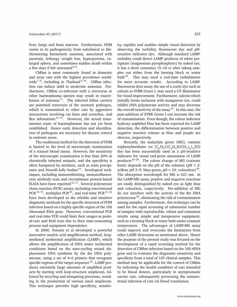
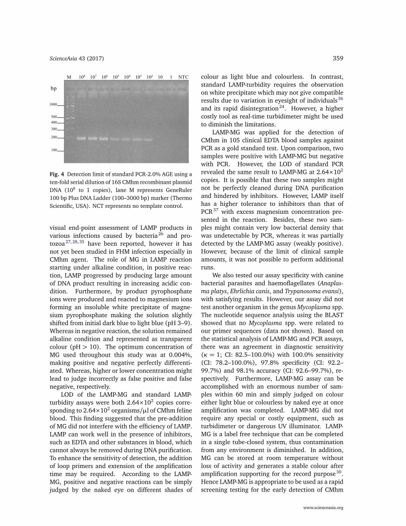
(c) M 108 107 106 105 104 103 102 10 1 NTC
bp
1000
500
400
300
200
100
Fig. 3 Detection limit of (a) LAMP-MG assay, (b) stan-dard LAMP-turbidity, and (c) LAMP-2.0% AGE; lane Mrepresents GeneRuler DNA Ladder mix (100–10 000 bp)marker (Thermo Scientific, USA) by using a ten-fold serialdilution of 16S CMhm recombinant plasmid DNA (108 to1 copies). NCT represents no template control.
DISCUSSION
With an increasing number of stray cats in Thailandsince 2005 as reported by Department of Communi-cable Disease Control, FIA has become an emerginginfectious disease that can be transmitted amongstray and domestic cats. Hence asymptomatic do-mestic cats that intend to be blood donors can becarriers for the disease. Thus the early detectionof CMhm infection is required and essential for thescreening of blood donors and controlling FHM inendemic areas. Herein, an LAMP-MG colorimetricassay has been developed as a rapid, sensitive andspecific tool for the detection of CMhm based on the16S rRNA gene due to its uniqueness in individualspecific sequence species with a single copy geneand distinct size from Mhf and CMtc. Furthermore,studies of FHM infection based on 16S rRNA genecoupled with internal control as feline 28S rRNAgene33, or feline glyceraldehyde-3-phosphate dehy-drogenase (FG3PDH) gene34 have been reported.While FHM sequence information on other genesneed to be further investigated.
Using of MG as pH-sensitive indicator dye for
www.scienceasia.org
ScienceAsia 43 (2017) 359
M 108 107 106 105 104 103 102 10 1 NTC
bp
1000
500
400
300
200
100
Fig. 4 Detection limit of standard PCR-2.0% AGE using aten-fold serial dilution of 16S CMhm recombinant plasmidDNA (108 to 1 copies), lane M represents GeneRuler100 bp Plus DNA Ladder (100–3000 bp) marker (ThermoScientific, USA). NCT represents no template control.
visual end-point assessment of LAMP products invarious infections caused by bacteria26 and pro-tozoa27, 28, 35 have been reported, however it hasnot yet been studied in FHM infection especially inCMhm agent. The role of MG in LAMP reactionstarting under alkaline condition, in positive reac-tion, LAMP progressed by producing large amountof DNA product resulting in increasing acidic con-dition. Furthermore, by product pyrophosphateions were produced and reacted to magnesium ionsforming an insoluble white precipitate of magne-sium pyrophosphate making the solution slightlyshifted from initial dark blue to light blue (pH 3–9).Whereas in negative reaction, the solution remainedalkaline condition and represented as transparentcolour (pH > 10). The optimum concentration ofMG used throughout this study was at 0.004%,making positive and negative perfectly differenti-ated. Whereas, higher or lower concentration mightlead to judge incorrectly as false positive and falsenegative, respectively.
LOD of the LAMP-MG and standard LAMP-turbidity assays were both 2.64×10² copies corre-sponding to 2.64×10² organisms/µl of CMhm felineblood. This finding suggested that the pre-additionof MG did not interfere with the efficiency of LAMP.LAMP can work well in the presence of inhibitors,such as EDTA and other substances in blood, whichcannot always be removed during DNA purification.To enhance the sensitivity of detection, the additionof loop primers and extension of the amplificationtime may be required. According to the LAMP-MG, positive and negative reactions can be simplyjudged by the naked eye on different shades of
colour as light blue and colourless. In contrast,standard LAMP-turbidity requires the observationon white precipitate which may not give compatibleresults due to variation in eyesight of individuals36
and its rapid disintegration24. However, a highercostly tool as real-time turbidimeter might be usedto diminish the limitations.
LAMP-MG was applied for the detection ofCMhm in 105 clinical EDTA blood samples againstPCR as a gold standard test. Upon comparison, twosamples were positive with LAMP-MG but negativewith PCR. However, the LOD of standard PCRrevealed the same result to LAMP-MG at 2.64×10²
copies. It is possible that these two samples mightnot be perfectly cleaned during DNA purificationand hindered by inhibitors. However, LAMP itselfhas a higher tolerance to inhibitors than that ofPCR37 with excess magnesium concentration pre-sented in the reaction. Besides, these two sam-ples might contain very low bacterial density thatwas undetectable by PCR, whereas it was partiallydetected by the LAMP-MG assay (weakly positive).However, because of the limit of clinical sampleamounts, it was not possible to perform additionalruns.
We also tested our assay specificity with caninebacterial parasites and haemoflagellates (Anaplas-ma platys, Ehrlichia canis, and Trypanosoma evansi),with satisfying results. However, our assay did nottest another organism in the genus Mycoplasma spp.The nucleotide sequence analysis using the BLASTshowed that no Mycoplasma spp. were related toour primer sequences (data not shown). Based onthe statistical analysis of LAMP-MG and PCR assays,there was an agreement in diagnostic sensitivity(κ = 1; CI: 82.5–100.0%) with 100.0% sensitivity(CI: 78.2–100.0%), 97.8% specificity (CI: 92.2–99.7%) and 98.1% accuracy (CI: 92.6–99.7%), re-spectively. Furthermore, LAMP-MG assay can beaccomplished with an enormous number of sam-ples within 60 min and simply judged on coloureither light blue or colourless by naked eye at onceamplification was completed. LAMP-MG did notrequire any special or costly equipment, such asturbidimeter or dangerous UV illuminator. LAMP-MG is a label free technique that can be completedin a single tube-closed system, thus contaminationfrom any environment is diminished. In addition,MG can be stored at room temperature withoutloss of activity and generates a stable colour afteramplification supporting for the record purpose30.Hence LAMP-MG is appropriate to be used as a rapidscreening testing for the early detection of CMhm
www.scienceasia.org

360 ScienceAsia 43 (2017)
infection worldwide.In conclusion, the present study demonstrates
that we developed the LAMP-MG assay for therapid detection of CMhm infection in cats. Limitof detection of LAMP-MG was equal to standardLAMP-turbidity and PCR assays corresponding to2.64×102 organisms/µl of CMhm feline blood. Out-standingly, LAMP-MG did not hinder DNA synthe-sis activity and maintained good reaction even inpresenting of inhibitors. Further, LAMP-MG can beeasily performed as well as simply detected by nakedeye given satisfying results as robustness, high sensi-tivity, specificity and accuracy within 60 min of reac-tion time. This technique can be used as alternativemethod for detection of feline CMhm infection.
Acknowledgements: This work was financially sup-ported by the Thailand Research Fund through theRoyal Golden Jubilee Ph.D. Programme under Grant No.PHD/0003/2553 and co-financed by the Higher Edu-cation Research Promotion under Grant No. 140/2559and the Graduate School of Srinakharinwirot University,Thailand.
REFERENCES
1. Willi B, Boretti FS, Tasker S, Meli ML, Wengi N,Reusch CE, Lutz H, Hofmann-Lehmann R (2007)From Haemobartonella to hemoplasma: molecularmethods provide new insights. Vet Microbiol 125,197–209.
2. Flint JC, Moss LC (1953) Infectious anemia in cats.J Am Vet Med Assoc 122, 45–8.
3. Foley JE, Pedersen NC (2001) Candidatus My-coplasma haemominutum, a low-virulence epiery-throcytic parasite of cats. Int J Syst Evol Microbiol 51,815–7.
4. Cooper SK, Berent LM, Messickv JB (1999) Compet-itive, quantitative PCR analysis of Haemobartonellafelis in the blood of experimentally infected cats.J Microbiol Meth 34, 235–44.
5. Inokuma H, Taroura S, Okuda M, Hisasue M, ItamotoK, Une S, Nakaichi M, Taura Y (2004) Molecularsurvey of Mycoplasma haemofelis and ‘CandidatusMycoplasma haemominutum’ infection in cats in Ya-maguchi and surrounding areas. J Vet Med Sci 66,1017–20.
6. Chungpivat S, Sarikaputi M, Jirasukprasert S, Jen-changkol S, Viseshakul N (2007) The molecular iden-tification of Mycoplasma haemofelis and Mycoplasmahaemominutum in cats suffering from hemoplasmosisin Thailand. Thai J Vet Med 37, 33–40.
7. Bauer N, Balzer HJ, Thüre S, Moritz A (2008) Preva-lence of feline hemotropic mycoplasmas in conve-nience samples of cats in Germany. J Feline Med Surg10, 252–8.
8. Peters IR, Helps CR, Willi B, Hofmann-Lehmann R,Tasker S (2008) The prevalence of three speciesof feline haemoplasmas in samples submitted to adiagnostics service as determined by three novel real-time duplex PCR assays. Vet Microbiol 126, 142–50.
9. Jiyipong T, Amavisit P, Tasker S, Peters I, Stich RW,Jittapalapong S (2008) Molecular detection of felinehemoplasma in stray cats in Bangkok, Thailand.Infect Genet Evol 8, S21–2.
10. Jiyipong T, Amavisit P, Wongnarkpet S, Tasker S,Jittapalapong S (2008) Molecular epidemiology ofMycoplasma haemofelis and ‘Candidatus Mycoplasmahaemominutum’ of stray cats in Bangkok. In: Pro-ceedings of the 15th Congress of FAVA, Bangkok, Thai-land, pp O23–4.
11. Tasker S, Braddock JA, Baral R, Helps CR, Day MJ,Gruffydd-Jones TJ, Malik R (2004) Diagnosis of fe-line haemoplasma infection in Australian cats usinga real-time PCR assay. J Feline Med Surg 6, 345–54.
12. Lappin MR, Griffin B, Brunt J, Riley A, Burney D,Hawley J, Brewer MM, Jensen WA (2006) Prevalenceof Bartonella species, haemoplasma species, Ehrlichiaspecies, Anaplasma phagocytophilum, and Neorickett-sia risticii DNA in the blood of cats and their fleas inthe United States. J Feline Med Surg 8, 85–90.
13. Shaw SE, Kenny MJ, Tasker S, Birtles RJ (2004)Pathogen carriage by the cat flea Ctenocephalides felis(Bouche) in the United Kingdom. Vet Microbiol 102,183–8.
14. Woods JE, Brewer MM, Hawley JR, Wisnewski N,Lappin MR (2005) Evaluation of experimental trans-mission of Candidatus Mycoplasma haemominutumand Mycoplasma haemofelis by Ctenocephalides felisto cats. Am J Vet Res 66, 1008–12.
15. Tasker S, Lappin MR (2002) Haemobartonella felis:recent developments in diagnosis and treatment.J Feline Med Surg 4, 3–11.
16. Alleman AR, Pate MG, Harvey JW, Gaskin JM, BarbetAF (1999) Western immunoblot analysis of the anti-gens of Haemobartonella felis with sera from experi-mentally infected cats. J Clin Microbiol 37, 1474–9.
17. Wolf-Jäckel GA, Jäckel C, Museux K, Hoelzle K,Tasker S, Lutz H, Hofmann-Lehmann R (2010) Iden-tification, characterization, and application of a re-combinant antigen for the serological investigationof feline hemotropic Mycoplasma infections. ClinVaccine Immunol 17, 1917–25.
18. Jensen WA, Lappin MR, Kamkar S, Reagan WJ(2001) Use of a polymerase chain reaction assay todetect and differentiate two strains of Haemobar-tonella felis in naturally infected cats. Am J Vet Res62, 604–8.
19. Berent LM, Messick JB, Cooper SK (1998) Detec-tion of Haemobartonella felis in cats with experimen-tally induced acute and chronic infections, using apolymerase chain reaction assay. Am J Vet Res 59,1215–20.
www.scienceasia.org

ScienceAsia 43 (2017) 361
20. Suksai P, Sangkachai N, Chatsiriwech J, Kanthasae-wee O, Sariya L, Chaichoun K (2010) Developmentof multiplex polymerase chain reaction for detectionof feline hemotropic mycoplasma in blood and tissuespecimens. Southeast Asian J Trop Med Publ Health41, 1447–53.
21. Tasker S, Helps CR, Day MJ, Gruffydd-Jones TJ,Harbour DA (2003) Use of real-time PCR to detectand quantify Mycoplasma haemofelis and CandidatusMycoplasma haemominutum DNA. J Clin Microbiol41, 439–41.
22. Lobetti RG, Tasker S (2004) Diagnosis of felinehaemoplasma infection using a real-time PCR assay.J S Afr Vet Assoc 75, 94–9.
23. Notomi T, Okayama H, Masubuchi H, Yonekawa T,Watanabe K, Amino N, Hase T (2000) Loop-mediatedisothermal amplification of DNA. Nucleic Acids Res28, e63.
24. Almasi MA, Ojaghkandi MA, Hemmatabadi A,Hamidi F, Aghaei S (2013) Development of colori-metric loop-mediated isothermal amplification assayfor rapid detection of the Tomato Yellow Leaf Curlvirus. J Plant Pathol Microbiol 4, 153.
25. Tanner NA, Zhang Y, Evans TC Jr (2015) Visualdetection of isothermal nucleic acid amplificationusing pH-sensitive dyes. BioTechniques 58, 59–68.
26. Farnia P, Masjedi MR, Mohammadi F, Tabarsei P,Farnia P, Mohammadzadeh AR, Baghei P, VarahramM, et al (2008) Colorimetric detection of multidrug-resistant or extensively drug-resistant tuberculosis byuse of malachite green indicator dye. J Clin Microbiol46, 796–9.
27. Nzelu CO, Cáceres AG, Guerrero-Quincho S, Tineo-Villafuerte E, Rodriquez-Delfin L, Mimori T, UezatoH, Katakura K, et al (2016) A rapid molecular di-agnosis of cutaneous leishmaniasis by colorimetricmalachite green-loop-mediated isothermal amplifi-cation (LAMP) combined with an FTA card as a directsampling tool. Acta Trop 153, 116–9.
28. Lucchi NW, Ljolje D, Silva-Flannery L, Udhayaku-mar V (2016) Use of malachite green-loop mediatedisothermal amplification for detection of Plasmodiumspp. parasites. PLoS One 11, e0151437.
29. Elaziouti A, Laouedj N, Ahmed B (2011) Effect of pHsolution on the optical properties of cationic dyes indye/Maghnia montmorillonite suspensions. J ChemEng Process Tech 2, 113–7.
30. Nzelu CO, Gomez EA, Cáceres AG, Sakurai T, Martini-Robles L, Uezato H, Mimori T, Katakura K, et al(2014) Development of a loop-mediated isothermalamplification method for rapid mass-screening ofsand flies for Leishmania infection. Acta Trop 132,1–6.
31. Guimaraes AM, Santos AP, do Nascimento NC,Timenetsky J, Messick JB (2014) Comparative ge-nomics and phylogenomics of hemotrophic my-coplasmas. PLoS One 9, e91445.
32. Kewish KE, Appleyard GD, Myers SL, Kidney BA,Jackson ML (2004) Mycoplasma haemofelis and My-coplasma haemominutum detection by polymerasechain reaction in cats from Saskatchewan and Al-berta. Can Vet J 45, 749–52.
33. Aquino LC, Hicks CAE, Scalon MC, Lima MGM,Lemos MS, Paludo GR, Helps CR, Tasker S (2014)Prevalence and phylogenetic analysis of haemoplas-mas from cats infected with multiple species. J Mi-crobiol Meth 107, 189–96.
34. Watanabe M, Hisasue M, Souma T, Ohshiro S, Ya-mada T, Tsuchiya R (2008) Molecular detection ofMycoplasma haemofelis and ‘Candidatus Mycoplasmahaemominutum’ infection in cats by direct PCR usingwhole blood without DNA extraction. J Vet Med Sci70, 1095–9.
35. Sriworarat C, Phumee A, Mungthin M, Leelayoova S,Siriyasatien P (2015) Development of loop-mediatedisothermal amplification (LAMP) for simple detec-tion of Leishmania infection. Parasites Vectors 8, 591.
36. Sappat A, Jaroenram W, Puthawibool T, Lomas T,Tuantranont A, Kiatpathomchai W (2011) Detectionof shrimp Taura syndrome virus by loop-mediatedisothermal amplification using a designed portablemulti-channel turbidimeter. J Virol Meth 175, 141–8.
37. Aryan E, Meshkat Z, Mirbagheri SZ, Alvandi AH, Saf-dari H, Riahi B, Momen-Heravi M (2014) Improveddetection of mycobacterium tuberculosis using aninhibitor-tolerant loop-mediated isothermal amplifi-cation. Eur Respir J 44, Suppl 58, P2588.
www.scienceasia.org