Development of Kraków external microbeam – single ion hit ...
18
Report No 1955/AP Development of Kraków external microbeam – single ion hit facility W. Polak, R.Hajduk, S. Lebed, J.Lekki, T. Horwacik, S. Maranda, T.Pieprzyca, C. Sarnecki, Z.Stachura, Z. Szklarz, O. Veselov, J.Styczeń Abstract: The main purpose of building the microbeam setup for the external (i.e out-of-vacuum) measurements is single cell with single ions irradiation. The single-ion hit facility is based on the existing Kraków microbeam setup characterized by the spatial resolutions of about 3 µm. Present work introduces the setup and the measurement chamber that was developed, constructed and assembled in IFJ PAN. The passage of single ions from vacuum to atmosphere (where the cell dish is located) is registered using the channeltron detector installed inside the chamber. Channeltron registers secondary electrons emitted from CsI layer covering the Si 3 N 4 exit window. The system of very precise diaphragms reduces the beam intensity down to a fluence of about 10 3 protons/sec. The beam blanking, correlated with single proton passage is provided by the fast, electrostatic deflecting system. The precise 3D table, installed outside the chamber, allows positioning the cell dish at a 200 µm distance from the exit window and change the targeted cell with a sub-micrometer precision within the dish. The paper shows results of the preliminary investigations aimed towards optimization of the most important issues: resolution of external microbeam, proton registration efficiency, efficiency of deflecting system and accuracy of single-proton-hit system. Henryk Niewodniczański Institute of Nuclear Physics Polish Academy of Science Kraków 2004
Transcript of Development of Kraków external microbeam – single ion hit ...

Report No 1955/AP
Development of Kraków external microbeam – single ion hit facility
W. Polak, R.Hajduk, S. Lebed, J.Lekki, T. Horwacik, S. Maranda,
T.Pieprzyca, C. Sarnecki, Z.Stachura, Z. Szklarz, O. Veselov, J.Styczeń
Abstract: The main purpose of building the microbeam setup for the external (i.e out-of-vacuum) measurements is single cell with single ions irradiation. The single-ion hit facility is based on the existing Kraków microbeam setup characterized by the spatial resolutions of about 3 µm. Present work introduces the setup and the measurement chamber that was developed, constructed and assembled in IFJ PAN. The passage of single ions from vacuum to atmosphere (where the cell dish is located) is registered using the channeltron detector installed inside the chamber. Channeltron registers secondary electrons emitted from CsI layer covering the Si3N4 exit window. The system of very precise diaphragms reduces the beam intensity down to a fluence of about 103 protons/sec. The beam blanking, correlated with single proton passage is provided by the fast, electrostatic deflecting system. The precise 3D table, installed outside the chamber, allows positioning the cell dish at a 200 µm distance from the exit window and change the targeted cell with a sub-micrometer precision within the dish. The paper shows results of the preliminary investigations aimed towards optimization of the most important issues: resolution of external microbeam, proton registration efficiency, efficiency of deflecting system and accuracy of single-proton-hit system.
Henryk Niewodniczański Institute of Nuclear Physics
Polish Academy of Science Kraków 2004

The aim of the project Our understanding of the biological effects of radiation exposure to very low doses is still
far from complete, despite of a tremendous progress in recent years. Any estimation of
radiation hazards requires profound knowledge on interaction of ions with biological cells
and tissue. In particular, there is a still unsolved debate about the influence of a very low
dose radiation on living organisms, which was up to now only estimated by high dose
data extrapolation. Results of the extrapolation based on irradiation cell by conventional
broad beams are of low statistical value because of insufficient information about number
of irradiated cells, number of particles traversed particular cell and spontaneous cell death
in control cell cultures.
Therefore, some laboratories have introduced the use of collimated ion beams [Savant
(2001), Watson (2000), Belyakov (2001), Belyakov (2002), Nagashawa (1999), Zhou
(2001), Folkard part I,II (1997), Moretto (2001), Folkard (2001)], where cells, or even
certain cell compartments, can be individually irradiated with an exact ion number. The
precise information of hit and non-hit cells enables also studies of the radiation damage
transfer from hit cell to neighboring cells, a so called “bystander effect”. The genotoxic
effect of charged particle radiation on living cells is a result of interactions among
biological matter; primary particles traversing the cell, positive ions and secondary
electrons produced along the track of charged particles and of chemical reactions with
free radicals or other reactive oxygen species created by radiolysis of water in the cell.
Those interactions can be tracked down to a level of a single chromosome or even of a
single DNA strand. The development of focused microbeams allows achieving of a better
linear energy transfer (LET) definition, greater range of LET using heavy ion beams, and
higher aiming accuracy.
The main aim of this project is to investigate cell response after hitting it with the certain
number of ions. Biological effects of ion irradiation will be studied as a function of
energy and atomic number of the irradiating ions, number of the ions traversing the cell,
the cell species and the cell state (alterations of the cell cycle and the functional status).
Measurements connected with the bystander effect, especially the number of non-hit dead
cells and the spatial range of the phenomena will be tested for various cell cultures,
various locating of the ion traversal through the cell and the different cell status. The
ultimate aim of the project is to understand the processes relevant to particle radiation-
induced cancer as well as medical and industrial applications of the radiation which cause

occupational hazards. Investigation over bystander effect phenomena may provide vital
knowledge for radiation cancer therapy. This new branch of nuclear microprobe
application grows very fast thanks to the great importance of the area under investigation
and is supported by the European Commission through a Marie Curie Research Training
Project CELLION, grouping ten scientific European institutions and coordinated by IFJ
PAN.
The measurement setup To enable the irradiations of living cells using single ions, the new measurement setup
has been developed in Kraków Institute of Nuclear Physics. This new microbeam facility
allows cell irradiations in atmosphere using protons with energy up to 2.5 MeV from the
Van de Graaff accelerator [Lekki (2002)]
Proton beam from the accelerator, after passing the 90° analysing magnet, is pre-focused
by 3 quadrupoles before entering the microprobe system (Fig 1). Further on, the precise
diaphragms reduce the beam to the current of about 0.16fA, what corresponds to about
1000 protons/s. Two electrostatic deflecting plates (made by Technisches Büro S. Fisher)
mounted after the slits allow rapid beam blanking (reaction time of single microseconds).
Final focusing of the beam is provided by the two quadrupoles manufactured in the Micro
Analytical Research Centre (MARC, Melbourne, Australia) [Lebed (2001),
Fig. 1 Schematic view of microbeam single-hit-facility in Kraków

Lekki (2002)]. The best spatial resolution of about 3 x 3 µm2 was achieved; however, 5 x
5 µm2 resolution is a standard operational value. However, in experiments described in
the present work, only resolution of about 12 x 12 µm2 was applied. Design of the
measurement chamber permits to focus proton microbeam close to the exit window. The
window is made of a thin (200 nm) Si3N4 membrane, and separates the vacuum inside the
chamber from the air where the biological sample is hold during investigations. Protons
passing to the air are counted by detection of the secondary electrons emitted from the
window foil by the channeltron [Sjuts Electronics] installed in the chamber. The
computer controlled, precise 2D table (Physik Instrumente, Voice Coil type V-106 2S,
resolution of 0.1µm) allows positioning the cells in the focus. The distance of the sample
from exit window is controlled by a micrometer screw and, to minimize protons
scattering, should be set to a value not exceeding 200 µm.
Fig. 2 Top and side view of the measurement chamber
The chamber (Fig. 2) has a possibility to rotate that enables precise focusing of the beam
using additional quartz (Fig. 2a), located 1 cm aside from exit window. After focusing the
beam and reducing its intensity, the chamber position is changed back to central axis to
allow passing the ions through the outlet window (Fig.2a).

Quartz Exit window
Fig. 2a Back side of the measurement Fig. 2b Picture of the chamber chamber and the microbeam facility
The first step – the external microbeam. In order to irradiate the living biological material, the proton beam must leave the ion guide and experimental chamber and enter the atmosphere.
Fig. 3 Picture of ionized air due to proton beam entering the atmosphere

The photo in Fig. 3 proves that the proton beam actually enters the atmosphere. Protons
passing through the window to the air are scattered in the Si3N4 window membrane and
thus their direction diverges from central axis. This effect, and not the scattering in
atmosphere, is the main cause of loosing the targeting accuracy of the irradiation, what
was proven using SRIM (The Stopping and Range of Ions in Matter) [Ziegler (1998)]
simulations. The further from the exit window the sample is situated the worse the
targeting resolution can be achieved. The SRIM simulations show that at 200 µm distance
and 200 nm thick exit window, the targeting resolution decreases to 4 µm from the ideal
case. This result concerns the estimation of angular spread of the beam that was initially
parallel. For a 1 mm distance, the resolution deteriorates by a factor of about 4 that makes
a significant difference and is unacceptable for the planned application. These estimations
were supported by direct measurements using scanning transmission ion microscopy
(STIM). The calibration grid (400 mesh) located at several distances from the exit
window was scanned using the external beam in air. The PIN diode positioned behind the
grid was used as a proton detector. The achieved resolution at a distance of 200 µm was
about 11 µm (Fig. 4a), which was in good agreement with calculations, taking into
account that resolution in vacuum was of about 10 µm and the partial variances added in
square values.

Fig. 4b Image of the same grid at 1mm distance from the exit window; resolution worse than 25µm
Fig. 4a Image of the calibration grid at 200µm distance from exit window; resolution ≈11µm
Proton registration efficiency Protons which had passed through the Si3N4 membrane (commercially available at Silson
company, UK) were registered by a semiconductor, ion implanted silicon detector
characterized by the 100% efficiency for the particle registration. Those protons passing
through the window ejected the secondary electrons from the membrane and the electrons
were registered by a channeltron. The channeltron signals were collected in coincidence
with the silicon detector signals in two ways:
- Time spectra were recorded using SILENA TDC with 1200ns base and 4096
channels and then were extracted and evaluated in the microbeam data acquisition
program CMB [Lekki (2000)].
- Coincidences were registered by an electronic coincidence logic unit and counted
in a scaler

I- Fast track (timing) II- Slow track (energy) 1. Channeltron (electrons detector) –
Sjuts Electronics 2. Si particle detector – Ortec 3. and 3*. Ortec Preamplifiers 4. Timing Filter Amplifier - Ortec 836 5. Constant Fraction Discriminator –
Caen mod.N415 6. Delay and Gate Generator- Ortec
8010 7. NIM – ECL – NIM translator-
Caen mod.92 8. Time to Digital Converter- Silena
4418/T 9. Coincidence logic unit – Caen
mod.N454 10. Scaler – Polon 401 11. Active Filter Amplifier – Ortec
572A 12. Active Filter Amplifier – Ortec 572 13. Amplitude to Digital Converter –
Caen mod.C420
Fig. 5 Electronic system to register protons and electrons signal
Both of the above approaches have some advantages. Using the first way, the time spectra
of protons, electrons and the coincidences of both signals can be easily extracted from
event–by–event data files and displayed in a computer screen; therefore we have good
control over experiment. The second way gives just the number of registered
coincidences but is completely independent from computer system and a dead time
created by the software of the acquisition system. Such a cross-checking of the results is
very valuable, as it must be taken into account that the computer controlled data
acquisition system has certain, not well defined reaction time (e.g. the Windows operating
system is characterized by the rather high interrupt latency, the TDC unit is closed for
pulses during time when computer reads data from it, etc.). Therefore some event losses
are unavoidable. Efficiency of the proton detection using the secondary electrons
registered in the channeltron was defined as the ratio of the coincident events to the total
number of the particle detector signals.
Apart from the fast timing tracks, slow energetic signals were also collected by Caen
ADC (Fig. 5). The electronic system enabled to register protons and electrons from both
tracks (energetic and fast), additionally protons and electrons number were counted in the
scaler. Therefore, the coincidences of slow energetic proton and electron signals could
have been compared with coincidences between fast timing signals and simultaneously
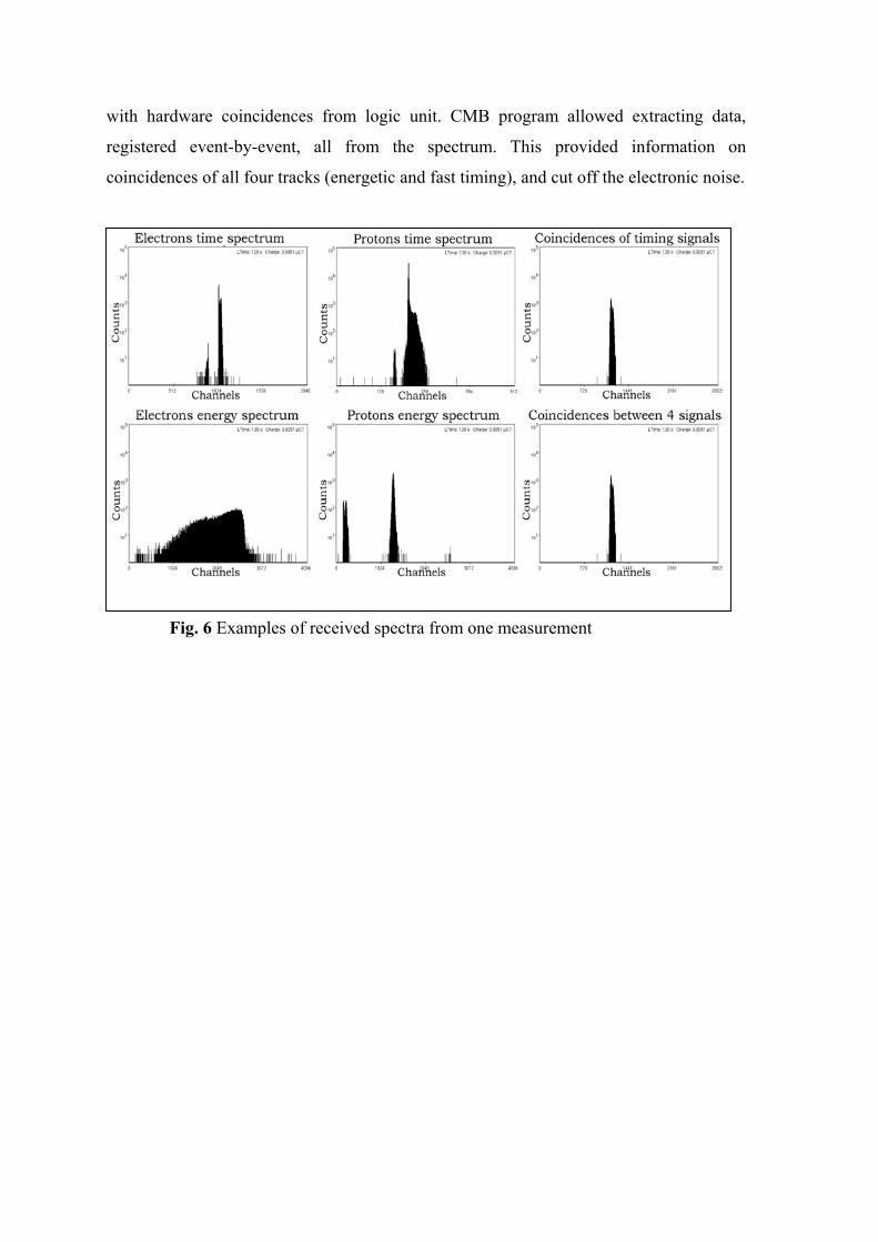
with hardware coincidences from logic unit. CMB program allowed extracting data,
registered event-by-event, all from the spectrum. This provided information on
coincidences of all four tracks (energetic and fast timing), and cut off the electronic noise.
Fig. 6 Examples of received spectra from one measurement

Table 1.
Table 1 gives an example of results from one of the measurements, spectra of which are
presented in the Fig. 6.
Number of counts in proton energy spectrum
Number of counts in electron energy spectrum
Number of counts in proton time spectrum
Number of counts in electron time spectrum
Number of protons counted in scaler
Number of electrons counted in scaler
Number of coincidences between time protons and time electrons impulses
Number of coincidences between all 4 signals
Number of coincidences counted by Coincidence Logic Unit
73102 56689 73234 56195 89964 58317 43940 43302 50190
The efficiency evaluated from the particular spectrum (shown in fig. 6) amounts to
- 57% considering fast timing tracks, and
- 56% considering counts in Coincidence Logic Unit
We observed that the number of the coincidences between all four signals was
comparable to the number of coincidences between two timing signals from protons and
electrons (discrepancy smaller than 0.02%).
From the previous experiments [Cholewa (2001), Fisher(2001), Polak (2003)] the Si3N4
membrane covered with the CsI layer proved to be more efficient than Si3N4 itself. The
similar results were obtained in the present work. The Si3N4 membrane efficiency for
proton registration was 10% only, while for Si3N4 covered with CsI the best result
achieved was 61%. The 61% efficiency is not sufficient for the investigation purposes,
where efficiency well above 90% is vital to hit single cell with single proton.
Furthermore, we observe that the efficiency is deteriorated in time as shown in the graph
below (Fig. 7). This means that the use of a new window is recommended each day. In
the figure below each bar corresponds to a slightly different beam spot position on the
window surface; therefore efficiency degradation can not be attributed to local destruction
of the window surface due to the proton bombardment.
E f f i c i e n c y d e t e r i o r a t i g i n t i m e
0
10
20
30
40
50
60
70
1 2 5 6D a y s
E f f
i c i
e n
c y [
%]
Fig. 7 Efficiency deteriorating in time

We will attempt to solve the problem of poor efficiency of proton registration in two
different ways.
1. The further test with Si3N4 covered with the CsI layer will be performed in better
geometrical conditions inside the standard vacuum chamber. Additionally, a mesh
with positive voltage will be applied in front of the channeltron. The vacuum
conditions also proved to be important to register secondary electrons [Cholewa
(1998)]. The vacuum level during our measurements was about 2.3x10⁻⁵ Tr while
following conclusions from abovementioned Cholewa’s publication at least
5.0x10⁻⁶ Tr seems to be crucial. If those future improvements allow registering
protons with efficiency better than 90%, this method will be used in the single-hit-
system.
2. Protons will be registered by a detector (Si or PIN diode) behind the cell dish.
This method, although giving about 100% efficiency, has some disadvantages:
- During the time of the experiment medium must be taken out from the cell dish,
otherwise protons will not reach the detector. The SRIM evaluations prove that
100% of protons will be transmitted to the detector considering: i) 200µm
distance from the outlet window to the bottom of the cell dish, ii) 3µm mylar
foil thickness, iii) 30µm of the cell thickness, iv) 5mm distance from the cell to
the detector). This approach also decreases the possible experiment time to less
than 10 minutes which guarantees survival of the cells.
- Observation of the cell dish is more difficult because the detector must be
positioned in front of the microscope objective for the time of measurement.
The single-event system The fast blanking of the beam (Fig. 8) is very important to obtain just one proton hit per
cell (providing 100% efficiency of proton registration was achieved).
Both the theoretical calculations and the experimental data proved that 440V applied to
deflecting plates was enough to divert protons from entering the chamber. In time the
blanking system was switched on, neither the protons nor electrons were registered by the
corresponding detectors. The electronic system was rearranged to perform testing
experiments for the beam blanking. Pulse from amplifier (electrons or protons) went to
Single Channel Analyzer SCA (Polon 1201) where the applied thresholds (Fig. 9) cut off
the electronic noise.

1. Silicon detector 2. Ortec Preamplifier 3. Active Filter Amplifier – Ortec 572 4. Single Channel Analizer (SCA)
– Polon 1201 5. Scaler – Polon 401 6. Computer with Analog Output Board
card which runs the blanking process and collects data from experiment
7. Switching off system – Technisches Büro S. Fisher
Fig. 8 Electronic system for measuring deflection efficiency
The computer after obtaining
the outgoing TTL signal,
created in SCA, rapidly
switched the beam off. After
500 ms the beam was
switched on until the proton
pulse was registered and the
beam was switched off again.
From calculations we found
out that even for 50 µs
resolution of the blanking
system, the probability of
registration two protons was about 0.1% for 0.1 fA beam current. Measured delay
between the pulse from detector to the moment of application of 440 V onto deflecting
plates was of about 13 µs.
Fig. 9 Picture shows the window set with SCA on energy of protons to cut off electronic noise
The beam blanking efficiency was tested using two different methods:
Method 1.
Switching the beam off after registering pulse from the proton detector located just
behind the exit window and, then, measuring the number of protons still reaching the
detector after the beam blanking signal was sent. To assure proper statistics, those
measurements were repeated many times and for various beam currents.

In most occasions the
deflecting system managed to
switch the beam off fast
enough to prevent more than
one proton hitting the
detector. However, there were
cases that more than one
count was accumulated
before next blanking of the
beam (Fig.10). Fig. 11 Deflecting Protons efficiency using silicon detector signals - beam intensity 800 protons/sec
Beam blanking pulses
The experiment presented in Fig.11 shows that increasing the beam current causes
malfunction of the system and more than one proton hit detector in many occasions.
The graph shows mean values for the particular beam current for the whole
measurement when real results (Fig.10) vary from one to few protons per one switch
off event. For beam intensity smaller than 3000 protons/sec the blanking system has a
constant performance of about 20% away from the ideal case (the ideal case is 1 one
proton per one blanking
event that is a value of 1 in
Figure 11, while currently
this value is about 1.2). The
cases when more than one
proton hit the detector are
due to the data acquisition
system performance that can
be improved by switching
the beam off using just
electronic hardware (not by a
computer system). The abovementioned beam intensity of 3000 protons/sec is more
than sufficient for successful cell irradiation investigations.
Protons deflecting efficiecy using Si detector for various beam curretnts
1.8
2
2.2
er o
ne ev
ent
1
1.2
1.4
1.6
0 5000 10000 15000 20000
beam intensity [protons/sec]
mea
n pr
oton
val
ue p
Fig. 11 Efficiency of deflecting proton using silicon detector for various beam intensities

Method 2.
Beam blanking after registering an event of proton passage through the exit window (i.e.
secondary pulse from the channeltron) and measuring the number of protons reaching the
detector afterwards. If a sufficient,
close to 100% proton passage
detection were achieved, this would
be the principal method of carrying
out the irradiation. However, due to
the use of this particular exit
window for five consecutive days,
the measured efficiency of
registering protons via registering
secondary electrons was only of
about 25%. Consequently, the
expected value of successful beam
blanking was close to this number, what was confirmed by the results shown in Figure 12
where, in average, 4.4 protons were registered per one blanking event. Unfortunately, the
corresponding proton number distribution extended from one proton up to even 35 in
extreme cases. That showed that at the moment the described system did not match
experimental requirements whatsoever (Fig. 12)
Fig. 11a Deflecting Protons efficiency using chaneltron signals - beam intensity 250 protons/sec
Beam blanking pulses
Single hit resolution The results of Fig. 12 may be illustrated using a different approach, used in another test
experiment. To estimate the resolution of single-proton-hit system and the performance of
positioning system, a solid state CR-39 detector (1.5 mm thick) was installed on a 3D
moving table at a distance of about 1 mm from the exit window and then irradiated with
protons. Unfortunately, the CR-39 detector was too thick to simultaneously register the
protons with another particle detector located behind the CR-39. The beam was switched
off using the secondary electrons signal from the channeltron. A pattern of 10 x 10 grid
was created by changing the table position by 100 µm after each blanking event. After the
irradiation process, the CR-39 detector was etched in a water solution of NaOH (6.25
mol/dm3) at the temperature of 70°C for 6 hours. Next, after washing in ethanol, the
sample was investigated using an optical microscope.

Fig. 13 Picture showing proton tracks obtained after etching CR-39 detector
As shown in Fig.13, every spot corresponding to a temporary beam position contains
from one up to 50 proton track aggregations. The method allows displaying single proton
tracks. The beam targeting resolution estimated from the picture is of about 40 µm which
is not yet good enough to target the single cell. Poor spatial resolution of the beam is
caused by a large (1mm) distance of the CR-39 detector from the exit window. Moreover,
behaviour of one of the quadrupole lenses during the present experiment was not
“normal” and the dumping of vibrations of the vacuum connection to the turbo-molecular
pump was not perfect. To improve the accuracy, primarily, the good beam resolution (in
vacuum) should be restored to 3 x 3 µm2. The distance between sample and window must
be reduced (cf. results shown in Fig. 4a), after installing emergency vacuum alarm (task
currently in progress) preventing consequences following the possible exit window
rupture. The anti-vibrating system must be applied because the quivering may alter the
table movement and the precision of beam targeting during measurement.

Conclusions The experiments described above show preliminary results of the performance of the first
external microbeam in Poland. Although the system is not yet ready to irradiate single
cells with single ions in a regular way, many useful and necessary tests can be carried out.
The resolution of 40µm in the single hit mode is a good and promising start. The
following experiments with modified Si3N4 windows in the vacuum chamber should give
an answer which system of proton detection will be used in our facility. Electronic
systems of detecting protons and electrons have been successfully tested as well as the
deflecting system which allows switching into single-hit mode. The information gathered
so far will be used in near future to improve the system performance and to make the first
cell irradiation possible.

References: Sawant SG, Randers-Pehrson G, Geard CR, Brenner DJ and Hall EJ: The bystander effect in radiation oncogenesis: I. Transformation in C3H 10T1/2 cells in vitro can be initiated in the unirradiated neighbors of irradiated cells. - Radiat Res, (2001) 155:3, 397-401. Watson GE, Lorimore SA, Macdonald DA and Wright EG: Chromosomal instability in unirradiated cells induced in vivo by a bystander effect of ionizing radiation. - Cancer Res, (2000) 60:20, 5608-11.
S.Lebed, Z.Stachura, M.Cholewa, G.J.F.Legge, J.Lekki, S.Maranda, A.Potempa, C.Sarnecki, Z.Szklarz, J.Styczeń, B.Sulkio–Cleff – „The new Cracow scanning nuclear microprobe”, Nucl. Instr. Meth. In Phys. Res. B 181 (2001) 95–98. Belyakov OV, Malcolmson AM, Folkard M, Prise KM and Michael BD: Direct evidence for a bystander effect of ionizing radiation in primary human fibroblasts. - Br J Cancer, (2001) 84:5, 674-679. Nagasawa H and Little JB: Unexpected Sensitivity to the Induction of Mutations by Very Low Doses of Alpha-Particle Radiation: Evidence for a Bystander Effect. - Radiat Res, (1999) 152:5, 552-557. Zhou H, Suzuki M, Randers-Pehrson G, Vannais D, Chen G, Trosko JE, Waldren CA and Hei TK: Radiation risk to low fluences of alpha particles may be greater than we thought. – Proc Natl Acad Sci USA, (2001) 98:25, 14410-14415. M.Folkard, K.M.Prise, B.Vojnovic, S.Gilchrist, G.Schettino, O.V.Belyakov, A.Ozols, B.D.Michael, “The impact of microbeams in radiation biology”, Nucl. Instr. Meth. In Phys. Res. B 181 (2001) 426–430 Folkard M, Vojnovic B, Prise KM, Bowey AG, Schettino G, Michael BD: A charged paticle microbeam: I. Development of an experimental system for targeting cells individually with counted particles - Int. J. Radiat. Biol. (1997) 72:375 Folkard M, Vojnovic B, Prise KM, Bowey AG, Schettino G, Michael BD: A charged paticle microbeam: II. Development of an experimental system for targeting cells individually with counted particles - Int. J. Radiat. Biol. (1997) 72:387 Moretto Ph. et al: Development of a single ion irradiation system at CENBG for applications in radiation biology – , Nucl. Instr. Meth. In Phys. Res. B 181 (2001) 104-109 W. Polak, J. Lekki, J. Gryboś, R. Hajduk, M. Cholewa, O. Kukharenko, Z. Stachura: Testing the Efficiency of the Si3N4 Membranes for Charged Particles Registration, Nukleonika 48 (2003) 25 Cholewa M, Kamiya T, Saint A, Prawer S, Legge G, Butler JE, Vestyck DJ Diamond membranes: applications for single ion detection using secondary electron emission - Diamond and Related Materials 7 (1998) 510-512

Fischer BE, Cholewa M, Hitoshi Noguchi Some experiences on the way to biological ingle ion experiments: ”, Nucl. Instr. Meth. In Phys. Res.B 181 (2001) 60-65 Cholewa M, Heiß M: Preparatory experiments for a single ion hit facility at GSI ”, Nucl. Instr. Meth. In Phys. Res.B 210 (2003) 296–301 Belyakov OV, Folkard M, Mothersill C, Prise KM and Michael BD: Bystander-induced apoptosis and premature differentiation in primary urothelial explants after charged particle microbeam irradiation. - Radiation Protection Dosimetry, (2002) 99:1-4, 249-251. J. Lekki, R. Hajduk, S. Lebed, A. Potempa, T. Pieprzyca, Z. Stachura, M. Ziębliński, and J. Styczeń: CMB v. 1.1 Data Acqusition and Evaluation System of the Cracow Nuclear Microprobe" – IFJ Report No 1856/AP (2000) J.F.Ziegler – SRIM version from 1998 Technisches Büro S.Fisher, Am Berg 9, D-64 372 Ober-Ramstadt, Germany, tel. +49-6154-4818 J.Lekki, M.Cholewa1, E.Dutkiewicz, A.Gwiazdowska, R.Hajduk, K.Irzyńska, S.Lebed2, P.Mazur, W.Polak, A.Potempa, T.Pieprzyca, C.Sarnecki, Z.Szklarz, A.Veselov, R.Zając, Z.Stachura, J.Styczeń: SPH IFJ Project of Single Proton Irradiation of Biological Cells- IFJ Report No 1915 (2002) http://www.sjuts.com http://www.silson.com