Development of ELISAs for diagnosis of acute typhoid fever ... · Development of ELISAs for...
24
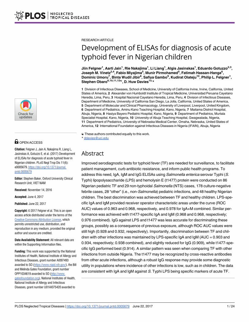
RESEARCH ARTICLE Development of ELISAs for diagnosis of acute typhoid fever in Nigerian children Jiin Felgner 1 , Aarti Jain 1 , Rie Nakajima 1 , Li Liang 1 , Algis Jasinskas 1 , Eduardo Gotuzzo 2,3 , Joseph M. Vinetz 2,4 , Fabio Miyajima 5 , Munir Pirmohamed 5 , Fatimah Hassan-Hanga 6 , Dominic Umoru 7 , Binta Wudil Jibir 8 , Safiya Gambo 9 , Kudirat Olateju 10 , Philip L. Felgner 1 , Stephen Obaro 6,10,11,12☯ , D. Huw Davies 1☯ * 1 Division of Infectious Diseases, School of Medicine, University of California Irvine, Irvine, California, United States of America, 2 Alexander von Humboldt Institute of Tropical Medicine, Universidad Peruana Cayetano Heredia, Lima, Peru, 3 Hospital Nacional Cayetano Heredia, Lima, Peru, 4 Division of Infectious Diseases, Department of Medicine, University of California San Diego, La Jolla, California, United States of America, 5 Department of Molecular and Clinical Pharmacology, University of Liverpool, Liverpool, United Kingdom, 6 Department of Pediatrics, Aminu Kano Teaching Hospital, Kano, Nigeria, 7 Maitama District Hospital, Abuja, Nigeria, 8 Hasiya Bayero Pediatric Hospital, Kano, Nigeria, 9 Department of Pediatrics, Murtala Specialist Hospital, Kano, Nigeria, 10 University of Abuja Teaching Hospital, Gwagwalada, Nigeria, 11 Department of Pediatrics, University of Nebraska Medical Center, Omaha, Nebraska, United States of America, 12 International Foundation against Infectious Diseases in Nigeria (IFAIN), Abuja, Nigeria ☯ These authors contributed equally to this work. * [email protected] Abstract Improved serodiagnostic tests for typhoid fever (TF) are needed for surveillance, to facilitate patient management, curb antibiotic resistance, and inform public health programs. To address this need, IgA, IgM and IgG ELISAs using Salmonella enterica serovar Typhi (S. Typhi) lipopolysaccharide (LPS) and hemolysin E (t1477) protein were conducted on 86 Nigerian pediatric TF and 29 non-typhoidal Salmonella (NTS) cases, 178 culture-negative febrile cases, 28 “other” (i.e., non-Salmonella) pediatric infections, and 48 healthy Nigerian children. The best discrimination was achieved between TF and healthy children. LPS-spe- cific IgA and IgM provided receiver operator characteristic areas under the curve (ROC AUC) values of 0.963 and 0.968, respectively, and 0.978 for IgA+M combined. Similar per- formance was achieved with t1477-specific IgA and IgM (0.968 and 0.968, respectively; 0.976 combined). IgG against LPS and t1477 was less accurate for discriminating these groups, possibly as a consequence of previous exposure, although ROC AUC values were still high (0.928 and 0.932, respectively). Importantly, discrimination between TF and chil- dren with other infections was maintained by LPS-specific IgA and IgM (AUC = 0.903 and 0.934, respectively; 0.938 combined), and slightly reduced for IgG (0.909), while t1477-spe- cific IgG performed best (0.914). A similar pattern was seen when comparing TF with other infections from outside Nigeria. The t1477 may be recognized by cross-reactive antibodies from other acute infections, although a robust IgG response may provide some diagnostic utility in populations where incidence of other infections is low, such as in children. The data are consistent with IgA and IgM against S. Typhi LPS being specific markers of acute TF. PLOS Neglected Tropical Diseases | https://doi.org/10.1371/journal.pntd.0005679 June 22, 2017 1 / 24 a1111111111 a1111111111 a1111111111 a1111111111 a1111111111 OPEN ACCESS Citation: Felgner J, Jain A, Nakajima R, Liang L, Jasinskas A, Gotuzzo E, et al. (2017) Development of ELISAs for diagnosis of acute typhoid fever in Nigerian children. PLoS Negl Trop Dis 11(6): e0005679. https://doi.org/10.1371/journal. pntd.0005679 Editor: Stephen Baker, Oxford University Clinical Research Unit, VIET NAM Received: November 14, 2016 Accepted: June 4, 2017 Published: June 22, 2017 Copyright: © 2017 Felgner et al. This is an open access article distributed under the terms of the Creative Commons Attribution License, which permits unrestricted use, distribution, and reproduction in any medium, provided the original author and source are credited. Data Availability Statement: All relevant data are within the Supporting Information files. Funding: This work was supported by the National Institutes of Health, National Institute of Allergy and Infectious Diseases, grant number AI097493 awarded to SO (https://www.niaid.nih.gov/), the Bill and Melinda Gates Foundation, grant number OPP1034619 awarded to SO (http://www. gatesfoundation.org), National Institutes of Health, National Institute of Allergy and Infectious Diseases, grant number U01AI075420 awarded to
Transcript of Development of ELISAs for diagnosis of acute typhoid fever ... · Development of ELISAs for...
RESEARCH ARTICLE
Development of ELISAs for diagnosis of acute
typhoid fever in Nigerian children
Jiin Felgner1, Aarti Jain1, Rie Nakajima1, Li Liang1, Algis Jasinskas1, Eduardo Gotuzzo2,3,
Joseph M. Vinetz2,4, Fabio Miyajima5, Munir Pirmohamed5, Fatimah Hassan-Hanga6,
Dominic Umoru7, Binta Wudil Jibir8, Safiya Gambo9, Kudirat Olateju10, Philip L. Felgner1,
Stephen Obaro6,10,11,12☯, D. Huw Davies1☯*
1 Division of Infectious Diseases, School of Medicine, University of California Irvine, Irvine, California, United
States of America, 2 Alexander von Humboldt Institute of Tropical Medicine, Universidad Peruana Cayetano
Heredia, Lima, Peru, 3 Hospital Nacional Cayetano Heredia, Lima, Peru, 4 Division of Infectious Diseases,
Department of Medicine, University of California San Diego, La Jolla, California, United States of America,
5 Department of Molecular and Clinical Pharmacology, University of Liverpool, Liverpool, United Kingdom,
6 Department of Pediatrics, Aminu Kano Teaching Hospital, Kano, Nigeria, 7 Maitama District Hospital,
Abuja, Nigeria, 8 Hasiya Bayero Pediatric Hospital, Kano, Nigeria, 9 Department of Pediatrics, Murtala
Specialist Hospital, Kano, Nigeria, 10 University of Abuja Teaching Hospital, Gwagwalada, Nigeria,
11 Department of Pediatrics, University of Nebraska Medical Center, Omaha, Nebraska, United States of
America, 12 International Foundation against Infectious Diseases in Nigeria (IFAIN), Abuja, Nigeria
☯ These authors contributed equally to this work.
Abstract
Improved serodiagnostic tests for typhoid fever (TF) are needed for surveillance, to facilitate
patient management, curb antibiotic resistance, and inform public health programs. To
address this need, IgA, IgM and IgG ELISAs using Salmonella enterica serovar Typhi (S.
Typhi) lipopolysaccharide (LPS) and hemolysin E (t1477) protein were conducted on 86
Nigerian pediatric TF and 29 non-typhoidal Salmonella (NTS) cases, 178 culture-negative
febrile cases, 28 “other” (i.e., non-Salmonella) pediatric infections, and 48 healthy Nigerian
children. The best discrimination was achieved between TF and healthy children. LPS-spe-
cific IgA and IgM provided receiver operator characteristic areas under the curve (ROC
AUC) values of 0.963 and 0.968, respectively, and 0.978 for IgA+M combined. Similar per-
formance was achieved with t1477-specific IgA and IgM (0.968 and 0.968, respectively;
0.976 combined). IgG against LPS and t1477 was less accurate for discriminating these
groups, possibly as a consequence of previous exposure, although ROC AUC values were
still high (0.928 and 0.932, respectively). Importantly, discrimination between TF and chil-
dren with other infections was maintained by LPS-specific IgA and IgM (AUC = 0.903 and
0.934, respectively; 0.938 combined), and slightly reduced for IgG (0.909), while t1477-spe-
cific IgG performed best (0.914). A similar pattern was seen when comparing TF with other
infections from outside Nigeria. The t1477 may be recognized by cross-reactive antibodies
from other acute infections, although a robust IgG response may provide some diagnostic
utility in populations where incidence of other infections is low, such as in children. The data
are consistent with IgA and IgM against S. Typhi LPS being specific markers of acute TF.
PLOS Neglected Tropical Diseases | https://doi.org/10.1371/journal.pntd.0005679 June 22, 2017 1 / 24
a1111111111
a1111111111
a1111111111
a1111111111
a1111111111
OPENACCESS
Citation: Felgner J, Jain A, Nakajima R, Liang L,
Jasinskas A, Gotuzzo E, et al. (2017) Development
of ELISAs for diagnosis of acute typhoid fever in
Nigerian children. PLoS Negl Trop Dis 11(6):
e0005679. https://doi.org/10.1371/journal.
pntd.0005679
Editor: Stephen Baker, Oxford University Clinical
Research Unit, VIET NAM
Received: November 14, 2016
Accepted: June 4, 2017
Published: June 22, 2017
Copyright: © 2017 Felgner et al. This is an open
access article distributed under the terms of the
Creative Commons Attribution License, which
permits unrestricted use, distribution, and
reproduction in any medium, provided the original
author and source are credited.
Data Availability Statement: All relevant data are
within the Supporting Information files.
Funding: This work was supported by the National
Institutes of Health, National Institute of Allergy and
Infectious Diseases, grant number AI097493
awarded to SO (https://www.niaid.nih.gov/), the Bill
and Melinda Gates Foundation, grant number
OPP1034619 awarded to SO (http://www.
gatesfoundation.org), National Institutes of Health,
National Institute of Allergy and Infectious
Diseases, grant number U01AI075420 awarded to
Author summary
In many African countries, clinical management of children that present with symptoms
of bacterial sepsis, such as typhoid fever (TF) caused by Salmonella Typhi, consists of
empiric broad spectrum antibiotics. Blood culture remains the gold-standard for diagno-
sis, but is slow, suffers from poor sensitivity, and often unavailable. Consequently multi-
drug resistant bacteria have emerged that are difficult to manage with antibiotics. There is
an urgent need to develop rapid, sensitive and affordable tests for patient diagnosis, help
curb antibiotic resistance, and inform public health preventive strategies such as the
deployment of vaccines. Here, we have assessed antibodies to S. Typhi lipopolysaccharide
(LPS) and hemolysin E (HylE, t1477) in the sera of Nigerian children with acute TF and
compared them with heathy children, children with other febrile infections, and adults
from around the world with a variety of other bacterial infections. The key finding con-
cerns LPS. This is a common cell-wall component present in many bacterial species. Yet
despite this, S. Typhi LPS-specific IgA and IgM are excellent markers of acute TF in Nige-
rian children, and insensitive to other non-salmonelloses. This surprising finding suggests
a rapid point-of-care test for TF can be developed based on detection of LPS-specific IgA
+IgM.
Introduction
Salmonelloses are a group of potentially fatal bacteremias caused by different serovars of Sal-monella enterica. Typhoid fever (TF), caused by the human-specific serovar S. Typhi, is a global
health problem, especially in developing countries [1, 2]. In 2010 there were an estimated 26.9
million TF episodes worldwide, with a case-fatality rate of ~ 1% [3]. Salmonellosis caused by
nontyphoid Salmonella (NTS) serovars are caused predominantly by the zoonotic serovars, S.
Typhimurium and S. Enteritidis [4–7]. These are emerging in sub-Saharan Africa as an impor-
tant cause of bacteremia in young children, typically when associated with malnutrition,
malaria, severe anemia, and/or HIV co-infection [6, 8–11]. Case-fatality rates for blood-borne,
or invasive, NTS (iNTS) infection is higher than that for typhoid, typically ~20% [4, 6, 12],
although the antibiotic treatment regimen is the same. Over-use of empiric broad-spectrum
antibiotic treatment for undifferentiated febrile disease has led to an increase in antibiotic
resistance in both typhoidal and nontyphoidal serovars, and the potential for new antibiotics is
not encouraging [1, 13, 14]. The development of effective vaccines to prevent invasive salmo-
nellosis is therefore an important global health priority [1, 15]
Accurate diagnosis of salmonellosis remains a challenge in endemic settings. Clinically, ini-
tial presentation with typhoid or NTS disease is usually with non-differentiating fever alone,
and often without symptoms of gastroenteritis that would indicate a Salmonella infection [7].
Bacterial culture is the gold standard for diagnosis of both typhoid and iNTS disease. However,
culture suffers from poor sensitivity, and culture facilities are very limited in resource poor set-
tings such as Nigeria and other countries in Africa. Even when such facilities are available, the
time to a laboratory diagnosis is around 48 hours, and is often unaffordable for most patients.
An inactivated-Salmonella agglutination test, developed by Widal >100 years ago, is a rapid
and affordable single-step test. It remains the mainstay of diagnosis in many developing coun-
tries, even when culture facilities are available. However, the Widal’s test has poor specificity,
thought to be caused by antigens shared between S. enterica serovars, and between other spe-
cies of bacteria, such as Brucella melitnesis [16]. The Widal’s test also fails to discriminate
ELISA for typhoid serodiagnosis
PLOS Neglected Tropical Diseases | https://doi.org/10.1371/journal.pntd.0005679 June 22, 2017 2 / 24
JMV, Fogarty International grant D43TW007120
awarded to JMV (https://www.fic.nih.gov/Pages/
Default.aspx), and the UK Medical Research
Council, Grant ref. MR/K000551/1 awarded to FM
and MP (http://www.mrc.ac.uk/). The funders had
no role in study design, data collection, data
analysis, decision to publish, or preparation of the
manuscript.
Competing interests: DHD discloses receiving
salary from Antigen Discovery Inc., which has
licensed the protein microarray technology. DHD
and the University of California, Irvine may
financially benefit from this interest if the company
is successful in marketing its products that are
related to this research. The terms of this
arrangement has been reviewed and approved by
the University of California, Irvine, in accordance
with its conflict of interest policies. The other
authors have declared that no competing interests
exist.
between current and previous exposure, thus requiring two samples to be taken 7–10 days
apart to monitor for an increase in titer. In practice, the decision to treat with antibiotics has to
be made on the basis of the first test, and confirmatory convalescent testing is often not practi-
cable or irrelevant for immediate patient management. It is also less sensitive in the acute stage
of infection when IgG titers are lower.
The lack of accurate tests for surveillance also has resulted in only limited understanding of
epidemiology of salmonellosis in Africa. The high mortality, particularly in children with iNTS
infections, and the recent emergence of drug resistance, emphasize the need for a better under-
standing of the epidemiology before the rational design and implementation of control mea-
sures, including vaccines, can be effectively deployed.
In this study we have addressed the development of improved serodiagnostics with well-
defined serum samples collected from febrile children in Nigeria. Based on proteome microar-
ray screening data published recently [17], we hypothesized that LPS and/or the hemolysin E
(HylE, t1477) antigen may have diagnostic utility for TF. However, it was unknown from the
original study if these antigens were cross-reactive for other bacteremias. Here we have evalu-
ated IgG, IgM and IgA ELISAs using purified S. Typhi LPS and HlyE using culture-confirmed
pediatric bacteremias, including typhoid, iNTS disease, and ‘other’ febrile diseases, as well as
healthy Nigerian children, and healthy adults from the U.S. We find LPS-specific IgA and IgA
+M ELISA, in particular, are sensitive in diagnosing acute typhoid in these children, and
descriminate well between typhoid and healthy, and other febrile bacteremias commonly
encountered in Nigeria.
Results
Pilot study with multi-LPS microarray
In a previous study [17] we confirmed the potential utility of S. Typhi LPS-specific IgA for the
diagnosis of acute typhoid in sera from Nigerian children. Before pursuing this further, we
wished to investigate potential cross-recognition of LPS by antibodies from other acute infec-
tions that might lead to a false-positive diagnosis of typhoid. For this we conducted a pilot
study using a microarray displaying LPS from 7 different bacterial pathogens (S. Typhi, S.
Typhimurium, F. tularensis, B. pseudomallei, B. melitensis, V. cholerae and E. coli), and probed
it with sera from Nigerian pediatric samples and adult samples available from other febrile dis-
eases, and controls. The data for IgA and IgG reactivity are summarized in the box plots in
Figs 1 and 2, respectively. Panels A-D show Nigerian pediatric samples. As reported previ-
ously, IgA reactivity for S. Typhi LPS was strongest in typhoid cases (N = 16; Fig 1A), largely
absent from ‘No Growth’ (N = 16; Fig 1C) and healthy control (N = 16; Fig 1D) samples, while
present in a few individuals with culture-confirmed NTS (N = 16; Fig 1B) presumably owing
to the antigenic similarities between LPS from related Salmonella serovars. Although there is a
range of signals from the typhoid cases, only one sample was negative. We then examined the
reactivity of sera from other bacteremias for other locations outside Nigeria, as follows: tulare-
mia from Spain (N = 12; Fig 1E), melioidosis from Thailand (N = 7 acute, and N = 7 conva-
lescent; Fig 1F), brucellosis from Peru (N = 12 acute, and N = 16 convalescent; Fig 1G),
cholera from Bangladesh (N = 7 acute, and N = 7 convalescent; Fig 1H), and C. difficile infec-
tions (CDI) from the UK (N = 16; Fig 1I). Also probed were malaria samples from Mali, PNG
and Kenya (N = 16; Fig 1J) and healthy controls from the U.S. (N = 20; Fig 1K). With the
exception of two melioidosis cases and two malaria cases, IgA from these other infections did
not cross-react with S. Typhi or S. Typhimurium LPS in this study.
Of note, IgA from other gram negative bacteremias did recognize the LPS appropriate to
the infecting organism. Thus, IgA in individuals with acute tularemia specifically recognized F.
ELISA for typhoid serodiagnosis
PLOS Neglected Tropical Diseases | https://doi.org/10.1371/journal.pntd.0005679 June 22, 2017 3 / 24
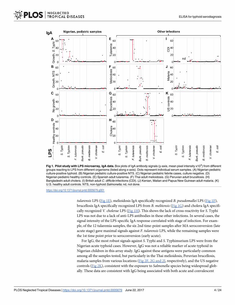
tularensis LPS (Fig 1E), melioidosis IgA specifically recognized B. pseudomallei LPS (Fig 1F),
brucellosis IgA specifically recognized LPS from B. melitensis (Fig 1G) and cholera IgA specifi-
cally recognized V. cholerae LPS (Fig 1H). This shows the lack of cross-reactivity for S. Typhi
LPS was not due to a lack of anti-LPS antibodies in these other infections. In several cases, the
signal intensity of the LPS-specific IgA response correlated with stage of infection. For exam-
ple, of the 12 tularemia samples, the six 2nd time-point samples after MA seroconversion (late
acute stage) gave maximal signals against F. tularensis LPS, while the remaining samples were
the 1st time point prior to seroconversion (early acute).
For IgG, the most robust signals against S. Typhi and S. Typhimurium LPS were from the
Nigerian acute typhoid cases. However, IgG was not a reliable marker of acute typhoid in
Nigerian children in this array study. IgG against these antigens were particularly common
among all the samples tested, but particularly in the Thai melioidosis, Peruvian brucellosis,
malaria samples from various locations (Fig 2F, 2G and 2J, respectively), and the US negative
controls (Fig 2K), consistent with the exposure to Salmonella species being widespread glob-
ally. These data are consistent with IgG being associated with both acute and convalescent
Fig 1. Pilot study with LPS microarray, IgA data. Box plots of IgA antibody signals (y-axis, mean pixel intensity x103) from different
groups reacting to LPS from different organisms (listed along x-axis). Dots represent individual serum samples. (A) Nigerian pediatric
culture-positive typhoid. (B) Nigerian pediatric culture-positive NTS. (C) Nigerian pediatric febrile cases, culture negative. (D)
Nigerian pediatric healthy controls. (E) Spanish adult tularemia. (F) Thai adult melioidosis. (G) Peruvian adult brucellosis. (H)
Bangladeshi adult cholera. (I) British adult C. difficile infections (CDI). (J) Kenian, Malian and Papua New Guinean adult malaria. (K)
U.S. healthy adult controls. NTS, non-typhoid Salmonella; nd, not done.
https://doi.org/10.1371/journal.pntd.0005679.g001
ELISA for typhoid serodiagnosis
PLOS Neglected Tropical Diseases | https://doi.org/10.1371/journal.pntd.0005679 June 22, 2017 4 / 24
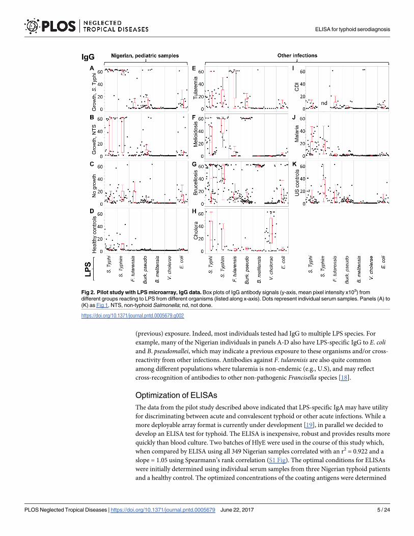
(previous) exposure. Indeed, most individuals tested had IgG to multiple LPS species. For
example, many of the Nigerian individuals in panels A-D also have LPS-specific IgG to E. coliand B. pseudomallei, which may indicate a previous exposure to these organisms and/or cross-
reactivity from other infections. Antibodies against F. tularenisis are also quite common
among different populations where tularemia is non-endemic (e.g., U.S), and may reflect
cross-recognition of antibodies to other non-pathogenic Francisella species [18].
Optimization of ELISAs
The data from the pilot study described above indicated that LPS-specific IgA may have utility
for discriminating between acute and convalescent typhoid or other acute infections. While a
more deployable array format is currently under development [19], in parallel we decided to
develop an ELISA test for typhoid. The ELISA is inexpensive, robust and provides results more
quickly than blood culture. Two batches of HlyE were used in the course of this study which,
when compared by ELISA using all 349 Nigerian samples correlated with an r2 = 0.922 and a
slope = 1.05 using Spearmann’s rank correlation (S1 Fig). The optimal conditions for ELISAs
were initially determined using individual serum samples from three Nigerian typhoid patients
and a healthy control. The optimized concentrations of the coating antigens were determined
Fig 2. Pilot study with LPS microarray, IgG data. Box plots of IgG antibody signals (y-axis, mean pixel intensity x103) from
different groups reacting to LPS from different organisms (listed along x-axis). Dots represent individual serum samples. Panels (A) to
(K) as Fig 1. NTS, non-typhoid Salmonella; nd, not done.
https://doi.org/10.1371/journal.pntd.0005679.g002
ELISA for typhoid serodiagnosis
PLOS Neglected Tropical Diseases | https://doi.org/10.1371/journal.pntd.0005679 June 22, 2017 5 / 24
by titration to be 1.25μg/ml for LPS, and 2.5μg/ml for HlyE (t1477). Two serum dilutions, 1/
100 and 1/200, were evaluated for the highest ratio when comparing heathy controls and cul-
ture-confirmed typhoid. For t1477 ELISA, 1/100 was selected, while for LPS ELISA, 1/200 was
found to give the higher ratio and selected for subsequent studies. Two secondary antibody
dilutions recommended by the manufacturer, 1/12,500 and 1/25,000, were evaluated for opti-
mal signal to background ratio. For IgA and IgM ELISA, 1/12500 dilution was selected, while
for IgG ELISA 1/25000 dilution was used. Once established, a standard operating procedure
was used throughout the study. Batches of ELISA plates were prepared by pre-coating plates
with antigen, blocking, and then storing dried at 4˚C in desiccated pouches until required for
use.
Detection of LPS-specific antibodies in serum samples by ELISA
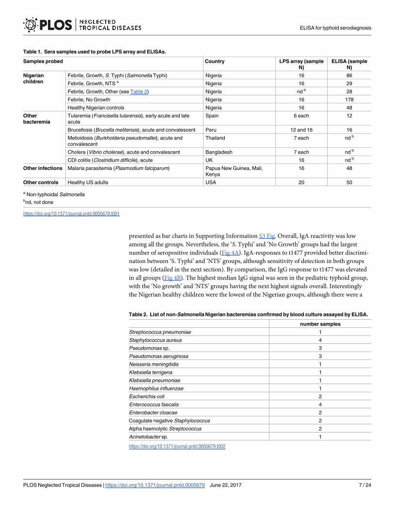
A total of 495 serum samples were used (Table 1) and tested for LPS-specific IgG, IgA, IgM
and IgA+IgM in separate ELISAs. The samples comprised 369 Nigerian pediatric samples,
consisting of culture-confirmed typhoid (“S. Typhi”, n = 86), non-typhoid Salmonella (“NTS”)
disease (n = 29) or other bacteremias (“Other”, n = 28; listed in Table 2), as well as febrile cases
that were blood culture-negative for any bacteria (“No Growth”, n = 178), and healthy Nige-
rian control children (“Healthy”, n = 48). Also tested by ELISA were well-defined sera from
tularemia, brucellosis, and malaria cases, as well as U.S. controls.
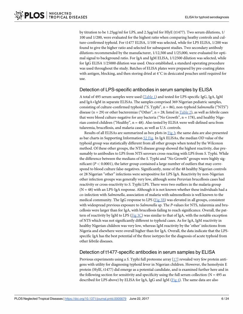
Results of all ELISAs are summarized as box plots in Fig 3; the same data are also presented
as bar charts in Supporting Information S2 Fig. In IgA ELISAs, the median OD value of the
typhoid group was statistically different from all other groups when tested by the Wilcoxon
method. Of these other groups, the NTS disease group showed the highest reactivity, due pre-
sumably to antibodies to LPS from NTS serovars cross-reacting with LPS from S. Typhi. While
the difference between the medians of the S. Typhi and “No Growth” groups were highly sig-
nificant (P< 0.0001), the latter group contained a large number of outliers that may corre-
spond to blood culture false-negatives. Significantly, none of the 48 healthy Nigerian controls
or 28 Nigerian “other” infections were seropositive for LPS IgA. Reactivity by non-Nigerian
other infection groups was generally very low, although some Peruvian brucellosis cases had
reactivity or cross-reactivity to S. Typhi LPS. There were two outliers in the malaria group
(N = 48) with an LPS-IgA response. Although it is not known whether these individuals had a
co-infection with Salmonella, association of malaria with salmonellosis is well known to the
medical community. The IgG response to LPS (Fig 3B) was elevated in all groups, consistent
with widespread previous exposure to Salmonella sp. The P-values for NTS, tularemia and bru-
cellosis were larger than for IgA, with brucellosis failing to reach significance. Overall, the pat-
tern of reactivity by IgM to LPS (Fig 3C) was similar to that of IgA, with the notable exception
of NTS which was not significantly different to typhoid cases. As for IgA, IgM reactivity in
healthy Nigerian children was very low, whereas IgM reactivity by the ‘other’ infections from
Nigeria and elsewhere were overall higher than for IgA. Overall, the data indicate that the LPS-
specific IgA has the best potential of the three isotypes for the diagnosis of acute typhoid from
other febrile diseases.
Detection of t1477-specific antibodies in serum samples by ELISA
Previous experiments using a S. Typhi full proteome array [17] revealed very few protein anti-
gens with utility for diagnosing typhoid fever in Nigerian children. However, the hemolysin E
protein (HylE, t1477) did emerge as a potential candidate, and is examined further here and in
the following section for sensitivity and specificity using the full serum collection (N = 495 as
described for LPS above) by ELISA for IgA, IgG and IgM (Fig 4). The same data are also
ELISA for typhoid serodiagnosis
PLOS Neglected Tropical Diseases | https://doi.org/10.1371/journal.pntd.0005679 June 22, 2017 6 / 24
presented as bar charts in Supporting Information S3 Fig. Overall, IgA reactivity was low
among all the groups. Nevertheless, the ‘S. Typhi’ and ‘No Growth’ groups had the largest
number of seropositive individuals (Fig 4A). IgA-responses to t1477 provided better discrimi-
nation between ‘S. Typhi’ and ‘NTS’ groups, although sensitivity of detection in both groups
was low (detailed in the next section). By comparison, the IgG response to t1477 was elevated
in all groups (Fig 4B). The highest median IgG signal was seen in the pediatric typhoid group,
with the ‘No growth’ and ‘NTS’ groups having the next highest signals overall. Interestingly
the Nigerian healthy children were the lowest of the Nigerian groups, although there were a
Table 1. Sera samples used to probe LPS array and ELISAs.
Samples probed Country LPS array (sample
N)
ELISA (sample
N)
Nigerian
children
Febrile, Growth, S. Typhi (Salmonella Typhi) Nigeria 16 86
Febrile, Growth, NTS a Nigeria 16 29
Febrile, Growth, Other (see Table 2) Nigeria nd b 28
Febrile, No Growth Nigeria 16 178
Healthy Nigerian controls Nigeria 16 48
Other
bacteremia
Tularemia (Francisella tularensis), early acute and late
acute
Spain 6 each 12
Brucellosis (Brucella melitensis), acute and convalescent Peru 12 and 16 16
Melioidosis (Burkholderia pseudomallei), acute and
convalescent
Thailand 7 each nd b
Cholera (Vibrio cholerae), acute and convalescent Bangladesh 7 each nd b
CDI colitis (Clostridium difficile), acute UK 16 nd b
Other infections Malaria parasitemia (Plasmodium falciparum) Papua New Guinea, Mali,
Kenya
16 48
Other controls Healthy US adults USA 20 50
a Non-typhoidal Salmonellabnd, not done
https://doi.org/10.1371/journal.pntd.0005679.t001
Table 2. List of non-Salmonella Nigerian bacteremias confirmed by blood culture assayed by ELISA.
number samples
Streptococcus pneumoniae 1
Staphylococcus aureus 4
Pseudomonas sp. 3
Pseudomonas aeruginosa 3
Neisseria meningitidis 1
Klebsiella terrigena 1
Klebsiella pneumoniae 1
Haemophilus influenzae 1
Escherichia coli 2
Enterococcus faecalis 4
Enterobacter cloacae 2
Coagulate negative Staphylococcus 2
Alpha haemolytic Streptococcus 2
Acinetobacter sp. 1
https://doi.org/10.1371/journal.pntd.0005679.t002
ELISA for typhoid serodiagnosis
PLOS Neglected Tropical Diseases | https://doi.org/10.1371/journal.pntd.0005679 June 22, 2017 7 / 24
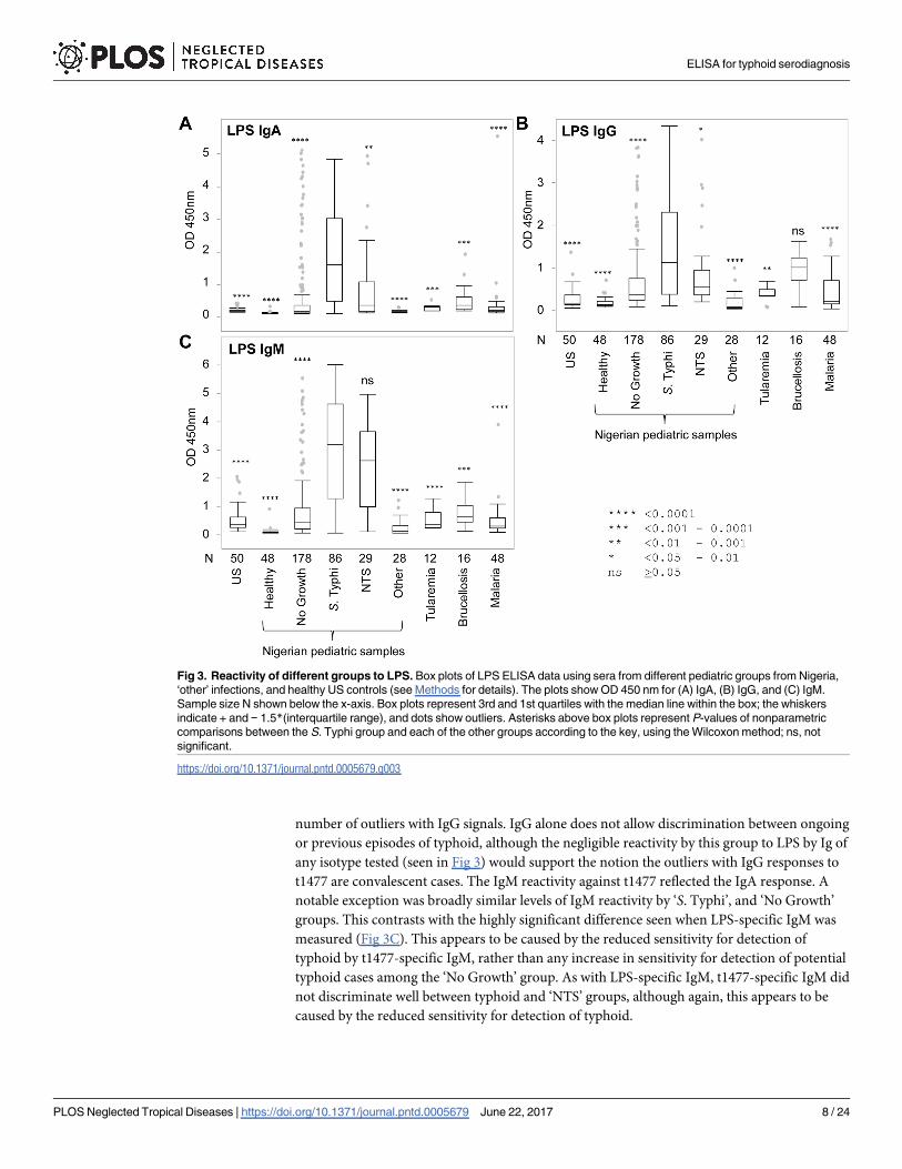
number of outliers with IgG signals. IgG alone does not allow discrimination between ongoing
or previous episodes of typhoid, although the negligible reactivity by this group to LPS by Ig of
any isotype tested (seen in Fig 3) would support the notion the outliers with IgG responses to
t1477 are convalescent cases. The IgM reactivity against t1477 reflected the IgA response. A
notable exception was broadly similar levels of IgM reactivity by ‘S. Typhi’, and ‘No Growth’
groups. This contrasts with the highly significant difference seen when LPS-specific IgM was
measured (Fig 3C). This appears to be caused by the reduced sensitivity for detection of
typhoid by t1477-specific IgM, rather than any increase in sensitivity for detection of potential
typhoid cases among the ‘No Growth’ group. As with LPS-specific IgM, t1477-specific IgM did
not discriminate well between typhoid and ‘NTS’ groups, although again, this appears to be
caused by the reduced sensitivity for detection of typhoid.
Fig 3. Reactivity of different groups to LPS. Box plots of LPS ELISA data using sera from different pediatric groups from Nigeria,
‘other’ infections, and healthy US controls (see Methods for details). The plots show OD 450 nm for (A) IgA, (B) IgG, and (C) IgM.
Sample size N shown below the x-axis. Box plots represent 3rd and 1st quartiles with the median line within the box; the whiskers
indicate + and − 1.5*(interquartile range), and dots show outliers. Asterisks above box plots represent P-values of nonparametric
comparisons between the S. Typhi group and each of the other groups according to the key, using the Wilcoxon method; ns, not
significant.
https://doi.org/10.1371/journal.pntd.0005679.g003
ELISA for typhoid serodiagnosis
PLOS Neglected Tropical Diseases | https://doi.org/10.1371/journal.pntd.0005679 June 22, 2017 8 / 24
Discrimination between groups by Receiver Operating Characteristics
(ROC) analysis
The accuracy of LPS and t1477 ELISAs to discriminate between Nigerian pediatric S. Typhi
patients and controls were determined by ROC analysis. Plots of true positive rate (sensitivity)
and false positive rate (1-specificity) for discriminating between typhoid cases and healthy chil-
dren are shown for LPS and t1477 in Fig 5A and 5B, respectively. Table 3 shows corresponding
percent specificity and sensitivity with either set at 90%, and areas under the curve (AUC).
With LPS, IgA and IgM both gave 94% sensitivity (at fixed specificity) when used alone, which
was increased slightly (to 95%) by combining the detection of IgA and IgM in the assay. Com-
bining IgA and IgM could also be achieved in silico by summing the OD450nm data for IgA
and IgM ELISAs performed individually (S4 Fig). LPS-specific IgA and IgM also give identical
specificity when used alone (98% at fixed sensitivity) which was unchanged by combining
Fig 4. Reactivity of different groups to hemolysin E (t1477). Box plots of t1477 ELISA data using sera from different pediatric
groups from Nigeria, ‘other’ infections, and healthy US controls (see Methods for details). The plots show OD 450 nm for (A) IgA, (B)
IgG, and (C) IgM. Sample size N shown below the x-axis. Box plots represent 3rd and 1st quartiles with the median line within the
box; the whiskers indicate + and − 1.5*(interquartile range), and dots show outliers. Asterisks above box plots represent P-values of
nonparametric comparisons between the S. Typhi group and each of the other groups according to the key, using the Wilcoxon
method; ns, not significant.
https://doi.org/10.1371/journal.pntd.0005679.g004
ELISA for typhoid serodiagnosis
PLOS Neglected Tropical Diseases | https://doi.org/10.1371/journal.pntd.0005679 June 22, 2017 9 / 24
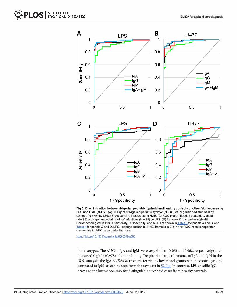
both isotypes. The AUC of IgA and IgM were very similar (0.963 and 0.968, respectively) and
increased slightly (0.978) after combining. Despite similar performance of IgA and IgM in the
ROC analysis, the IgA ELISAs were characterized by lower backgrounds in the control groups
compared to IgM, as can be seen from the raw data in S2 Fig. In contrast, LPS-specific IgG
provided the lowest accuracy for distinguishing typhoid cases from healthy controls.
Fig 5. Discrimination between Nigerian pediatric typhoid and healthy controls or other febrile cases by
LPS and HylE (t1477). (A) ROC plot of Nigerian pediatric typhoid (N = 86) vs. Nigerian pediatric healthy
controls (N = 48) by LPS. (B) As panel A, instead using HylE. (C) ROC plot of Nigerian pediatric typhoid
(N = 86) vs. Nigerian pediatric ‘other’ infections (N = 28) by LPS. (D) As panel C, instead using HylE.
Corresponding values for % sensitivity, % specificity, and AUC are shown in Table 3 for panels A and B, and
Table 4 for panels C and D. LPS, lipopolysaccharide; HylE, hemolysin E (t1477); ROC, receiver operator
characteristic; AUC, area under the curve.
https://doi.org/10.1371/journal.pntd.0005679.g005
ELISA for typhoid serodiagnosis
PLOS Neglected Tropical Diseases | https://doi.org/10.1371/journal.pntd.0005679 June 22, 2017 10 / 24
In the t1477 ELISAs, although AUC values of IgA and IgM were identical (0.968), IgA pro-
vided superior sensitivity than IgM (94% and 86%, respectively, at fixed specificity) and speci-
ficity (96% and 88%, respectively, at fixed sensitivity). Multiplexing IgA and IgM did not
increase sensitivity or specificity over IgA alone, although there was a modest increase in AUC
(to 0.976). As with LPS, t1477-specific IgG also gave lower accuracy than IgA or IgM for diag-
nosing acute typhoid. These data indicate both LPS and t1477-specific IgA and IgM provide
good discrimination between healthy Nigerian children and those with acute typhoid fever,
which is improved by detection of both IgA and IgM isotypes together.
We then compared acute typhoid with 28 Nigerian ‘other’ (non-Salmonella) infections
(listed in Table 2), since this is more relevant to the diagnosis of typhoid in the clinical setting.
ROC Plots are shown in Fig 5C and 5D, with corresponding AUC, and percent sensitivity and
specificity given in Table 4. Here, LPS-specific IgA and IgM give comparable sensitivity when
used alone (86% and 87%, respectively, at fixed specificity), which is increased to 90% when
IgA and IgM are combined. LPS-specific IgM provided considerably greater specificity than
IgA when used alone (82% and 75%, respectively, at fixed sensitivity), which is dramatically
increased (to 96%) when combined. The AUC is also increased slightly by combining IgA and
IgM to 0.938. LPS-specific IgG provides the lowest sensitivity and specificity of all three
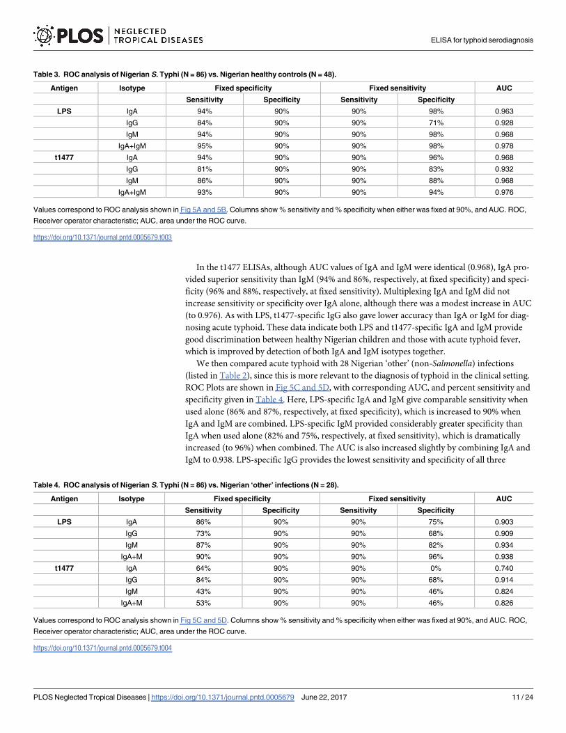
Table 3. ROC analysis of Nigerian S. Typhi (N = 86) vs. Nigerian healthy controls (N = 48).
Antigen Isotype Fixed specificity Fixed sensitivity AUC
Sensitivity Specificity Sensitivity Specificity
LPS IgA 94% 90% 90% 98% 0.963
IgG 84% 90% 90% 71% 0.928
IgM 94% 90% 90% 98% 0.968
IgA+IgM 95% 90% 90% 98% 0.978
t1477 IgA 94% 90% 90% 96% 0.968
IgG 81% 90% 90% 83% 0.932
IgM 86% 90% 90% 88% 0.968
IgA+IgM 93% 90% 90% 94% 0.976
Values correspond to ROC analysis shown in Fig 5A and 5B. Columns show % sensitivity and % specificity when either was fixed at 90%, and AUC. ROC,
Receiver operator characteristic; AUC, area under the ROC curve.
https://doi.org/10.1371/journal.pntd.0005679.t003
Table 4. ROC analysis of Nigerian S. Typhi (N = 86) vs. Nigerian ‘other’ infections (N = 28).
Antigen Isotype Fixed specificity Fixed sensitivity AUC
Sensitivity Specificity Sensitivity Specificity
LPS IgA 86% 90% 90% 75% 0.903
IgG 73% 90% 90% 68% 0.909
IgM 87% 90% 90% 82% 0.934
IgA+M 90% 90% 90% 96% 0.938
t1477 IgA 64% 90% 90% 0% 0.740
IgG 84% 90% 90% 68% 0.914
IgM 43% 90% 90% 46% 0.824
IgA+M 53% 90% 90% 46% 0.826
Values correspond to ROC analysis shown in Fig 5C and 5D. Columns show % sensitivity and % specificity when either was fixed at 90%, and AUC. ROC,
Receiver operator characteristic; AUC, area under the ROC curve.
https://doi.org/10.1371/journal.pntd.0005679.t004
ELISA for typhoid serodiagnosis
PLOS Neglected Tropical Diseases | https://doi.org/10.1371/journal.pntd.0005679 June 22, 2017 11 / 24
isotypes. By comparison, the relative accuracy of t1477 in ELISAs for diagnosing acute typhoid
was lower for all three Ig isotypes compared to LPS, and also reduced relative to discrimination
of typhoid vs. healthy controls. Unexpectedly IgG emerged as the isotype with the highest sen-
sitivity and specificity of t1477-specific Igs. It is possible this is restricted to childhood, where
there is relatively less lifetime exposure to Salmonella than older children and adults, combined
with a robust IgG response during typhoid fever.
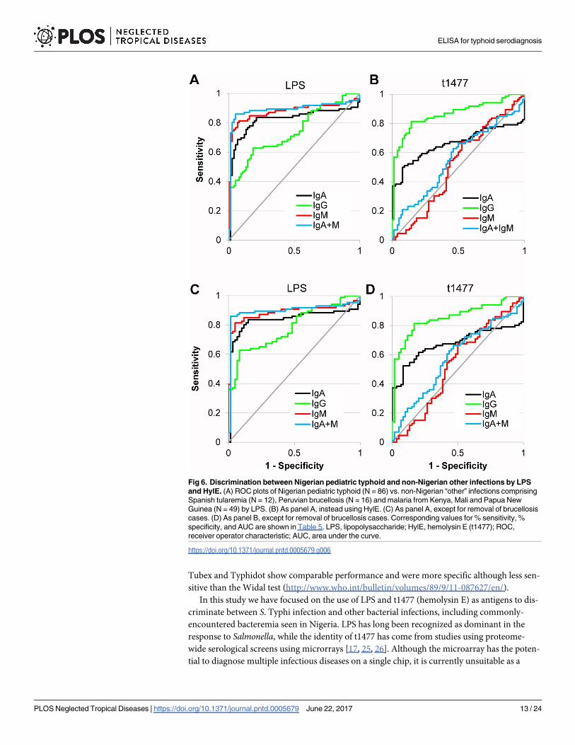
We also compared the ability of LPS and t1477 to discriminate between Nigerian pediatric
typhoid and additional samples from ‘other’ non-Salmonella infections obtained from loca-
tions outside Nigeria, namely tularemia (Spain, N = 12), brucellosis (Peru, N = 16) and malaria
(various sources, N = 48). ROC plots are shown in Fig 6A (LPS) and 6B (t1477), with corre-
sponding AUC and percent sensitivity and specificity given in Table 5. Data were broadly sim-
ilar to that seen with Nigerian ‘other’ infections, with combined IgA+IgM providing the most
accurate test when using LPS, and IgG providing the most accurate test when using t1477. As
noted earlier, brucellosis samples were prominent among the ‘other’ infections for cross-reac-
tivity to S. Typhi LPS. If these samples were removed from the analysis (Fig 6C and 6D) there
was a slight increase in sensitivity and specificity in almost all situations, with the exception of
t1477-specific IgG (Table 5, values in parenthesis).
Finally we explored the effect of multiplexing LPS and t1477 antigens on the accuracy of the
test for typhoid compared to each antigen alone (Fig 7 and Table 6). Multiplexing LPS IgA
with t1477IgG in silico increased accuracy compared to either alone, while multiplexing LPS
IgA+IgM (as mixed secondary antibodies) with t1477 IgG in silico increased the accuracy
further.
Discussion
In countries in Sub-Saharan Africa, where typhoid and non-typhoidal salmonellosis are major
causes of bacterial sepsis in children, accurate and rapid point-of-care tests are urgently needed
to replace existing diagnostic methods. Culture of S. Typhi organisms from bone marrow is
the gold standard, but because it is invasive, blood culture is often a more practical, albeit less
sensitive, alternative. Blood or bone marrow culture is also slow (2–3 days to arrive at a diag-
nosis), and empiric broad-spectrum antibiotic treatment is often initiated without a diagnosis
being made.
The traditional Widal’s test, which is based on the agglutination of inactivated SalmonellaTyphi and Paratyphi A organisms by antibodies to flagellin and LPS (H and O antigens,
respectively) is rapid, inexpensive and requires no instrumentation. However, interpretation
of the results must be made with caution. Sensitivity of the Widal’s test is lower in the early
stage of infection when antibody titers are low. The test also fails to discriminate between
acute from convalescent infection, leading to reduced sensitivity in endemic settings [20].
Although sensitivity can be improved if a follow-up sample is tested [21], this is not an option
for rapid diagnosis. The test also lacks specificity owing to cross-reactivity with antibodies
against closely-related NTS serovars [22] and other bacteria, notably Brucella [16]. Misuse of
the Widal’s test has contributed to over-diagnosis of Salmonella infection, inappropriate anti-
biotic use, and the emergence of drug resistance [23].
Recent alternatives for serodiagnosis of typhoid include the Tubex test for LPS-specific IgM
and the Typhidot test for IgG or IgM against a 50kDa outer membrane protein [24]. The
Tubex test format is based on the interference by patient serum antibodies with the agglutina-
tion of latex beads coated with O9-specific monoclonal antibody and S. Typhi LPS-coated
magnetic beads. The Typhidot test is a pre-dotted antigen strip. Neither test is currently con-
figured for detection of IgA. Both have been evaluated in several Asian and African study sites;
ELISA for typhoid serodiagnosis
PLOS Neglected Tropical Diseases | https://doi.org/10.1371/journal.pntd.0005679 June 22, 2017 12 / 24
Tubex and Typhidot show comparable performance and were more specific although less sen-
sitive than the Widal test (http://www.who.int/bulletin/volumes/89/9/11-087627/en/).
In this study we have focused on the use of LPS and t1477 (hemolysin E) as antigens to dis-
criminate between S. Typhi infection and other bacterial infections, including commonly-
encountered bacteremia seen in Nigeria. LPS has long been recognized as dominant in the
response to Salmonella, while the identity of t1477 has come from studies using proteome-
wide serological screens using microrrays [17, 25, 26]. Although the microarray has the poten-
tial to diagnose multiple infectious diseases on a single chip, it is currently unsuitable as a
Fig 6. Discrimination between Nigerian pediatric typhoid and non-Nigerian other infections by LPS
and HylE. (A) ROC plots of Nigerian pediatric typhoid (N = 86) vs. non-Nigerian “other” infections comprising
Spanish tularemia (N = 12), Peruvian brucellosis (N = 16) and malaria from Kenya, Mali and Papua New
Guinea (N = 49) by LPS. (B) As panel A, instead using HylE. (C) As panel A, except for removal of brucellosis
cases. (D) As panel B, except for removal of brucellosis cases. Corresponding values for % sensitivity, %
specificity, and AUC are shown in Table 5. LPS, lipopolysaccharide; HylE, hemolysin E (t1477); ROC,
receiver operator characteristic; AUC, area under the curve.
https://doi.org/10.1371/journal.pntd.0005679.g006
ELISA for typhoid serodiagnosis
PLOS Neglected Tropical Diseases | https://doi.org/10.1371/journal.pntd.0005679 June 22, 2017 13 / 24
point-of-care test for many clinics in its current format. An accurate, more deployable test,
particularly if configured into a format able to provide a result in<30 minutes, could help
curb the inappropriate use of antibiotics and stem the rise in antibiotic resistance in Nigeria.
The data presented here indicate LPS-specific IgA (or IgA+M combined) discriminate well
between Nigerian children with typhoid and healthy Nigerian children (AUC = 0.963 and
0.978, respectively; Table 3). More importantly for the clinical setting, LPS-specific IgA (or IgA
+M combined) also discriminates between Nigerian children with typhoid and children with
‘other’ (non-Salmonella) infections (AUC = 0.903 and 0.938, respectively; Table 4). Similarly,
discrimination between typhoid cases and healthy children using t1477-specific IgA (Table 3)
was comparable to that obtained with LPS-specific IgA, although discrimination between
typhoid and ‘other’ cases using t1477-specific IgA (Table 4) was far less accurate than for LPS-
specific IgA. One possibility is proteins antigenically related to S. Typhi t1477 hemolysin E are
found in one or more of the other bacterial infections represented in the collection (see
Table 2). Such potential cross-reactivity would reduce the diagnostic utility of the antigen for
typhoid.
LPS-specific IgG provided less accuracy for discriminating between Nigerian pediatric
typhoid and healthy Nigerian children (Table 3), which was reduced further when discriminat-
ing typhoid with ‘other’ infections (Table 4). It is possible that IgG titers remain elevated for
longer than IgA, thereby making it more difficult to discriminate between acute and previous
or convalescent infections using IgG. This may be less of an issue in children where lifetime
exposure to Salmonella species will likely be less than in adults. Although we have not exam-
ined Nigerian adults in this study, the expectation is they will have higher and more durable
IgG titers to both LPS and t1477 than in children. This notion is supported for LPS by the pilot
LPS array (Fig 2) in which all the non-Nigerian samples (i.e., panels E through K) were from
adults. Thus, the median IgG signal of the healthy Nigerian children was lowest among all the
groups tested, including adults from two non-endemic sites, the US (Fig 2K) and UK (Fig 2I).
It remains to be determined whether LPS- and/or t1477-specific IgA has any utility for diag-
nosing typhoid in adults.
Unexpectedly, t1477-specific IgG performed better than LPS-specific IgG for discriminat-
ing between Nigerian pediatric typhoid and healthy Nigerian children (Table 3), and between
Nigerian pediatric typhoid with ‘other’ infections (Table 4). Indeed, t1477-specific IgG also
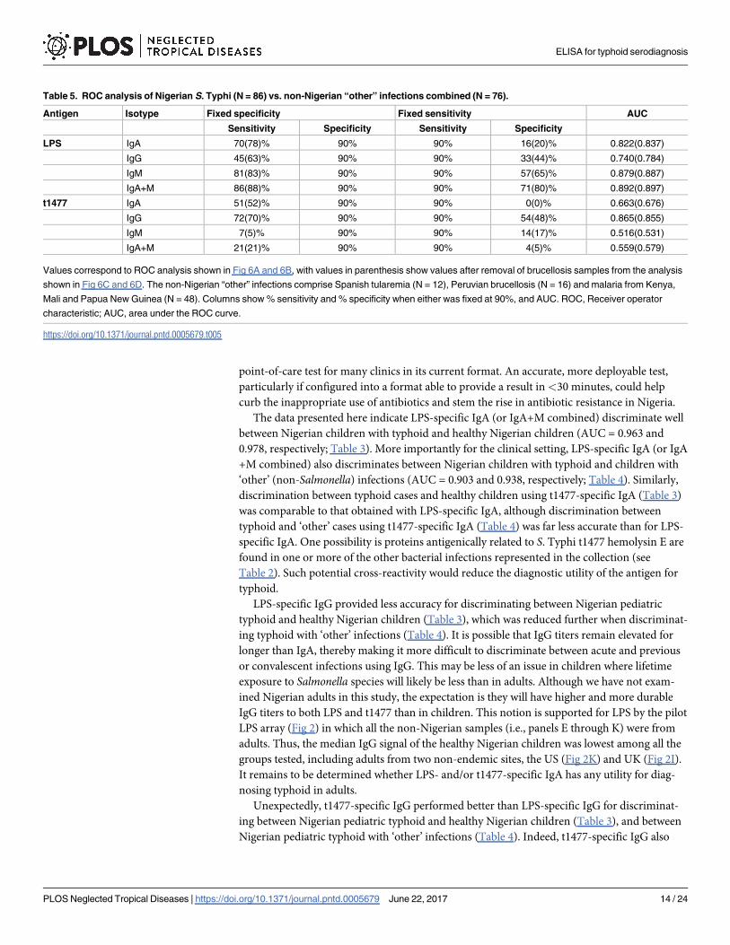
Table 5. ROC analysis of Nigerian S. Typhi (N = 86) vs. non-Nigerian “other” infections combined (N = 76).
Antigen Isotype Fixed specificity Fixed sensitivity AUC
Sensitivity Specificity Sensitivity Specificity
LPS IgA 70(78)% 90% 90% 16(20)% 0.822(0.837)
IgG 45(63)% 90% 90% 33(44)% 0.740(0.784)
IgM 81(83)% 90% 90% 57(65)% 0.879(0.887)
IgA+M 86(88)% 90% 90% 71(80)% 0.892(0.897)
t1477 IgA 51(52)% 90% 90% 0(0)% 0.663(0.676)
IgG 72(70)% 90% 90% 54(48)% 0.865(0.855)
IgM 7(5)% 90% 90% 14(17)% 0.516(0.531)
IgA+M 21(21)% 90% 90% 4(5)% 0.559(0.579)
Values correspond to ROC analysis shown in Fig 6A and 6B, with values in parenthesis show values after removal of brucellosis samples from the analysis
shown in Fig 6C and 6D. The non-Nigerian “other” infections comprise Spanish tularemia (N = 12), Peruvian brucellosis (N = 16) and malaria from Kenya,
Mali and Papua New Guinea (N = 48). Columns show % sensitivity and % specificity when either was fixed at 90%, and AUC. ROC, Receiver operator
characteristic; AUC, area under the ROC curve.
https://doi.org/10.1371/journal.pntd.0005679.t005
ELISA for typhoid serodiagnosis
PLOS Neglected Tropical Diseases | https://doi.org/10.1371/journal.pntd.0005679 June 22, 2017 14 / 24

Fig 7. Discrimination between Nigerian pediatric typhoid and Nigerian other febrile cases by data
multiplexed in silico. ROC plots of Nigerian typhoid patients (N = 86) vs. Nigerian ‘other’ infections (N = 28).
Corresponding values for % sensitivity, % specificity and AUC are shown in Table 6. Antigens are ranked in
ascending AUC. LPS, lipopolysaccharide; t1477, (hemolysin E, HylE); ROC, receiver operator characteristic;
AUC, area under the curve.
https://doi.org/10.1371/journal.pntd.0005679.g007
Table 6. Discrimination between Nigerian pediatric typhoid and Nigerian other febrile cases by data multiplexed in silico.
Antigen and Isotype Fixed specificity Fixed sensitivity AUC
Sensitivity Specificity Sensitivity Specificity
LPS IgA 86% 90% 90% 75% 0.903
LPS IgA+M 90% 90% 90% 96% 0.929
t1477 IgG 84% 90% 90% 68% 0.914
LPS IgA + t1477 IgG 87% 90% 90% 82% 0.939
LPS IgA+M + t1477 IgG 92% 90% 90% 93% 0.950
Values correspond to ROC analysis shown in Fig 7. Columns show % sensitivity and % specificity when either was fixed at 90%, and AUC. ROC, Receiver
operator characteristic; AUC, area under the ROC curve.
https://doi.org/10.1371/journal.pntd.0005679.t006
ELISA for typhoid serodiagnosis
PLOS Neglected Tropical Diseases | https://doi.org/10.1371/journal.pntd.0005679 June 22, 2017 15 / 24
performed better than t1477-specific IgA and IgM for discriminating between typhoid and
‘other’ infections in Nigerian children. It is possible this diagnostic performance occurs only in
children, where there is less lifetime exposure to Salmonella. It is anticipated that t1477-specific
IgG will have less utility for diagnosing acute typhoid in older children and adults where the
IgG titers from convalescent infections are likely to be much higher.
Finally we also compared the ability of LPS and t1477 to discriminate between typhoid and
non-Nigerian ‘other’ infections from other locations around the world. In the ELISA, LPS-spe-
cific IgA+M provided excellent sensitivity and specificity, although we did notice detection of
some Peruvian brucellosis cases using S. Typhi LPS (Fig 3 and Table 5). There are accounts in
the literature of antigenic cross-reactivity between Brucella sp. and S. enterica serotype Urbana
[16, 27, 28] which raises the possibility of cross-reactivity between antibodies generated during
human brucellosis and Salmonella antigens. Although brucellosis is rare in Nigeria, if discrimi-
nation between acute typhoid and brucellosis is necessary, one option might be to utilize sero-
diagnostic B. melitensis antigens discovered previously [29–32] or B. melitensis LPS (Fig 1G) to
assist in positive identification of brucellosis cases. The accuracy of t1477-specific IgA (or IgA
+M) was lower than for LPS, consistent with its performance in discriminating between
typhoid and Nigerian “other” infections.
LPS has received considerable interest as a potential diagnostic antigen for typhoid and for
the basis of alternative assays to the Widal’s test. In one longitudinal study [33], anti-LPS IgA
and IgM titers were seen to peak around d11-21 and decline thereafter, whereas IgG titers
remained elevated and did not decline as rapidly. Other studies have also shown the transient
nature of anti-LPS IgA in typhoid in saliva samples [34, 35] as well as in sera of gastroenteritis
caused by non-typhoidal Salmonella serovars [36]. Thus, LPS-specific IgA appears to be a use-
ful marker of acute Salmonellosis owing to its transient appearance after infection. The tran-
sient nature of IgA appears to be a peculiarity of LPS, and possibly other T-independent
antigens, since serum and mucosal IgA responses to bacteria are generally long-lived [37–39].
In the present study, the LPS molecule did not discriminate well between typhoid and NTS,
presumably because of the presence of shared epitopes present in the conserved lipid A and
core oligosaccharide regions [40]. However, the more variant O-polysaccarides where serovar-
specific epitopes of the O-antigen are located may discriminate between antibodies engen-
dered by typhoid and NTS serovars. Salmonella O-polysaccharides have been produced from
bacterial extracts and conjugated to protein carriers for use as subunit vaccines [41–43],
although their utility as specific diagnostics is less well explored. In one such study, the S.
Typhi O-polysaccharide O:1,9,12 performs well in IgG dot blots as a discriminator between
typhoid and other acute infections or healthy controls, although IgA and the ability to discrim-
inate between typhoid and NTS were not examined [44]. Neither the use of LPS nor measure-
ment of IgA for diagnosis of typhoid is novel, but when used together appear to represent a
good marker for acute infection in Nigerian children.
The t1477/hemolysin E (HylE, also known as cytolysin A or CylA) protein is a known dom-
inant antigen in the antibody response to S. Typhi infection [17, 25, 26, 45]. Its utility as a
potential serodiagnostic for typhoid has been demonstrated independently in a study of differ-
ent Ig isotypes in 50 culture-confirmed typhoid cases [46]. In that study, IgA was the most sen-
sitive, detecting 28/50 cases using a cut-off defined by the isotype-matched responses by
healthy controls and other febrile infections. IgG was second most sensitive (19/50), and IgM
least sensitive (3/50). A subsequent pilot study has demonstrated the utility of anti-HylE IgA in
saliva as a biomarker for acute typhoid fever [47]. S. Typhi HylE is a 302 amino-acid long
transmembrane protein with a helix hydrophobic segment located between residues 179 and
199. Along with homologs in other bacteria, such as the prototypic ClyA in E. coli, S. Typhi
HylE belongs to a family of important pore-forming virulence factors of bacterial pathogens
ELISA for typhoid serodiagnosis
PLOS Neglected Tropical Diseases | https://doi.org/10.1371/journal.pntd.0005679 June 22, 2017 16 / 24
that assemble in cell membranes [48]. The HylE gene (t1477) is present in human-specific
typhoid serovars (Typhi and Paratyphi) but absent from others (e.g., S. Typhimurium). In Fig
4, IgA reactivity by the 29 Nigerian children with invasive NTS (iNTS) is negligible with the
exception of two outliers with low reactivity. However, sensitivity of t1477-specific IgA for
detection of typhoid is also low, indicating this antigen is unlikely to have utility for discrimi-
nating iNTS and typhoid.
Methods
Ethics statement
This study was conducted with informed consent and approved by the Ethics Committees of
the Federal Capital Territory of Nigeria, Federal Medical Center Keffi, Aminu Kano Teaching
Hospital and University of Nebraska Medical Center (UNMC), Omaha Institutional Review
Board (IRB). We used written consent provided by parent or guardian of each child. The pro-
cess was approved by both local IRB and UNMC IRB.
Sera from Spanish tularemia cases were provided by Drs. Raquel Escudero and Pedro
Anda, Instituto de Salud Carlos III, Madrid, Spain. Human subjects approval from Comite de
Bioetica y Bienestar Animal, Instituto de Salud Carlos III (approval no. PI 33). Sera from Thai
melioidosis cases were provided by Direk Limmathurotsakul and Narisara Chantratita, Mahi-
dol University, Thailand. Ethical approval for the study was from the Ministry of Public
Health, Royal Government of Thailand, and the Oxford Tropical Research Ethics Committee.
Sera from Peruvian brucellosis cases were collected with human subjects approval from the
Human Research Protection Committee of the University of California San Diego, the Comite
de Etica of Universidad Peruana Cayetano Heredia, Lima, Peru, and the Comite de Etica of
Asociacion Benefica PRISMA, Lima, Peru. Sera from Bangladeshi cholera cases were provided
by Drs. Edward Ryan, Richelle Charles and Firdausi Qadri, Massachusetts General Hospital,
Boston, MA. Human subjects approval by IRB protocol # 1999P009116 and International Cen-
tre for Diarrhoeal Disease Research, Bangladesh (ICDDRB) #PR-11041. Sera from Clostridiumdifficile infections were collected with ethical approval from the University of Liverpool
Research Ethics Committee (#08/H1005/32), and each patient provided written informed con-
sent prior to recruitment. Malaria sera were collected with human subjects approvals from
Institutional Review Boards at University Hospitals Case Medical Center and the Kenya Medi-
cal Research Institute Ethical Review Committee [49], the Medical Research Advisory Council,
PNG [50], and the Ethics Committee of the Faculty of Medicine, Pharmacy, and Odonto-Sto-
matology and the Institutional Review Board at the National Institute of Allergy and Infectious
Diseases, National Institutes of Health [51]. Sera from healthy US adults were collected under
UCI IRB protocol #2007–5896. Sera were provided to the University of California Irvine
(UCI) for assay without patient identifiers and were classified as exempt status by the UCI
Institutional Review Board.
Sera
A retrospective study was designed using a convenience series of sera samples from Nigerian
pediatric febrile cases and healthy controls, as well as other infectious diseases from other loca-
tions outside Nigeria, which were assayed by ELISA and/or LPS microarray (Table 1). The
Nigerian samples were collected between 2009 and 2014 from children aged 8 months—13
years (median approximately 4 years) who presented to primary or secondary health centers in
central and northwest Nigeria with an acute febrile illness and other symptoms that were sug-
gestive of bacteremia. The duration of symptoms ranged from about 3–10 days with a median
of 5 days, as documented in the clinical data captured during enrollment. S. Typhi is the
ELISA for typhoid serodiagnosis
PLOS Neglected Tropical Diseases | https://doi.org/10.1371/journal.pntd.0005679 June 22, 2017 17 / 24
leading cause of childhood bacteremia in this area [52]. Baseline demographics of this popula-
tion have been described previously [52, 53]. Following informed consent from the parent or
guardian, blood was obtained aseptically from a peripheral vein for blood culture and simulta-
neously an aliquot for serum separation was saved. Blood sampling and processing were as
previously described [52, 53]. Briefly, only aerobic blood culture bottles were used and held in
a Bactec 9050 incubator (Becton Dickinson, Temse, Belgium) for a maximum of 5 days. Bacte-
ria were identified by morphology, and for Enterobacteriacae, by use of an API 20 E rapid
identification system (BioMerieux, Marcy-l’Etoile, France). Bacterial isolates were stored in
skimmed milk at -70˚C, and further characterized at the Clinical Microbiology laboratory,
University of Nebraska Medical Center. Bacteremia was defined as the isolation of at least 1
noncontaminant bacteria from the admission blood culture. These samples comprised chil-
dren with typhoid (N = 86), non-typhoid Salmonella (NTS) infections (N = 29), other bacter-
emias (N = 28), and febrile cases that were culture negative (‘No Growth’, N = 178). Samples
sizes were determined by availability during the collection period. No samples with missing or
indeterminate culture test results were used in this study. In addition, we also obtained sera
from healthy Nigerian children enrolled from immunization clinics in the same facilities as
controls (N = 48). These children present for routine immunizations and typically are in a sta-
ble state of health. Only children who were asymptomatic and did not have a history of a
febrile illness in the past month, or had taken any antibiotic during the same period, were eligi-
ble. No blood cultures were performed on the healthy controls.
For the pilot LPS array (detailed below), an expanded collection of samples from “other”
control infections from other countries were tested in addition to Nigerian samples discussed
above, as follows. 1) Tularemia sera (N = 12) from a 2007 Spanish outbreak of Francisella tular-ensis subsp. holarctica. These consisted of paired samples from 6 acute cases that were seronega-
tive by microagglutination (MA) test at the 1st time point at presentation and which
seroconverted by the 2nd time point approximately 2 weeks later. These samples were found
previously to be seropositive for F. tularensis subsp. tularensis (FTT) strain Schu S4 antigens at
both time points using a proteome microarray [18]. 2) Melioidosis sera from Thailand
(N = 14). Samples were collected in 2004 from patients presenting with symptoms of melioido-
sis, and were diagnosed by indirect hemagglutination assay (IHA) and blood and throat swab
culture, as described previously [54]. 3) Brucellosis sera collected prior to 2008 from an endemic
region of Peru (N = 28), previously shown to be seropositive using a Brucella melitensis prote-
ome microarray [31, 32]. Samples probed were culture positive/Rose-Bengal positive (N = 12)
and culture negative/Rose Bengal positive (N = 16). These correspond to samples taken on the
first day (acute infection) and within 6 weeks after obtaining the first sample (convalescent
infection).4) Cholera sera from Bangladesh collected between 2008 and 2010 presenting to the
International Centre for Diarrhoeal Disease Research, Bangladesh (ICDDRB) hospital with
acute watery and stool culture confirmed V. cholerae O1 infection. Following informed consent,
venous blood was collected from adults (age 18–55 years) at the acute phase of infection (N = 7)
after clinical stabilization (day 2), and again at convalescent phases of infection (d7 and 30;
N = 7). 5) Sera from Clostridium difficile infections (CDI) from diagnosed acute cases in the UK
collected between 2008 and 2012 (N = 16) [55]. Each patient was followed-up for minimum
period of 30 days initially and then for 1 year from notes for the collection of additional demo-
graphics clinical outcome information. 6) Symptomatic malaria cases from Kenya, Papua New
Guinea and Mali (N = 48). These were diagnosed with Plasmodium falciparum parasitemia, and
all defined as seropositive using different iterations of P. falciparum protein arrays derived from
strain 3D7 [56]. 7) Healthy US adults from a non-endemic area (Orange County, CA), Adher-
ence to Standards for Reporting of Diagnostic Accuracy Studies (STARD) is shown by the flow-
chart (S5 Fig) and checklist (S1 Text) in the Supporting Information.
ELISA for typhoid serodiagnosis
PLOS Neglected Tropical Diseases | https://doi.org/10.1371/journal.pntd.0005679 June 22, 2017 18 / 24
LPS microarray
Lipopolysaccharides (LPS) were obtained as follows: 1) LPS from S. Typhosa (= S. Typhi) was
purchased from Sigma-Aldrich (Cat. #L2387); 2) LPS from S. Typhimurium was purchased
from Sigma-Aldrich (Cat. #L6511); 3) LPS from Francisella tularensis Subsp. novicida was puri-
fied from the live vaccine strain (LVS) (DSTL batch #B07/3564), as described [57]; 4) LPS
from Burkholderia pseudomallei was purified from strain K96243 (DSTL batch #B07/3558), as
described [58]; 5) LPS from Brucella melitensis was purified from strain 16M, as described
[31]; 6) LPS from V. cholerae O1 was purified from Ogawa (strain X-25049) and Inaba (strain
T19479) serotypes, as described [59]; 7) Escherichia coli 055:B5 LPS was purchased from
Sigma-Aldrich (Cat. #L2880). Each LPS species was diluted in PBS buffer, pH 7.3–7.5 (EMD
Millipore Corp., Billerica, MA; Cat. #6506-OP) and printed on nitrocellulose-coated glass
slides (Oncyte Avid from Grace Bio-Labs, Bend, OR) using a GeneMachines Omnigrid 100
array printer, and printed at a concentration of 0.1 μg/ml. This concentration was determined
previously in titration experiments to be the lowest concentration able to provide near maxi-
mal signals. Performers of the LPS microarray assays were blinded to the identity of the sam-
ples until after the assays were completed. LPS arrays were probed for 18h at 4˚C with sera
diluted 1/100 in protein microarray blocking buffer (Maine Manufacturing, GVS North Amer-
ica, Sanford, ME) supplemented with E. coli lysate (Antigen Discovery Inc, Irvine, CA). Bound
IgG and IgA were then detected using secondary antibodies conjugated to biotin followed by
streptavidin conjugated to quantum dots, and then visualized in an ArrayCAMarray imager,
as described previously [19].
ELISA
Hemolysin E protein (HylE, gene t1477 from S. Typhi Ty2 strain) was expressed in E. coli and
purified as described previously [17]. LPS from S. Typhi was as described above for microar-
rays. ELISAs were performed as described [60]. Briefly, antigens were coated onto microtiter
plates (ThermoScientific, Walham, MA) at concentrations 1.25 μg/ml (LPS) and 2.5 μg/ml
(HylE) in TBS (100μl/well) overnight at 4˚C. The coating concentrations were determined pre-
viously for each antigen by serial dilution experiments. The following day, plates were washed
4 times in 1x TBS containing 0.05% Tween20 (T-TBS; ThermoScientific) and blocked with
casein/TBS blocking buffer (ThermoScientific) for 1-2h (300 μl/well). Blocking buffer was then
decanted, and the plates air-dried and stored in desiccated foil pouches at 4˚C until required
for use. Performers of the ELISAs were blinded to the identity of the samples until after the
assays were completed. For ELISA assay, sera were diluted to 1/200 (LPS) and 1/100 (HylE) in
casein/TBS blocking buffer containing E. coli lysate (GenScript, Piscataway, NJ) at 1.5 mg/ml
final concentration, and incubated for 30 min prior to placing into the plates. Plates were incu-
bated for 45 min with gentle rocking at room temperature (RT). After washing with T-TBS
goat anti-human IgG-, IgA- or IgM-HRP conjugates (Bethyl Laboratories, Inc., Montgomery,
TX) diluted 1/25,000 (IgG) or 1/12,500 (IgA, IgM) in Guardian Stabilizer (ThermoScientific)
were added to wells (100 μl /well) and incubated for 45 min at RT (100 μl/well). After washing
with T-TBS, plates were developed by adding 100 μl/well SureBlueReserve TMB developer
(Kirkegaard and Perry Laboratories, Inc., Gaithersburg, MD) for 10 min in the dark. Develop-
ment was stopped by addition of 100 μl/well of 0.2M H2SO4 and OD read at 450 nm in a Mul-
tiskan FC plate reader.
Statistical methods
ELISA data were collected at OD450nm and data were corrected by the positive control
between runs. Dot plots and comparisons between medians of different groups using the
ELISA for typhoid serodiagnosis
PLOS Neglected Tropical Diseases | https://doi.org/10.1371/journal.pntd.0005679 June 22, 2017 19 / 24
Wilcoxon method, were produced in JMP (SAS Institute, Inc., Cary, NC, USA). Receiver oper-
ator characteristic (ROC) analyses were performed between patient groups for each antigen
with a varying threshold cut off in the R statistical environment using ROCR. Plots of false pos-
itive vs. true positive plots were made, from which areas under the curve (AUC) and sensitivity
and 1-specificity values were calculated for each antigen(s).
Supporting information
S1 Fig. Scatter plot comparison of IgG ELISA data using two different batches of hemoly-
sin E antigen (HylE, t1477) each assayed using the same 349 Nigerian samples; r2 = 0.922
and slope = 1.05 using Spearmann’s rank correlation.
(BMP)
S2 Fig. Bar chart of LPS ELISA data; data provided in file “S2 DATA_ELISA”.
(BMP)
S3 Fig. Bar chart of hemolysin E (HylE, t1477) ELISA data; data provided in file “S2
DATA_ELISA”.
(BMP)
S4 Fig. Scatter plot of IgA+IgM multiplex ELISAs using Nigerian typhoid and healthy
pediatric samples obtained by either combining secondary antibodies in the assay
(“combo”, x-axis) or by combining data of IgA and IgM obtained individually (“in silico”,
y-axis); data provided in file “S2 DATA_ELISA”.
(BMP)
S5 Fig. STARD flow chart.
(BMP)
S1 Text. STARD checklist.
(DOCX)
S1 DATA_LPS_ARRAY. LPS microarray data, Excel file.
(XLSX)
S2 DATA_ELISA. ELISA data, Excel file.
(XLSX)
Acknowledgments
The authors wish to thank the following collaborators: Dr. Raquel Escudero and Dr. Pedro
Anda (Instituto de Salud Carlos III, Madrid, Spain) for Spanish tularemia serum samples;
Richard Titball and Joann L. Prior (Defence Science and Technology Laboratory, Porton
Down, UK) for gifts of LPS from F. tularensis LVS and B. pseudomallei; Edward Ryan, Richelle
Charles and Firdausi Qadri (Massachusetts General Hospital, Boston, MA) for gifts of V. cho-lerae LPS and Bangladesh cholera plasma samples; James Kazura (Case Western Reserve Uni-
versity); for gift of malaria serum samples from Kenya and Papua New Guinea; Peter
Crompton (NIH/NIAID) for gift of malaria serum samples from Mali; Direk Limmathurotsa-
kul and Narisara Chantratita (Mahidol University, Thailand) for gift of Thai melioidosis sera.
We also thank and Renee Tsolis (UC Davis) for stimulating discussion.
Author Contributions
Conceptualization: PLF SO DHD.
ELISA for typhoid serodiagnosis
PLOS Neglected Tropical Diseases | https://doi.org/10.1371/journal.pntd.0005679 June 22, 2017 20 / 24
http://journals.plos.org/plosntds/article/asset?unique&id=info:doi/10.1371/journal.pntd.0005679.s001
http://journals.plos.org/plosntds/article/asset?unique&id=info:doi/10.1371/journal.pntd.0005679.s002
http://journals.plos.org/plosntds/article/asset?unique&id=info:doi/10.1371/journal.pntd.0005679.s003
http://journals.plos.org/plosntds/article/asset?unique&id=info:doi/10.1371/journal.pntd.0005679.s004
http://journals.plos.org/plosntds/article/asset?unique&id=info:doi/10.1371/journal.pntd.0005679.s005
http://journals.plos.org/plosntds/article/asset?unique&id=info:doi/10.1371/journal.pntd.0005679.s006
http://journals.plos.org/plosntds/article/asset?unique&id=info:doi/10.1371/journal.pntd.0005679.s007
Formal analysis: AJai LL DHD.
Funding acquisition: SO.
Investigation: JF AJas RN DHD.
Methodology: JF RN AJas DHD.
Project administration: PLF SO DHD.
Resources: EG JMV FHH DU BWJ SG KO FM MP.
Supervision: DHD.
Visualization: DHD.
Writing – original draft: DHD.
Writing – review & editing: SO.
References1. Crump JA, Mintz ED. Global trends in typhoid and paratyphoid Fever. Clin Infect Dis. 2010; 50(2):241–
6. https://doi.org/10.1086/649541 PMID: 20014951.
2. Iperepolu OH, Entonu PE, Agwale SM. A review of the disease burden, impact and prevention of
typhoid fever in Nigeria. West Afr J Med. 2008; 27(3):127–33. PMID: 19256314.
3. Buckle GC, Walker CL, Black RE. Typhoid fever and paratyphoid fever: Systematic review to estimate
global morbidity and mortality for 2010. J Glob Health. 2012; 2(1):010401.
4. Reddy EA, Shaw AV, Crump JA. Community-acquired bloodstream infections in Africa: a systematic
review and meta-analysis. Lancet Infect Dis. 2010; 10(6):417–32. Epub 2010/06/01. https://doi.org/10.
1016/S1473-3099(10)70072-4 PMID: 20510282.
5. Feasey NA, Dougan G, Kingsley RA, Heyderman RS, Gordon MA. Invasive non-typhoidal salmonella
disease: an emerging and neglected tropical disease in Africa. Lancet. 2012;. Epub 2012/05/17. https://
doi.org/10.1016/S0140-6736(11)61752-2 PMID: 22587967.
6. Graham SM, Molyneux EM, Walsh AL, Cheesbrough JS, Molyneux ME, Hart CA. Nontyphoidal Salmo-
nella infections of children in tropical Africa. Pediatr Infect Dis J. 2000; 19(12):1189–96. Epub 2001/01/
06. PMID: 11144383.
7. Tennant SM, MacLennan CA, Simon R, Martin LB, Khan MI. Nontyphoidal salmonella disease: Current
status of vaccine research and development. Vaccine. 2016; 34(26):2907–10. https://doi.org/10.1016/j.
vaccine.2016.03.072 PMID: 27032517.
8. Gilks CF, Brindle RJ, Otieno LS, Simani PM, Newnham RS, Bhatt SM, et al. Life-threatening bacterae-
mia in HIV-1 seropositive adults admitted to hospital in Nairobi, Kenya. Lancet. 1990; 336(8714):545–9.
Epub 1990/09/01. PMID: 1975046.
9. Gordon MA, Banda HT, Gondwe M, Gordon SB, Boeree MJ, Walsh AL, et al. Non-typhoidal salmonella
bacteraemia among HIV-infected Malawian adults: high mortality and frequent recrudescence. AIDS.
2002; 16(12):1633–41. Epub 2002/08/13. PMID: 12172085.
10. Peters RP, Zijlstra EE, Schijffelen MJ, Walsh AL, Joaki G, Kumwenda JJ, et al. A prospective study of
bloodstream infections as cause of fever in Malawi: clinical predictors and implications for management.
Trop Med Int Health. 2004; 9(8):928–34. Epub 2004/08/12. https://doi.org/10.1111/j.1365-3156.2004.
01288.x PMID: 15304000.
11. MacLennan CA, Gondwe EN, Msefula CL, Kingsley RA, Thomson NR, White SA, et al. The neglected
role of antibody in protection against bacteremia caused by nontyphoidal strains of Salmonella in Afri-
can children. J Clin Invest. 2008; 118(4):1553–62. Epub 2008/03/22. https://doi.org/10.1172/JCI33998
PMID: 18357343.
12. Graham SM, Walsh AL, Molyneux EM, Phiri AJ, Molyneux ME. Clinical presentation of non-typhoidal
Salmonella bacteraemia in Malawian children. Trans R Soc Trop Med Hyg. 2000; 94(3):310–4. Epub
2000/09/07. PMID: 10975008.
13. Becker D, Selbach M, Rollenhagen C, Ballmaier M, Meyer TF, Mann M, et al. Robust Salmonella
metabolism limits possibilities for new antimicrobials. Nature. 2006; 440(7082):303–7. Epub 2006/03/
17. https://doi.org/10.1038/nature04616 PMID: 16541065.
ELISA for typhoid serodiagnosis
PLOS Neglected Tropical Diseases | https://doi.org/10.1371/journal.pntd.0005679 June 22, 2017 21 / 24
14. Gordon MA, Graham SM, Walsh AL, Wilson L, Phiri A, Molyneux E, et al. Epidemics of invasive Salmo-
nella enterica serovar enteritidis and S. enterica Serovar typhimurium infection associated with multi-
drug resistance among adults and children in Malawi. Clin Infect Dis. 2008; 46(7):963–9. Epub 2008/05/
01. https://doi.org/10.1086/529146 PMID: 18444810.
15. Levine MM. Enteric infections and the vaccines to counter them: future directions. Vaccine. 2006; 24
(18):3865–73. Epub 2006/04/11. https://doi.org/10.1016/j.vaccine.2006.03.039 PMID: 16603279.
16. Nielsen K, Smith P, Yu WL, Halbert G. Salmonella enterica Serotype Urbana Interference with Brucello-
sis Serology. J Immunoassay Immunochem. 2007; 28(3):289–96. https://doi.org/10.1080/
15321810701454904 PMID: 17613674.
17. Davies DH, Jain A, Nakajima R, Liang L, Jasinskis A, Supnet M, et al. Serodiagnosis of Acute Typhoid
Fever in Nigerian Pediatric Cases by Detection of Serum IgA and IgG against Hemolysin E and Lipo-
polysaccharide. The American journal of tropical medicine and hygiene. 2016. https://doi.org/10.4269/
ajtmh.15-0869 PMID: 27215295.
18. Nakajima R, Escudero R, Molina DM, Rodriguez-Vargas M, Randall A, Jasinskas A, et al. Towards
development of improved serodiagnostics for tularemia using Francisella tularensis proteome microar-
rays. J Clin Microbiol. 2016. https://doi.org/10.1128/JCM.02784-15 PMID: 27098957.
19. Jain A, Taghavian O, Vallejo D, Dotsey E, Schwartz D, Bell FG, et al. Evaluation of Quantum dot immu-
nofluorescence and a digital CMOS imaging system as an alternative to conventional organic fluores-
cence dyes and laser scanning for quantifying protein microarrays. Proteomics. 2016. Epub 2016/02/
05. https://doi.org/10.1002/pmic.201500375 PMID: 26842269.
20. Olopoenia LA, King AL. Widal agglutination test—100 years later: still plagued by controversy. Postgrad
Med J. 2000; 76(892):80–4. https://doi.org/10.1136/pmj.76.892.80 PMID: 10644383.
21. House D, Chinh NT, Diep TS, Parry CM, Wain J, Dougan G, et al. Use of paired serum samples for ser-
odiagnosis of typhoid fever. J Clin Microbiol. 2005; 43(9):4889–90. https://doi.org/10.1128/JCM.43.9.
4889-4890.2005 PMID: 16145168.
22. Reynolds DW, Carpenter RL, Simon WH. Diagnostic specificity of Widal’s reaction for typhoid fever.
JAMA. 1970; 214(12):2192–3. PMID: 4921576.
23. Andrews JR, Ryan ET. Diagnostics for invasive Salmonella infections: Current challenges and future
directions. Vaccine. 2015; 33 Suppl 3:C8–15. https://doi.org/10.1016/j.vaccine.2015.02.030 PMID:
25937611.
24. Choo KE, Oppenheimer SJ, Ismail AB, Ong KH. Rapid serodiagnosis of typhoid fever by dot enzyme
immunoassay in an endemic area. Clin Infect Dis. 1994; 19(1):172–6. PMID: 7948526.
25. Liang L, Juarez S, Nga TV, Dunstan S, Nakajima-Sasaki R, Davies DH, et al. Immune profiling with a
Salmonella Typhi antigen microarray identifies new diagnostic biomarkers of human typhoid. Scientific
reports. 2013; 3:1043. https://doi.org/10.1038/srep01043 PMID: 23304434.
26. Charles RC, Liang L, Khanam F, Sayeed MA, Hung C, Leung DT, et al. Immunoproteomic analysis of
antibody in lymphocyte supernatant in patients with typhoid fever in Bangladesh. Clin Vaccine Immunol.
2014; 21(3):280–5. Epub 2013/12/29. https://doi.org/10.1128/CVI.00661-13 PMID: 24371257.
27. Emmerzaal A, de Wit JJ, Dijkstra T, Bakker D, van Zijderveld FG. The Dutch Brucella abortus monitor-
ing programme for cattle: the impact of false-positive serological reactions and comparison of serologi-
cal tests. Vet Q. 2002; 24(1):40–6. https://doi.org/10.1080/01652176.2002.9695123 PMID: 11924560.
28. Corbel MJ. The relationship between the protective and cross-reacting antigens of Brucella spp., Yersi-
nia enterocolitica 0:9 and Salmonella serotypes of Kauffmann-White group N. Contrib Microbiol Immu-
nol. 1979; 5:50–63. PMID: 119612.
29. Cannella AP, Tsolis RM, Liang L, Felgner PL, Saito M, Sette A, et al. Antigen-specific acquired immunity
in human brucellosis: implications for diagnosis, prognosis, and vaccine development. Front Cell Infect
Microbiol. 2012; 2:1. https://doi.org/10.3389/fcimb.2012.00001 PMID: 22919593.
30. Cannella AP, Lin JC, Liang L, Atluri V, Gotuzzo E, Felgner PL, et al. Serial kinetics of the antibody
response against the complete Brucella melitensis ORFeome in focal vertebral brucellosis. J Clin Micro-
biol. 2012; 50(3):922–6. https://doi.org/10.1128/JCM.05298-11 PMID: 22219303.
31. Liang L, Tan X, Juarez S, Villaverde H, Pablo J, Nakajima-Sasaki R, et al. Systems biology approach
predicts antibody signature associated with Brucella melitensis infection in humans. J Proteome Res.
2011; 10(10):4813–24. Epub 2011/08/26. https://doi.org/10.1021/pr200619r PMID: 21863892.
32. Liang L, Leng D, Burk C, Nakajima-Sasaki R, Kayala MA, Atluri VL, et al. Large scale immune profiling of
infected humans and goats reveals differential recognition of Brucella melitensis antigens. PLoS Negl Trop
Dis. 2010; 4(5):e673. Epub 2010/05/11. https://doi.org/10.1371/journal.pntd.0000673 PMID: 20454614.
33. Nardiello S, Pizzella T, Russo M, Galanti B. Serodiagnosis of typhoid fever by enzyme-linked immuno-
sorbent assay determination of anti-Salmonella typhi lipopolysaccharide antibodies. J Clin Microbiol.
1984; 20(4):718–21. PMID: 6386877.
ELISA for typhoid serodiagnosis
PLOS Neglected Tropical Diseases | https://doi.org/10.1371/journal.pntd.0005679 June 22, 2017 22 / 24
34. Herath HM. Early diagnosis of typhoid fever by the detection of salivary IgA. J Clin Pathol. 2003; 56
(9):694–8. PMID: 12944555.
35. Zaka-ur-Rab Z, Abqari S, Shahab T, Islam N, Shukla I. Evaluation of salivary anti-Salmonella typhi lipo-
polysaccharide IgA ELISA for serodiagnosis of typhoid fever in children. Archives of disease in child-
hood. 2012; 97(3):236–8. https://doi.org/10.1136/adc.2011.300622 PMID: 22215815.
36. Strid MA, Dalby T, Molbak K, Krogfelt KA. Kinetics of the human antibody response against Salmonella
enterica Serovars Enteritidis and Typhimurium determined by lipopolysaccharide enzyme-linked immu-
nosorbent assay. Clin Vaccine Immunol. 2007; 14(6):741–7. https://doi.org/10.1128/CVI.00192-06
PMID: 17329442.
37. Xiong N, Hu S. Regulation of intestinal IgA responses. Cellular and molecular life sciences: CMLS.
2015; 72(14):2645–55. https://doi.org/10.1007/s00018-015-1892-4 PMID: 25837997.
38. Hapfelmeier S, Lawson MA, Slack E, Kirundi JK, Stoel M, Heikenwalder M, et al. Reversible microbial
colonization of germ-free mice reveals the dynamics of IgA immune responses. Science. 2010; 328
(5986):1705–9. https://doi.org/10.1126/science.1188454 PMID: 20576892.
39. Macpherson AJ, Geuking MB, Slack E, Hapfelmeier S, McCoy KD. The habitat, double life, citizenship,
and forgetfulness of IgA. Immunol Rev. 2012; 245(1):132–46. https://doi.org/10.1111/j.1600-065X.
2011.01072.x PMID: 22168417.
40. Erridge C, Bennett-Guerrero E, Poxton IR. Structure and function of lipopolysaccharides. Microbes
Infect. 2002; 4(8):837–51. PMID: 12270731.
41. Watson DC, Robbins JB, Szu SC. Protection of mice against Salmonella typhimurium with an O-specific
polysaccharide-protein conjugate vaccine. Infect Immun. 1992; 60(11):4679–86. PMID: 1383154.
42. Jazani NH, Worobec E, Shahabi S, Nejad GB. Conjugation of tetanus toxoid with Salmonella typhimur-
ium PTCC 1735 O-specific polysaccharide and its effects on production of opsonizing antibodies in a
mouse model. Canadian journal of microbiology. 2005; 51(4):319–24. https://doi.org/10.1139/w05-008
PMID: 15980894.
43. Micoli F, Ravenscroft N, Cescutti P, Stefanetti G, Londero S, Rondini S, et al. Structural analysis of O-
polysaccharide chains extracted from different Salmonella Typhimurium strains. Carbohydr Res. 2014;
385:1–8. https://doi.org/10.1016/j.carres.2013.12.003 PMID: 24384528.
44. Zuniga J, Lillo L, Shin JJ, Machavarapu RL, Quiroga T, Goycoolea M, et al. Salmonella enterica serovar
Typhi O:1,9,12 polysaccharide-protein conjugate as a diagnostic tool for typhoid fever. J Clin Microbiol.
2005; 43(9):4545–50. https://doi.org/10.1128/JCM.43.9.4545-4550.2005 PMID: 16145105.
45. Charles RC, Sheikh A, Krastins B, Harris JB, Bhuiyan MS, LaRocque RC, et al. Characterization of anti-
Salmonella enterica serotype Typhi antibody responses in bacteremic Bangladeshi patients by an
immunoaffinity proteomics-based technology. Clin Vaccine Immunol. 2010; 17(8):1188–95. https://doi.
org/10.1128/CVI.00104-10 PMID: 20573880.
46. Ong EB, Ignatius J, Anthony AA, Aziah I, Ismail A, Lim TS. Multi-isotype antibody responses against
the multimeric Salmonella Typhi recombinant hemolysin E antigen. Microbiology and immunology.
2015; 59(1):43–7. https://doi.org/10.1111/1348-0421.12211 PMID: 25399538.
47. Chin KL, Redhuan NE, Balaram P, Phua KK, Ong EB. Detection of Salivary IgA Antibodies Against the
HlyE Antigen as a Diagnosis of Typhoid Fever. J Clin Diagn Res. 2016; 10(6):DM01–3. https://doi.org/
10.7860/JCDR/2016/17801.7909 PMID: 27504289.
48. Hunt S, Green J, Artymiuk PJ. Hemolysin E (HlyE, ClyA, SheA) and related toxins. Advances in experi-
mental medicine and biology. 2010; 677:116–26. PMID: 20687485.
49. Dent AE, Nakajima R, Liang L, Baum E, Moormann AM, Sumba PO, et al. Plasmodium falciparum Pro-
tein Microarray Antibody Profiles Correlate With Protection From Symptomatic Malaria in Kenya. J
Infect Dis. 2015; 212(9):1429–38. https://doi.org/10.1093/infdis/jiv224 PMID: 25883384.
50. Stanisic DI, Fowkes FJ, Koinari M, Javati S, Lin E, Kiniboro B, et al. Acquisition of antibodies against
Plasmodium falciparum merozoites and malaria immunity in young children and the influence of age,
force of infection, and magnitude of response. Infect Immun. 2015; 83(2):646–60. https://doi.org/10.
1128/IAI.02398-14 PMID: 25422270.
51. Crompton PD, Kayala MA, Traore B, Kayentao K, Ongoiba A, Weiss GE, et al. A prospective analysis
of the Ab response to Plasmodium falciparum before and after a malaria season by protein microarray.
Proc Natl Acad Sci U S A. 2010; 107(15):6958–63. Epub 2010/03/31. https://doi.org/10.1073/pnas.
1001323107 PMID: 20351286.
52. Obaro SK, Hassan-Hanga F, Olateju EK, Umoru D, Lawson L, Olanipekun G, et al. Salmonella Bacter-
emia Among Children in Central and Northwest Nigeria, 2008–2015. Clin Infect Dis. 2015; 61 Suppl 4:
S325–31. https://doi.org/10.1093/cid/civ745 PMID: 26449948.
ELISA for typhoid serodiagnosis
PLOS Neglected Tropical Diseases | https://doi.org/10.1371/journal.pntd.0005679 June 22, 2017 23 / 24
53. Obaro S, Lawson L, Essen U, Ibrahim K, Brooks K, Otuneye A, et al. Community acquired bacteremia
in young children from central Nigeria—a pilot study. BMC Infect Dis. 2011; 11:137. https://doi.org/10.
1186/1471-2334-11-137 PMID: 21595963.
54. Cheng AC, Peacock SJ, Limmathurotsakul D, Wongsuvan G, Chierakul W, Amornchai P, et al. Pro-
spective evaluation of a rapid immunochromogenic cassette test for the diagnosis of melioidosis in
northeast Thailand. Trans R Soc Trop Med Hyg. 2006; 100(1):64–7. https://doi.org/10.1016/j.trstmh.
2005.04.019 PMID: 16168447.
55. Swale A, Miyajima F, Roberts P, Hall A, Little M, Beadsworth MB, et al. Calprotectin and lactoferrin fae-
cal levels in patients with Clostridium difficile infection (CDI): a prospective cohort study. PLoS One.
2014; 9(8):e106118. https://doi.org/10.1371/journal.pone.0106118 PMID: 25170963.
56. King CL, Davies DH, Felgner P, Baum E, Jain A, Randall A, et al. Biosignatures of Exposure/Transmis-
sion and Immunity. The American journal of tropical medicine and hygiene. 2015; 93(3 Suppl):16–27.
Epub 2015/08/12. https://doi.org/10.4269/ajtmh.15-0037 PMID: 26259938.
57. Thomas RM, Titball RW, Oyston PC, Griffin K, Waters E, Hitchen PG, et al. The immunologically distinct
O antigens from Francisella tularensis subspecies tularensis and Francisella novicida are both virulence
determinants and protective antigens. Infect Immun. 2007; 75(1):371–8. https://doi.org/10.1128/IAI.
01241-06 PMID: 17074846.
58. Nelson M, Prior JL, Lever MS, Jones HE, Atkins TP, Titball RW. Evaluation of lipopolysaccharide and
capsular polysaccharide as subunit vaccines against experimental melioidosis. J Med Microbiol. 2004;
53(Pt 12):1177–82. https://doi.org/10.1099/jmm.0.45766-0 PMID: 15585494.
59. Xu P, Alam MM, Kalsy A, Charles RC, Calderwood SB, Qadri F, et al. Simple, direct conjugation of bac-
terial O-SP-core antigens to proteins: development of cholera conjugate vaccines. Bioconjug Chem.
2011; 22(10):2179–85. https://doi.org/10.1021/bc2001984 PMID: 21899371.
60. Felgner J, Juarez S, Hung C, Liang LI, Jain A, Doskaya M, et al. Identification of Toxoplasma gondii anti-
gens associated with different types of infection by serum antibody profiling. Parasitology. 2015; 142
(6):827–38. https://doi.org/10.1017/S0031182014001978 PMID: 25586591.
ELISA for typhoid serodiagnosis
PLOS Neglected Tropical Diseases | https://doi.org/10.1371/journal.pntd.0005679 June 22, 2017 24 / 24