Development of computational pregnant female and …pinlab.hcuge.ch/pdf/EJNMMI2016_dosimetry.pdf ·...
11
ORIGINAL ARTICLE Development of computational pregnant female and fetus models and assessment of radiation dose from positron-emitting tracers Tianwu Xie 1 & Habib Zaidi 1,2,3,4 Received: 8 April 2016 /Accepted: 16 June 2016 /Published online: 28 June 2016 # Springer-Verlag Berlin Heidelberg 2016 Abstract Purpose Molecular imaging using PET and hybrid (PET/CT and PET/MR) modalities nowadays plays a pivotal role in the clinical setting for diagnosis and staging, treatment response monitoring, and radiation therapy treatment planning of a wide range of oncologic malignancies. The developing embryo/fetus presents a high sensitivity to ionizing radiation. Therefore, estimation of the radiation dose delivered to the embryo/fetus and pregnant patients from PET examinations to assess potential radiation risks is highly praised. Methods We constructed eight embryo/fetus models at vari- ous gestation periods with 25 identified tissues according to reference data recommended by the ICRP publication 89 representing the anatomy of the developing embryo/fetus. The developed embryo/fetus models were integrated into re- alistic anthropomorphic computational phantoms of the preg- nant female and used for estimating, using Monte Carlo cal- culations, S-values of common positron-emitting radionuclides, organ absorbed dose, and effective dose of a number of positron-emitting labeled radiotracers. Results The absorbed dose is nonuniformly distributed in the fetus. The absorbed dose of the kidney and liver of the 8- week-old fetus are about 47.45 % and 44.76 % higher than the average absorbed dose of the fetal total body for all inves- tigated radiotracers. For 18 F-FDG, the fetal effective doses are 2.90E-02, 3.09E-02, 1.79E-02, 1.59E-02, 1.47E-02, 1.40E- 02, 1.37E-02, and 1.27E-02 mSv/MBq at the 8th, 10th, 15th, 20th, 25th, 30th, 35th, and 38th weeks of gestation, respectively. Conclusion The developed pregnant female/fetus models matching the ICRP reference data can be exploited by dedi- cated software packages for internal and external dose calcu- lations. The generated S-values will be useful to produce new standardized dose estimates to pregnant patients and embryo/ fetus from a variety of positron-emitting labeled radiotracers. Keywords PET . Radiation dosimetry . Pregnant female models . Monte Carlo . Simulation Introduction PET makes use of positron-emitting labeled tracers to visual- ize and quantify in vivo biochemical processes and molecular events occurring in the human body. It has been widely adopted as an important tool for clinical diagnosis, prognosis, staging and restaging, monitoring response to treatment, and radiation therapy planning of a variety of oncologic malignan- cies [1]. Similar to concerns raised for the pediatric population [2, 3], radiation exposure of pregnant or potentially pregnant patients is becoming an increasingly important and concerning issue in diagnostic imaging owing to the high risks related to radiation exposure of the developing fetus. Overall, Electronic supplementary material The online version of this article (doi:10.1007/s00259-016-3448-8) contains supplementary material, which is available to authorized users. * Habib Zaidi [email protected] 1 Division of Nuclear Medicine and Molecular Imaging, Geneva University Hospital, CH-1211 Geneva, Switzerland 2 Geneva Neuroscience Center, Geneva University, Geneva, Switzerland 3 Department of Nuclear Medicine and Molecular Imaging, University of Groningen, University Medical Center Groningen, Groningen, Netherlands 4 Department of Nuclear Medicine, University of Southern Denmark, DK-500 Odense, Denmark Eur J Nucl Med Mol Imaging (2016) 43:2290–2300 DOI 10.1007/s00259-016-3448-8
Transcript of Development of computational pregnant female and …pinlab.hcuge.ch/pdf/EJNMMI2016_dosimetry.pdf ·...

ORIGINAL ARTICLE
Development of computational pregnant female and fetus modelsand assessment of radiation dose from positron-emitting tracers
Tianwu Xie1 & Habib Zaidi1,2,3,4
Received: 8 April 2016 /Accepted: 16 June 2016 /Published online: 28 June 2016# Springer-Verlag Berlin Heidelberg 2016
AbstractPurpose Molecular imaging using PET and hybrid (PET/CTand PET/MR) modalities nowadays plays a pivotal role in theclinical setting for diagnosis and staging, treatment responsemonitoring, and radiation therapy treatment planning of awide range of oncologic malignancies. The developingembryo/fetus presents a high sensitivity to ionizing radiation.Therefore, estimation of the radiation dose delivered to theembryo/fetus and pregnant patients from PET examinationsto assess potential radiation risks is highly praised.Methods We constructed eight embryo/fetus models at vari-ous gestation periods with 25 identified tissues according toreference data recommended by the ICRP publication 89representing the anatomy of the developing embryo/fetus.The developed embryo/fetus models were integrated into re-alistic anthropomorphic computational phantoms of the preg-nant female and used for estimating, using Monte Carlo cal-culations, S-values of common positron-emitt ing
radionuclides, organ absorbed dose, and effective dose of anumber of positron-emitting labeled radiotracers.Results The absorbed dose is nonuniformly distributed in thefetus. The absorbed dose of the kidney and liver of the 8-week-old fetus are about 47.45 % and 44.76 % higher thanthe average absorbed dose of the fetal total body for all inves-tigated radiotracers. For 18F-FDG, the fetal effective doses are2.90E-02, 3.09E-02, 1.79E-02, 1.59E-02, 1.47E-02, 1.40E-02, 1.37E-02, and 1.27E-02 mSv/MBq at the 8th, 10th,15th, 20th, 25th, 30th, 35th, and 38th weeks of gestation,respectively.Conclusion The developed pregnant female/fetus modelsmatching the ICRP reference data can be exploited by dedi-cated software packages for internal and external dose calcu-lations. The generated S-values will be useful to produce newstandardized dose estimates to pregnant patients and embryo/fetus from a variety of positron-emitting labeled radiotracers.
Keywords PET . Radiation dosimetry . Pregnant femalemodels .Monte Carlo . Simulation
Introduction
PET makes use of positron-emitting labeled tracers to visual-ize and quantify in vivo biochemical processes and molecularevents occurring in the human body. It has been widelyadopted as an important tool for clinical diagnosis, prognosis,staging and restaging, monitoring response to treatment, andradiation therapy planning of a variety of oncologic malignan-cies [1]. Similar to concerns raised for the pediatric population[2, 3], radiation exposure of pregnant or potentially pregnantpatients is becoming an increasingly important andconcerning issue in diagnostic imaging owing to the high risksrelated to radiation exposure of the developing fetus. Overall,
Electronic supplementary material The online version of this article(doi:10.1007/s00259-016-3448-8) contains supplementary material,which is available to authorized users.
* Habib [email protected]
1 Division of Nuclear Medicine and Molecular Imaging, GenevaUniversity Hospital, CH-1211 Geneva, Switzerland
2 Geneva Neuroscience Center, Geneva University,Geneva, Switzerland
3 Department of Nuclear Medicine andMolecular Imaging, Universityof Groningen, University Medical Center Groningen,Groningen, Netherlands
4 Department of Nuclear Medicine, University of Southern Denmark,DK-500 Odense, Denmark
Eur J Nucl Med Mol Imaging (2016) 43:2290–2300DOI 10.1007/s00259-016-3448-8

the gestational age and the fetal absorbed dose level determinethe risks associated with the risks of using ionizing radiation.During 8–15 weeks of gestation (early pregnancy), theembryo/fetus presents the highest sensitivity to ionizing radi-ation where radiation exposure might induce non-stochasticeffects, i.e. embryonic death, growth retardation, anatomicmalformation, and microcephaly at a threshold between0.35-0.5 Gy [4]. For a fetus of 16–40 weeks, radiation expo-sure is associated with non-stochastic effects of growth retar-dation, decreased brain size, and mental retardation at athreshold of 1.5 Gy and stochastic effects of childhood cancerwhen fetal absorbed dose exceeds 100 mGy [5]. The fetalnervous system exhibits a long period of sensitivity to ionizingradiation during the whole gestation period and its develop-ment is known to be affected by radiation exposure above50 cGy [6]. In this context, accurate estimation of the radiationdose plays an essential role in risk analysis during the decisionmaking process when attempting to balance the benefits ofradiologic imaging for diseased pregnant patients with theradiation risks to the developing fetus.
Several computational models of pregnant women andfetus have been developed and incorporated within MonteCarlo calculations for assessing fetal radiation dose in radio-logic imaging procedures [7]. Stabin et al. [8] and Russellet al. [9] constructed the first complete set of pregnant femalemodels using simple mathematical surface equations to esti-mate the fetal dose at early pregnancy and at the 3rd trimesterfor a large number of radiopharmaceuticals. The developedmathematical pregnant female models have been integratedin the MIRDOSE and OLINDA/EXM personal computersoftware packages for internal dose assessment. They wereused by Zanotti-Fregonara et al. [10–12] and Takalkar et al.[13] to estimate the fetal absorbed dose at various gestationperiods from 18F-FDG examinations of pregnant patients.However, mathematical models are commonly used for esti-mating radiation dose to the embryo/fetus in published litera-tures [9, 14, 15] while the organ-level radiation dose of thedeveloping fetus is rarely reported. With the development ofanthropomorphic computational phantoms and the continu-ously increasing number of PET radiotracers targeting variousmolecular targets, the assessment of the fetal radiation dosefrom common positron-emitting radiotracers using new gen-eration pregnant female phantoms providing detailed anatom-ic description of fetal internal organs is needed.
In this work, we develop a complete set of embryo/fetusmodels at the 8th, 10th, 15th, 20th, 25th, 30th, 35th, and 38thweeks of gestation with 25 identified tissues according to thereference fetal anatomic data recommended by the ICRP [16].The developed fetus models were integrated within the anthro-pomorphic pregnant female phantoms to run Monte Carlo-based particle transport simulations of common positron-emitting radionuclides. S-values, absorbed and effective dosesto the pregnant female and fetus from positron-emitting
radionuclides and commonly used positron-emitting labeledradiotracers were calculated. The produced radiation dosime-try database provides a systematic estimate of fetal organ-levelradiation dose from positron-emitting labeled radiopharma-ceuticals, whereas the calculated S-values can be used forthe assessment of the radiation dose to the fetus from newPET-based molecular imaging probes.
The pregnant female phantom series of the RensselaerPolytechnic Institute (RPI) (12th, 24th, and 36th weeks ofgestation) [17], the Fetal and Mother Numerical Models(FEMONUM) developed by Telecom ParisTech (8th, 10th,26th, 30th, and 35th weeks of gestation) [18], the Katja phan-tom [19] (24th week of gestation), as well as the voxel new-born model [20] of Helmholtz Zentrum München were usedin this work to construct new series of fetal/pregnant femalemodels [21]. The FEMONUM utero-fetus models at the 8thand 10th weeks of gestation were constructed based on med-ical images acquired using 3-D ultrasound imaging while theFEMONUM utero-fetus models of the 26th, 30th, and 35thweeks of gestation were constructed using 3-D magnetic res-onance imaging (MRI) data. The RPI pregnancy phantomswere constructed based on CT images of a pregnant female(7th month of gestation) and the VIP-Man model. The Katjafetus model was constructed based on abdominal MRI imagesof a pregnant patient at 24th week of pregnancy, while thenewborn baby model of Helmholtz Zentrum München wasconstructed from CT scans of a donated baby body.
Materials and methods
Development of pregnant female phantom series
In this work, the voxel-based Katja phantom and newbornphantom of Helmholtz Zentrum München were transformedto corresponding non-uniform rational basis spline (NURBS)surface representation models using an in-house developedC++ code and the Rhinoceros™ package to develop a newset of fetal/pregnant female models. The body contour andmaternal internal organs of RPI phantoms were adapted.FEMONUM phantoms were used for defining the placenta,umbilical cord, vesicle Vitelline, uterine wall, amniotic fluid,and fetal body contours of the constructed models at the 8thand 10th weeks of gestation. The bone marrow and skeletonof the fetus phantoms were scaled from the fetal skeleton ofthe RPI phantom at 9 months of gestation to match the refer-ence organ mass of the ICRP [16]. The fetal organs, includingadrenal, pancreas, spleen, and thymus, were scaled from thevoxel newborn model of Helmholtz Zentrum München tomatch the reference organ mass of ICRP [16]. For the lung,brain, heart, eyes, stomach, gall bladder, the organs of thefetus model at 8–20 weeks of gestation were scaled from theKatja model. The organs of the fetus model at 25–35 weeks of
Eur J Nucl Med Mol Imaging (2016) 43:2290–2300 2291

gestation were scaled from FEMONUM models, whereas theorgans of the fetus model at 38 weeks of gestation were scaledfrom organs of the voxel newborn model. A pipe model alongthe fetal spine was constructed to represent the spinal cord.The fetal liver and kidney models at 8–30 weeks of gestationand 35–38 weeks of gestation were scaled from the Katjamodel and the newborn model, respectively. A descriptionof the sources of the constructedmaternal body and fetal organmasses is listed in Supplemental Table S1. The target massesof fetal organs at different gestation periods were obtainedfrom the ICRP reference dataset. The maternal breast, contourof maternal abdomen, maternal bladder, maternal small intes-tine (SI), and large intestine (LI) at different gestation periodswere manually adjusted using the Rhinoceros™ package. Atotal of 35 maternal tissues and 25 fetal regions were includedin the surface representing pregnant female phantom seriesconstructed in this work. Figure 1 shows 3D visualization ofthe front views and side views of the developed computationalpregnant phantoms at various gestation periods. Figure 2shows a 3D visualization of the developed embryo/fetusmodels with the placenta and umbilical cord.
Radiation dose calculations
The developed computational pregnant female phantomswerevoxelized using the Binvox package [22] and imported to theMCNPX code [23] for radiation transport simulations. S-values of uniformly distributed positron-emitting sources in
all identified maternal and fetal tissues were calculated.Absorbed dose and effective dose delivered to fetal and ma-ternal body organs from a number of PET radiotracers, includ-ing 11C-Acetate, 11C- and 18F-Amino acids, 11C-Methionine,11C- and 18F-Brain receptor substances, 11C (Realistic maxi-mum model), [Methyl-11C]-Thymidine, 11C-Thymidine, 11C-1-(3,4-dimethoxyphenethyl)-4-(3-phenylprophyl)piperazine(11C-SA4503), 11C-8-dicyclopropylmethyl-1-methyl-3-p ropy lxan th ine ( 11C-MPDX) , 11C-(E) -8- (3 ,4 ,5 -trimethoxystyryl)-1,3,7-trimethylxanthine (11C-TMSX),4-11C-methylphenyl-1,4-diazabicyclo[3.2.2.]nonane-4-car-boxylate (11C-CHIBA-1001), 11C-4'-thiothymidine (11C-4DST), 15O-water, 2-[18F]Fluoro-2-deoxy-D-glucose (18F-FDG), 6-[18F]Fluoro-L-dopa (18F-L-dopa), 4-borono-2-18F-fluoro-L-phenylalanine (18F-FBPA), 6-[18F]Fluorodopamine(18F-FDOPA), 68Ga-ethylenediaminetetraacetic acid (68Ga-EDTA), and 68Ga-[1,4,7,10-Tetraazacyclododecane-1,4,7,10-tetraacetic acid]-1-Nal3-octreotide (68Ga-DOTANOC) werecalculated based on biokinetic data reported in ICRP publica-tions [24, 25] and other published literature [26, 27]. TheMedical Internal Radiation Dose (MIRD) formalism [28],ICRP 103 recommendations [29] and the estimated fetal meanresidence times (MRTs) from our previous work [15] wereemployed to calculate S-values, radiation absorbed dose andeffective dose in the new series of fetal/pregnant female phan-toms. For each radiotracer, maternal organs are classified intotwo types: organs of type I, which include organs with MRTsgiven in the ICRP reports or in the literature, and organs of
Fig. 1 3D visualization of thedeveloped computationalpregnant female phantoms atdifferent gestations showing (a)side views and (b) front views,respectively. The uterine was settransparent to exhibit the fetus
2292 Eur J Nucl Med Mol Imaging (2016) 43:2290–2300

type II that encompass all the other organs. For organs of typeI of the maternal body, the MRTs were calculated as:
MRTType Imaternal ¼ MRTType I
R � VolumeTBmaternal
VolumeTBmaternal þ VolumeTBfetal;
For maternal organs of type II, the MRTs were calculatedas:
MRTType IImaternal ¼ MRTSum
R −X
MRTType IR
� �
� VolumeTBmaternal
VolumeTBmaternal þ VolumeTBfetal
!
� VolumeType IImaternal
VolumeTBmaternal−X
VolumeType Imaternal
0
@
1
A:
For fetal organs, the MRTs are given by:
MRTorganfetus ¼ MRTSum
R � VolumeTBfetalVolumeTBmaternal þ VolumeTBfetal
!
� Volumeorganfetus
VolumeTBfetal;
where MRTRSum refers to the sum of reported mean resi-
dence times of certain radiotracers in the adult body. The de-veloping embryo/fetus may uptake the injected compoundsfrom maternal blood. In accordance with Benveniste et al.[30], which suggested similar uptake values for fetal and ma-ternal activity for FDG, an equal average activity concentra-tion in maternal and fetal tissues was assumed for all investi-gated radiotracers [15].
Fetal dose from 18F-FDG in clinical scans
18F-FDG is the workhorse of PET scanning and the mostcommonly used PET tracer in oncology. The diagnosis ofcancer using FDG-PET during pregnancy is relatively rare.However, few studies [12, 13] reported on a series of elevenpatients at 5–30 weeks of pregnancy that underwent 18F-FDGscans for diagnostic workup for cancer. In both studies, FDGtime-integrated activity coefficients of the fetus were
calculated based on PET images and combined with four an-thropomorphic phantoms of pregnant women at early preg-nancy, first, second, and third trimesters, respectively, to cal-culate fetal doses. In this work, we recalculated the fetal dosein the eleven reported cases based on the time-integrated ac-tivity coefficients in the fetal bodies provided reported in theabove referenced works [12, 13] and the new series of anthro-pomorphic pregnant female phantoms at eight gestation pe-riods. The ICRP biokinetic data were used to determine thetime-integrated activity coefficients of FDG in maternalorgans.
Results
Developed pregnant female phantom series
Organ and tissue masses of the developed computational preg-nant female phantoms are listed in Supplemental Table S2.The fetal skin was generated by assigning a skin tag to theoutermost voxel layer of the fetal body contour. Percent dif-ferences between organ masses of the developed series ofpregnant female phantoms and ICRP 89 reference values arealso shown. The total body masses of most fetus models werematched to ICRP reference data to within 0.2 % while thefetus model at 8 weeks of pregnancy has a total mass12.98 % higher than the reference value. All maternal tissueswere matched to reference masses within 1 %, except thebreasts, eyeballs, and eye lens. For fetus models above15 weeks of gestation, all fetal tissues were matched toICRP reference masses to within 0.3 %. For fetus models atearly pregnancy (8th week and 10th week), the fetal total bodyweight was set at its reference value and, consequently, it wasnot possible to accommodate a larger mass for bone marrow,thyroid, kidneys, adrenals, and pancreas in the fetus modelswithout concomitant expansion of the fetal body contour.
S-values for positron-emitting radionuclides
S-values of 69 target regions from nine positron-emitting ra-dionuclides (C-11, N-13, O-15, F-18, Cu-64, Ga-68, Rb-82,Y-86, and I-124) calculated for the constructed female
Fig. 2 3D visualization of the fetal phantoms at different gestation periods
Eur J Nucl Med Mol Imaging (2016) 43:2290–2300 2293

phantom series are provided in the Supplemental Table S3.Figure 3 compares the self-absorbed S-values from F-18 torepresentative targeted fetal regions. At 8 weeks of pregnancy,the fetal thyroid presents the highest self-absorbed S-valuesfrom F-18 (3.11 mGy/MBq.s), while the fetal brain presentsthe lowest self-absorbed S-value from F-18 (1.08E-02 mGy/MBq.s).
Figure 4 shows the self-absorbed S-values for maternaltotal body and fetal total body. The self-absorbed S-valuesof the maternal total body are almost constant at differentgestation periods while self-absorbed S-values of the fetaltotal body decrease with gestational age. The self-absorbedS-values of fetal organs decrease with gestational age. Forthe considered positron-emitting radionuclides, the averagerelative difference of fetal self-absorbed S-values per kg dif-ference in fetal weight (%/kg) between the 8th and 38th weekof pregnancy is −28.3 %/kg. Figure 5 shows the cross-absorbed S-values of F-18 and Ga-68 of representative fetalorgans from the maternal total body and maternal urinarybladder. For F-18, the cross-absorbed S-values for maternaltotal body irradiating fetal total body and organs are mostlycontributed by the two annihilation photons and follow a lin-ear relationship with gestational age.
Absorbed and effective doses
Absorbed doses of 35 maternal organs and 25 fetal organsfrom 21 common positron-emitting radiotracers were evaluat-ed (Supplemental Tables S4-S5). Figure 6 shows the absorbeddose from 18F-FDG to representative fetal tissues and mater-nal organs. For most targeted maternal regions, except thebladder wall, the absorbed dose from radiotracers decreasesslightly with the gestational age. The absorbed dose is non-uniformly distributed in the fetus. The absorbed dose of thefetal kidney and liver of the 8-week-old fetus are about47.45 % and 44.76 % higher than the average absorbed doseof the fetal total body for the 21 evaluated radiotracers. The
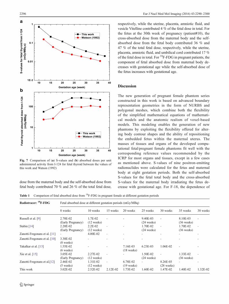
fetal kidney and liver receive the highest dose from 18F-FDG,namely 4.38E-02 mGy/MBq and 4.04E-02 mGy/MBq, re-spectively, at the 8th week of gestation. For the fetus above10 weeks of gestation, the bone marrow, brain, and thyroidreceive the highest absorbed radiation doses from 18F-FDGthan other fetal tissues. The absorbed doses to the fetal bonemarrow and thyroid from 18F-FDG are about 35.1 % and22.4 % higher than the average absorbed dose of fetal totalbody at 10–38 weeks of pregnancy. Figure 7 compares the S-values and absorbed dose per unit administered activity fromI-124 for fetal thyroid between the values of this work andthose reported in Watson [31]. The discrepancies betweenfetal thyroid S-values in these two works can be attributed toJohnson’s radiation transport model for photons [32] used byWatson [31], which clearly results in underestimation of thesevalues. In addition, differences between residence times usedin the two works might result in additional discrepancies be-tween absorbed dose estimates. Table 1 compares the estimat-ed radiation doses per unit administered activity to the fetaltotal body for 18F-FDG between this work and results reportedin the literature. The absorbed doses from 18F-FDG to the fetaltotal body in this work are 3.02E-02, 2.52E-02, 2.12E-02,1.73E-02, 1.60E-02, 1.47E-02, 1.40E-02, and 1.32E-02 mGy/MBq at the 8th, 10th, 15th, 20th, 25th, 30th, 35th,and 38th week of pregnancy, respectively. The fetal absorbeddoses obtained in this work are higher than those reported byStabin et al. [14] at early pregnancy and at the 3rd month ofgestation, but lower than the corresponding values for thefetus at 6 and 9 months of gestation. For the 3 months preg-nant female phantom, the fetal mass in the work of Stabin et al.is 458 g while the ICRP suggested a value of 85 g. This mightresult in an underestimation of the fetal absorbed dose.
The effective dose provides a single number for estimating theradiobiological detriment from radiation exposure and allowscomparisons of radiation risks associated with different imagingtechniques/scenarios. However, the effective dose of the fetushas never been reported in the literature. In this work, the
Fig. 3 Self-absorbed S-values torepresentative fetal organs fromF-18
2294 Eur J Nucl Med Mol Imaging (2016) 43:2290–2300

effective dose of the fetus was calculated at various gestationsfrom common positron-emitting radiotracers (Table 2). Since18F-FDG, 18F-FDOPA, 11C-labeled metahydroxephedrine and68Ga-DOTANOC are commonly used for PET imaging of pheo-chromocytoma in pregnant patients, we observed that 18F-FDOPA produces the highest fetal effective dose. Figure 8 com-pares the effective doses of the fetus and pregnant women from18F-FDG and 68Ga-DOTANOC. For the pregnant female, theeffective dose changes slightly during the whole gestation periodwhile the fetal effective doses are significantly higher at the 8thand 10th weeks of pregnancy than the other gestation periods.For 18F-FDG, the fetal effective doses at the 8th and 10th weeksof pregnancy are 2.28 and 2.42 times larger than those of the38thweek of pregnancy. The fetal effective dose at the 10thweekof pregnancy is higher than at the 8thweek of pregnancy becausemore critical organs are considered in the phantom at the10thweek of pregnancy.
Fetal dose from 18F-FDG in clinical scans
Table 3 compares the fetal dose in the new series of pregnantfemale phantoms with those reported in the literature.Patient#1 (5th week pregnancy) receives the highest absorbeddose (9.69 mGy) from 18F-FDG. The fetal doses estimated inthis work are 1.1–2.1 times higher than the correspondingreported values. For early pregnancy (5–15 weeks of pregnan-cy), the recalculated fetal dose is about 28.1 % higher thanthose reported by Zanotti-Fregonara et al. [12]. For the fetus atthe 8th week of pregnancy (patient#1), the cross-absorbed
Fig. 4 Self-absorbed S-values for various radionuclides for (a) maternaltotal body and (b) fetal total body
Fig. 5 Cross-absorbed S-values for (a) maternal body and (b) maternalbladder irradiating representative fetal organs
Fig. 6 Absorbed dose per unit administered activity from 18F-FDG for(a) fetal tissues and (b) maternal tissues
Eur J Nucl Med Mol Imaging (2016) 43:2290–2300 2295
dose from the maternal body and the self-absorbed dose fromfetal body contributed 70 % and 26 % of the total fetal dose,
respectively, while the uterine, placenta, amniotic fluid, andvesicle Vitelline contributed 4 % of the fetal dose in total. Forthe fetus at the 30th week of pregnancy (patient#10), thecross-absorbed dose from the maternal body and the self-absorbed dose from the fetal body contributed 36 % and47 % of the total fetal dose, respectively, while the uterine,placenta, amniotic fluid, and umbilical cord contributed 17 %of the fetal dose in total. For 18F-FDG in pregnant patients, thecomponent of fetal absorbed dose from maternal body de-creases with gestational age while the self-absorbed dose ofthe fetus increases with gestational age.
Discussion
The new generation of pregnant female phantom seriesconstructed in this work is based on advanced boundaryrepresentation geometries in the form of NURBS andpolygonal meshes, which combine both the flexibilityof the simplified mathematical equations of mathemati-cal models and the anatomic realism of voxel-basedmodels. This modeling enables the generation of newphantoms by exploiting the flexibility offered for alter-ing body contour shapes and the ability of repositioningthe embedded fetus within the maternal uterus. Themasses of tissues and organs of the developed compu-tational fetal/pregnant female phantoms fit well with thecorresponding reference values recommended by theICRP for most organs and tissues, except in a few casesas mentioned above. S-values of nine positron-emittingradionuclides were calculated for the fetus and maternalbody at eight gestation periods. Both the self-absorbedS-values for the fetal total body and the cross-absorbedS-values for the maternal body irradiating the fetus de-crease with gestational age. For F-18, the dependence of
Table 1 Comparison of fetal absorbed dose from 18F-FDG in pregnant female at different gestation periods
Radiotracer: 18F-FDG Fetal absorbed dose at different gestation periods (mGy/MBq)
8 weeks 10 weeks 15 weeks 20 weeks 25 weeks 30 weeks 35 weeks 38 weeks
Russell et al. [9] 2.70E-02(Early Pregnancy)
1.7E-02(12 weeks)
- - 9.40E-03(24 weeks)
- 8.10E-03(36 weeks)
-
Stabin [14] 2.20E-02(Early Pregnancy)
2.2E-02(12 weeks)
- - 1.70E-02(24 weeks)
- 1.70E-02(36 weeks)
-
Zanotti-Fregonara et al. [11] - 4.00E-02 - - - - - -
Zanotti-Fregonara et al. [10] 3.30E-02(8 weeks)
- - - - - - -
Takalkar et al. [13] 1.55E-02(6 weeks)
- - 7.16E-03(18 weeks)
6.23E-03 1.06E-02 - -
Xie et al. [15] 3.05E-02(Early Pregnancy)
2.27E-02(12 weeks)
- - 1.50E-02(24 weeks)
- 1.33E-02(36 weeks)
-
Zanotti-Fregonara et al.[12] 2.46E-02(5 weeks)
1.31E-02(12 weeks)
- 6.78E-02(19 weeks)
- 8.26E-03(28 weeks)
- -
This work 3.02E-02 2.52E-02 2.12E-02 1.73E-02 1.60E-02 1.47E-02 1.40E-02 1.32E-02
Fig. 7 Comparison of (a) S-values and (b) absorbed doses per unitadministered activity from I-124 for fetal thyroid between the values ofthis work and Watson (1992)
2296 Eur J Nucl Med Mol Imaging (2016) 43:2290–2300

the self-absorbed S-value of fetal total body on gesta-tional age are given by:
SFetus→FetusFetal Body ¼ 79:61� GWð Þ−4:483 ð1Þ
whereas the dependence of fetal total body on the cross-absorbed S-value on gestational age can be given as:
SMaternal Body→FetusFetal Body ¼ −1:697� 10−8 � GWð Þ þ 1:284� 10−6 ð2Þ
where GW is the gestation period of fetus (in weeks), varyingfrom 8 to 38 weeks. These rules can be used for a quick andrough estimation of the fetal total body absorbed dose fromF-18 labeled radiotracers:
DFetal Body ¼ MRTMaternal Body � SMaternal Body→FetusFetal Body
þMRTFetus � SFetus→FetusFetal Body ð3Þ
where D is the absorbed dose in mGy/MBq and MRT is themean residence time or time-integrated activity (MBq.s/MBq)of F-18 labeled tracers in the maternal total body or the fetus.
The absorbed and effective doses from 21 positron-emitting labeled radiotracers were estimated for 25 fetal tis-sues and the fetal total body. In this work, the standard MIRDformalism was adopted for estimating radiation doses to each
organ. However, some molecules are designed to be deliveredspecifically to the blood vessels for diagnostic and/or thera-peutic applications. As such, the mean residence time of theseradiotracers in the blood would be higher than the MIRDestimates, thus resulting in higher absorbed dose to the endo-thelial walls, especially for small vessels.
For radiation dosimetry of radiotracers based on anthropo-morphic computational phantoms, the effective dose presentsa lower uncertainty compared to absorbed doses derived fromclinical data [33]. However, the published literature indicatesthat the absorbed dose of the fetal total body from radiotracersis frequently considered while the fetal effective dose is rarelyreported. The effective dose of the fetus estimated in this workcan be used for comparison of the risks associated with differ-ent diagnostic imaging techniques/scenarios in regard to thepotential radiobiological detriment to various developing fetaltissues. The fetal effective doses at the 8th and 10th weeks ofgestation are significantly higher than those at other gestationperiods because of the significantly lower fetal weight at earlypregnancy.
For 18F-FDG, the difference of organ absorbed dosescalculated from different mathematical and voxel-basedphantoms can be greater than 150 % [33]. We calculatedfetal absorbed dose for a number of clinical studies report-ed in the literature based on reported time-integrated
Table 2 Comparison of fetal effective doses from investigated radiotracers in pregnant female at different gestation periods
Radiotracers Fetal effective dose at different gestation periods (mSv/MBq)
8 weeks 10 weeks 15 weeks 20 weeks 25 weeks 30 weeks 35 weeks 38 weeks
11C-acetate 2.46E-03 2.27E-03 2.10E-03 2.13E-03 2.15E-03 2.06E-03 2.05E-03 1.98E-0311C-amino acids 5.55E-03 4.94E-03 3.84E-03 3.65E-03 3.59E-03 3.38E-03 3.35E-03 3.18E-0311C brain receptor substances 7.49E-03 6.64E-03 4.22E-03 3.81E-03 3.62E-03 3.39E-03 3.34E-03 3.12E-0311C-methionine 1.29E-02 1.16E-02 5.41E-03 4.38E-03 3.77E-03 3.47E-03 3.38E-03 3.04E-0311C (realistic maximum model) 2.22E-02 1.98E-02 8.03E-03 6.01E-03 4.82E-03 4.34E-03 4.15E-03 3.58E-03
[Methyl-11C]thymidine 3.30E-03 3.02E-03 2.81E-03 2.85E-03 2.89E-03 2.78E-03 2.79E-03 2.71E-0311C-thymidine 3.37E-03 2.93E-03 2.63E-03 2.60E-03 2.61E-03 2.48E-03 2.46E-03 2.36E-0311C-SA4503 3.77E-03 3.49E-03 3.19E-03 3.15E-03 3.17E-03 3.01E-03 3.03E-03 2.92E-0311C-MPDX 4.50E-03 4.01E-03 3.52E-03 3.41E-03 3.41E-03 3.23E-03 3.21E-03 3.07E-0311C-TMSX 4.46E-03 3.96E-03 3.48E-03 3.41E-03 3.42E-03 3.25E-03 3.23E-03 3.09E-0311C-CHIBA-1001 4.42E-03 4.30E-03 3.89E-03 3.55E-03 3.45E-03 3.15E-03 3.20E-03 3.05E-0311C-4DST 5.75E-03 5.13E-03 3.80E-03 3.61E-03 3.53E-03 3.35E-03 3.31E-03 3.15E-0315O-water 4.75E-04 5.38E-04 4.73E-04 4.78E-04 4.67E-04 4.40E-04 4.38E-04 4.23E-0418F-amino acids 2.15E-02 2.52E-02 1.69E-02 1.57E-02 1.49E-02 1.43E-02 1.40E-02 1.31E-0218F brain receptor substances 2.54E-02 2.81E-02 1.73E-02 1.59E-02 1.50E-02 1.43E-02 1.41E-02 1.32E-0218F-FDG 2.90E-02 3.09E-02 1.79E-02 1.59E-02 1.47E-02 1.40E-02 1.37E-02 1.27E-0218F-L-DOPA 5.06E-02 4.84E-02 2.26E-02 1.85E-02 1.57E-02 1.46E-02 1.40E-02 1.25E-0218F-FBPA 4.01E-02 4.28E-02 2.20E-02 1.88E-02 1.68E-02 1.59E-02 1.53E-02 1.41E-0218F-FDOPA 5.52E-02 5.40E-02 2.61E-02 2.16E-02 1.86E-02 1.73E-02 1.66E-02 1.49E-0268Ga-EDTA 4.43E-02 4.57E-02 2.23E-02 1.90E-02 1.68E-02 1.57E-02 1.50E-02 1.38E-0268Ga-DOTANOC 1.71E-02 1.93E-02 1.31E-02 1.25E-02 1.18E-02 1.13E-02 1.10E-02 1.05E-02
Eur J Nucl Med Mol Imaging (2016) 43:2290–2300 2297

activity coefficients for the fetus and the developed anthro-pomorphic pregnant female phantoms. The fetal absorbeddoses calculated in this work are about 10.3–110.3 %higher than the corresponding values reported in the liter-ature. For early pregnancy, the dose to the uterus wasused as a surrogate of the dose to the fetus the calcula-tions performed by Zanotti-Fregonara et al. [12] andTakalkar et al. [13]. However, the reference fetal weightrecommended by the ICRP for early pregnancy, i.e. the8th week of gestation (4.7 g) is significantly lower thanthe mass of the adult female uterus (80 g). The replace-ment of the fetus with maternal uterus in radiation dosecalculations at early pregnancy will result in underestima-tion of S-values for the fetal total body. In this context,the new pregnant female phantoms can provide more ac-curate fetal absorbed dose from radiopharmaceuticals atearly pregnancy.
Conclusion
A new series of computational anthropomorphic pregnant fe-male phantoms for representing reference fetus and adult fe-male at various gestation periods were constructed. The devel-oped computational phantoms were used to conduct a system-atic study for evaluation of radiation dose to the fetus andgravida from common positron-emitting radionuclides and ra-diotracers based on updated tissue weighting factors andbiodistribution data. The latest generation computationalmodels can provide more accurate radiation dose estimatesfor the growing fetus. The produced S-values can be used forassessment of radiation dose to pregnant patients and the fetusat different gestational ages from various positron-emitting ra-diotracers. The calculated dosimetric database can be exploited
Fig. 8 Effective dose per unit administered activity for the fetus andpregnant female from (a) 18F-FDG and (b) 68Ga-DOTANOC
Table 3 Comparison offetal absorbed dosesfrom 18F-FDG for thereported pregnantpatients
Patient no. Stage ofgestation (weeks)
Administeredactivity (MBq)
Fetal absorbed dose from 18F-FDG
Values reported in literatures This work
Dose to fetus(mGy/MBq)
Total dose tofetus (mGy)
Dose to fetus(mGy/MBq)
Total dose tofetus (mGy)
1a 5 296 2.46E-02 7.28 3.27E-02 9.692a 12 385 1.31E-02 5.04 1.62E-02 6.253a 12 350 1.36E-02 4.76 1.73E-02 6.064a 19 296 6.78E-03 2.01 1.43E-02 4.225a 19 348 6.29E-03 2.19 1.29E-02 4.506a 28 296 8.26E-03 2.44 1.38E-02 4.087b 18 200 1.03E-02 2.06 1.14E-02 2.278b 25 337 7.41E-03 2.50 1.33E-02 4.499b 28 174 6.93E-03 1.21 9.70E-03 1.6910b 30 229 1.17E-02 2.68 1.38E-02 3.1711b 23 181 7.27E-03 1.32 1.30E-02 2.35
a Patients reported by Zanotti-Fregonara et al. [12]b Patients reported by Takalkar et al. [13]
2298 Eur J Nucl Med Mol Imaging (2016) 43:2290–2300
for evaluation and comparison of radiation risks to pregnantpatients and unborn embryo/fetus associated with different im-aging techniques/scenarios. The fetal organ-level dose is rarelyreported in literature. As the radio-sensitivity and radiation risksof developing fetal organs vary across different trimesters, theestimation of organ-level radiation dose to the fetus may pavethe way for a detailed investigation of the correlations betweenradiation exposure of the uterus and organ-specific childhoodcancer after birth. The developed fetus models and pregnantfemale phantoms matching the ICRP reference data can beused for calculation of internal/external radiation absorbed doseto provide standardized dose estimates for various radiotracersin clinical and research settings.
Acknowledgments This work was supported by the Swiss NationalScience Foundation under Grant SNSF 31003A-149957. The authorswould like to thank Prof. George Xu (Rensselaer Polytechnic Institute,New York), Dr Isabelle Bloch (Telecom ParisTech) and Dr Maria Zankl(Helmholtz ZentrumMünchen) for providing the pregnant and fetal com-putational models.
Compliance with ethical standards 1. Disclosure of potential con-flicts of interest: none of the authors have affiliations that present financialor non-financial competing interests for this work.
2. Research involving human participants: All procedures performedin studies involving human participants were in accordance with theethical standards of the institutional and/or national research committeeand with the 1964 Helsinki declaration and its later amendments or com-parable ethical standards.
3. Informed consent: informed consent was obtained from all individ-ual participants included in the study.
Conflict of Interest The authors declare that they have no conflict ofinterest.
References
1. Gambhir SS. Molecular imaging of cancer with positron emissiontomography. Nat Rev Cancer. 2002;2:683–93.
2. Xie T, Bolch WE, Lee C, Zaidi H. Pediatric radiation dosimetry forpositron-emitting radionuclides using anthropomorphic phantoms.Med Phys. 2013;40:102502–14.
3. Xie T, Zaidi H. Evaluation of radiation dose to anthropomorphicpaediatric models from positron-emitting labelled tracers. PhysMed Biol. 2014;59:1165–87.
4. Kusama T, Ota K. Radiological protection for diagnostic examina-tion of pregnant women. Congenit Anom (Kyoto). 2002;42:10–4.
5. Stabin MG. Radiation dose concerns for the pregnant or lactatingpatient. Semin Nucl Med. 2014;44:479–88.
6. Altman KI, Lett JT. Relative radiation sensitivities of human organsystems: Vol. 15, Elsevier; 2013.
7. Zaidi H, Xu XG. Computational anthropomorphic models of thehuman anatomy: The path to realistic Monte Carlo modeling inmedical imaging. Annu Rev Biomed Eng. 2007;9:471–500.
8. Stabin MG, Watson E, Cristy M, Ryman J, Eckerman K, Davis J,et al. Mathematical models and specific absorbed fractions of pho-ton energy in the nonpregnant adult female and at the end of each
trimester of pregnancy. Oak Ridge National Lab., TN (UnitedStates); 1995.
9. Russell JR, Stabin MG, Sparks RB, Watson E. Radiation absorbeddose to the embryo/fetus from radiopharmaceuticals. Health Phys.1997;73:756–69.
10. Zanotti-Fregonara P, Jan S, Champion C, Trébossen R, Maroy R,Devaux J-Y, et al. In vivo quantification of 18F-FDG uptake inhuman placenta during early pregnancy. Health Phys. 2009;97:82–5.
11. Zanotti-Fregonara P, Jan S, Taieb D, Cammilleri S, Trébossen R,Hindié E, et al. Absorbed 18F-FDG dose to the fetus during earlypregnancy. J Nucl Med. 2010;51:803–05.
12. Zanotti-Fregonara P, Laforest R, Wallis JW. Fetal radiation dosefrom 18F-FDG in pregnant patients imaged with PET, PET/CT,and PET/MR. J Nucl Med. 2015;56:1218–22.
13. Takalkar AM, Khandelwal A, Lokitz S, Lilien DL, Stabin MG.18F-FDG PET in pregnancy and fetal radiation dose estimates. JNucl Med. 2011;52:1035–40.
14. Stabin MG. Proposed addendum to previously published fetal doseestimate tables for 18F-FDG. J Nucl Med. 2004;45:634–35.
15. Xie T, Zaidi H. Fetal and maternal absorbed dose estimates forpositron-emitting molecular imaging probes. J Nucl Med.2014;55:1459–66.
16. ICRP. Publication 89: Basic anatomical and physiological data foruse in radiological protection: reference values. Ann ICRP.2002;32:1–277.
17. Xu XG, Taranenko V, Zhang J, Shi C. A boundary-representationmethod for designing whole-body radiation dosimetry models:pregnant females at the ends of three gestational periods—RPI-P3, −P6 and -P9. Phys Med Biol. 2007;52:7023–44.
18. Bibin L, Anquez J, de la Plata Alcalde JP, Boubekeur T, AngeliniED, Bloch I. Whole-body pregnant woman modeling by digitalgeometry processing with detailed uterofetal unit based on medicalimages. IEEE Trans Biomed Eng. 2010;57:2346–58.
19. Becker J, Zankl M, Fill U, Hoeschen C. Katja—the 24th week ofvirtual pregnancy for dosimetric calculations. Pol J Med Phys Eng.2008;14:13–20.
20. Petoussi-Henss N, Zankl M, Fill U, Regulla D. The GSF family ofvoxel phantoms. Phys Med Biol. 2002;47:89.
21. Xie T, Zaidi H. Construction of pregnant female phantoms at dif-ferent gestation periods for radiation dosimetry. Proc of the 5thInternational Workshop on Computational Phantoms forRadiation Protection, Imaging and Radiotherapy. Seoul, Korea,19–22 July 2015; 2015; p. 4–5.
22. Patrick M. Binvox code - 3Dmesh voxelizer. Princeton University:Princeton University.
23. Pelowitz DB. MCNPX User's Manual Version 2.5.0. Los AlamosNational Laboratory: Los Alamos, NM; 2005.
24. ICRP. ICRP publication 80: Radiation dose to patients from radio-pharmaceuticals (addendum 2 to ICRP publication 53). Ann ICRP.1998;28:1–126.
25. ICRP. ICRP Publication 106: Radiation dose to patients from ra-diopharmaceuticals. Addendum 3 to ICRP Publication 53. AnnICRP. 2008;38:1–197.
26. Sakata M, Oda K, Toyohara J, Ishii K, Nariai T, Ishiwata K. Directcomparison of radiation dosimetry of six PET tracers using humanwhole-body imaging and murine biodistribution studies. Ann NuclMed. 2013;27:285–96.
27. Pettinato C, Sarnelli A, Di DonnaM, Civollani S, Nanni C,MontiniG, et al. 68Ga-DOTANOC: biodistribution and dosimetry in pa-tients affected by neuroendocrine tumors. Eur J Nucl Med MolImaging. 2008;35:72–9.
28. Bolch WE, Eckerman KF, Sgouros G, Thomas SR. MIRDPamphlet No. 21: A generalized schema for radiopharmaceuticaldosimetry - Standardization of nomenclature. J Nucl Med.2009;50:477–84.
Eur J Nucl Med Mol Imaging (2016) 43:2290–2300 2299

29. ICRP. ICRP Publication 103: The 2007 Recommendations of theInternational Commission on Radiological Protection. Ann ICRP.2007;37:1–332.
30. Benveniste H, Fowler JS, Rooney WD, Moller DH, Backus WW,Warner DA, et al. Maternal-fetal in vivo imaging: a combined PETand MRI study. J Nucl Med. 2003;44:1522–30.
31. Watson E. Radiation absorbed dose to the human fetal thyroid.Proc. Fifth International Radiopharmaceutical Dosimetry
Symposium: Oak Ridge Associated Universities Oak Ridge, TN;1992; p. 179–87.
32. Johnson J. Fetal thyroid dose from intakes of radioiodine by themother. Health Phys. 1982;43:573–82.
33. Spielmann V, Li WB, Zankl M, Oeh U, Hoeschen C. Uncertaintyquantification in internal dose calculations for seven selected radio-pharmaceuticals. J Nucl Med. 2016;57:122–28.
2300 Eur J Nucl Med Mol Imaging (2016) 43:2290–2300