Development of an Advanced Retinal Imaging System · ... optical coherence tomography ... • The...
20
9/9/2016 ©UWMRF 2016 1 Development of an Advanced Retinal Imaging System (OTT ID 1181) Inventors: Hao Zhang, Ph.D., Shuliang Jiao, Ph.D., Lihong Wang, Ph.D. Department of Biomedical Engineering, Department of Ophthalmology, Northwestern University, Department of Biomedical Engineering, Florida International University, Department of Biomedical Engineering, Washington University For further information please contact: Jessica Silvaggi, Ph.D. Senior Licensing Manager 1440 East North Avenue Milwaukee, WI 53202 Tel: 414-906-4654 [email protected]
Transcript of Development of an Advanced Retinal Imaging System · ... optical coherence tomography ... • The...

9/9/2016©UWMRF 2016 1
Development of an Advanced Retinal Imaging System
(OTT ID 1181)
Inventors: Hao Zhang, Ph.D., Shuliang Jiao, Ph.D., Lihong Wang, Ph.D.
Department of Biomedical Engineering, Department of Ophthalmology, Northwestern University, Department of Biomedical Engineering, Florida International University,
Department of Biomedical Engineering, Washington University
For further information please contact: Jessica Silvaggi, Ph.D.
Senior Licensing Manager1440 East North Avenue
Milwaukee, WI 53202Tel: 414-906-4654

Current Problems: Retinal Disease
2
Current problems• Retinal diseases are the major cause of blindness in developed countries
• Diabetic retinopathy (DR) and age-related macular degeneration (AMD) are two major retinal diseases that account for about 20 million patients and the numbers continue to rise
• Diabetic patients are suggested to have annual eye exams. However, there is no clinical applicable method that is able to diagnose diabetic retinopathy or AMD in an early stage
• Up to 50% of patients are not getting their eyes examined or are diagnosed too late for effective treatment
• Existing technologies, such a fundus camera, optical coherence tomography (OCT), and scanning laser ophthalmology are limited in that they cannot measure functional variations in retinal vessels
©UWMRF 2016 9/9/2016

Solutions
Solutions of this invention• The progression of diabetic retinopathy is proven to be associated with functional
changes in retinal vessels, more specifically, the metabolism of transported oxygen by blood.
• This diagnostic device enables early-stage detection and pathological investigation of diabetic retinopathy (DR), glaucoma, and age-related macular degeneration (AMD) through in vivo non‐invasive imaging of blood hemoglobin oxygenation in the eye and retinal pigment epithelium imaging.
• The device integrates three imaging modalities: photoacoustic ophthalmoscopy (PAOM, ultrasound detection), optical coherence tomography (OCT), and autofluorescence (AF). All modalities can be added to a slit lamp bio‐microscope or a commercial OCT. PAOM can also be integrated with a fundus camera
• Visual and quantitative data is collected in one pass and presented in an integrated view for research or diagnostic uses.
• This system provides high imaging speed and precision in localization.
3©UWMRF 2016 9/9/2016

DR and AMD Facts and Figures
• Diabetic retinopathy affects more than 5 million Americans age 18 and older
• There were 25.8 million people with diabetes in 2011 in the U.S.
• Most patients with diabetes will eventually develop diabetic retinopathy, requiring ophthalmologic diagnosis and treatment
• Diabetic retinopathy causes 12,000 to 24,000 new cases of blindness each year
• When diabetic retinopathy is in the mild or moderate stage, good blood sugar control can slow the progression of diabetic retinopathy
• Surgery often slows or stops the progression of diabetic retinopathy, but it's not a cure; types of surgery include laser treatment and vitrectomy
• More than 1.6 million Americans over age 60 have advanced macular degeneration, with 10 million total cases across all age groups
• AMD is the leading cause of vision loss and blindness among Americans who are age 65 and older
• There is no outright cure for age-related macular degeneration, but some treatments may delay its progression or even improve vision; treatments for macular degeneration depend on whether the disease is in its early-stage, dry form or in the more advanced, wet form that can lead to serious vision loss
4
http://www.diabetes.org/, http://www.allaboutvision.com/conditions/amd.htm, http://www.diabetes.org/
.http://www.mayoclinic.com/health/diabetic-retinopathy/DS00447/DSECTION=treatments-and-drugs
©UWMRF 2016 9/9/2016

Intellectual Property and Licensing
Issued Patents
• 8,016,419: Systems and methods for photoacoustic opthalmoscopy
• 8,025,406: Systems and methods for photoacoustic ophthalmoscopy
Licensing
• This technology is part of an active and ongoing research program and is seeking partners for development of the final product
• It is available for licensing under exclusive or non-exclusive terms
• This device can be designed as a standalone PAOM/OCT module or the PAOM can be engineered as an add-on device for currently manufactured OCT units.
5©UWMRF 2016 9/9/2016
Potential Applications:
6
• This device can be designed as a standalone PAOM/OCT module or the PAOM can be engineered as an add-on device for currently manufactured OCT units
• Retinal diseases that can be treated include DR and AMD
• Use in the study of glaucoma in which OCT is utilized to diagnose central retinal vein occlusion
• Detection of early signs of glaucoma
• Diagnoses of diseases in which morphology and function of retinal vessels are affected including: stroke, Alzheimer’s disease, and hypertensive retinopathy
©UWMRF 2016 9/9/2016

7
Market Potential
Market:
• 1/3 of the medical expenses in the USA are spent on diabetes and diabetic complications.
• Between 40-45% of Americans diagnosed with diabetes have some stage of diabetic retinopathy
• The global diabetes market is estimated to double from $32 billion to $64 billion over the next 15 years, including new diagnoses and therapies.
• The global macular degeneration market was valued at $1 billion in 2009 and is expected to grow to $2 billion with a compound annual growth rate (CAGR) of 10.3% by 2016. This growth is primarily attributed to the increase in aged population across the world.
• It is estimated that there are approximately 9,000-10,000 fundus cameras in clinical use in the U.S., with 800-1200 new units sold annually worldwide and a market growth of 7% annually
• A potential market of 9,500 OCT cameras are estimated to be in use by ophthalmologists in the U.S.
“Early Stage Assessment of Diabetic Retinopathy.” Foresight Science and Technology, Inc. October 27, 2008.
“Strategic Analysis of Optical Imaging Technologies in U.S. Clinical Diagnostics and Drug Discovery Markets.”
May 24, 2009. Frost and Sullivan web site.
http://www.pr-inside.com/macular-degeneration-drug-pipeline-analysis-r1820893.htm
©UWMRF 2016 9/9/2016

8
Existing Retinal Imaging Modalities
Fundus photographyScanning laser
Ophthalmoscopy (SLO/cSLO)
Autofluorescence imaging Fluorescein angiography
Images are from internet©UWMRF 2016 9/9/2016

Photoacoustic Ophthalmoscopy (PAOM)
• Photoacoustic imaging is a novel technology that measures the optical absorption contrast in biological tissue non-invasively
• It is based on the detection of laser induced ultrasonic waves
• When short laser pulses irradiate biological tissue, optical energy is absorbed by blood vessels, for example, and converted to heat (causing only a milli-degree temperature rise)
• Then, thermoelastic expansions occur, which lead to the vibration of local tissue and the generation of wideband ultrasonic waves
• Such ultrasonic waves (referred to as PA waves) are detected to form an image according to their amplitudes and time-of-arrival to the ultrasonic detector
9©UWMRF 2016 9/9/2016

10
Detailed Design of PAOM
Left: •Schematic of the optical illumination and ultrasonic detection. •Optical illumination is the same as the confocal scanning laser ophthalmology and OCT. •During scanning, the ultrasonic detectors are kept stationary and only touch the eye lid after applying medical ultrasonic gel.Right: •Photograph of the slit-lamp based PAOM+OCT
©UWMRF 2016 9/9/2016

• PAOM measures the structural and functional retinal vascular information, such as the retinal vessel morphology and the retinal blood hemoglobin oxygen saturation (sO2) non-invasively with high precision.
• A uniquely designed combination of contact lens and ultrasonic transducer is to be worn during examinations.
• The integration of the contact lens with ultrasonic transducer is the first of its kind and solves the clinical applicability issues (relative movement of the eye and the ultrasonic transducer). The reason is that the contact tends to move together with the eye during the examination so that only optical eye tracking is required.
Important Features/Benefits of PAOM Design
5/28/201311 Schematic for fused PAOM and OCT
• A natural fusion of PAOM with clinical ophthalmic optical coherence tomography (OCT).
• The capability of providing comprehensive physiological information including vessel diameter (as small as 15 μm), blood flow velocity, and sO2, which give the metabolic rate of a specific retinal region.
©UWMRF 2016

12
PAOM Imaging provides more advanced imaging compared to other modalities
PAOM OCT Autofluorescence
532 nm 870 nm Excitation: 500 nm
Emission: 600 nm
Optics Express 18, 3967-3972 (2010)
In vivo imaging of a pigmented rat eye, which is centered at the optical disc. The OCT image is acquired simultaneously with the PAOM.
Explanation of Technology
©UWMRF 2016 9/9/2016

13
Volumetric Visualization of PAOM retinal images
Volumetric movie
OSA Virtual Journal of Biomedical Optics Vol. 5, issue 5 “Feature Image”; OCT New.org “Feature of the week 03/14/2010”; Optics Express 18, 3967-3972 (2010)
Pseudo-color after segmentation
Left: retinal vessels and the retinal pigmented epithelium (RPE) are separated using an automatic segmentation algorithm and colored in red and blue, respectively. Right: 3D visualization.
©UWMRF 2016 9/9/2016

14
Imaging retinal and choroidal vascular network simultaneously for the first time
RNFL IS/OS RPE+CC+CH
PAOM
OCT
Journal of Modern Optics 58, 1997 (2011)
©UWMRF 2016 9/9/2016
15
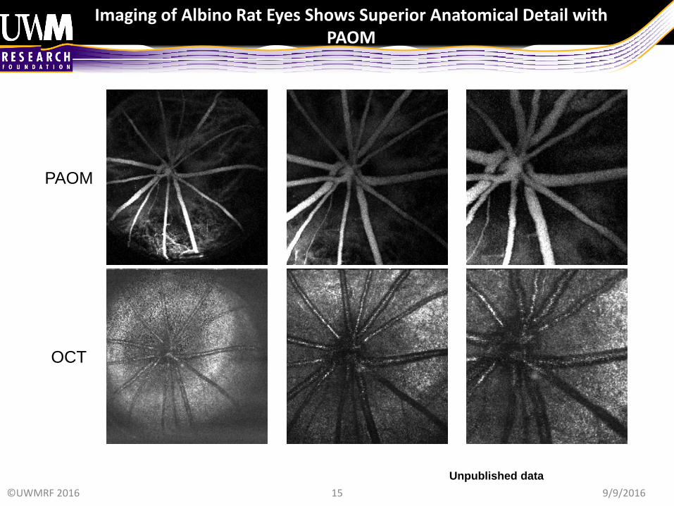
Imaging of Albino Rat Eyes Shows Superior Anatomical Detail with PAOM
Unpublished data
PAOM
OCT
©UWMRF 2016 9/9/2016

16
OCTPAOM SLO
Albino
Pigmented
(c)(a)RV
CV
(b)
(d) (f)(e)
PAOM is integrated with SLO and OCT for truly multi-functional imaging
Journal of Biomedical Optics 17, 061206 (2012)
2012 top downloaded paper©UWMRF 2016 9/9/2016

17
SPOT
Laser
Amplifier
2D Galvo Mirrors
Control PC
DAQ PC
Scan Control
PD
X Scan
Y Scan
M2
Laser
Trigger
Fiber
Acquisition
Trigger
BBO
LLM
M1
SP1
SP2
L2 L1
FilterL3
L4
Bulb
Fundus image
PA
UT
OBJ1OBJ2
Iris
Lens
L5
ANU
STOP
Current Eye Research, in press (2013)
Explanation of Technology
PAOM is integrated with fundus camera for real-time video feed-back
©UWMRF 2016 9/9/2016

Next Experiments
18
1
2
3
45
6
7
8
910
1. Mapping of the global and local
retinal metabolic rate
2. Imaging larger eyes
3. Safety evaluation
4. Patient tests
©UWMRF 2016 9/9/2016

19
Next Steps
• Optimize the system to improve image quality, specificity, and sensitivity
• Develop prototype for human imaging and seek collaborators for patient clinical trials
Approximately $500,000 will be needed to engineer a standalone prototype suitable for human clinical trials
Subsequent prototypes are expected to cost $100,000-$200,000 to manufacture
• License technology to companies manufacturing contact lens and ophthalmic imaging devices
• Collaborations; SBIR Partner Companies for a Start-up company
• Partnering for development and/or licensing
©UWMRF 2016 9/9/2016

1
Development of an Advanced Retinal Imaging System
(OTT ID 1181)
For further information please contact:
Jessica Silvaggi, Ph.D.Senior Licensing Manager1440 East North Avenue
Milwaukee, WI 53202Tel: 414-906-4654
©UWMRF 2016 9/9/2016