Development of a Hybrid Nanoprobe for Triple-Modality MR ...
15
Development of a Hybrid Nanoprobe for Triple-Modality MR/SPECT/Optical Fluorescence Imaging Madru, Renata; Svenmarker, Pontus; Ingvar, Christian; Ståhlberg, Freddy; Andersson- Engels, Stefan; Knutsson, Linda; Strand, Sven-Erik Published in: Diagnostics DOI: 10.3390/diagnostics4010013 2014 Link to publication Citation for published version (APA): Madru, R., Svenmarker, P., Ingvar, C., Ståhlberg, F., Andersson-Engels, S., Knutsson, L., & Strand, S-E. (2014). Development of a Hybrid Nanoprobe for Triple-Modality MR/SPECT/Optical Fluorescence Imaging. Diagnostics, 4(1), 13-26. https://doi.org/10.3390/diagnostics4010013 Total number of authors: 7 General rights Unless other specific re-use rights are stated the following general rights apply: Copyright and moral rights for the publications made accessible in the public portal are retained by the authors and/or other copyright owners and it is a condition of accessing publications that users recognise and abide by the legal requirements associated with these rights. • Users may download and print one copy of any publication from the public portal for the purpose of private study or research. • You may not further distribute the material or use it for any profit-making activity or commercial gain • You may freely distribute the URL identifying the publication in the public portal Read more about Creative commons licenses: https://creativecommons.org/licenses/ Take down policy If you believe that this document breaches copyright please contact us providing details, and we will remove access to the work immediately and investigate your claim.
Transcript of Development of a Hybrid Nanoprobe for Triple-Modality MR ...
LUND UNIVERSITY
PO Box 117221 00 Lund+46 46-222 00 00
Development of a Hybrid Nanoprobe for Triple-Modality MR/SPECT/OpticalFluorescence Imaging
Madru, Renata; Svenmarker, Pontus; Ingvar, Christian; Ståhlberg, Freddy; Andersson-Engels, Stefan; Knutsson, Linda; Strand, Sven-ErikPublished in:Diagnostics
DOI:10.3390/diagnostics4010013
2014
Link to publication
Citation for published version (APA):Madru, R., Svenmarker, P., Ingvar, C., Ståhlberg, F., Andersson-Engels, S., Knutsson, L., & Strand, S-E. (2014).Development of a Hybrid Nanoprobe for Triple-Modality MR/SPECT/Optical Fluorescence Imaging. Diagnostics,4(1), 13-26. https://doi.org/10.3390/diagnostics4010013
Total number of authors:7
General rightsUnless other specific re-use rights are stated the following general rights apply:Copyright and moral rights for the publications made accessible in the public portal are retained by the authorsand/or other copyright owners and it is a condition of accessing publications that users recognise and abide by thelegal requirements associated with these rights. • Users may download and print one copy of any publication from the public portal for the purpose of private studyor research. • You may not further distribute the material or use it for any profit-making activity or commercial gain • You may freely distribute the URL identifying the publication in the public portal
Read more about Creative commons licenses: https://creativecommons.org/licenses/Take down policyIf you believe that this document breaches copyright please contact us providing details, and we will removeaccess to the work immediately and investigate your claim.
Diagnostics 2014, 4, 13-26; doi:10.3390/diagnostics4010013
diagnosticsISSN 2075-4418
www.mdpi.com/journal/diagnostics/ Article
Development of a Hybrid Nanoprobe for Triple-Modality MR/SPECT/Optical Fluorescence Imaging
Renata Madru 1,*, Pontus Svenmarker 2, Christian Ingvar 3, Freddy Ståhlberg 1,4,5,Stefan-Andersson Engels 2, Linda Knutsson 1 and Sven-Erik Strand 1,4
1 Department of Medical Radiation Physics, Lund University, Barngatan 2, 221 85 Lund, Sweden; E-Mails: [email protected] (F.S.); [email protected] (L.K.); [email protected] (S.-E.S.)
2 Department of Physics, Lund University, Professorsgatan 1, 223 63 Lund, Sweden; E-Mails: [email protected] (P.S.); [email protected] (S.-A.E.)
3 Department of Surgery, Skane University Hospital, Entrégatan 7, 221 85 Lund, Sweden; E-Mail: [email protected]
4 Lund University Bioimaging Center (LBIC), Kliniggatan 32, 222 42 Lund, Sweden 5 Department of Radiology, Skane University Hospital, Entrégatan 7, 221 85 Lund, Sweden
* Author to whom correspondence should be addressed; E-Mail: [email protected]; Tel.: +46-46-222-0839; Fax: +46-46-17-8540.
Received: 29 January 2014; in revised form: 28 February 2014 / Accepted: 4 March 2014 / Published: 10 March 2014
Abstract: Hybrid clinical imaging is an emerging technology, which improves disease diagnosis by combining already existing technologies. With the combination of high-resolution morphological imaging, i.e., MRI/CT, and high-sensitive molecular detection offered by SPECT/PET/Optical, physicians can detect disease progression at an early stage and design patient-specific treatments. To fully exploit the possibilities of hybrid imaging a hybrid probe compatible with each imaging technology is required. Here, we present a hybrid nanoprobe for triple modality MR/SPECT/Fluorescence imaging. Our imaging agent is comprised of superparamagnetic iron oxide nanoparticles (SPIONs), labeled with 99mTc and an Alexa fluorophore (AF), together forming 99mTc-AF-SPIONs. The agent was stable in human serum, and, after subcutaneous injection in the hind paw of Wistar rats, showed to be highly specific by accumulating in the sentinel lymph node. All three modalities clearly visualized the imaging agent. Our results show that a single imaging agent can be used for hybrid imaging. The use of a single hybrid contrast agent permits simultaneous hybrid imaging and, more conventionally, allow for single modality imaging at different
OPEN ACCESS

Diagnostics 2014, 4 14
time points. For example, a hybrid contrast agent enables pre-operative planning, intra-operative guidance, and post-operative evaluation with the same contrast agent.
Keywords: SPION; SPECT; optical; fluorescence imaging; magnetic resonance imaging; MR; SLN
1. Introduction
Traditional emission tomography and radiological imaging technologies provide valuable physiological, metabolic and morphological information for diagnosing of disease. A single imaging modality cannot, by itself, give complete information of disease and the underlying molecular aberrations causing it. A combination of modalities may give the necessary information that physicians need to detect disease progression at an early stage, design patient-specific treatments, and monitor in vivo post-therapeutic effects for therapy optimization [1]. For these reasons, improved multimodality imaging with molecular sensitivity could help pave the way for personalized medicine [2–4].
Multimodality imaging combines two or more imaging technologies in order to increase the diagnostic potential and to overcome the limitations offered by each single modality. A high-resolution imaging technology, providing morphological information, is typically combined with a high-sensitivity imaging technology sensing molecular aberrations. Examples of such systems include SPECT/CT, PET/CT, PET/MR, and MR/optical imaging modalities [5–8]. Advances in the development of SPECT/MR scanners and their distinct advantages have previously been reported [9,10]. SPECT and PET are highly sensitive but are limited by poor spatial resolution and lack of morphological information. A combination of SPECT or PET with CT imaging, which has the ability to acquire complementary whole body, high-resolution morphological images, has rapidly evolved as a new golden standard technique within oncology [11–13]. MR imaging, on the other hand, provides non-invasive, high-resolution functional and morphological images with better soft tissue contrast than CT. Its drawbacks are longer imaging time and the low sensitivity to molecular targets. Combining PET/SPECT or optical imaging with MR technology will enable a unique feature for detection and monitoring of diseases [14,15]. Emerging optical imaging technologies, such as fluorescence, bioluminescence and Cerenkov luminescence are fast (images can be acquired in <1 min), inexpensive and easy to implement in a clinical setting [16,17]. In order to fully exploit the advantages of such multimodality technologies, multimodal imaging agents are warranted.
Superparamagnetic iron oxide nanoparticles (SPIONs) possess unique properties that make them well-suited for multimodality molecular imaging [18]. Initially, SPIONs were introduced as MR contrast agents and clinically approved for visualization of liver metastasis (Resovist®), angiography, and sentinel node mapping (Feridex®, Combidex®). The large surface to volume ratio also enables addition of different ligands for active targeting of, i.e., immune cells, angiogenesis or plaque [19–22]. Furthermore, SPIONs show a great potential for in vivo stem cell tracking and monitoring gene delivery [23,24]. Labeled with radionuclides, SPIONs can be used for combined PET/MR or SPECT/MR imaging [25–28].

Diagnostics 2014, 4 15
In our previous study, we suggested to use radiolabeled iron oxide nanoparticles (99mTc-SPIONs) for early detection/staging of cancer by SPECT and MR [28]. Herein, we improved the properties of the 99mTc-SPIONs by labeling them with a fluorescent dye, and not only 99mTc and SPIONS for SPECT/MR, but also for optical fluorescence imaging. This triple-modality molecular imaging agent identifies the target by whole body SPECT and MR imaging, and highlight the target intra-operatively by fluorescence imaging. The radiolabeled and fluorescent SPIONs can also be visualized by microscopy during ex vivo pathological evaluation and the molecular targets can be quantified, e.g., by autoradiography. Here, we demonstrate that the attached dye does not affect the size and in vivo stability of the nanoparticles. The targeting ability of SLNs and biodistribution of the agent is demonstrated in Wistar rats.
2. Experimental Section
2.1. SPIONs
The SPIONs used in this study have been described elsewhere [28]. Briefly, the SPIONs consist of a monodispersed superparamagnetic iron oxide core (Fe3O4) coated with polyethylene glycol and including primary amino groups. The coating prevents aggregation and enables functionalization.
2.2. Labeling of SPIONs with Fluorescent Dye and Radioactivity
The SPIONs (0.5 mL, 3.4 mg/mL Fe) were labeled with the amino-reactive Alexa Fluor 647 (AF) carboxylic acid dye (Molecular probes, Life Technologies) by succinimidyl (NHS) ester coupling. The dye (10 mg) was dissolved in DMSO (1 mL) and incubated with the SPIONs for 2 h. The AF–SPIONs were washed three times with saline (0.9% v/v) and filtered after each wash by using a MACS MS column (Miltenyi Biotech, Germany) and a strong cylindrical magnet. When the magnet was attached to the column, SPIONs got trapped in the filter while the fluid ran through. By removing the magnet, the nanoparticles could be eluted with saline.
The remaining free amino groups on the SPIONs surface were used to label the nanoparticles with 99mTc. 99mTc-pertechnetate (99mTcO4
�) was obtained from a 99Mo/99mTc-generator (DRN 4329 Ultratechnekow FM). The radiolabeling followed a previously developed protocol [28] and was slightly modified to obtain the highest labeling efficiency at pH 4–4.2. The protocol stated that stannous chloride (10 mg) was to be dissolved in 10 mL sterile water and stirred for 1 min. A part of the solution (0.5 mL) was passed through a 22 μm Millipore filter and added to the 99mTcO4
� (278 MBq, 1 mL), which was kept in a vacuumcollecting vial. The hydrogen ion activity was adjusted to pH 4 by adding NaOH (1 M, ~20 μL), after which, AF–SPIONs were added (0.1 mL) and gently shaken for 30 s. The mixture was incubated for 1 h at room temperature. The fluorescence and radiolabeled SPIONs (99mTc-AF–SPIONs) were filtered through MACS columns as described previously and eluted in saline (0.9% v/v, ~0.25 mL).
2.3. Quality Control
The radiolabeling yield was measured with instant-thin layer chromatography (ITLC). A small volume (0.8 μL) of reaction mixture was dropped on silica-gel impregnated glass fiber sheets
Diagnostics 2014, 4 16
(5 × 10 cm ITLC-SG, Pall ITLC Media, Pall Life Sciences) and aqueous methanol (85% v/v) was used as mobile phase for developing. The sheets were dried and cut in two strips, of 3 cm and 7 cm. The two segments were compared for radioactivity, as measured by a NaI(Tl) well type detector (1282 CompuGamma CS, LKB Wallac).
The reaction mixture, containing 99mTc-AF-SPIONs, was filtered and pre-concentrated using MACS MS columns (Miltenyi Biotech, Germany). The radioactivity of the buffer solution was compared to the radioactivity of the filtered 99mTc-AF-SPIONs.
Two 99mTc-AF-SPION mixtures (10 μL) were diluted in human serum (500 μL) and incubated for 24 h at pH 7 in room temperature. The ITLC method was then used to measure any release of free 99mTc. SPIONs and 99mTc-AF-SPIONs were imaged with transmission electron microscopy (TEM, Philips CM 10 System, USA) using 400 mesh carbon grids to measure the size of the nanoparticles and to evaluate possible aggregation.
2.4. Animal Studies
To show the in vivo feasibility for imaging, two white Wistar rats weighing 250–300 g were used. The experiments were completed in compliance with local and national regulations approved by the Local Ethics Committee for Animal Research. The animals were slightly anesthetized with isoflurane and injected subcutaneously in the right hind paw, with 99mTc-AF-SPIONs (0.07–0.1 mL) dispersed in physiological saline buffer (~40 MBq, ~0.3 mg Fe). The animals returned to their cages and, 5 h post injection, imaged with MR, SPECT and Optical modalities. To assess the activity distribution the animals were sacrificed and tissues/organs were measured in the NaI(Tl) well type detector (1282 CompuGamma CS, LKB Wallac).
2.4.1. MR Imaging
The animals were imaged with a 3T-MR system (Philips Achieva) equipped with a Sense-Flex-M coil. The images were acquired using Spin Echo (SE) and 3D Gradient Echo (GRE) pulse sequences with the following parameters. SE: TE/TR 80/3000 ms; FOV 100 mm; voxel size 0.5 × 0.5 × 1.3 mm3; flip angle 90° and number of average 10. GRE: TE/TR 14.6/2000 ms; voxel size 0.8 × 0.8 × 1.3 mm3; flip angle 90° and number of averages 10.
2.4.2. SPECT Imaging
A scintillation camera (GE Hawkeye; GE Healthcare) captured anterior and posterior images in 15 min. The camera was equipped with a low energy, high-resolution parallel-hole collimator. SPECT/CT projections were acquired for 1 h, at 60 s per projection, with a 20% energy window centered at 140 keV.
2.4.3. Optical Imaging
The animals were shaved prior to imaging in an effort to minimize autofluorescence. Imaging of the animals was performed in epifluorescence mode. Laser induced fluorescence of the 99mTc-AF-SPIONs was accomplished by a Helium-Neon laser (633 nm, 5 mW). The laser source illuminated the animal

Diagnostics 2014, 4 17
with a slightly divergent beam with a spot size of approximately 2 cm2. The illumination spot was raster-scanned in an overlapping pattern across the region of interest, and one image was acquired for each illumination position. An air-cooled charge coupled device camera (Andor iXon DU-897) captured the image after having passed through a combination of a long-pass filter (Schott, RG665) and a dielectric band-pass filter centered at 670 nm. All images were background subtracted, subjected to a threshold, and summed to give the resulting image. The resulting image was overlaid a photograph of the animal to give an anatomical reference.
2.5. Biodistribution
The animals were dissected; the lymph nodes, kidneys, spleen, liver, and the hind paw were weighed and measured for activity in the NaI(Tl) well-type detector (1282 CompuGamma CS; LKB Wallac). The radioactivity measurement was corrected for background and decay. The uptake of the 99mTc-AF-SPIONs within the dissected organs was expressed in % of injected activity (% IA/g).
The lymph nodes were frozen, cut in 100 μm and 20 μm thick frozen coronal sections using a cryostat (Microme, Vacutome HM 500 OM; Microme International) and mounted on microscope slides and air dried at room temperature. The 100 μm sections were imaged and analyzed by digital autoradiography (DARG) with a silicon strip detector (Biomolex 700 Imager; Biomolex AS). Each second cryo-slice was hematoxilyn and eosin (H&E) stained and used as a reference for the DARG images. The 20 μm sections were DAPI stained and used for microscopy analysis (Zeiss Axiovert 200M).
3. Results and Discussion
Advances in development of hybrid imaging technologies have created an enormous interest in the design and preparation of novel multimodality imaging agents [29–31]. Among these imaging agents the nuclear/MR and MR/optical imaging probes are the most successful combinations due to its high degree of complementarity.
In this work we propose combining information from MR, nuclear and optical images in order to have the full scale from whole-body imaging down to cellular and molecular imaging with use of one single agent. The major advantages to work with SPIONs are the easy production of the spherical core within narrow size range distribution, the simple coating procedure and the low in vivo toxicity. As the SPIONs are small in size and have a large area to volume ratio, a single nanoparticle can be labeled with a large number of radionuclides, fluorophores, and/or targeting ligands. In addition, polyethylene glycol coated SPIONs circulate in the blood pool/vasculature for a longer period of time compared with the low molecular weight gadolinium-based MR contrast imaging agents [32]. The labeling of SPIONs with a fluorescent dye and radionuclides may change the nanoparticles surface, physical and/or chemical properties. The final size of the 99mTc-AF-SPIONs is crucial for targeting because it may influence the pharmacokinetic and biodistribution of the agent.
3.1. Labeling of the SPIONs with Fluorescent Dye and 99mTc
Despite the low cost of optical instrumentation and easy implementation for intra-operative surgery, there are only two fluorescent dyes (ICG and fluorescein sodium) approved by the FDA [33]. In basic

Diagnostics 2014, 4 18
research and preclinical imaging, the most commonly used fluorophores are those in the range of NIR [34]. The long excitation and emission wavelengths can penetrate deeper in tissue and give less autofluorescence. Cyanine dye Cy5 and Alexa Fluor (650 nm excitation, 670–680 nm emission) are almost as good at penetrating tissue as true NIR radiation. In addition, they are very stable, and can be visualized with standard light microscopy for pathological evaluation of samples and have easy conjugation chemistry. In this study, a simple and highly efficient conjugation protocol was developed for labeling SPIONs with AF. The ratio of fluorophore molecules to nanoparticles was calculated to be 300:1, leaving more than 200 free binding sites for labeling SPIONs with 99mTc in order to evaluate the biodistribution of the agent in vivo. 99mTc is a generator produced isotope, highly available and the physical properties such as half-life and emitted energy is well-suited for imaging and quantification purposes.
3.2. Quality Control
The radiolabeling yield of the 99mTc-AF-SPIONs, determined by ITLC, was 99%. 99mTc-AF-SPIONs remained at the origin whereas free 99mTc moved with the solvent front (Rf 1.0). Control measurements with 99mTc-pertechnetate have been performed to validate the method. Additionally, the high radiolabeling efficiency was confirmed by magnetic separation and found to be 99.7%.
Figure 1. Transmission electron microscopy images of nanoparticles. (a) SPIONs in 0.9% saline buffer, pH 7 and (b) 99mTc-AF-SPIONs after incubation in human serum at room temperature. The images demonstrate that the size of the nanoparticles is not affected by the labeling procedure including both radionuclides and fluorescent dye. No aggregations of the particles have been observed after incubation of 99mTc-AF-SPIONs in human serum.
99mTc-AF-SPIONs incubated in serum showed a good dispersion and was found to be stable. After
24 h incubation of the 99mTc-AF-SPIONs, the amount of free 99mTc in human serum was less than 3%, analyzed with the ITLC method. The stability of the 99mTc-AF-SPIONs was also confirmed by the TEM images as demonstrated in Figure 1. The TEM images show that the size of the 99mTc-AF-SPIONs is not affected by the labeling procedure; the spherical shape of the iron oxide core can be
Diagnostics 2014, 4 19
easily recognized and the surrounding polyethylene coating, which was shown to be intact. Both unlabeled (Figure 1A) and 99mTc-AF SPIONs (Figure 1B) show a mean diameter of 11 nm ± 2 nm and no aggregation could be observed. Dynamic Light Scattering (DLS) is another method that can be used to measure the size of the nanoparticles as we presented in our previous work [28]. In this study DLS could not be applied because the fluorophores attached to the SPIONs have similar emission wave lengths as the laser used in the instrument and the size measurements may be corrupted. However, the attached fluorophores consists of very small molecules and have little effect of the overall size of the SPIONs. On the other hand, it can affect the stability (SPIONs can aggregate because the charge of the nanoparticles may change) and, therefore, we used TEM in this study.
3.3. Animal Studies
3.3.1. MR Imaging
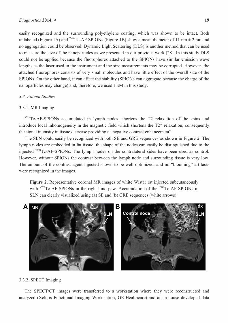
99mTc-AF-SPIONs accumulated in lymph nodes, shortens the T2 relaxation of the spins and introduce local inhomogeneity in the magnetic field which shortens the T2* relaxation; consequently the signal intensity in tissue decrease providing a “negative contrast enhancement”.
The SLN could easily be recognized with both SE and GRE sequences as shown in Figure 2. The lymph nodes are embedded in fat tissue; the shape of the nodes can easily be distinguished due to the injected 99mTc-AF-SPIONs. The lymph nodes on the contralateral sides have been used as control. However, without SPIONs the contrast between the lymph node and surrounding tissue is very low. The amount of the contrast agent injected shown to be well optimized, and no “blooming” artifacts were recognized in the images.
Figure 2. Representative coronal MR images of white Wistar rat injected subcutaneously with 99mTc-AF-SPIONs in the right hind paw. Accumulation of the 99mTc-AF-SPIONs in SLN can clearly visualized using (a) SE and (b) GRE sequences (white arrows).
3.3.2. SPECT Imaging
The SPECT/CT images were transferred to a workstation where they were reconstructed and analyzed (Xeleris Functional Imaging Workstation, GE Healthcare) and an in-house developed data

Diagnostics 2014, 4 20
visualization software (Interactive Data Language, IDL) was used. The representative image clearly visualized the injection site and inguinal lymph node (right side) Figure 3. Small amounts of the 99mTc-AF-SPIONs could also be detected bilaterally in the inguinal lymph node (left side, second node) as well as in the renal (third) lymph node, 5 h post injection.
Figure 3. SPECT/CT image of the same animal shown by MR in Figure 2. The arrows depict the injection site and accumulation of the 99mTc-AF-SPIONs in SLN. SPECT is less affected by attenuation compared with optical imaging, therefore, is an invaluable tool to quantify and study the biodistribution of the newly developed agent.
3.3.3. Optical Imaging
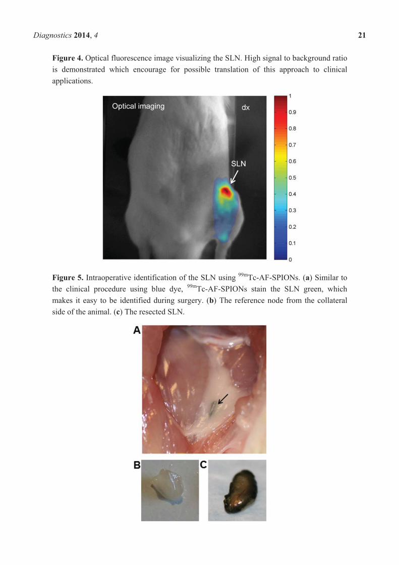
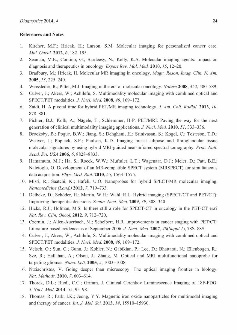
Figure 4 displays an image of the fluorescence signal originating from the 99mTc–Alexa Fluor 647 labeled SPIONs. An accumulation of the fluorescence signal was observed and its location matches that of the inguinal lymph node. The injection site was not within the imaged region. Lymph nodes deeper than 2 cm have not been detected. However, intraoperatively, the agent showed to be useful to identify the SLN. 99mTc-AF-SPIONs stains the SLN green (Figure 5) and can provide a real time visual guidance during surgery just like methylene blue administrated to patients in the clinical procedure [35,36]. In addition, 99mTc-AF-SPIONs can be administrated in a single injection, compared with the world-wide accepted SLN standard procedure, injecting the radiocolloids and dye separately.
Diagnostics 2014, 4 21
Figure 4. Optical fluorescence image visualizing the SLN. High signal to background ratio is demonstrated which encourage for possible translation of this approach to clinical applications.
Figure 5. Intraoperative identification of the SLN using 99mTc-AF-SPIONs. (a) Similar to the clinical procedure using blue dye, 99mTc-AF-SPIONs stain the SLN green, which makes it easy to be identified during surgery. (b) The reference node from the collateral side of the animal. (c) The resected SLN.

Diagnostics 2014, 4 22
Figure 6. Biodistribution of 99mTc-AF-SPIONs in two white Wistar rats, 5 h post injection.
Figure 7. Microscopy images of a cryosectioned SLN. (a) Coronal section (20 μm) of a half SLN indicating the microdistribution of the nanoparticles within the SLN. The blue structures are cell nucleus and the red light is emitted from the 99mTc-AF-SPIONs. The nanoparticles accumulate in the cortex and within the medullary sinuses. (b) Image visualizing the cortex of the SLN and indicating that the nanoparticles are mostly located extracellularly in comparison with the medullary sinus where the nanoparticles seem to be located within the macrophages. (c) and (d) bright field images of the SLN corresponding to (a) respectively (b) which show the orientation and anatomy.
0.0
0.1
1.0
10.0
100.0
1,000.0
SLNInguinal
right
Inguinalleft LN
Renal LN Spleen Kidney R Kidney L Liver
% IA
/g

Diagnostics 2014, 4 23
3.4. Biodistribution
The uptake of the 99mTc-AF-SPIONs within lymph nodes (LN) was confirmed by the biodistribution analysis. The highest mean uptake was found in the inguinal LN (right side), 330 ± 120% IA/g, followed by the inguinal left side (150 ± 20% IA/g), renal LN side (100 ± 40% IA/g), spleen (1.3 ± 0.02% IA/g), kidneys (0.56 ± 0.08% IA/g) and liver (0.1 ± 0.2% IA/g) as presented in Figure 6. These results suggest a higher uptake of the 99mTc-AF-SPIONs in SLN compared with our previous study using 99mTc-SPIONs (211 ± 225% IA/g) [28]. The higher uptake is often correlated with higher accuracy in localization of SLN.
Digital autoradiography and microscopy showed that 99mTc-AF-SPIONs indeed localized to SLN. The fluorescence dye enabled localization and studies of microdistribution of the nanoparticles when the 99mTc radioactivity was no longer measurable. Furthermore, a non-homogenous uptake of the nanoparticles was found within the SLN as shown in (Figure 7). The nanoparticles were found both extracellularly and intracellular within the cortex and in sinuses.
4. Conclusions
In conclusion, we developed a new triple-modality imaging agent, 99mTc-AF-SPIONs, for MR, SPECT and optical fluorescence imaging. The easy and straight forward labeling procedure and stability of the agent can be used for detection and intra-operative localization of the SLNs.
Acknowledgments
This study was performed with generous support from the Swedish Cancer Foundation, the Swedish Science Council, Berta Kamprad’s Foundation, Gunnar Nilsson’s Foundation, and the ALF Foundation of the Medical Faculty of Lund University. The SPIONs were provided as a generous gift from Genovis AB, Sweden. The authors gratefully thank Johan Olsrud, Jimmy Lätt for the technical support on MR and Anders Örbom for his help with the DARG instrument. Lund University Bioimaging Center (LBIC), Lund University, is gratefully acknowledged for providing experimental resources.
Author Contributions
Renata Madru: design of study, experimental work, writing. Pontus Svenmarker: contribution in design and whole body optical imaging experiments, proofreading. Christian Ingvar: design of the study and proofreading. Freddy Ståhlberg: design, support related MR imaging, proofreading. Stefan-Andersson Engels: contribution in design and whole body optical imaging experiments, proofreading. Linda Knutsson: design, support related to MR imaging, proofreading. Sven-Erik Strand: design of study, analyzing results, writing, and proofreading.
Conflicts of Interests
The SPIONs were a generous gift by Genovis AB.
Diagnostics 2014, 4 24
References and Notes
1. Kircher, M.F.; Hricak, H.; Larson, S.M. Molecular imaging for personalized cancer care. Mol. Oncol. 2012, 6, 182–195.
2. Seaman, M.E.; Contino, G.; Bardeesy, N.; Kelly, K.A. Molecular imaging agents: Impact on diagnosis and therapeutics in oncology. Expert Rev. Mol. Med. 2010, 15, 12–20.
3. Bradbury, M.; Hricak, H. Molecular MR imaging in oncology. Magn. Reson. Imag. Clin. N. Am. 2005, 13, 225–240.
4. Weissleder, R.; Pittet, M.J. Imaging in the era of molecular oncology. Nature 2008, 452, 580–589. 5. Culver, J.; Akers, W.; Achilefu, S. Multimodality molecular imaging with combined optical and
SPECT/PET modalities. J. Nucl. Med. 2008, 49, 169–172. 6. Zaidi, H. A pivotal time for hybrid PET/MR imaging technology. J. Am. Coll. Radiol. 2013, 10,
878–881. 7. Pichler, B.J.; Kolb, A.; Nägele, T.; Schlemmer, H-P. PET/MRI: Paving the way for the next
generation of clinical multimodality imaging applications. J. Nucl. Med. 2010, 51, 333–336. 8. Brooksby, B.; Pogue, B.W.; Jiang, S.; Dehghani, H.; Srinivasan, S.; Kogel, C.; Tosteson, T.D.;
Weaver, J.; Poplack, S.P.; Paulsen, K.D. Imaging breast adipose and fibroglandular tissue molecular signatures by using hybrid MRI-guided near-infrared spectral tomography. Proc. Natl. Acad. Sci. USA 2006, 6, 8828–8833.
9. Hamamura, M.J.; Ha, S.; Roeck, W.W.; Muftuler, L.T.; Wagenaar, D.J.; Meier, D.; Patt, B.E.; Nalcioglu, O. Development of an MR-compatible SPECT system (MRSPECT) for simultaneous data acquisition. Phys. Med. Biol. 2010, 55, 1563–1575.
10. Misri, R.; Saatchi, K.; Häfeli, U.O. Nanoprobes for hybrid SPECT/MR molecular imaging. Nanomedicine (Lond.) 2012, 7, 719–733.
11. Delbeke, D.; Schöder, H.; Martin, W.H.; Wahl, R.L. Hybrid imaging (SPECT/CT and PET/CT): Improving therapeutic decisions. Semin. Nucl. Med. 2009, 39, 308–340.
12. Hicks, R.J.; Hofman, M.S. Is there still a role for SPECT-CT in oncology in the PET-CT era? Nat. Rev. Clin. Oncol. 2012, 9, 712–720.
13. Czernin, J.; Allen-Auerbach, M.; Schelbert, H.R. Improvements in cancer staging with PET/CT: Literature-based evidence as of September 2006. J. Nucl. Med. 2007, 48(Suppl 1), 78S–88S.
14. Culver, J.; Akers, W.; Achilefu, S. Multimodality molecular imaging with combined optical and SPECT/PET modalities. J. Nucl. Med. 2008, 49, 169–172.
15. Veiseh, O.; Sun, C.; Gunn, J.; Kohler, N.; Gabikian, P.; Lee, D.; Bhattarai, N.; Ellenbogen, R.; Sze, R.; Hallahan, A.; Olson, J.; Zhang, M. Optical and MRI multifunctional nanoprobe for targeting gliomas. Nano. Lett. 2005, 5, 1003–1008.
16. Ntziachristos, V. Going deeper than microscopy: The optical imaging frontier in biology. Nat. Methods. 2010, 7, 603–614.
17. Thorek, D.L.; Riedl, C.C.; Grimm, J. Clinical Cerenkov Luminescence Imaging of 18F-FDG. J. Nucl. Med. 2014, 55, 95–98.
18. Thomas, R.; Park, I.K.; Jeong, Y.Y. Magnetic iron oxide nanoparticles for multimodal imaging and therapy of cancer. Int. J. Mol. Sci. 2013, 14, 15910–15930.

Diagnostics 2014, 4 25
19. Su, H.; Mou, Y.; An, Y.; Han, W.; Huang, X.; Xia, G.; Ni, Y.; Zhang, Y.; Ma, J.; Hu, Q. The migration of synthetic magnetic nanoparticle labeled dendritic cells into lymph nodes with optical imaging. Int. J. Nanomed. 2013, 8, 3737–3744.
20. Cai, W.; Chen, X. Multimodality imaging of vascular endothelial growth factor and vascular endothelial growth factor receptor expression. Front. Biosci. 2007, 12, 4267–4279.
21. Cai, W.; Chen, X. Multimodality molecular imaging of tumor angiogenesis. J. Nucl. Med. 2008, 49, 113–128.
22. Zhou, Q.; Yang, K.R.; Gao, P.; Chen, W.L.; Yang, D.Y.; Liang, M.J.; Zhu, L. An experimental study on MR imaging of atherosclerotic plaque with SPIO marked endothelial cells in a rabbit model. J. Magn. Reson. Imag. 2011, 34, 1325–1332.
23. Cromer, B.S.M.; Walczak, P.; Bulte, J.W. Tracking stem cells using magnetic nanoparticles. Wiley Interdiscip. Rev. Nanomed. Nanobiotechnol. 2011, 3, 343–355.
24. Herranz, F.; Almarza, E.; Rodríguez, I.; Salinas, B.; Rosell, Y.; Desco, M.; Bulte, J.W.; Ruiz-Cabello J. The application of nanoparticles in gene therapy and magnetic resonance imaging. Microsc. Res. Tech. 2011, 74, 577–591.
25. Xie, J.; Chen, K.; Huang, J.; Lee, S.; Wang, J.; Gao, J.; Li, X.; Chen, X. PET/NIRF/MRI triple functional iron oxide nanoparticles. Biomaterials 2010, 31, 3016–3022.
26. Lee, H.; Yu, M.K.; Park, S.; Moon, S.; Min, J.J.; Jeong, Y.Y.; Kang, H-W, Jon, S. Thermally cross-linked superparamagnetic iron oxide nanoparticles:� Synthesis and application as a dual imaging probe for cancer in vivo. J. Am. Chem. Soc. 2007, 129, 12739–12745.
27. Glaus, C.; Rossin, R.; Welch, M.J.; Bao, G. In vivo evaluation of 64Cu-labeled magnetic nanoparticles as a dual-modality PET/MR imaging agent. Bioconjug. Chem. 2010, 21, 715–722.
28. Madru, R.; Kjellman, P.; Olsson, F.; Wingårdh, K.; Ingvar, C.; Ståhlberg, F.; Olsrud, J.; Lätt, J.; Fredriksson, S.; Knutsson, L.; Strand, S.-E. 99mTc-labeled superparamagnetic iron oxide nanoparticles for multimodality SPECT/MRI of sentinel lymph nodes. J. Nucl. Med. 2012, 53, 459–463.
29. Dave, S.R.; Gao, X. Monodisperse magnetic nanoparticles for biodetection, imaging, and drug delivery: A versatile and evolving technology. Wiley Interdiscip. Rev. Nanomed. Nanobiotechnol. 2009, 1, 583–609.
30. Choi, H.S.; Frangioni, J.V. Nanoparticles for biomedical imaging: fundamentals of clinical translation. Mol. Imag. 2010, 9, 291–310.
31. Jokerst, J.V.; Gambhir, S.S. Molecular Imaging with Theranostic Nanoparticles. Acc. Chem. Res. 2011, 44, 1050–1060.
32. Xie, J.; Huang, J.; Li, X.; Sun, S.; Chen, X. Iron oxide nanoparticle platform for biomedical applications. Curr. Med. Chem. 2009, 16, 1278–1294.
33. Nguyen, T.Q. Fluorescence-guided surgery with live molecular navigation—A new cutting edge. Nat. Rev. Cancer 2013, 13, 653–662.
34. Luo, S.; Zhang, E.; Su, Y.; Cheng, T.; Shi, C. A review of NIR dyes in cancer targeting and imaging. Biomaterials 2011, 32, 7127–7138.
35. Varghese, P.; Abdel-Rahman, A.T.; Akberali, S.; Mostafa, A.; Gattuso, J.M.; Carpenter, R.Methylene blue dye-a safe and effective alternative for sentinel lymph node localization. Breast J. 2008, 14, 61–67.

Diagnostics 2014, 4 26
36. Buscombe, J.; Paganelli, G.; Burak, Z.E.; Waddington, W.; Maublant, J.; Prats, E.; Palmedo, H.; Schillaci, O.; Maffioli, L.; Lassmann, M.; Chiesa, C.; Bombardieri, E.; Chiti, A. Sentinel node in breast cancer procedural guidelines. Eur. J. Nucl. Med. Mol. Imag. 2007, 34, 2154–2159.
© 2014 by the authors; licensee MDPI, Basel, Switzerland. This article is an open access article distributed under the terms and conditions of the Creative Commons Attribution license (http://creativecommons.org/licenses/by/3.0/).