Development and validation of magnetic bead pentaplex ... · and tetanus toxoid was supplied by...
11
Contents lists available at ScienceDirect Methods journal homepage: www.elsevier.com/locate/ymeth Development and validation of magnetic bead pentaplex immunoassay for simultaneous quantification of murine serum IgG antibodies to acellular pertussis, diphtheria and tetanus antigens used in combination vaccines Laxmikant Kadam, Krunal Patel, Manish Gautam ⁎ , Shrikant Thorat, Prathamesh Kale, Arvind Kumar Ghule, Akansha Gairola, Harish Rao, Yojana Shinde, Umesh Shaligram, Sunil Gairola Serum Institute of India Pvt Ltd., 212/2, Hadapsar, Pune, Maharashtra 411028, India ARTICLE INFO Keywords: Mutiplex ELISA Acellular pertussis Magnetic beads Combination vaccines Immunogenicity ABSTRACT We describe here a magnetic bead-based multiplex (pentaplex) immunoassay (MIA) platform developed as an alternative to enzyme-linked immunosorbent assays (ELISA) used in immunogenicity testing of DTaP/TdaP vaccine in animals. MIA simultaneously measures the concentration of serum (IgG) antibodies against B. Pertussis antigens; pertussis toxin, filamentous hemagglutinin (FHA), pertactin (PRN) and tetanus (T) and diphtheria (D) toxoid in the Tdap vaccine immunized animals. Assay validation experiments were done using a panel of serum samples. The results are expressed in IU/ml using WHO reference mice serum. The standard curve was linear with 4PL logistic fit over an eight 2-fold dilution range with LOQ of 0.003, 0.022, 0.005 IU/ml for PT, FHA and PRN and 0.016 U/ml for T and D antigens indicating sensitivity. No interference was observed in monoplex versus multiplex measurements. Specificity was demonstrated by ≥90% homologous and ≤15% heterologous inhibition for all the antigens. The assay was reproducible, with a mean coefficient of variation (CV) of ≤10% for intra-assay duplicates and ≤25% for interassays using different lots of beads and analyst. Accuracy was demonstrated wherein the ratio of observed vs. assigned unitages were within 80–120%. The study suggests that the Pentaplex (MIA) platform meets all the criteria for the serological assay combination vaccines with addi- tional advantages of high throughput, reduced sample volumes, faster analysis with reduced manpower in contrast to conventional monoplex ELISA. 1. Introduction Combination or multivalent vaccines are the cornerstones of pae- diatric and adult immunization programs. Diphtheria (D), pertussis (whole cell; WP) and tetanus (T) antigens combined into a single pro- duct with an adjuvant represent the first successful example of combi- nation vaccine to achieve public health benefits [1–3]. The substitution of whole cell pertussis antigens with acellular pertussis (aP) antigens has further paved the path for even more complex combination vac- cines [4]. The compositions of available acellular pertussis vaccines vary widely, yet most of them contain detoxified pertussis toxin, alone or in combination with filamentous haemagglutinin (FHA), pertactin 69 kDa, PRN and fimbriae antigens (fim 2, 3) [5]. The preclinical evaluation of aP antigens to predict their clinical efficacy is a major challenge. WHO and European Pharmacopeia re- commends a non-lethal immunogenicity test in mice for monitoring lot to lot consistency during the manufacturing. The assay involves im- munization of mice with serial dilutions of the vaccine and serum IgG antibody response against each pertussis vaccine component is then measured by enzyme-linked immunosorbent assay (ELISA). Consequently, a large number of different ELISAs have to be performed to estimate antibody levels for all the antigens in the vaccine, which involves higher costs, logistics and most importantly requires con- siderable serum volumes [6,7]. The Multiple Analyte Profiling technology (xMAP®; Luminex Corp., Austin, TX) is a flow cytometry-based system (19) based on the use of distinct fluorescent microspheres as the carrier of different antigens enables the simultaneous detection of up to 100 analytes in a single well of a 96-well flat-bottom plate. Several studies have previously described the accuracy and high-throughput advantage of this platform for eval- uating vaccine-elicited binding antibodies [8–11]. For example, Pav- liakova D et al reported a 13-Plex Luminex assay for quantification of https://doi.org/10.1016/j.ymeth.2019.01.015 Received 18 September 2018; Received in revised form 27 December 2018; Accepted 23 January 2019 ⁎ Corresponding author. E-mail address: [email protected] (M. Gautam). Methods 158 (2019) 33–43 Available online 25 January 2019 1046-2023/ © 2019 Elsevier Inc. All rights reserved. T
Transcript of Development and validation of magnetic bead pentaplex ... · and tetanus toxoid was supplied by...

Contents lists available at ScienceDirect
Methods
journal homepage: www.elsevier.com/locate/ymeth
Development and validation of magnetic bead pentaplex immunoassay forsimultaneous quantification of murine serum IgG antibodies to acellularpertussis, diphtheria and tetanus antigens used in combination vaccines
Laxmikant Kadam, Krunal Patel, Manish Gautam⁎, Shrikant Thorat, Prathamesh Kale,Arvind Kumar Ghule, Akansha Gairola, Harish Rao, Yojana Shinde, Umesh Shaligram,Sunil GairolaSerum Institute of India Pvt Ltd., 212/2, Hadapsar, Pune, Maharashtra 411028, India
A R T I C L E I N F O
Keywords:Mutiplex ELISAAcellular pertussisMagnetic beadsCombination vaccinesImmunogenicity
A B S T R A C T
We describe here a magnetic bead-based multiplex (pentaplex) immunoassay (MIA) platform developed as analternative to enzyme-linked immunosorbent assays (ELISA) used in immunogenicity testing of DTaP/TdaPvaccine in animals. MIA simultaneously measures the concentration of serum (IgG) antibodies against B. Pertussisantigens; pertussis toxin, filamentous hemagglutinin (FHA), pertactin (PRN) and tetanus (T) and diphtheria (D)toxoid in the Tdap vaccine immunized animals. Assay validation experiments were done using a panel of serumsamples. The results are expressed in IU/ml using WHO reference mice serum. The standard curve was linearwith 4PL logistic fit over an eight 2-fold dilution range with LOQ of 0.003, 0.022, 0.005 IU/ml for PT, FHA andPRN and 0.016 U/ml for T and D antigens indicating sensitivity. No interference was observed in monoplexversus multiplex measurements. Specificity was demonstrated by ≥90% homologous and ≤15% heterologousinhibition for all the antigens. The assay was reproducible, with a mean coefficient of variation (CV) of ≤10%for intra-assay duplicates and ≤25% for interassays using different lots of beads and analyst. Accuracy wasdemonstrated wherein the ratio of observed vs. assigned unitages were within 80–120%. The study suggests thatthe Pentaplex (MIA) platform meets all the criteria for the serological assay combination vaccines with addi-tional advantages of high throughput, reduced sample volumes, faster analysis with reduced manpower incontrast to conventional monoplex ELISA.
1. Introduction
Combination or multivalent vaccines are the cornerstones of pae-diatric and adult immunization programs. Diphtheria (D), pertussis(whole cell; WP) and tetanus (T) antigens combined into a single pro-duct with an adjuvant represent the first successful example of combi-nation vaccine to achieve public health benefits [1–3]. The substitutionof whole cell pertussis antigens with acellular pertussis (aP) antigenshas further paved the path for even more complex combination vac-cines [4]. The compositions of available acellular pertussis vaccinesvary widely, yet most of them contain detoxified pertussis toxin, aloneor in combination with filamentous haemagglutinin (FHA), pertactin69 kDa, PRN and fimbriae antigens (fim 2, 3) [5].
The preclinical evaluation of aP antigens to predict their clinicalefficacy is a major challenge. WHO and European Pharmacopeia re-commends a non-lethal immunogenicity test in mice for monitoring lot
to lot consistency during the manufacturing. The assay involves im-munization of mice with serial dilutions of the vaccine and serum IgGantibody response against each pertussis vaccine component is thenmeasured by enzyme-linked immunosorbent assay (ELISA).Consequently, a large number of different ELISAs have to be performedto estimate antibody levels for all the antigens in the vaccine, whichinvolves higher costs, logistics and most importantly requires con-siderable serum volumes [6,7].
The Multiple Analyte Profiling technology (xMAP®; Luminex Corp.,Austin, TX) is a flow cytometry-based system (19) based on the use ofdistinct fluorescent microspheres as the carrier of different antigensenables the simultaneous detection of up to 100 analytes in a single wellof a 96-well flat-bottom plate. Several studies have previously describedthe accuracy and high-throughput advantage of this platform for eval-uating vaccine-elicited binding antibodies [8–11]. For example, Pav-liakova D et al reported a 13-Plex Luminex assay for quantification of
https://doi.org/10.1016/j.ymeth.2019.01.015Received 18 September 2018; Received in revised form 27 December 2018; Accepted 23 January 2019
⁎ Corresponding author.E-mail address: [email protected] (M. Gautam).
Methods 158 (2019) 33–43
Available online 25 January 20191046-2023/ © 2019 Elsevier Inc. All rights reserved.
T

human serum antibodies to Streptococcus pneumoniae capsular Poly-saccharides used in conjugate vaccine [12]. In another study, usingLuminex-based mPlex-Flu assay, influenza -specific IgG antibodymediated cross-reactivity to adjuvanted recombinant influenza he-magglutinin (rHA) was studied in ferrets and mice [13]. Further inanother application to Human papiloma Virus (HPV) quadrivalentvaccine, a multiplexed luminex assay was used to assess long termantibody to responses to serotypes 6, 11, 16 and 18 in humans [14]. Anumber of studies are also available for multiplexed analysis of diph-theria, tetanus and acellular pertussis antigens. However, a majority ofthese reports are for quantification of human antibodies with applic-ability to seroprevalence or evaluating pre and post vaccination sam-ples [15,16]. Very few reports on MIAs are described for applicability tothe preclinical assessment of DTaP or Tdap based combination vaccinesin animals, as required by regulatory agencies [17].
We report here development and validation of a magnetic beadbased pentaplex immunoassay for combined quantification of mouseserum antibodies against pertussis components (Ptx, FHA and PRN),diphtheria and tetanus toxoid. The immunoassay is intended for directapplicability to mouse immunogenicity test as recommended inEuropean, British and Indian pharmacopeia for quality control testingof aP antigens in Tdap or DTaP vaccine. The described Penta-Plexmagnetic microsphere based fluorescent immunoassay (MIA) measurestotal IgG levels in Tdap immunized animals against an internationalreference standard. The study indicates that assay exhibits a wide dy-namic range for all the five antigens. The assay was validated as per theinternational regulatory guidance provided by US FDA, EMEA and ICHon validation of bioanalytical methods and assessed key characteristics:quantifiable range, precision, specificity and ruggedness and dilutionallinearity. The multiplex assay satisfies all the requirements for a quality
control assay and will be useful for measuring immune responses toTdap or DTaP combination vaccines.
2. Materials and methods
2.1. Purified antigens, reagents, vaccines
Pertussis toxin, Filamentous hemagglutinin, Pertactin, diphtheriaand tetanus toxoid was supplied by Serum Institute of India Pvt Ltd.Protein content of antigens were estimated by a validated BCA assay.The antigens are QC tested to meet all the purity and integrity re-quirements. R-phycoerthyryn (R-PE) - conjugated anti-mouse was ob-tained from Jackson Immunoresearch. Beads (carboxylated micro-spheres) were obtained from Bio-Rad laboratories. Sulfo-NHS wasprocured from Thermofisher and EDC was purchased from Bio-Rad.BSA was obtained from Sigma Aldrich. Tween-20 was purchased fromMerck. Tdap Vaccine manufactured by Serum Institute of India Ltdcomprising of following antigens; Tetanus toxoid not less than 2.5 Lf,diphtheria toxoid- not less than 5 Lf; Pertussis Toxoid 8 mcg, detoxifiedfilamentous hemagglutinin- 8 mcg and Pertactin- 2.5 mcg in 0.5 ml dosewas used in the study.
2.2. Standard sera
WHO international reference mouse serum (NIBSC 97/642) wasused as reference standard. The international reference sera were de-veloped by pooling mouse serum samples following vaccination withDTaP vaccine. The sera have following assigned unitages: 17 units; anti-PT, 143 units’ anti-FHA, 30 units of anti-PRN and 32 units of anti-FIM 2and 3 per vial. One lyophilized vial was reconstituted with 500 µL of 1X
Table 1Description of serum panels used in the validation. Assigned unitages to panel sera are also provided, which was used for precision and accuracy studies.
Panel Validationparameter
Description Details
1 Specificity Following panel of sera samples was used for establishing specificity1A Specificity Sera obtained by immunization of whole cell pertussis
vaccine. The sera will be positive for only pertussisantigens
Pertussis vaccine manufactured at SII. was immunized on day 0, and 14, 28 days.The animals were bled on day 42 for the sera. Sera of 30 mice were pooled todevelop the panel
1A
1B Specificity Sera containing antibodies to Diphtheria antigens only Sera was obtained by immunization of NIH Harlan mice with SII manufacturedPneumo conjugate vaccine. The vaccine used diphtheria protein as carrierprotein. Animals were immunized on day 0, 14 and 28. Sera were collected onday 42. (Serum of 30 mice was pooled to develop the panel)
1C Specificity Sera containing antibodies to Diphtheria and Tetanusantigens only
Sera were obtained by immunization of Harlan mice with a commerciallyavailable Pneumoconjugate vaccine (Synflorix vaccine). This vaccine uses carrierprotein of tetanus and diphtheria in the conjugate vaccine. The immunizationprotocols were similar of what was followed for above panel. (Pool of N=30)
1D Specificity Negative control sera The sera pool is obtained by immunization with placebo buffer (Tdap vaccine).The immunization protocol is similar to above. (Pool of N=30)
2 Precision Post vaccinated sera samples2A Precision Obtained by immunization 1/2.5 of human dose of
Tdap vaccineSingle dose immunization.IU/ml U/ml
PT FHA PRN DT TT37 50 10 13 99
2B Precision Obtained by immunization 1/5.0 of human dose ofTdap vaccine
Single dose immunization.IU/ml U/ml
PT FHA PRN DT TT18 26 5 3 56
2C Precision Obtained by immunization of 1/10th of human dose ofTdap vaccine
Single dose immunization.IU/ml U/ml
PT FHA PRN DT TT8 22 1 1 24
2D Precision High titer sera obtained by immunization of 1/2.5 ofhuman dose Tdap vaccine on day 0, 14 and 28. Seracollection on day 42
Multiple immunizations.IU/ml U/ml
PT FHA PRN DT TT40 159 33 44 244
L. Kadam, et al. Methods 158 (2019) 33–43
34

PBS. Aliquots were prepared and stored at −20 °C till further use. Theconcentrations of anti-diphtheria and anti-tetanus IgG were unknownand so these were subjectively set at 100 U/ml for validation purpose inthe study.
2.3. Animals
NIH mice (male or female) were housed and used for experimentaccording to the institutional ethics guidelines for animal experimentsat Serum Institute of India Pvt Ltd. Mouse immune sera were generatedby immunization of groups of 6–8week old mice (N=10/group). Theretroorbital bleeding method was used for sera collection on on re-spective day after immunization. Details of serum panel developmentwith respect to animal experiment is given in Table 1. All the experi-ments were regulated under National Institutes of Health guide for thecare and use of Laboratory animals (NIH Publications No. 8023, revised1978).
2.4. Sera panel for validation testing
The QC control sera were used for validation testing. Separateserum panels for specificity and precision studied were developed. Thedetails of the development of these serum panels are given in Table 1and Fig. 1.
The study also included testing ofn serum samples obtained byimmunization of Tdap vaccine at four dilutions of 2.5, 5, 10 and 20 onday 0 and sera collection on day 35. For each dilution (N=10) animalswere used. Antibody concentrations for all the five antigens were de-termined for each mice and geometric mean concentrations (GMC)were reported.
2.5. Coupling of purified antigens to polystyrene beads
Purified Ptx, FHA, PRN, Dtx and Ttx were coupled to activatedmicrospheres (beads) of regions; 35, 42, 14, 44 and 48 respectivelyfollowing a procedure according to Van Gageldonk et al. [18]. Briefly,2.5 to 12.5×10⁶ beads were activated by incubating it with 100 ug of
EDC (1-ethyl-3 dimethyl amino propyl carbodiimide hydrochloride)and N-hydroxysulfosuccinmide (sulfo-NHS) (pH=6.1) for 20min. Thiswas followed by washing steps using magnetic separator. Respectiveantigens were added to wash activated beads and kept in dark for 2 hunder constant mixing (15–30 rpm). The resulting mixture is washedand the supernatant is discarded. After three steps of pelleting andwashing, coupled beads are blocked using 1% BSA buffer for 30minand are finally kept in storage buffer (0.1% w/v BSA in PBS containing0.05% sodium azide and 0.02% tween 20). Final count of beads is en-umerated using neubauer chamber.
2.6. Pentaplex assay
Eight steps of 2 fold dilutions of NIBSC reference standard serumwere prepared in luminex assay buffer. Sera samples were diluted inassay buffer containing PBS (1X), BSA (0.2%), tween 20 (0.1%) andsodium azide (0.01%). Each dilution of reference and sample weremixed with 1:1 (50 µl) conjugated beads to attain 4000 beads/region/well in a 96 well multiscreen HTS filter plate (Millipore Corporation,USA) and incubated for 1 h at 37 degree celcius in the dark on the plateshaker at 150 rpm. Suitable blanks (negative control, sera obtainedfrom un-immunized animals) and assay blanks (without serum) wereincluded in every plate. The beads were washed three times with PBSby filtration using magnetic plate assembly. Subsequently, beads werefurther washed three times using luminex assay buffer. This was fol-lowed by addition of PE labeled secondary anti mouse antibody (re-commended dilution of 1:250) and followed by incubation at 37 °C for30min with 150 rpm shaking. The beads were washed and read on theflow cytometer (Bioplex-200). The events were acquired using Bio-plexmanager software. The system classifies beads on the basis of its uniquespectral pattern analyzed as median fluorescent intensity (MFI) of thesignal of the reporter antibody. For each analyte (5 plex), MFI is con-verted to U/mL by interpolation from a 4 PL standard curve for everybead region/standard. Each sample/standard dilution is read in dupli-cates and % CV is monitored.
Fig. 1. Sera panels used for validation testing. The panels were designed to demonstrate assay specificity and precision. The specificity panel was designed todemonstrate assay specificity and selectivity for all the five antigens. Precision panel had five different sera samples with different unitages of all the five antigens.
L. Kadam, et al. Methods 158 (2019) 33–43
35

2.7. Assay development
The assay optimization experiments focused on determining theoptimal condition of major controllable parameters. The major assayconditions such as concentration of the blocking buffer, secondary an-tibody as well as optimization of incubation time for the secondaryantibody and samples was evaluated. Positive control serum with as-signed unitage was used in the optimization experiments. The experi-mental conditions involving using BSA at the more than 0.1%, PEconjugated antibody concentration at 1/250 and incubation time of30min for both secondary antibody and serum samples resulted insignificant improvement in assay performance.
2.8. Assay validation parameters
2.8.1. Assay specificityInhibition experiments were performed to establish the specificity of
the method. A high titre serum with known high concentrations of IgGantibodies specific to all the five antigens (Ptx, FHA, PRN, Dtx and Ttx)were serially diluted and homologous and heterologous inhibition wasdetermined by addition of one of the antigens to the reaction mixture.Homologous and heterologous inhibition is reported as the percentagecompared to the control. The assay specificity was also demonstratedusing a panel of serum samples, which were obtained by immunizationof one or two of the antigens used in the assay. Details of the panel aregiven in Table 1.
2.8.2. Assay linearity and rangeMean fluorescent intensity (MFI) in response to the anti-mouse re-
ference standard serum (NIBSC 97/642) serially diluted in 2 folds andwere analyzed for the quantifiable range. The data obtained from lin-earity data sets were analyzed using 4 PL curve fitting using Bioplexsoftware. Curve constants such as ‘a’ (estimated response at zero con-centration), ‘b’ (denotes slope factor), ‘c’ (denotes mid range con-centration) and‘d’ (denotes estimated response at infinite concentra-tion) were used for trending and 95% CI. ere calculated. The fit wascarried out using Bioplex-200 system manager software. The quantifi-able range is the concentration range of the standard curve over whichback fits (residuals) is within the predetermined acceptance criteria.Acceptance criteria of back fits (70–130%) were used [19,20].
2.8.3. Assay sensitivityThe lower and upper limits of the quantification (LOQs) were de-
termined using a negative serum sample and positive serum controlsrespectively. Using negative serum, readings from individual wells(n= 50), mean MFI, standard deviation (SD) and mean ± 2SDs MFIvalues for all the five antigens are calculated. The lower limit of de-tection (LLOD) for each analyte was determined by interpolation ofmean ± 2SD target values from the reference curve and represented asconcentration in IU/mL for PT, FHA and PRN and U/mL for T and Dantigens. From the LLOD; the lower limit of quantification (LLOQ) iscalculated is 3× (LLOD) following guidance from EMEA [16].
2.8.4. Assay precisionPrecision of the assay was assessed by determination of repeatability
and ruggedness. Repeatability was assessed by evaluating variabilitywithin a single bead lot and analyst. Ruggedness was performed toassess variability at different analysts and different lots of antigencoupled beads. Panel 2 as per Table 1 was used for the assessment.Interassay precision was estimated by testing the panel samples acrossmultiple assay runs by analyst-1 and intra-assay precision was esti-mated on the basis of runs carried out by analyst 2 using different beadlot and on a different day. % CV of results for each analyte was cal-culated and presented.
2.8.5. Dilutional integrityDilutability depicts the equivalence of the dilution corrected con-
centrations across the test sample through a series. The minimum re-quired test sample dilution for serum sample is 1:100 and 1:50 forstandard serum and positive control serum. Dilutability of the assay wasevaluated using 2 fold dilutions until the serum sample was foundquantifiable. The quantifiable criterion was based on % RSD of dupli-cates and dilution corrected concentrations to be within 70–130% cri-terion.
2.8.6. AccuracyAccuracy is a measure of closeness of agreement of test results ob-
tained by the analytical method to an assigned value. Four controlserum samples were used for determination of accuracy. These fourserum samples were designed to cover range of IU/mL values and ob-tained by immunizing animal at three different dilutions of TdaP vac-cine. 40 animals for each dilution (1/2.5. 1/5 and 1/10 of human doseof Tdap vaccine) were immunized on day 0 and sera was collected onday 35. Sera samples were pooled dilution wise and three controlsamples representing the three respective dilutions were used for ac-curacy studies. These sera samples were assigned unitages against in-ternational reference serum using a pharmacopeia method. 4 sera panelsamples were run across six runs (3 runs by analyst 1 and 3 runs byanalyst 2) and observations of concentrations were compared againstassigned values and assay was considered accurate if the values werewithin 70–130% of assigned values.
2.9. Data analysis
Pearson coefficient analysis and linear regression were used toanalyze relationship between data sets. For pearson coefficient analysis,statistical software (JASP version 0.9.0.1) was used. Variability analysisof precision and accuracy sets was done using % CV values which iscalculated using Microsoft Excel functions. Geometric CVs were used torepresent variability among animals in applicability studied. GeometricCV is calculated using (GCV= (10 s− 1)× 100%). S is the standarddeviation of the log10 transformed potency estimates.
3. Results
3.1. Pentaplex MIA assay development and optimization
Five antigens i.e. diphtheria, tetanus, pertussis toxin, Filamentoushemagglutinin and pertactin antigen were coupled to respective beadsusing a reported procedure [15]. Preliminary experiments were per-formed for confirmation of bead to antigen ratio for all the five anti-gens. The reactivity in the negative control wells was monitored foreach bead lot and signal-to-noise ratios were then determined for eachcoupling condition. The one that gave the best signal to noise ratio withthe minimum amount of antigen was selected. Bead to antigen ratio of5 µg of antigens per 6.25*106 activated beads gave the best resultswhich was further in agreement with findings of Prince et al. [21].Different lots of antigens were evaluated in order to demonstrate therobustness and consistency of the coupling process. Purity (more than95%) of antigens was found to be critical with respect to performance ofthe beads in the assay. The coupling method was further standardizedwith respect to vortexing and rpm speeds to minimize aggregation. Itwas observed that rpm speeds up to 100 were suitable for uniformityand critical to maintaining aggregation below 5%. Table 2 provides theoverall performance of conjugation method with respect to recoveries.The effect of different dilution buffers and blocking agents were alsostudied. A minimum concentration of 0.2% BSA and tween 20 wasfound optimum for blocking for all the antigens. The assay protocol isguided by recommendations from Luminex Cook book [22] wherein the4000 beads/antigen were incubated with the minimum volume of di-luted serum (50 µl) for 60min. NIBSC reference serum, which is
L. Kadam, et al. Methods 158 (2019) 33–43
36

positive for all the five antigens, was used for evaluation of linearity.Excellent linearity was observed for all the five antigens in the referenceserum (Fig. 1). Assay specificity was also established by studying pos-sible interference among bead sets by comparing the reference standardcurves generated from monoplex MIA versus the multiplex MIA in threedifferent sets of experiments. Fig. 3 shows the correlation (r > 0.99)between monoplex versus multiplex measurements using NIBSC re-ference serum.
3.2. Assay specificity and selectivity
3.2.1. Assay specificity and selectivity was studied using two methodsMethod 1: Specific panels of sera were designed to establish the
antigen specificity on the beads (Table 1). A panel of four sera sampleswas used to demonstrate the specificity. The panel details are men-tioned in Table 1. Panel 1A showed the positive reaction for PT, FHAand PRN coupled beads only and no reaction was observed at D and Tbead regions. Sera B is generated by using Diphtheria toxoid and assayshowed positive response for only D antigen, while a negative responsewas observed for all the other bead regions. Sera C is specific to D and Tantigens and assay showed positive correlations wherein no responsewas detected for pertussis antigens and positive response was observedfor D and T antigens. Sera D is negative serum control and assay showednear to baseline response for all the five antigens confirming the assayspecificity and selectivity (Table 3).
Method 2: Homologous and heterologous inhibition experiment:Inhibition experiments were also performed in order to demonstrateselectivity and specificity. Homologous and heterologous binding ofreference serum to beads were evaluated by pre-incubating 25 µg eachof the antigens with the fixed dilution of serum (1:50). Homologousinhibition of more than 90% was observed for all the antigens, whileheterologous inhibition was below 20%. Table 4 provides the results ofthe inhibition experiment.
3.3. Standard curve quantifiable range
The assay aims to determine antibody response to all the five anti-gens of Tdap vaccine in a single well. International reference serumNIBSC (97/642) was used for establishment of assay range. Each vial ofmouse reference serum (NIBSC code; 97/642) contains 17 units of anti-PT, 143 unit of anti-FHA, and 30 units of anti-PRN. International re-ference serum is positive for T and D antibodies, a concentration of
100 U/mL was given for both the antigens due to non-availability ofpre-assigned unitages. The standard curve was fitted using 4 PL fit.During the assay development, curve fitting was evaluated both using 4PL and 5 PL logistic fits. Both the fitting models were found equallygood to meet pre-determined criteria on curve residuals (back fits) andprecision of replicates. Four parametric curve fitted back-calculatedconcentrations of the standard in the defined range met the acceptancecriteria of mean accuracy within the range of 80–120% and imprecisionless than 20%. LOQ of the assay was determined as the lowest cali-bration point for which the concentration can be back-calculated on theregression curve with 70–130% accuracy and a CV below 25%. TheULOQ is the upper calibration point that meets these criteria. The dy-namic range thus extends from the LOQs to ULOQs. The standard curveranges for all the five antigens are shown in Table 5 and Fig. 2.
3.4. Assay reproducibility
Tables 6 and 7 summarizes the intra and inter-assay variability data.Mean % CV of intra-assay variations obtained from analysis of serumpanel samples in independent runs by a single analyst and using a singlebead lot ranged between 3 and 15% for all the analytes. The mean % CVfor inter-assay variations (different day, different analyst and differentbead lot) for all the different antigens ranged between 3 and 20%. Theassay displayed good repeatability and intermediate precision for allthe antigens.
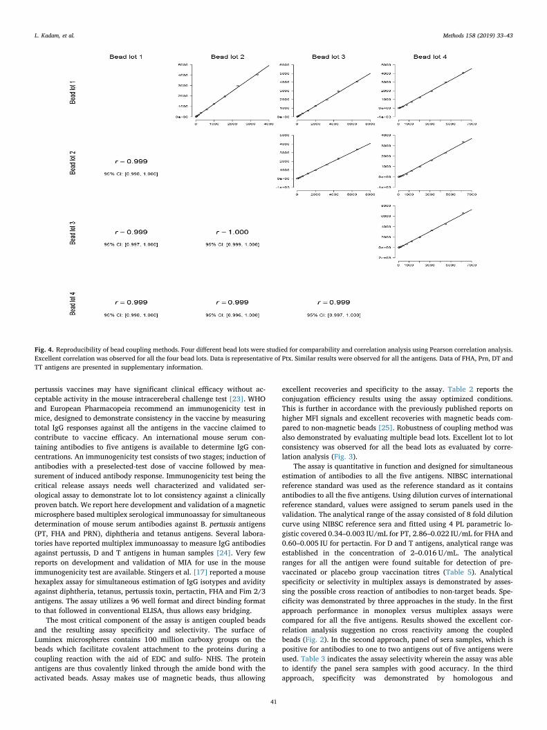
Assay ruggedness was also studied with respect to assessing varia-tions due to use of different lots of antigen coupled beads. Four differentlots of beads were used for assessment for all the antigens. The lot to lotvariability was studied using Pearson correlation analysis (Fig. 4). The
Table 2Performance of bead coupling method. Bead counts were counted on hemocytometer pre and post coupling and percent recovery is reported. The percent recoveriesare representative of 4 coupling reactions.
Sr.No Antigen Bead region no. Initial Beads Taken Bead count post coupling % Recovery
1 Pertussis Toxin (PT) 35 3.75× 10⁶ 3.52×10⁶ 952 Filamentous Hemagglutinin (FHA) 42 3.75× 10⁶ 3.12×10⁶ 833 Pertactin (PRN) 14 3.75× 10⁶ 3.30×10⁶ 884 Diphtheria Toxin (PT) 44 3.75 X 10⁶ 3.40 X 10⁶ 915 Tetanus Toxin (TT) 48 3.75× 10⁶ 3.72×10⁶ 99
Table 3Method Selectivity. Specificity/selectivity of Pentaplex MIA: Panel sera samplesrepresenting antibodies positivity for combinations among PT, FHA, PRN, DT &TT antibodies. Values in tables are representative of Mean fluorescence in-tensity (N=3) which was observed for target and non-target bead regions forthe respective serum panel.
Panel Sera PT FHA PRN DT TT
Panel 1A (PT, FHA PRN) 826 3942 329 129 89Panel 1B (DT) 18 34 10 7249 11Panel 1C (DT, TT) 15 42 7 5426 12,482Panel 1D (Negative serum) 55 42 47 26 44
Table 4Method specificity of pentaplex MIA: percentage inhibition on addition ofhomologous and heterologous inhibitor. Concentration used: 25 µg/ml for allthe antigens.
Inhibitor/Beads PT FHA PRN DT TT
PT 87 0 0 0 0FHA 0 90 4 0 0PRN 13 0 99 1 15DT 0 1 5 99 0TT 10 7 2 5 95
Table 5Standard Curve ranges for all the analyte giving the limits of quantification. Thecurve fitting was evaluated with both 5 and 4 PL logistic curves. 4 PL logisiticcurves were selected for further studies. Correlation coefficients observed for 4and 5 PL fits are also presented.
Analyte/Assay
Penta-Plex Assay R2 value obtainedusing 4PL curvefit
R2 value obtainedusing 5PL curvefitIgG (ULOQ) IgG (LLOQ)
PT 0.34 0.003 0.995 0.995FHA 2.86 0.022 0.997 0.998PRN 0.6 0.005 0.999 0.999DT 2 0.016 0.995 0.998TT 2 0.0016 0.998 0.999
L. Kadam, et al. Methods 158 (2019) 33–43
37

Regression type: Logistic ‐ 4PL Regression Type: Logistic ‐ 4PL
Std Curve: ‐5.03916 + (10209.1 + 5.03916) Std. Curve: FI = 21.1876 + (13701.2 ‐ 21.1876) / (1 + (Conc / 0.472973)^‐0.938901) / (1 + (Conc / 0.113708)^‐0.962804) FitProb. = 0.9560, ResVar. = 0.1653 FitProb. = 0.8610, ResVar. = 0.3256
Regression type: Logistic ‐ 4PL Regression Type: Logistic ‐ 4PL Std Curve: ‐16.498 + (22254 + 16.498) Std. Curve: FI = ‐10.9987 + (15138 + 10.9987) / (1 + (Conc / 0.305051)^‐0.936422) / (1 + (Conc / 0.369441)^‐0.865262) FitProb. = 0.6486, ResVar. = 0.6195 FitProb. = 0.2532, ResVar. = 1.3376
Regression type: Logistic ‐ 4PL Std Curve: ‐12.4986 + (16850 + 12.4986) / (1 + (Conc / 0.279298)^‐0.806481) FitProb. = 0.6289, ResVar. = 0.6471
Fig. 2. Pentaplex assay reference curves for all the five antigens. Dashed lines represent lower and upper LOQs.
L. Kadam, et al. Methods 158 (2019) 33–43
38

y = 0.9724x - 31.894R² = 0.9992
0
1000
2000
3000
4000
5000
6000
7000
8000
9000
0 2000 4000 6000 8000 10000
MFI
obt
aine
d fro
m M
ultip
lex
assa
y
Assay selectivity for Pertussis toxin (PT)
y = 0.9943x - 7.1862R² = 0.9996
0
2000
4000
6000
8000
10000
12000
14000
16000
18000
20000
0 5000 10000 15000 20000
MFI
obt
aine
d fro
m M
ultip
lex
assa
y
MFI obtained from Monoplex assay
Assay selectivity for FHA
y = 2.1342x + 43.14R² = 0.9971
0
100
200
300
400
500
600
700
800
900
1000
0 100 200 300 400 500
MFI
obt
aine
d fro
m M
ultip
lex
assa
y
MFI obtained from Monoplex assay
Assay selectivity for Pertactin
y = 0.9912x - 156.18R² = 0.9984
0
2000
4000
6000
8000
10000
12000
14000
16000
0 5000 10000 15000 20000
MFI
obt
aine
d fro
m m
ultip
lex
assa
y
MFI obtained from monoplex assay
Assay selectivity for TT
y = 0.9912x - 156.18R² = 0.9984
02000400060008000
10000120001400016000
0 5000 10000 15000 20000MFI
obt
aine
d fro
m m
ultip
lex
assa
y
MFI obtained from monoplex assay
Assay selectivity for DT
Fig. 3. Comparison of MFI signals generated from monoplex and pentaplex MIA with reference serum titration for Ptx, FHA, PRN in IU/ml and Dtx and Ttx in U/ml.
L. Kadam, et al. Methods 158 (2019) 33–43
39

results indicated that four bead lots were consistent and an excellentcorrelation coefficient was observed amongst all the bead lots for all thefour antigens (Fig. 4) demonstrating the ruggedness of couplingmethod.
3.5. Assay accuracy
Tables 6 and 7 depicts the assay accuracy for all the antigens. Theestimated values were compared with assigned concentrations and %accuracy was calculated for the sera panel. The % accuracy estimateswere found within 80–130%, thereby meeting the criteria for con-cordance and accuracy.
3.6. Dilutional integrity
The effect of dilution was studied on high titre sera wherein dilu-tions ranging from 6.25 to 20,0000 to determine any bias. No sig-nificant bias was observed with studied dilutions and the estimate ateach dilution was found within the acceptance criteria of 70–130% ofexpected value (Fig. 5).
3.7. Assay applicability
The applicability of the developed MIA in an mouse immunogenicytest was confirmed by confirming the dose response in Tdap immunizedanimals. Mouse Immunogenicity test involves the dose response ana-lysis of reference and test vaccine. For routine batch analysis, aminimum of three dilutions of vaccine are chosen. Animals were im-munized with three test dilution of Tdap vaccine and sera samples wereanalyzed on day 35 for IgG responses using pentaplex assay. The assaywas able to predict the dose wise trend in the geometric mean con-centrations of antibodies against all the antigens in the vaccine. Dosedependent responses for the antigens in the vaccine is presented inFig. 6.
4. Discussion and conclusion
The objective of quality control testing of acelluar pertussis antigensis the laboratory evaluation of their immunological efficiency to protectagainst human disease. The mouse intracereberal challenge assay,which is accepted assay for whole cell pertussis vaccines have chal-lenges in interpreting the potency of acellular pertussis antigens. Thereare reports from clinical trials which demonstrated that acellular
Table 6Precision and accuracy estimates of the MIA assay for PT, FHA and PRN. Intra-assay (with same bead lot and analyst-1) and inter-assay variation (different day,different analyst and different bead lot). Accuracy represents concordance with assigned value in both inter and intra-assay formats.
Pertussis toxin Precision Accuracy
Panel Sera used for study Assigned potency in(IU/ml)
Intra assay variative(Mean % CV)
Inter assay variative(Mean % CV)
% Interassay accuracy (%agreement with assigned value)
% Intrassay Accuracy (%agreement with assigned value)
2A 37 10 17 117 852B 18 9 16 106 842C 8 6 13 104 882D 40 9 19 100 81
Filamentous hemaagglutinin(FHA)
Assigned potency(IU/ml)
Intra assay variation(Mean % CV)
Inter assay variation(Mean % CV)
% Interassay accuracy % Intrassay Accuracy
2A 50 4 9 106 1092B 26 4 17 106 1032C 22 4 18 95 902D 159 1 0 99 83
Pertactin Assigned potency(IU/ml)
Intra assay variation(Mean % CV)
Inter assay variation(Mean % CV)
% Interassay accuracy % Intrassay Accuracy
2A 10 5 0 107 1002B 5 3 3 96 1042C 1 0 0 100 1002D 33 3 11 105 86
Table 7Precision and accuracy estimates of the MIA assay for DT and TT: Intra-assay (with same bead lot and analyst-1) and inter-assay variation (different day, differentanalyst and different bead lot). Accuracy represents concordance with assigned value in both inter and intra-assay formats.
Diphtheria Precision Accuracy
Panel Sera used forstudy
Assigned potency in(IU/ml)
Intra assay variation(Mean % CV)
Inter assay variation(Mean % CV)
% Interassay accuracy (%agreement with assigned value)
% Intrassay Accuracy (%agreement with assigned value)
2A 13 6 15 100 852B 3 13 20 130 702C 1 0 0 100 1002D 44 15 5 80 105
Tetanus Assigned potency(IU/ml)
Intra assay variation(Mean % CV)
Inter assay variation(Mean % CV)
% Interassay accuracy % Intrassay Accuracy
2A 99 6 9 100 1032B 56 6 11 95 902C 24 3 14 103 1012D 244 13 10 82 100
L. Kadam, et al. Methods 158 (2019) 33–43
40
pertussis vaccines may have significant clinical efficacy without ac-ceptable activity in the mouse intracereberal challenge test [23]. WHOand European Pharmacopeia recommend an immunogenicity test inmice, designed to demonstrate consistency in the vaccine by measuringtotal IgG responses against all the antigens in the vaccine claimed tocontribute to vaccine efficacy. An international mouse serum con-taining antibodies to five antigens is available to determine IgG con-centrations. An immunogenicity test consists of two stages; induction ofantibodies with a preselected-test dose of vaccine followed by mea-surement of induced antibody response. Immunogenicity test being thecritical release assays needs well characterized and validated ser-ological assay to demonstrate lot to lot consistency against a clinicallyproven batch. We report here development and validation of a magneticmicrosphere based multiplex serological immunoassay for simultaneousdetermination of mouse serum antibodies against B. pertussis antigens(PT, FHA and PRN), diphtheria and tetanus antigens. Several labora-tories have reported multiplex immunoassay to measure IgG antibodiesagainst pertussis, D and T antigens in human samples [24]. Very fewreports on development and validation of MIA for use in the mouseimmunogenicity test are available. Stingers et al. [17] reported a mousehexaplex assay for simultaneous estimation of IgG isotypes and avidityagainst diphtheria, tetanus, pertussis toxin, pertactin, FHA and Fim 2/3antigens. The assay utilizes a 96 well format and direct binding formatto that followed in conventional ELISA, thus allows easy bridging.
The most critical component of the assay is antigen coupled beadsand the resulting assay specificity and selectivity. The surface ofLuminex microspheres contains 100 million carboxy groups on thebeads which facilitate covalent attachment to the proteins during acoupling reaction with the aid of EDC and sulfo- NHS. The proteinantigens are thus covalently linked through the amide bond with theactivated beads. Assay makes use of magnetic beads, thus allowing
excellent recoveries and specificity to the assay. Table 2 reports theconjugation efficiency results using the assay optimized conditions.This is further in accordance with the previously published reports onhigher MFI signals and excellent recoveries with magnetic beads com-pared to non-magnetic beads [25]. Robustness of coupling method wasalso demonstrated by evaluating multiple bead lots. Excellent lot to lotconsistency was observed for all the bead lots as evaluated by corre-lation analysis (Fig. 3).
The assay is quantitative in function and designed for simultaneousestimation of antibodies to all the five antigens. NIBSC internationalreference standard was used as the reference standard as it containsantibodies to all the five antigens. Using dilution curves of internationalreference standard, values were assigned to serum panels used in thevalidation. The analytical range of the assay consisted of 8 fold dilutioncurve using NIBSC reference sera and fitted using 4 PL parametric lo-gistic covered 0.34–0.003 IU/mL for PT, 2.86–0.022 IU/mL for FHA and0.60–0.005 IU for pertactin. For D and T antigens, analytical range wasestablished in the concentration of 2–0.016 U/mL. The analyticalranges for all the antigen were found suitable for detection of pre-vaccinated or placebo group vaccination titres (Table 5). Analyticalspecificity or selectivity in multiplex assays is demonstrated by asses-sing the possible cross reaction of antibodies to non-target beads. Spe-cificity was demonstrated by three approaches in the study. In the firstapproach performance in monoplex versus multiplex assays werecompared for all the five antigens. Results showed the excellent cor-relation analysis suggestion no cross reactivity among the coupledbeads (Fig. 2). In the second approach, panel of sera samples, which ispositive for antibodies to one to two antigens out of five antigens wereused. Table 3 indicates the assay selectivity wherein the assay was ableto identify the panel sera samples with good accuracy. In the thirdapproach, specificity was demonstrated by homologous and
Fig. 4. Reproducibility of bead coupling methods. Four different bead lots were studied for comparability and correlation analysis using Pearson correlation analysis.Excellent correlation was observed for all the four bead lots. Data is representative of Ptx. Similar results were observed for all the antigens. Data of FHA, Prn, DT andTT antigens are presented in supplementary information.
L. Kadam, et al. Methods 158 (2019) 33–43
41

heterologous inhibition method (Table 4). Excellent specificity can beattributed to the quality of the antigens used in the coupling andmagnetic beads in the assay. Chemically well defined and purified an-tigen batch for each analyte is necessary for a an optimum couplingprocess. There are reports which have shown that use of the aceullarpertussis purified toxins significantly improves the assay performanceand concordance with the values obtained by conventional ELISAmethods. The study made use of purified toxins (PT, FHA and PRN) andtoxoids (D and T) for the coupling process. Commercial availability ofpurified D and T toxins is always a challenge and have batch to batchvariability. As the intended objective was to evaluate the immune re-sponses against vaccines which use toxoids as antigens, the study fo-cused on validating the assay with use of D and T toxoids as capture
antigens. Specificity results clearly suggest that results were compar-able to performance reported in published results of similar multiplexassays which used purified toxins of D and T for the coupling [18]. Thevalidation studies also assessed the precision and analytical accuracy ofthe assay. Inter-assay and intra-assay variability was found to be below20% and excellent concordance was observed between assigned andpredicted values.
Towards applicability and implementation to mouse im-munogenicity test, the assay was applied to analysis of sera samplesobtained from animals which were immunized with different dilutionsof Tdap vaccine. The assay was able to predict the dose dependent trendin IgG responses against all the five antigens, which will be important todefine the potency and quality of vaccine against a reference vaccine
Fig. 5. Dilution integrity assessment. Positive control serum with assigned value was used for the assessment. Graph is representative of assessment for all the fiveantigens. The dilution wise estimate of concentrations was within±30% criteria.
Fig. 6. Mouse Immunogenicity Test. Effect of im-munizing Tdap vaccine dose on mice antibody re-sponses against DT, TT, PT, PRN and FHA as ana-lyzed by pentaplex MIA. Animals (N=10/dilution)were immunized with different dilutions of Tdapvaccine (1/2.5, 1/5. 1/10 and 1/20 dilution ofhuman dose) on day 0 and sera was collected on day35. Geometric mean concentrations (IU/ml) werecalculated using pentaplex MIA. The error bars arerepresentative of respective group standard devia-tions.
L. Kadam, et al. Methods 158 (2019) 33–43
42

batch of proven clinical efficacy (Fig. 5). The multiplex immunoassaydescribed here thus meets the necessary criteria for implementation asquality control tool for Tdap or DTaP containing vaccine. The assay isable to simultaneously quantify antibodies to all the five antigens usingthe minimum serum volumes and very small amounts of antigens of thefive antigen as compared to conventional ELISA platforms. The multi-plexed measurements further reduced the time and labor to assay largenumber of sera samples. For example, 100 samples can be assayed for 5antigens on five different plates in a single day. This will be a significantcost and time savings in quality control laboratory setting where mul-tiple vaccine batches are manufactured and each batch needs to beanalyzed for its immunogenicity potential.
In conclusion, the assay described here meets all the requirements ofspecificity, selectivity and reproducibility and accuracy and is thus aviable alternative to conventional ELISA for detection of DTaP3 anti-bodies in mouse serum samples. The robustness and assay format alsooffers opportunities to include additional antigens of DTaP combina-tions such as Hib and Hepatitis B or other pertussis antigens such asadenylate cyclase and Fim 2/3. The study reports development, quali-fication and validation of multiplex assay. For further use in the qualitycontrol testing of vaccines, detailed characterization and validationstudies are required
Declaration of interests
None.
Acknowledgement
Authors thanks Serum Institute of India Ltd for all the fundingsupport. All the authors are employees of Serum Institute of India PvtLtd and declare no conflict of interest. This research did not receive anyspecific grant from funding agencies in the public, commercial, or not-for-profit sectors.
References
[1] G.S. Marshall, L.E. Happe, O.E. Lunacsek, M.D. Szymanski, C.R. Woods, M. Zahn,et al., Use of combination vaccines is associated with improved coverage rates,Pediatr. Infect. Dis. J. 26 (2007) 496–500.
[2] H. Kalies, V. Grote, T. Verstraeten, L. Hessel, H.J. Schmitt, R. von Kries, The use ofcombination vaccines has improved timeliness of vaccination in children, Pediatr.Infect. Dis. J. 25 (25) (2006) 507–512.
[3] B. Schlingmann, K.R. Castiglia, C.C. Stobart, M.L. Moore, Polyvalent vaccines: high-maintenance heroes, PLoS Pathog. 14 (2018) 5, https://doi.org/10.1371/journal.ppat.1006904 eCollection 2018 Apr.
[4] E. Vidor, B. Soubeyrand, Manufacturing DTaP-based combination vaccines: in-dustrial challenges around essential public health tools, Expert Rev. Vaccines 15(2016) 1575–1582.
[5] S.S. Jadhav, S. Gairola, Composition of acellular pertussis and combination vac-cines: a general review, Biologicals 27 (1999) 105–110.
[6] World Health Organization. Recommendations to assure the quality, safety andefficacy of acellular pertussis vaccines. WHO Technical Report Series 878 57-76.< http://www.who.int/biologicals/publications/trs/areas/vaccines/acellular_pertussis/WHO_TRS_878_A2.pdf?ua=1> , 1998.
[7] Monographs for aP vaccines or aP-based combination vaccines: monographs 1356,1595, 1931, 1932, 1933,1934, 2065, 2067, 2329. Ph. Eur. 6th Edition. Strasbourg,France: Council of Europe, 2009.
[8] G. Panicker, I. Rajbhandari, B.M. Gurbaxani, T.D. Querec, E.R. Unger, Developmentand evaluation of multiplexed immunoassay for detection of antibodies to HPVvaccine types, J. Immunol. Methods 417 (2015) 107–114, https://doi.org/10.1016/j.jim.2014.12.013 Epub 2014 Dec 30.
[9] A.J. Basile, K. Horiuchi, A.J. Panella, J. Laven, O. Kosoy, R.S. Lanciotti,N. Venkateswaran, B.J. Biggerstaff, Multiplex microsphere immunoassays for thedetection of IgM and IgG to arboviral diseases, PLoS One 8 (2013), https://doi.org/10.1371/journal.pone.0075670. eCollection 2013 e75670.
[10] K. McCutcheon, A multiplex approach to isotyping antigen-specific antibodies usingbiotinylated antigen/streptavidin-phycoerythrin, Methods Mol. Biol. 418 (2008)187–208.
[11] S.D. Eletu, C.L. Sheppard, E. Thomas, K. Smith, P. Daniel, D.J. Litt, W.S. Lim,N.K. Fry, Development of an extended-specificity multiplex immunoassay for de-tection of streptococcus pneumoniae serotype-specific antigen in urine by use ofhuman monoclonal antibodies, Clin. Vaccine Immunol. 24 (2017) e00262–e317,https://doi.org/10.1128/CVI.00262-17.
[12] D. Pavliakova, P.C. Giardina, S. Moghazeh, S. Sebastian, M. Koster, V. Pavliak,V. Pavliak, A. McKeen, R. French, K.U. Jansen, M. Pride, Development and vali-dation of 13-plex luminex-based assay for measuring human serum antibodies toStreptococcus. pneumoniae capsular polysaccharides, mSphere 3 (2018), https://doi.org/10.1128/mSphere.00128-18.
[13] J. Wang, S.P. Hilchey, M. DeDiego, S. Perry, O. Hyrien, A. Nogales, J. Garigen,F. Amanat, N. Huertas, F. Krammer, L. Martinez-Sobrido, D.J. Topham,J.J. Treanor, M.Y. Sangster, M.S. Zand, Broad cross-reactive IgG responses elicitedby adjuvanted vaccination with recombinant influenza hemagglutinin (rHA) inferrets and mice, PLoS One 13 (2018) e0193680, , https://doi.org/10.1371/journal.pone.0193680.
[14] M. Nygård, A. Saah, C. Munk, L. Tryggvadottir, E. Enerly, M. Hortlund,L.G. Sigurdardottir, S. Vuocolo, S.K. Kjaer, J. Dillner, Evaluation of the long-termanti-human papillomavirus 6 (HPV6), 11, 16, and 18 immune responses generatedby the quadrivalent HPV vaccine, Clin. Vaccine Immunol. 22 (2015) 943–948,https://doi.org/10.1128/CVI.00133-15.
[15] H.L. Itell, E.P. McGuire, P. Muresan, C.K. Cunningham, E.J. McFarland, W.Borkowsky, S.R. Permar, G.G. Fouda. Development and application of a multiplexassay for the simultaneous measurement of antibody responses elicited by commonchildhood vaccines.
[16] R.N. Caboré, D. Piérard, K. Huygen, A Belgian Serosurveillance/Seroprevalencestudy of diphtheria, tetanus and pertussis using a luminex xMAP technology-basedpentaplex, Vaccines (Basel) 4 (2016) E16, https://doi.org/10.3390/vaccines4020016.
[17] R.M. Stenger, M. Smits, B. Kuipers, S.F. Kessen, C.J. Boog, C.A. van Els, Fast, an-tigen-saving multiplex immunoassay to determine levels and avidity of mouseserum antibodies to pertussis, diphtheria, and tetanus antigens, Clin. VaccineImmunol. 18 (2011) 595–603, https://doi.org/10.1128/CVI.00061-10 Epub 2011Feb 16.
[18] P.G. Van Gageldonk, F.G. van Schaijk, F.R. van der Klis, G.A. Berbers, Developmentand validation of a multiplex immunoassay for the simultaneous determination ofserum antibodies to Bordetella pertussis, diphtheria and tetanus, J. Immunol.Methods 335 (2008) 79–89, https://doi.org/10.1016/j.jim.2008.02.018 Epub 2008Mar 26.
[19] EMEA, Guideline on bioanalytical method validation. EMEA/CHMP/EWP/192217/2009 Rev. 1 Corr 2.< http://www.ema.europa.eu/docs/en_GB/document_library/Scientific_guideline/2011/08/WC500109686.pdf > , 2009 (accessed 15.09.2018).
[20] US FDA, Guidance for industry on bioanalytical Method Validation.< https://www.fda.gov/downloads/drugs/guidances/ucm070107.Pdf > , 2018.
[21] H.E. Prince, M. Lapé-Nixon, J. Matud, Evaluation of a tetraplex microsphere assayfor Bordetella pertussis antibodies, Clin. Vaccine Immunol. 13 (2006) 266–270.
[22] Luminex Cook book (4th edition).< http://info.luminexcorp.com/en-us/research/download-the-xmap-cookbook> (assessed 15/09/2018).
[23] M.J. Corbel, D.K.L. Xing, A consideration of control requirements of acellular per-tussis vaccines, Dev. Biol. Stand 89 (1997) 343–347.
[24] R.N. Caboré, D. Piérard, K. Huygen, The performance of multiplex immunoassaysfor antibody determination to diphtheria, tetanus and pertussis: a need for stan-dardisation, J. Vaccines Vaccin. 7 (2016) 338, https://doi.org/10.4172/2157-7560.1000338.
[25] M.S. Hansenová, A. van Belkum, H.P. Endtz, F.J. Bikker, E.C. Veerman, W.J. vanWamel, Comparison of non-magnetic and magnetic beads in bead-based assays, J.Immunol. Methods 436 (2016) 29–33, https://doi.org/10.1016/j.jim.2016.06.003Epub 2016. Jun 11. PubMed PMID: 27296810.
L. Kadam, et al. Methods 158 (2019) 33–43
43
![Tetanus Toxin Antibody Levels in Pre-School Nigerian ... · serum anti-tetanus antibody levels provides scope for an objective analysis of tetanus immunity [22]. Serological surveys](https://static.fdocuments.in/doc/165x107/5d389a8a88c99359198c7365/tetanus-toxin-antibody-levels-in-pre-school-nigerian-serum-anti-tetanus.jpg)