Determination of quantitative peptide-binding motifs of ... · PDF file4.1.2 Pathogenesis of...
126
Determination of quantitative peptide-binding motifs of four common equine MHC class I alleles, and identification of an equine herpesvirus type 1-derived cytotoxic T lymphocyte epitope Dissertation zur Erlangung des akademischen Grades eines Doktors der Naturwissenschaften (Dr. rer. nat.) am Fachbereich Biologie, Chemie, Pharmazie der Freien Universität Berlin vorgelegt von Tobias Bergmann Berlin, Deutschland Mai 2016
Transcript of Determination of quantitative peptide-binding motifs of ... · PDF file4.1.2 Pathogenesis of...
Determination of quantitative peptide-binding motifs of four
common equine MHC class I alleles, and identification of an
equine herpesvirus type 1-derived cytotoxic T lymphocyte epitope
Dissertation zur Erlangung des akademischen Grades eines
Doktors der Naturwissenschaften (Dr. rer. nat.)
am Fachbereich Biologie, Chemie, Pharmazie
der Freien Universität Berlin
vorgelegt von
Tobias Bergmann
Berlin, Deutschland
Mai 2016

Diese Promotionsarbeit wurde im Zeitraum vom Januar 2012 bis zum Mai 2016 am Institut
für Virologie der Freien Universität Berlin unter der Leitung von Prof. Dr. Nikolaus Osterrie-
der angefertigt.
1. Gutachter: Prof. Dr. Nikolaus Osterrieder
2. Gutachter: Prof. Dr. Petra Knaus
Tag der Disputation: 17.02.2017
III
1. Table of contents
1. Table of contents ........................................................................................................................ III
2. List of figures and tables .............................................................................................................. V
3. Abbreviations ............................................................................................................................. VI
4. Introduction ..................................................................................................................................8
4.1 Herpesviruses ............................................................................................................................9
4.1.1 Replication cycle of equine herpesvirus type 1 .................................................................. 10
4.1.2 Pathogenesis of EHV-1 infections ..................................................................................... 11
4.1.3 Immune response to EHV-1 infections .............................................................................. 13
4.2 The Major Histocompatibility Complex ................................................................................... 15
4.2.1 MHC class I genetics ........................................................................................................ 16
4.2.2 Structure of MHC class I molecules .................................................................................. 18
4.2.3 Antigen processing for the MHC class I pathway .............................................................. 20
4.3. Project introduction ................................................................................................................ 25
4.3.1 Equine MHC class I .......................................................................................................... 25
4.3.2 T cell frequencies after in vitro stimulation ....................................................................... 26
5. Materials and Methods ............................................................................................................... 28
5.1. Materials ................................................................................................................................ 28
5.1.1 Chemicals, consumables and equipment ........................................................................... 28
5.1.2 Enzymes and markers ....................................................................................................... 33
5.1.3 Antibodies ........................................................................................................................ 33
5.1.4 Cells ................................................................................................................................. 33
5.1.5 Virus ................................................................................................................................ 34
5.1.6 Kits for molecular biology ................................................................................................ 34
5.1.7 Buffers, media and antibiotics ........................................................................................... 34
5.2 Methods .................................................................................................................................. 37
5.2.1 Cell culture methods ......................................................................................................... 37
5.2.2 Biochemical Methods ....................................................................................................... 40
5.2.3 Cell assays ........................................................................................................................ 46
6. Results ......................................................................................................................................... 50
6.1 Equine MHC class I ................................................................................................................ 50
IV
6.1.1 Stabilization of equine MHC-I molecules by externally supplied peptides ......................... 50
6.1.2 Elution of natural equine MHC class I ligands .................................................................. 55
6.1.3 Development of high throughput equine MHC-I binding assays ........................................ 59
6.1.4 Determination of quantitative peptide-binding motifs for all ELA alleles .......................... 60
6.1.5 Identification of equine herpes virus type-1-derived binding peptides ................................ 64
6.1.6 Quantifying the binding repertoires of Eqca-1*00101 and Eqca-N*00101 ......................... 66
6.1.7 Screening for ex vivo T cell reactivity in PBMCs from vaccinated or infected horses......... 67
6.2 Detection of IFN-γ-secreting cells upon virus stimulus ............................................................ 69
6.2.1 RacL11-infected PBMCs co-express MHC-I..................................................................... 69
6.2.2 PBMCs from a horse with unknown infection history respond to RacL11 infection ........... 70
6.2.3 PBMCs from EHV-1 primed but not naïve horses respond to RacL11 stimulus ................. 71
6.2.4 PBMCs from vaccinated horses did not respond to RacL11 stimulus ................................. 73
7. Discussion .................................................................................................................................... 74
8. Outlook ........................................................................................................................................ 83
9. Summary ..................................................................................................................................... 85
10. Zusammenfassung ..................................................................................................................... 87
11. References ................................................................................................................................. 89
12. Publications ............................................................................................................................. 100
13. Acknowledgements .................................................................................................................. 101
14. Selbstständigkeitserklärung .................................................................................................... 102
15. Curriculum vitae ..................................................................................................................... 103
16. Supplemental data................................................................................................................... 106

V
2. List of figures and tables
Figure 1: A herpes simplex virus particle. ...........................................................................................9
Figure 2: Pathogenesis of EHV-1 infections. ..................................................................................... 12
Figure 3: Shared classical and non-classical MHC-I loci for 10 ELA-A haplotypes. .......................... 17
Figure 4: Schematic representation of the binding groove of an MHC class I molecule. ..................... 19
Figure 5: Antigen processing pathway for MHC class I molecules. .................................................... 23
Figure 6: In vitro binding assay. ........................................................................................................ 46
Figure 8: RMA-S MHC stabilization assay........................................................................................ 47
Figure 9: FACS histogram for MHC stabilization. ............................................................................. 50
Figure 10: Peptide stabilizers for Eqca-1*00101. ............................................................................... 51
Figure 11: Peptide stabilizers for Eqca-N*00101. .............................................................................. 52
Figure 12: Peptide stabilizers for Eqca-1*00201. ............................................................................... 53
Figure 13: Length distribution of eluted peptides. .............................................................................. 58
Figure 14: Summary of the binding motifs for ELA alleles ................................................................ 64
Figure 15: Percent of predicted peptides binding to Eqca-1*00101 or Eqca-N*00101 ........................ 65
Figure 16: ELISpot interferon-γ T cell responses to Eqca-1*00101 binding-peptides.. ....................... 68
Figure 17: ELISpot interferon-γ T cell responses to Eqca-N*00101 binding-peptides. ....................... 69
Figure 18: Equine PBMCs infected with EHV-1 strain RacL11 expressing the gene for GFP. ........... 70
Figure 19: PBMCs from a horse with unknown infection history respond to RacL11 stimulus. .......... 70
Figure 20: PBMCs from EHV-1 primed but not naive horses respond to RacL11 stimulus ............... 71
Figure 21: T cell frequencies of EHV-1 primed and naïve horses after RacL11 stimulus . .................. 72
Figure 22: T cell frequencies of additional EHV-1 primed and naïve horses ...................................... 72
Figure 23: T cell frequencies after vaccination. ................................................................................. 73
Table 1: ELA class I alleles used in this study ................................................................................... 25
Table 2: Peptide sequences for Eqca-1*00101 ................................................................................... 51
Table 3: Peptide sequences for Eqca-N*00101 .................................................................................. 52
Table 4: Peptide sequences for Eqca-1*00201 ................................................................................... 54
Table 5: Peptides eluted from Eqca-1*00101..................................................................................... 55
Table 6: Peptides eluted from Eqca-N*00101 .................................................................................... 56
Table 7: Peptides eluted from Eqca-16*00101 ................................................................................... 57
Table 8: Peptides eluted from Eqca-1*00201..................................................................................... 58
Table 9: Assay specifications of in vitro binding assays for all ELA alleles ....................................... 59
Table 10: PSCL-derived ARB matrix for Eqca-1*00101 ................................................................... 60
Table 11: PSCL-derived ARB matrix for Eqca-N*00101 .................................................................. 61
Table 12: PSCL-derived ARB matrix for Eqca-16*00101 ................................................................. 62
Table 13: PSCL-derived ARB matrix for Eqca-1*00201 ................................................................... 63
Table 14: EHV-1-derived peptides for Eqca-1*00101 and Eqca-N*00101 ......................................... 66
Table 15: Blood donors for ELISpot analyses .................................................................................... 67
Table 16: ICP4-derived MHC stabilizers and peptides for Eqca-1*00101 and Eqca-N*00101............ 75

VI
3. Abbreviations
aa amino acid
ABC ATP-binding cassette
APC antigen-presenting cell
ARB average relative binding
ATP adenosine triphosphat
BAC bacterial artificial chromosome
BHV bovine herpesvirus
BSA bovine serum albumin
CD cluster of differentiation
CNS central nervous system
ConA concanavalin A
cTEC cortical thymic epithelia cell
CTL cytotoxic T lymphocyte
ddH2O double distilled water
DMSO dimethyl sulfoxide
DMP dimethyl pimelimidate
DNA deoxyribonucleic acid
DRiPs defective ribosomal products
dsDNA double-stranded DNA
E early
EDTA ethylenediaminetetraacetic acid
EHV equine herpes virus
EIAV equine infectious anemia virus
ELA equine leukocyte antigen
ELISpot enzyme-linked immunospot
ER endoplasmic reticulum
ERAP ER-resident aminopeptidases
Eqca Equus caballus
FCS Fetal calf serum
FT flowthrough
gB glycoprotein B
gC glycoprotein C
gD glycoprotein D
gE glycoprotein E
gG glycoprotein G
GFP green fluorescent protein
gH/gL glycoprotein H/glycoprotein L complex
HBC hepatitis B virus
HCMV human cytomegalovirus
HLA human leukocyte antigen
HSV herpes simplex virus
VII
HVEM herpesvirus entry mediator
ICP0 infected cell protein 0
ICP4 infected cell protein 4
ICP47 infected cell protein 47
IE immediate early
IFN-γ interferon-γ
IR internal repeat
kbp kilo base pair
KSHV Kaposi sarcoma herpesvirus
L late
LPS lipopolysaccharide
MEM minimal essential medium Eagle
MHC major histocompatibility complex
MHC-I MHC class I molecule
MLV modified-live vaccine
NK cell natural killer cell
O/N over night
ORF open reading frame
RNA ribonucleic acid
rpm rotations per minute
rt room temperature
P position
PAMPs pathogen-associated molecular patterns
PBMC peripheral blood mononuclear cell
PBS phosphate buffered saline
PBST PBS Tween
PCR polymerase chain reaction
pi post infection
PLC peptide loading complex
PMSF phenylmethylsulfonyl fluoride
PRR pattern recognition receptors
PrV pseudorabies virus
PSCL positional scanning combinatorial libraries
P/S penicillin/streptomycin
SD standard deviation
SF specificity factor
SFC spot-forming cell
TAE Tris-acetate-EDTA buffer
TAP transporter associated with peptide processing
Th cell T helper cell
TLM translocation motif
TNF tumor necrosis factor
TPP tripeptidyl peptidase
UL unique long
US unique short
VZV varicella zoster virus

Introduction
8
4. Introduction
Every organism on earth is target of constant attacks by microorganisms seeking to invade
and procreate inside a host’s body. An infection can be associated with disease or remain sub-
clinical. The major defense against intruding pathogens is the immune system, which evolved
in a constant arms race with microorganisms. The immune system is based on three tightly
regulated columns. The first line of defense is the intrinsic immune system, which is always
present and does not have to be induced. It includes physical barriers like the skin and muco-
sae as well as protein molecules with more specific functions. The second column represents
the innate immune system composed of a variety of receptors, cytokines and immune cells. In
contrast to intrinsic resistance, the innate immune system has to be activated upon contact
with an antigen. Patrolling phagocytic cells internalize extracellular particles and recognize
conserved patterns associated with pathogens (PAMPs: Pathogen associated molecular pat-
terns), e.g. lipopolysaccharide of gram-positive bacteria, unmethylated CpG-DNA, or double-
stranded RNA as an intermediary replication product of some viruses, with their diverse pat-
tern recognition receptors (PRR). Recognition of PAMPs results in the production of pro-
inflammatory cytokines like interleukins, type I and II interferons as well as tumor necrosis
factor (TNF). That attracts the full complement of effector cells leading to activation of the
adaptive immune system, which constitutes the third column. The adaptive immune system
requires prolonged contact with the specific pathogen in order to mount a highly specific and
effective immune response. Key players are B lymphocytes, which produce soluble epitope-
specific immunoglobulins (Ig), and T lymphocytes, which specifically recognize intra- and
extracellular antigens presented by infected or professional antigen presenting cells (APCs),
respectively.
Once a cell has been identified as infected, CD8+ cytotoxic T lymphocytes (CTL) destroy the
particular cell. Key players in the process of recognition are major histocompatibility complex
(MHC) molecules, which present pathogen-derived peptides on the surface of the cell. Of
particular importance and interest are classical MHC class I and II (MHC-I, -II) molecules as
they are directly involved in the recognition of cell-associated as well as cell-free pathogens.
Both present peptide epitopes to T lymphocytes, which specifically recognize the complex via

Introduction
9
their polymorphic T cell receptor (TCR). Class I molecules mainly associate with endogenous
peptides, and in case of an intracellular infection, pathogen-derived peptides are presented to
CD8+ CTL, which upon binding proliferate and consequently destroy the infected cell. Thym-
ic self-intolerance ensures that only epitopes of pathogen origin will effectively activate effec-
tor functions of CTLs. Class II molecules act in a similar way as they present exogenous pep-
tides degraded in lysosomes to CD4+ helper T lymphocytes (Th cells). However, upon activa-
tion Th cells do not directly destroy the activating cell but secrete a bandwidth of immunolog-
ically active cytokines and chemokines to orchestrate the immune response (summarized from
[1]).
4.1 Herpesviruses
The order of Herpesvirales comprises a vast number of viruses capable of infecting virtually
all vertebrate and even some invertebrate species. They are grouped into three families termed
Herpesviridae, Alloherpesviridae and Malacoherpesviridae. Viruses infecting mammals, birds
and reptiles fall into three subfamilies of the Herpesviridae, namely alpha-, beta- and gamma-
herpesvirinae, which all share structural features in their virion composition, envelopes and
their genomes as well as their ability to cause latent infections [2, 3]. However, alphaherpesvi-
ruses have a fast replication cycle, a broad host range and establish latency in sensory ganglia
whereas betaherpesviruses replicate slowly and are more restricted in their host range. The
same is true for gammaherpesviruses, which mainly target immune cells for infection [4].
Figure 1: A herpes simplex virus particle. The DNA
genome is contained within the nucleocapsid, which is
surrounded by the amorphous tegument. A lipid
bilayer forms an envelope around it spiked with sur-face glycoproteins.
All herpesviruses have a linear, double-
stranded DNA genome with sizes ranging
from 108 – 300 kilo base pairs (kbp). For
alphaherpesviruses, it is divided into a long
and a short region, which harbor unique
sequences (UL: unique long, US: unique
short), flanked by internal and terminal
repeats. Their large genome can sustain
almost an entire DNA replication machi-
nery enabling them to replicate indepen-
dently from the cell cycle, and an array of
immune modulatory genes, which help
establish latent infections [3, 5].

Introduction
10
A virion of the prototypic herpes simplex virus (HSV) consists of an icosahedral nucleocapsid
formed by 162 capsomers associated with genomic DNA [3] surrounded by an amorphous
protein structure. This tegument forms an inner layer that is engulfed in a host cell-derived
membrane harboring several glycoproteins essential for adsorption and penetration as well as
immune modulation (Figure 1). Apart from having a mere scaffolding function, proteins of
the tegument are widely important for initiation and implementation of the infection [3].
4.1.1 Replication cycle of equine herpesvirus type 1
In equid species, nine different herpesviruses have been identified, which can be classified
into the subfamilies of alpha- and gammaherpesviruses. Horses are the natural hosts for al-
phaherpesviruses equine herpesvirus (EHV) -1, -3 and -4 as well as the gammaherpesviruses
EHV-2 and -5 [3, 6-8]. In economical, clinical and epidemiological terms most important are
EHV-1 and -4, which share a DNA sequence homology of up to 90 % [9]. EHV-1 is the caus-
ative agent of rhinopneumonitis, late-term abortion and neonatal foal disease as well as equine
herpesvirus myeloencephalopathy (EHM). As typical for herpes viruses, EHV-1 has a high
seroprevalence in domestic horse breeds ranging from 30 – 80 % [31].
EHV-1 is an alphaherpesvirus of the genus varicellovirus with a genome size of around 150
kbp, and harbors 80 open reading frames coding for at least 77 proteins [10, 11]. Entry into
cells follows the typical mechanism that is known for most herpesviruses including the proto-
typic HSV-1. Initial attachment is mediated through a weak interaction of the glycoproteins
gC and gB to cellular glucosaminoglycan and subsequent binding of gD to its specific cellular
receptor [12-14]. For HSV-1, nectins, herpesviral entry mediator (HVEM) and heparan sulfate
have been identified as gD-specific entry receptors [14]. EHV-1 can also use alternative re-
ceptors like MHC class I molecules preferably from alleles carrying alanine at position 172 in
the α2 region [15-17]. However, as MHC class I molecules are constitutively expressed on all
cell types, there may be other factors involved in cell tropism. Upon gD binding, it complexes
with gB and gH/gL, probably inducing conformational changes in the fusogenic gB [14]. Af-
ter attachment, the virus can enter a cell by either direct fusion with the cell membrane or
from within endosomes at acidic pH. Glycoprotein H and its cellular interactor α4β1 integrin
have been found to route the virion either way [18]. Once the nucleocapsid has been released
into the cytosol, it is transported along microtubules and the DNA genome is released into the

Introduction
11
nucleus where it circularizes [19]. During lytic replication, the whole set of viral genes is ex-
pressed allowing for a high production of progeny virus.
Herpes viral gene expression is temporally regulated and occurs in three phases. Immediate
early (IE) genes are the first to be transcribed and they include other transcriptional regulators.
The EHV-1 sole IE gene encodes an ICP4 (infected cell protein 4) homologue capable of
binding several other viral proteins as well as cellular transcription factors like TFIIB [20-23].
The first viral proteins present in the cytoplasm, however, are tegument proteins including
transactivators, which initiate immediate early gene expression, or modulating factors like the
viral host shut-off protein (vhs) that degrades cellular mRNAs in order for the virus to take
over cell metabolism [3, 24]. In the next step, proteins with early (E) kinetics are produced,
which are mainly associated with DNA replication [3, 25]. Structural proteins that form the
tegument, capsid and glycoproteins are produced as part of the late (L) phase [26]. One hall-
mark of herpes viruses is their ability to establish persistent infections that do not lead to the
generation of progeny since gene expression is reduced to a minimum. In this latency stage,
the viral genome is either present as a circular episome tethered to or for some herpesviruses
as a provirus integrated into the cell chromosomes [27]. As almost no viral proteins are pro-
duced and thusly presented to T lymphocytes, herpesviruses can persist for a prolonged time
in the host body without evoking an immune response. Primary cellular targets for latency are
trigeminal ganglia for most alphaherpesviruses, or lymphatic tissues for beta- and gammaher-
pesviruses [3]. EHV-1 although being an alphaherpesvirus primarily latently infects
CD5+/CD8
+ T lymphocytes, but trigeminal ganglia have also been described as site of latent
EHV-1 infections in horses [28, 29]. For the virus to infect a new host, however, it has to
reactivate and enter the lytic replication cycle to produce infectious progeny [3].
4.1.2 Pathogenesis of EHV-1 infections
The highly contagious EHV-1 is usually transmitted by direct contact through infected nasal
discharge and in rare cases through aerosols or contaminated food or water [30]. Respiratory
infection is characterized by shedding of virus through infected discharge, fever and anorexia
[31]. EHV-1 primarily replicates in the epithelium of the upper respiratory tract (URT) and
the nasal epithelium as early as 12 h post infection (pi). The virus cannot cross the basement
membrane directly, but modulation of the extracellular matrix proteins facilitates shuttling to
local lymph nodes via infected resident CD172+ monocytes where it infects more leukocytes
resulting in a cell-associated viraemia (Figure 2) [32, 33]. This is a prerequisite for the rapid
Introduction
12
dissemination of the infection to secondary sites of replication like the endothelia of the en-
dometrium and the central nervous system (CNS) [34-41]. An estimated 1 in 104 to 10
6 lym-
phocytes is infected leading to high levels of viraemia. Production of viral antigen indicative
of viral replication is predominantly found in monocytes, CD8+
and to a lesser extent in CD4+
T cells [35, 41, 42]. Consequently, experimental infection of pathogen free foals (SPF) results
in both a lymphopaenia and neutropaenia within the first week after infection [39].
Upregulation of adhesion molecules on the surface of infected lymphocytes facilitates adhe-
sion to endothelia and spread of the virus through cell-to-cell contacts, which is the primary
distribution route from peripheral blood mononuclear cells (PBMCs) to endothelial cells ra-
ther than by viral egress [43, 44]. In pregnant mares, infection of the endothelia in the endo-
metrium of the uterus might result in severe vasculitis, thrombosis and subsequent fetal
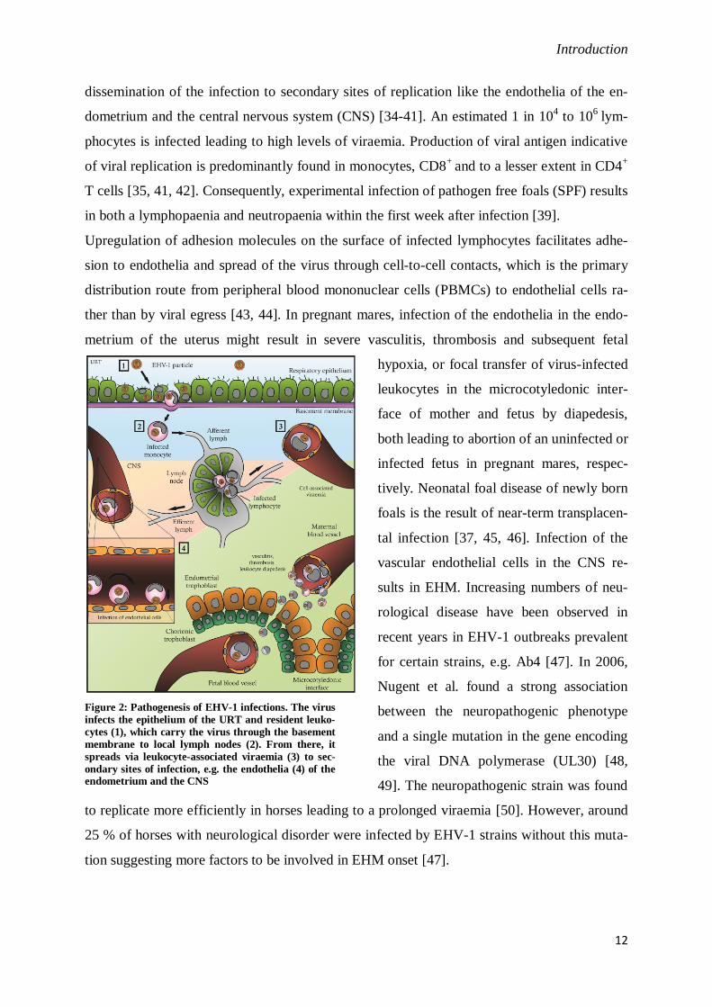
Figure 2: Pathogenesis of EHV-1 infections. The virus
infects the epithelium of the URT and resident leuko-
cytes (1), which carry the virus through the basement
membrane to local lymph nodes (2). From there, it
spreads via leukocyte-associated viraemia (3) to sec-
ondary sites of infection, e.g. the endothelia (4) of the endometrium and the CNS
hypoxia, or focal transfer of virus-infected
leukocytes in the microcotyledonic inter-
face of mother and fetus by diapedesis,
both leading to abortion of an uninfected or
infected fetus in pregnant mares, respec-
tively. Neonatal foal disease of newly born
foals is the result of near-term transplacen-
tal infection [37, 45, 46]. Infection of the
vascular endothelial cells in the CNS re-
sults in EHM. Increasing numbers of neu-
rological disease have been observed in
recent years in EHV-1 outbreaks prevalent
for certain strains, e.g. Ab4 [47]. In 2006,
Nugent et al. found a strong association
between the neuropathogenic phenotype
and a single mutation in the gene encoding
the viral DNA polymerase (UL30) [48,
49]. The neuropathogenic strain was found
to replicate more efficiently in horses leading to a prolonged viraemia [50]. However, around
25 % of horses with neurological disorder were infected by EHV-1 strains without this muta-
tion suggesting more factors to be involved in EHM onset [47].

Introduction
13
4.1.3 Immune response to EHV-1 infections
EHV-1 has an impressive capacity to evade the immune response of the host in order to stay
under the immunological radar. Subversion of recognition and destruction of infected cells by
antibody-dependent or CTL-mediated cell lysis as well as modulation of the host cytokine
response are potent immune evasive strategies [51]. In the humoral immune response, antibo-
dies specific for viral envelope proteins have two major functions. On the one hand, they rec-
ognize cell-free viral particles and associate with them, which by itself may neutralize viral
infectious abilities, and greatly increase receptor-mediated phagocytosis by activated macro-
phages [1]. Antibody-dependent cell-mediated cytotoxicity (ADCC), on the other hand, relies
on the presence of viral proteins on the surface of an infected cell. Natural killer cells (NK)
display an Fc receptor (CD16) capable of binding IgG associated with viral antigen, which
activates the NK cell to secrete type 2 interferon (IFN-γ), a potent antiviral immune modula-
tor, as well as granzymes that lyse the target cell [1]. In a similar process, antibody-
opsonization triggers the complement system to initiate its proteolytic cascade that results
either in direct destruction of the infected cell by formation of pores in the cell membrane
through the membrane-attack complex (MAC), or in enhanced phagocytosis [1].
As mentioned before, EHV-1 infection results in a leukocyte-associated viraemia, which
shields viral particles from antibody recognition. Additionally, no viral envelope proteins can
be detected on infected peripheral blood mononuclear cells (PBMCs), which could circum-
vent ADCC [52]. Several viral envelope proteins can function as decoy receptors. Glycoprote-
in C (gC) binds to the complement factor C3 interfering with the activation of the complement
cascade, and gG associates with a broad range of chemokines inhibiting their immunomodula-
tory functions [53, 54]. Protective immunity against infection of horses with respiratory pa-
thogens is mainly attributed to virus neutralizing mucosal IgA antibodies [55, 56]. Conse-
quently, challenge infection of horses with virulent EHV-1 strains results in a mucosal hu-
moral immune response with neutralizing IgA being the predominant antibody isotype [57].
Inactivated vaccines, however, fail to induce protective IgA titers [57]. This might explain
why circulating antibodies alone do not protect against infection and why mucosal as well as
cellular immunity is required [58, 59]. Cellular immunity, namely CD8+ CTL activity, is de-
pendent on viral epitopes being presented on MHC class I molecules of an infected cell. As
outlined later on, viral epitopes are generated in a multistep process, and in herpesvirus infec-
tions any step can be tackled by viral evasive strategies. EHV-1 codes for two proteins,
pUL49.5 and pUL56, which interfere with the antigen processing pathway [60, 61].

Introduction
14
Nevertheless, despite all those evasive strategies some degree of protection and reduction in
viral shedding can be achieved through vaccination reducing the number of abortion storms
and overall veterinary burden [59, 62]. Several inactivated vaccines are available, which can
induce virus neutralizing (VN) as well as complement-fixing (CF) antibody titers reducing the
severity and duration of infection, but their efficacy and their potential to prevent viraemia
and consequently abortion or EHM is very limited [58, 59, 63]. Frequent re-immunizations
are necessary since acquired immunity is particularly short-lived and re-infections can occur
within 3 – 6 months [59].
Increasing evidence suggests that cell-mediated immunity plays a major role in protection
from and control of EHV-1 infections and associated diseases. Virus-specific CTL frequen-
cies in peripheral blood of uninfected or with inactivated vaccine immunized horses are unde-
tectable or very low and those animals develop typical respiratory symptoms and viraemia,
and almost all pregnant mares abort upon challenge infection [64, 65]. Only multiply infected
horses are completely protected with no detectable viraemia but significantly increased virus-
specific precursor CTL frequencies [65]. This strong association suggests an EHV-1 infection
to be controlled predominantly by virus-specific cytotoxic T lymphocyte activity. Additional-
ly, CD4+ T cells also seem to play a role in protection, which might be attributed to direct
antiviral effects of IFN-γ [66, 67]. In these studies, the majority of IFN-γ-producing lympho-
cytes were CD8+, but also CD4
+ T cells were shown to contribute to IFN-γ production, which
increases after multiple EHV-1 infections. NK cells seem not to play a big role in the IFN-γ
response [68].
Vaccination strategies that target the induction of cellular immunity classically rely on atte-
nuated virus strains [1]. One modified-live vaccine (MLV) based on the attenuated strain
RacH is currently available for EHV-1 [69]. In regard to the virulent strain Ab4, it harbors
several deletions in its genome including ORF1, which codes for pUL56, and ORF2, glyco-
protein 2 and IR6 [70]. Although it confers protection against respiratory disease, its influence
on viraemia, abortion and EHM incidence is still controversial [59]. Several studies have
shown that it induces high titers of neutralizing antibodies but its effect on viraemia is very
limited [71-73]. Those limitations, in addition to the safety concerns associated with live vac-
cine applications particularly in pregnant mares, make the MLV for EHV-1 less than an ideal
vaccine.
To better understand the poor efficacy of vaccines, a closer look at the CTL response was
taken. For equine infectious anemia virus (EIAV), few CTL epitopes have been identified that
are restricted primarily by loci of the MHC class I haplotypes ELA-A5 and -A9 [74-76]. For

Introduction
15
EHV-1, the ICP4 (IE) protein encoded by gene 64 has been shown to contain CTL epitopes
restricted by MHC class I molecules of the ELA-A3 and –A2 haplotypes, however, a precise
peptide epitope remains to be defined [77-79]. Using a vaccinia virus-based vector vaccine
expressing the ICP4 gene (NYVAC-IE), or alternative approaches using particle-mediated
DNA vaccines coding for gC, gB, gD, ICP4 or an ICP27 homologue pUL5 failed to induce
sufficient protection against EHV-1 challenge infection [80, 81]. However, animals used in
these studies were not MHC-I haplotyped. A later study showed partial protection of ELA-
A3.1+ (Eqca-1*00101) ponies vaccinated with NYVAC-IE against EHV-1infection. The vac-
cinated ponies showed increased IFN-γ mRNA expression and reduced viraemia [82].
In conclusion, there is a variety of inactivated vaccines and one MLV in the market with sev-
eral alternative vaccine designs in clinical trials. Although most of them induce high serum
titers of neutralizing antibodies, induction of mucosal as well as cellular immunity, which are
considered key players in protection from and control of EHV-1 infections, is very poor.
4.2 The Major Histocompatibility Complex
Early in the 20th
century CC Little and EE Tyzzer working with Japanese waltzing mice ob-
served successful transplantation of tumors in F1 generations parented by mice of different
strains but rejection by almost all mice in the F2 generation. They concluded that the compa-
tibility of tissues is dependent on a number of different hereditable factors and that chances of
an F2 offspring to possess all those factors are very low [83, 84]. Subsequently, PA Gorer
found rejection reactions to be associated with four different antigens (antigen I, II, III and
IV) and GD Snell located them to a distinct genetic area, which he called H (histocompatibili-
ty) genes, defining the H-2 haplotype in mice [85, 86]. Decades later, Zinkernagel and Doher-
ty realized that T cell responses are restricted to major histocompatibility antigens, which be-
came known in humans as the human leukocyte antigen (HLA) located on chromosome 6
[87]. Soon it became clear that the genetic locus coding for HLA or H-2 molecules comprises
an abundance of genes and that this cluster is common to all jawed vertebrates [88], so it was
renamed to major histocompatibility complex (MHC).
As it is defined today, the human MHC encodes several hundred genes and pseudogenes par-
ticipating in the adaptive and innate immune response, and spans over 4 million base pairs
[89]. It can be subdivided into MHC I, II and III regions, which accumulate immunomodula-

Introduction
16
tory genes. The genes for classical as well as nonclassical MHC class I molecules are located
in the MHC I region, for class II molecules in the MHC II region, and the MHC III region
harbors genes, most of which are involved in the production of functional class I and II pro-
teins or take part in inflammatory processes [89].
4.2.1 MHC class I genetics
Ubiquitously found on all nucleated cells, primary function of MHC class I molecules (MHC-
I) is to convey the immune status of a cell by sampling the current proteome [1]. These mole-
cules bind intracellular peptides with certain corresponding chemical specificities and trans-
port them to the cell surface for T cell scrutiny. In context of an intracellular infection, for
example, T cell clones with TCRs specific for a given complex of MHC-I and viral peptide
bind, get activated and consequently destroy the target cell [1].
As a result, and perhaps not surprisingly, a strong selection pressure drives pathogen evolu-
tion towards escape variants that circumvent MHC-I and TCR specificities. To counter that,
the classical MHC-Ia locus is polygenic and highly polymorphic. In humans, three different
genetic loci encoding for classical MHC class Ia molecules are expressed. These loci, called
Human Leukocyte Antigen (HLA)-A, -B or –C, are inherited as a unit (haplotype) by Mende-
lian rules. The A, B and C loci are amongst the most diverse genes in the human genome,
with 3285, 4077 and 2801 alleles described to date, respectively, leading to a tremendous
amount of different allele combinations on population levels (http://hla.alleles.org/nomen-
clature/stats.html, September 2015). This high allelic variation is due to point mutations and
shared sequence motifs arisen by gene conversion events [90]. Most individuals are hetero-
zygous doubling the number of MHC-I molecules to six.
In humans, three nonclassical MHC-Ib loci (HLA-E, -F, -G), characterized by lower allele
numbers ranging from 18 to 51 (http://hla.alleles.org/nomenclature/stats.html, September
2015) are also expressed. The MHC-Ib molecules seem to have some peptide presenting
properties, as a few viral peptides have been found to stabilize HLA-E molecules at 26 °C,
although no evidence that they can interact with TCRs has been provided. Rather, they appear
to play immunomodulatory roles by regulating T cells, NK cells, macrophages, dendritic cells
and other immune cells [91].

Introduction
17
The equine MHC class I counterpart (Equine Leukocyte Antigen: ELA) is located on chromo-
some 20 and comprises up to 30 classical and nonclassical MHC class I loci as well as pseu-
dogenes spanning 4000 bp in eight exons [92, 93]. Serologically, 19 ELA-A haplotypes have
been distinguished, to date, each with similar levels of polymorphism estimated using intra-
MHC microsatellites and sequencing [94-96]. Using the same techniques, 27 microsatellite
haplotypes that did not correspond with any serological haplotype were identified [94]. The
number of expressed loci, however, may vary but up to seven different class I molecules can
be detected in horses homozygous for the ELA-A3 haplotype [97, 98]. In 2010, Tallmadge
and al. were able to assign almost 50 alleles to shared loci of ten different haplotypes (Figure
3), and to construct a phylogenetic tree based on sequence similarities [95].
Figure 3: Shared classical (blue numbers) and non-classical (black numbers) MHC-I loci for 10 ELA-A haplotypes [95].
As racing horse breeds, Thoroughbreds and Standardbreds are economically important. Sero-
logical as well as molecular typing of these breeds indicates genetic diversity that is
represented by only a limited number of MHC haplotypes (ELA-A2, -A3, -A5, -A9 and -A10)
[94, 99]. The ELA haplotypes A3, A2 and A9, for example, are present in 25 %, 17 %, and 14
%, respectively, of Thoroughbred horses [99, 94]. The evolution of the domestic horse lineage
is still controversially debated. The origin of wild horse Equus ferus domestication some 160
thousand years (kyr) ago can be located to the western Eurasian steppe followed by a demic
Introduction
18
spread of herds to the west adjoined by an east-to-west decline in genetic diversity [100]. The
high level of matrilineal diversity and low level of Y chromosome variability suggest a pre-
dominantly female introgression into domestic herds, which might reflect the strong patrili-
neal bottleneck in western central Eurasia around 125 kyr ago [101-103]. The discovery of the
fossils of a Pleistocene horse confirmed that the only still existing wild horse, the Przewalski
horse, falls into a different phylogenetic clade than modern domestic horses, and thusly is not
a direct ancestor. [104]. However, 29 genetic loci have been identified that show significantly
lower polymorphism in modern compared to Przewalski horses, probably due to domestica-
tion [104]. In contrast, evolution of dogs is the result of more than 500 individual domestica-
tion events of wolfs leading to a significantly higher MHC diversity [105]. In that light, it
seems possible that the limited equine MHC haplotype diversity might be a consequence of
millennia of breeding procedures aimed at optimizing particular characteristics of the horse,
MHC diversity not being one of them.
4.2.2 Structure of MHC class I molecules
The functional MHC class I molecule is a type I transmembrane protein consisting of an
α chain with three distinct domains (α1, α2 and α3) and a smaller β chain, the soluble β2-
microglobulin. The α1 and α2 domains together form a binding cleft or groove with each do-
main contributing an α helix forming the walls and a β sheet on the bottom [1]. The α3 do-
main spans the cellular membrane with a short C-terminal domain in the cytosol, and provides
an interaction site for the T lymphocyte co-receptor CD8 [1, 106]. The binding groove ac-
commodates peptide ligands of, typically, eight to ten amino acids in length, although longer
and rarely shorter peptides have been observed. As both ends of the class I groove are closed,
bound peptides generally fit their N- and C-termini completely into the groove, although in
some cases, as has been shown for a calreticulin-derived decamer, peptides may extend
beyond the N- or C-terminus. Peptides bind in an extended conformation, but may bulge out
in the middle, exposing certain residues for T cell scrutiny [107, 108].
The highly polymorphic genes of the MHC-I loci give rise to very diverse proteins where
most of the polymorphic positions can be found lining the peptide binding cleft [1]. In gener-
al, the binding cleft manifest six pockets (A-F), each engaging certain positions in the peptide
and mediating specific interactions [109]. The residues forming the pockets confer a high de-
gree of chemical specificity, with high affinity ligands generally possessing corresponding
Introduction
19
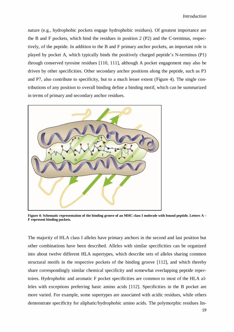
nature (e.g., hydrophobic pockets engage hydrophobic residues). Of greatest importance are
the B and F pockets, which bind the residues in position 2 (P2) and the C-terminus, respec-
tively, of the peptide. In addition to the B and F primary anchor pockets, an important role is
played by pocket A, which typically binds the positively charged peptide’s N-terminus (P1)
through conserved tyrosine residues [110, 111], although A pocket engagement may also be
driven by other specificities. Other secondary anchor positions along the peptide, such as P3
and P7, also contribute to specificity, but to a much lesser extent (Figure 4). The single con-
tributions of any position to overall binding define a binding motif, which can be summarized
in terms of primary and secondary anchor residues.
Figure 4: Schematic representation of the binding groove of an MHC class I molecule with bound peptide. Letters A – F represent binding pockets.
The majority of HLA class I alleles have primary anchors in the second and last position but
other combinations have been described. Alleles with similar specificities can be organized
into about twelve different HLA supertypes, which describe sets of alleles sharing common
structural motifs in the respective pockets of the binding groove [112], and which thereby
share correspondingly similar chemical specificity and somewhat overlapping peptide reper-
toires. Hydrophobic and aromatic F pocket specificities are common to most of the HLA al-
leles with exceptions preferring basic amino acids [112]. Specificities in the B pocket are
more varied. For example, some supertypes are associated with acidic residues, while others
demonstrate specificity for aliphatic/hydrophobic amino acids. The polymorphic residues lin-
Introduction
20
ing the wall of the MHC groove have an intimate impact on TCR recognition. As shown for
two alleles of the ELA-A1 haplotype, Eqca-N*00601 and Eqca-N*00602, which differ by
only one amino acid in the α2 domain, CTL activity was abolished by a single substitution of
E to A causing the EIAV peptide Gag-GW12 to bulge differently in the middle and conse-
quently abolish TCR binding [113, 114].
MHC-I binding specificities can be quantified as a function of the affinities associated with
the presence of specific amino acids in a respective position. These types of analyses can be
undertaken by assessing the frequency of various residues in specific positions of peptides
eluted from MHC molecules, or by screening larger panels of peptides for binding to purified
MHC molecules. For humans, mice, primates and some other species, an impressive array of
MHC class I specificities has been described, whereas for most species, including equids, data
is very limited [115].
4.2.3 Antigen processing for the MHC class I pathway
From its origin in a protein to loading onto an MHC-I molecule, a peptide ligand undergoes a
variety of processing steps that aim at generating a peptide pool with preferred specificities
for most MHC-I molecules. Many peptides in this pool have their origin in once functional
proteins that are recycled as part of normal protein turnover. However, cells infected with
influenza virus are recognized by T cells within 1.5 hours pi, rather than after 10 hours when
degradation of stable proteins takes place [116]. Due to defective transcription or translation,
alternative reading frame usage or missfolding, 30-70 % of all proteins are defective ribosom-
al products (DRiPs), which are immediately degraded providing real-time surveillance of pro-
teins that are being produced [117].
Proteins for degradation are often tagged with polyubiquitin molecules by E1, E2 and E3 pro-
teins to direct them to a multicatalytic protein complex, the 26S proteasome [118], which is
the major source of peptides in a cell. It is present in the cytoplasm as well as the nucleus of
all eukaryotic nucleated cells [119]. Structurally, it resembles a barrel with two 19S caps en-
gulfing the 20S catalytic core [120]. The actual proteolytic activity falls to two β rings of the
20S core, each of them containing three active sites with distinct but partially overlapping
cleaving specificities (chymotrypsin-, trypsin-, and post-glutamyl peptide hydrolase-like)
[121]. Human proteasomes have been found to cleave a substrate preferably after hydrophob-

Introduction
21
ic, basic and aromatic residues, generating preferred C-termini for the peptide transporter
TAP and MHC-I molecules [122].
Upon IFN-γ stimulation, subunits β1, β2 and β5 of the β rings are replaced by the alternative
subunits LMP1 and 7, which are encoded in the MHC, as well as MECL-1, forming the im-
munoproteasome [122]. This modification alters the catalytic activity towards an increased
production of N-terminally extended peptides to broaden downstream trimming by peptidases,
which is thought to increase the pool of putative MHC-I ligands [124]. However, even more
importantly, it seems that the proteolytic activity of the immunoproteasome is increased in an
inflammatory background. As part of the innate immune reaction, IFN-γ stimulated activity of
iNOS (inducible nitric oxide synthase) creates free nitric oxide radicals that aim at generally
promoting inflammation by vasodilation, and damaging the DNA and proteins of the patho-
gen [125-127]. The unselective oxidation of DNA and proteins also affects the cell leading to
an increased number of DRiPs, which have to be degraded in order to prevent protein aggre-
gation. The increased activity of the immunoproteasome could account for those high protein
contents and provide a way to quickly generate MHC-I ligands for detection by T cells. In a
subset of cortical thymic epithelial cells (cTEC), the immunoproteasome is constitutively ex-
pressed. However, predominantly present is the thymoproteasome that like the immunopro-
teasome harbors LMP-1 and -2, but also another alternative subunit β5i, whose structural dif-
ferences to the constitutive β5 subunit suggest a weaker chymotrypsin-like activity [128].
This subunit can also directly interact with the cytoplasmic domains of TAP1 and TAP2 sug-
gesting a direct transfer of peptides from the proteasome through TAP into the lumen of the
ER [129].
Many herpes viruses have adopted evasion strategies that target one or several steps in the
antigen processing machinery to conceal the infection from the adaptive immune system,
which is necessary to establish long-lived, latent infections. Both Epstein-Barr virus (EBV) as
well as Kaposi sarcoma associated herpesvirus (KSHV) encode at least one protein that is
present in latently infected cells, and yet no CTL response against epitopes derived from those
proteins can be detected [130]. The EBV nuclear antigen 1 (EBNA1) and KSHV latency-
associated nuclear antigen 1 (LANA1), respectively, may escape proteasomal degradation due
to long repeats of two or three amino acids, even though presentation of EBNA1 peptides can
be seen in some cell types [131-133]. Interestingly, EBNA1 and LANA1 share no sequence
homology, which is one reason why the exact mechanism of inhibition is yet unknown [133].
The proteasome is the major but not exclusive source of peptides as inhibition experiments
have shown [134]. Furthermore, even if cytosolic proteasomes interact with TAP, nuclear

Introduction
22
proteasomes, however, do not have direct contact with TAP as it is excluded from the nuclear
face of the membrane. Nuclear peptides, therefore, diffuse freely out into the cytosol where
they encounter cytosolic peptidases like IFN-γ-inducible leucine amino peptidase or tripep-
tidyl peptidase II (TPPII) [135]. Since degradation is a very rapid process many peptides will
be lost for antigen presentation. Thus, cytosolic peptidases contribute to the peptide pool by
trimming longer peptides, albeit their activity is not essential for the generation of most anti-
genic peptides [136-138]. To shuttle peptides to the place of MHC-I loading, the transporter
associated with peptide processing (TAP) located in the membrane of the ER translocates
cytosolic peptides of 8 – 16 amino acids length into the ER lumen [139]. The TAP complex is
a heterodimer consisting of two subunits, TAP1 and TAP2 that belong to the family of ABC
transporters. Both are encoded in the MHC class II region and share highly homologues re-
gions constituting the cytosolic ATP-binding and the N-terminal transmembrane domain [140,
141]. Both TAP subunits are polymorphic, which can influence substrate specificity as has
been shown for rat TAP and a human splice variant TAP2iso [142-144]. Both preferably
transport peptides with a hydrophobic and basic C-terminus [145]. Peptide binding and subse-
quent translocation leads to a conformational change and stimulation of ATP hydrolysis
[146]. The expression of TAP1 and 2 genes is greatly increased upon IFN-γ stimulation as is
the activity of the immunoproteasome and synthesis of MHC-I molecules upregulating anti-
gen presentation in inflammation [147].
HSV-1 ICP47 inhibits peptide binding to TAP by competing for its binding site with an af-
finity 10-1000 times higher than for most peptides [148, 149]. The US6 gene product of hu-
man cytomegalovirus (HCMV) is also capable of inhibiting TAP transport by associating with
its nucleotide-binding domain and thereby preventing ATP hydrolysis and conformational
rearrangement, which is imperative for peptide translocation [150]. The UL49.5 protein of
bovine herpesvirus-1 (BHV-1) targets TAP for proteasomal degradation whereas its EHV-1
and pseudorabies virus (PrV) homologues inhibit ATP binding [151, 152]. EBV BNFL2a
protein was shown to block peptide and ATP binding to TAP [153].
Inside the ER, peptides longer than the preferred length for MHC-I loading can be trimmed by
ER-resident amino peptidases (ERAP). Two peptidases in humans and one in mice have been
described, which have distinct cleavage preferences for the generation of certain N-termini.
IFN-γ inducible ERAP1 was the first to be identified and found to cleave all peptide bonds
except those upstream of proline residues, thereby generating ligands with P at the putative
anchor position 2 [154, 155]. ERAP1 acts in concert with ERAP2, which removes N-terminal

Introduction
23
basic residues [156]. Murine ERAAP on the other hand trims all peptides except those with
proline at the second position [157].
Figure 5: Antigen processing pathway for MHC class I molecules. For details see text. Briefly, defective ribosomal
products are degraded by the proteasome, shuttled into the ER by TAP, bound to MHC-I with the help of the PLC
and transported to the surface through the secretory pathway. Viral proteins interfere with this pathway at multiple sites.
The luminal domain of TAP interacts with the MHC-I chaperone tapasin, which with the help
of two additional chaperons, calreticulin and ERp57, stabilizes the partially folded MHC-I-
β2-microglobulin complex [158, 159]. This structure is commonly referred to as peptide load-
ing complex (PLC). ERp57 is a thiol oxidoreductase ensuring correct formation of disulfide
bonds in the MHC-I heavy chains and acts in concert with calreticulin [160]. The glycoprotein
tapasin not only bridges MHC-I to TAP but it also stabilizes class I molecules in a peptide
receptive state, enhances expression of TAP and prevents premature release of MHC-I [161-
163]. It is therefore crucial for peptide loading, and tapasin-deficient cells fail to assemble the
PLC, which abrogates antigen presentation for most HLA alleles [164]. That makes it a target
for viral evasion strategies.
HCMV encodes the protein pUS3, which inhibits tapasin by associating with it whereas the
adenoviral E3-19K binds to TAP preventing bridging with tapasin [166, 167].
The last step in the processing pathway is then loading of the peptide onto MHC-I molecules
according to its binding preferences.
Introduction
24
Some viruses target MHC-I molecules directly. US2 and US11 proteins of HCMV as well as
the murine CMV E3 ligase mK3 physically bind to HLA-A2 molecules and redirect them to
the ubiquitin-proteasome pathway or lysosomes for degradation [168, 169]. US3 and ORF66
products of HCMV and varicella zoster virus (VZV), respectively, both retain MHC-I in the
Golgi or ER preventing it from reaching its destination on the surface [170, 171]. EHV-1
downregulates MHC-I by dynamin-dependent endocytosis through the action of the gene
product of ORF1, an UL56 homologue [61]. Interestingly, MHC-I molecules of certain loci
and alleles seem to be more affected than others, suggesting a selective downregulation to
avoid complete depletion of MHC-I from the surface, as this would trigger NK cells to de-
stroy the cell [1, 172].
The antigen processing pathway involves a number of steps with certain substrate specificities
that aim at optimizing a putative MHC class I ligand pool. The importance of every step is
highlighted by the abundance of different viral evasive strategies, which although targeting
the same components show a remarkable diversity of actions.

Introduction
25
4.3. Project introduction
4.3.1 Equine MHC class I
As outlined before, infections of equids with the equine herpesvirus type 1 (EHV-1) are still a
major threat to animal health and a financial burden for the horse industry. Previous studies
established a correlation between precursor CD8+
cytotoxic T lymphocytes frequencies in
peripheral blood and clinical protection from challenge infection [64, 65]. Classical vaccina-
tion strategies with attenuated modified-live virus or inactivated vaccines, however, regularly
fail to induce protective CTL frequencies [71]. Since CTL responses are restricted to MHC
class I molecules, detailed knowledge of MHC specificities, which determine potential CTL
epitopes, is crucial in order to design a new generation of EHV-1 vaccines that would stimu-
late cellular immunity. For alleles of the MHC class I haplotypes A3 and A2, CTL epitopes
could be mapped to the EHV-1 ICP4 protein, but distinct peptides remained to be defined [77,
78].
This project focuses on elucidating the binding motifs of four equine classical MHC-I alleles
(Eqca-1*00101, -N*00101, -1*00201 and -16*00101) that are specific for the ELA haplo-
types A3, A2 and A9, respectively (Aim 1), and identification of their EHV-1-derived CTL
epitopes (Aim 2).
Table 1: ELA class I alleles used in this study
ELA haplotype Locus Systematic nom. Alternative nom. Allele
A3 1 Eqca-1*00101 ELA-A3.1 B2
A2 not assigned Eqca-N*00101 ELA-A2.x 8-9
A2 16 Eqca-16*00101 ELA-A2.16 1-29
A9 1 Eqca-1*00201 ELA-A9.1 -
Throughout this thesis, the systematic nomenclature will be used, albeit all three are promi-
nent in the literature. The alleles were chosen because they belong to common haplotypes in
common horse breeds and because they had already been expressed in different mouse cell
lines, RMA-S and P815.
RMA-S cells produce ‘empty’, temperature-sensitive MHC-I molecules, and this instability
can be overcome by binding of externally supplied peptides [173]. RMA-S clones expressing
the desired ELA alleles were used to screen a peptide library derived from the EHV-1 ICP4
protein for MHC-I binding peptides since previous studied had identified ICP4 as a source of

Introduction
26
EHV-1-specific CTL epitopes [77, 78]. Alignment of the peptide sequences might suggest a
putative binding motif and possible CTL epitope candidates. In a second, more refined ap-
proach, equine MHC-I molecules were purified from clonal P815 cells, their endogenous pep-
tide ligand repertoires eluted and sequenced, and quantitative binding motifs identified by in
vitro binding assays using positional scanning combinatorial libraries, which allow prediction
of peptide binders from any equine pathogen. Additionally, the binding-peptide repertoires of
Eqca-1*00101 and Eqca-N*00101 were determined.
Finally, binding motifs were used to predict EHV-1-derived peptide binders for Eqca-
1*00101 and Eqca-N*00101. Analyzed in in vitro binding studies, high affinity binders were
tested for T cell reactivity in peripheral blood of EHV-1 primed and naïve horses in ex vivo
ELISpot assays (Aim 2).
The results for Eqca-1*00101 were published in September 2015.
4.3.2 T cell frequencies after in vitro stimulation
As previously established, EHV-1-specific T lymphocytes play a critical role in virological
and clinical protection from EHV-1 infection [64, 65]. Preliminary data point to insufficient
induction of virus-specific T cell frequencies by commercial vaccines. For many years, the
gold standard for enumeration of CTL frequencies in peripheral blood has been the cytotoxic
T cell assay with limiting dilution analysis (LDA). However, detection of virus-specific pre-
cursor CTLs relies on the ability of individual T cells to undergo clonal proliferation and dif-
ferentiation, which might underestimate frequencies of responsive effector CTLs [174]. Fur-
thermore, detection is restricted to cells with cytolytic capacity. Additionally, LDAs are very
laborious and time-consuming to perform and strongly dependent on culture conditions [175].
Lately, new approaches to determine CTL frequencies have been established. Peptide-specific
MHC tetramers allow quantification of T cells harboring a TCR capable of binding that par-
ticular peptide-MHC complex. However, they do not provide any information about the func-
tional phenotype of these cells [176]. ELISpot assays are well suited to detect IFN-γ secreting
CTLs after stimulation with their cognate peptide antigen [177, 178]. IFN-γ is generally con-
sidered to be a surrogate marker for T cell activity, which are the only source of that cytokine
besides NK cells [179-181]. ELISpot analyses have been extensively used to characterize
T cell epitopes for various MHC class I and II alleles for many pathogens in a few species
[182-184]. Diagnostic ELISpot-based tools for infection with Mycobacterium tuberculosis or

Introduction
27
Borrelia burgdorferi are commercially available [185, 186]. Both rely on stimulation of
PBMCs with a pathogen-derived peptide antigen. However, peptide stimulation either in
LDAs or ELISpot as well as peptide-tetramer approaches limit T cell detection to clones spe-
cific for only one antigen and ignore the variety of pathogen-derived antigens. Since CTL-
mediated immune responses in vivo are likely to be directed against several epitopes restricted
by multiple MHC class I alleles, development of robust detection systems that take all patho-
gen-specific CTL populations into account would be highly desirable as it would enable re-
producible, multi-centered trials for determination of protective CTL frequencies against vari-
ous infectious diseases. Furthermore, CD4+ T lymphocytes also participate in cell-mediated
immunity, not only by orchestrating the immune response but also by secreting IFN-γ. In the
recent past, antiviral activity of the adaptive cellular immune system is not only attributed to
direct cytotoxic effects, but also to noncytopathic actions of antiviral cytokines, e.g. IFN-γ
and TNF-α, released by CD8+ and CD4
+ T cells [67, 187]. ELISpot analyses can easily detect
and enumerate cytokine-secreting cells, are easy to perform, only need a limited number of
input cells and almost no specialized equipment.
In theory, re-stimulation of EHV-1-primed PBMCs with replication-competent virus should
result in presentation of viral antigens on infected cells and professional APCs, and subse-
quent MHC-I- and -II-restricted activation of CD8+ and CD4
+ T cells, respectively. Since the
immune response against EHV-1 infections is associated with elevated INF-γ concentrations
in peripheral blood coinciding with a decline in cell-associated viraemia [67, 82, 188], regard-
less of the source of the cytokine (CD4+ or CD8
+ T lymphocytes), enumeration of IFN-γ-
producing cells could provide a useful correlate of protection from viraemia and consequently
abortion and EHM.
The aim of this project is to evaluate the potential of a modified ELISpot assay to quantify
T cell responses against EHV-1 of primed and naïve horses.

Materials
28
5. Materials and Methods
5.1. Materials
5.1.1 Chemicals, consumables and equipment
5.1.1.1 Chemicals
Name Type/Cat.No. Company
Acetic acid 20103.295 VWR, Radnor
Agarose – Standard Roti® grade [A160, 2500] Applichem, Darmstadt
Albumin Fraction V [A1391,0250] Applichem, Darmstadt
Aminoethylcarbazole (AEC) [A6926] Sigma-Aldrich, St. Louis
Aprotinin [A6106] Sigma-Aldrich, St. Louis
β-mercaptoethanol [28625] Serva, Heidelberg
β2-microglobulin Scripps Laboratories,
Chloramine T [857319] Sigma-Aldrich, St. Louis
cholamidopropyl dimethylammonio
propanesulfonate (CHAPS) [226947] Sigma-Aldrich, St. Louis
Diethylamine [471216] Sigma-Aldrich, St. Louis
Dimethyl formamide [UN2205] Carl Roth, Karlsruhe
Dimethyl sulfoxide (DMSO) [1.02952.2500] Merck Millipore, Darmstadt
Ethanol, absolute [34923] Sigma-Aldrich, St. Louis
Ethylendiaminetetraacetic acid [A2937,1000] Applichem, Darmstadt
(EDTA)

Materials
29
FACS rinse [340346] BD, San Jose
FACS clean [340345] BD, San Jose
FACS sheath fluid [B51503] Beckman Coulter, Krefeld
FACS FlowClean [A64669] Beckman Coulter, Krefeld
G-10 beads [GE 17-0010-01] GE Healthcare, Little Chalfont
Leupeptin [L2884] Sigma-Aldrich, St. Louis
Metabisulfite [S9000] Sigma-Aldrich, St. Louis
Methanol [UN1230] VWR, Radnor
Methyl cellulose [M0262] Sigma-Aldrich, St. Louis
Microscint [6013621] PerkinElmer, Waltham
NP40 [18896] Sigma-Aldrich, St. Louis
Octylglucoside [29836-26-8] Anatrace, Maumee
Pepstatin A [P5318] Sigma-Aldrich, St. Louis
Phosphatase inhibitor cocktail II [P5726] Sigma-Aldrich, St. Louis
Phosphatase inhibitor cocktail III [P0044] Sigma-Aldrich, St. Louis
phenylmethylsulfonyl fluoride [10837091001] Sigma-Aldrich, St. Louis
(PMSF)
Protein-A sepharose [P3391] Sigma-Aldrich, St. Louis
Sepharose [4B200] Sigma-Aldrich, St. Louis
Sodium acetate (NaAc) [A4279,0100] Applichem, Darmstadt
Sodium azide (NaAz) [UN1687] Applichem, Darmstadt
Sodium chloride (NaCl) A3597,1000] Applichem, Darmstadt
Sodium iodine 125 (NaI125
) [NEZ033002MC] PerkinElmer, Waltham

Materials
30
Tris(hydroxymethyl)aminomethane [443866G] VWR, Radnor
(Tris)
5.1.1.2 Consumables
Name Feature/Cat.No. Company
Acrodisc syringe filter [PN4187] Life Science Laboratories
Amicon Ultrafiltration unit [UFC900308] Merck Millipore, Darmstadt
Anti-static wipes [PP9226] Desco
Cell culture flasks 25 mL, 75 mL Sarstedt, Nümbrecht
Cell culture plate, 10 cm [83.1802] Sarstedt, Nümbrecht
Conical test tubes 17x120 Sarstedt, Nümbrecht
Conical test tubes 30x115 Sarstedt, Nümbrecht
Costar seals [3080] PerkinElmer, Waltham
Cryotubes Nunc, Kamstupvej
Econo-Column, 10 cm [7371512] Biorad, Munich
ELISpot PVDF plates [MSIPS410] Merck Millipore, Darmstadt
FACS tubes [55.1579] Sarstedt, Nümbrecht
Greiner 96-Well U-shape [650201] Greiner Bio-One
Greiner 96-Well white high binding [655074] Greiner Bio-One
Microtubes, low protein-binding [727.706.600] Sarstedt, Nümbrecht
Pipettes 5, 10, 25 ml Sarstedt, Nümbrecht
Pipettetips P1000, 200, 100, 10 VWR, West Chester
Roller bottles Cellmaster Greiner Bio-One

Materials
31
Spuncolumns [SP-25] ABT, USA
SuperFrost® Plus [J1800AMNZ] Menzel Glaser, Braunschweig
TopSeal [6050195] PerkinElmer, Waltham
U-bottom 96-well plates [92697] TPP, Trasadingen
5.1.1.3 Equipment
Name Feature/Cat.No. Company
Casy cell counter Casy TT 150 Roche, Berlin
Cell incubators Excella ECO-1 New Brunswick Scientific
Labofuge 400R Rotor 8177 Heraeus, Hanau
Centrifuge 5424R Rotor 5424 Eppendorf, Hamburg
CytoFlex flow cytometer BeckmanCoulter, Krefeld
Bioreader-6000 FZβ ELISPOT Biosys
FACSCalibur flow cytometer BD, San Jose
Freezer -20 °C Liebherr, Bulle
Freezer -80 °C GFL, Burgwedel
Gel electrophoresis chamber VWR International, West Chester
Ice machine AF100 Scotsman, Vernon Hills
INTEGRA Pipetboy IBS Integrated Bioscience,
Fernwald
Microscope AE20 AE20 Motic, Wetzlar
Nanodrop 1000 Peqlab, Erlangen
Newbauer counting chamber Assitant, Sondheim/Rhön
Materials
32
Nitrogen tank ARPEGE70 Air liquide, Düsseldorf
Oven Mennert, Schwabach
pH-meter RHBKT/C WTW Inolab, Weilheim
Refrigerator Erwin Bonn, Duisburg
Rollerbottle incubator Incudrive D-1 Schuettbiotec, Göttingen
Sterile laminar flow chambers Bleymehl, Inden
Thermocycler Professional Trio Biometra, Jena
Thermomixer comfort Eppendorf, Hamburg
TopCount microscintillator Packard Instrument Co.,
Ultracentrifuge L7-65 Beckman, Krefeld
UV Spectrophotometer SmartSpec Plus Biorad, Munich
UV transilluminator Bio-Vision-3026 Peqlab, Erlangen
Vortex Genie 2™ Bender&Hobein AG, Zürich
Waterbath TW2 Julabo, Seebach
5.1.1.4 Software
FlowJo V.7.6.5 TreeStar, Ashland
Graphpad Prism 5 Version 5 Graphpad Software inc, La Jolla
ND1000 V.3.0.7 Peqlab, Erlangen
Vector NTI 9 Version 9 Invitrogen Life Technologies, Grand
Island

Materials
33
5.1.2 Enzymes and markers
Name Cat.No. Company
Generuler TM 1kb Plus ladder [Cat. No. SM0311] Fermentas, Mannheim
Taq DNA-Polymerase [Cat. No.01-1020] PeqLab, Erlangen
5.1.3 Antibodies
Name Cat.No Company
Mouse MKD6 IgG [523-56-3-4]
Mouse anti-bovine IFNγ IgG [3115-3-1000] Mabtech, Nacka Strand
Mouse anti-bovine IFNγ-biotin IgG [3115-6-500] Mabtech, Nacka Strand
Mouse anti-equine MHC-I IgG
(CZ3)
Alexa goat anti-mouse IgG [A11001] Thermo Fisher, Waltham
(H+L) 488
Alexa goat anti-mouse IgG [A-21236] Thermo Fisher, Waltham
(H+L) 647
5.1.4 Cells
Name Features Reference
RMA-S Mus musculus [189]
lymphocytoma
TAP2 deficiency
P815 Mus musculus ATCC TIB-64
mastocytoma cell line
DBA/2 strain

Materials
34
RK13 Rabbit epithelia kidney cell ATCC CCL-37
line,
Equine PBMC mononuclear cells primary cells
5.1.5 Virus
Name Features Reference
EHV-1 strain RacL11 ΔORF1/2, Δgp2, ΔIR6, GFP+ [190]
5.1.6 Kits for molecular biology
Name Cat.No. Company
RTP® DNA/RNA Virus Mini Kit [10400100300] Statec
GF-1 Nucleic acid extraction Kit [GF-GC-100] Vivantis
Vectastain Kit [PK-6100] Biozol, Eching
5.1.7 Buffers, media and antibiotics
5.1.7.1 Buffers
1x Phosphate saline buffer 1x Tris-Acetate-EDTA buffer 1% Agarose Gel
2 mM KH2PO4 40 mM Tris 100 mM agarose
10 mM Na2HPO4 1 mM Na2EDTA x 2 H2O 1x TAE buffer
137 mM NaCl 20 mM Acetic acid 99 %, 1.5 µL ethidium bromide
2.7 mM KCl, pH 7.3 pH 8.0

Materials
35
1x PBST 1x PBSN
1x PBS 1x PBS
0.05 % Tween20 0.05 % NP40
Binding buffer (BB) Elution buffer (EB) Neutralization buffer
1.5 M Glycin 0,2 M Glycin, pH 2,5 1 M Tris/HCl, pH 9
3 M NaCl, pH 9
Lysis buffer (MHC) Wash buffer Elution buffer
20 mM Tris 10 mM Tris/HCl, pH 8 150 mM NaCl, pH 11.5
150 mM NaCl 1 % NP40 50 mM diethyl amine
1 % NP40 0.02 % sodium azide
200 mM PMSF 1 % octylglucoside
Tris/HCl buffer pH 7.4 HEPES buffer pH 7.4
1 M Tris/HCl, pH 7.4 1 M HEPES/HCl, pH 7.4
Tris saline buffer pH 8.0 Lysis buffer (peptide elution)
100 mM Tris/HCL, pH8 20 mM Tris/HCl, pH 8
150 mM NaCl
10 mg/mL CHAPS
High salt buffer Low salt buffer No salt buffer
20 mM Tris/HCl, pH 8 20 mM Tris/HCl, pH 8 20 mM Tris/HCl, pH 8
1 M NaCl 150 mM NaCl
Materials
36
AEC buffer
3.5 mM acetic acid
8.5 mM sodium acetate
5.1.7.2 Cell culture supplements
RPMI 1640 (bicarbonate buffered) [P04-18500] PAN, Aidenbach
RPMI 1640 (unbuffered) PAN, Aidenbach
Minimum essential medium [P04-09500] PAN, Aidenbach
Fetal calf serum (FCS) [P30-3306] PAN, Aidenbach
L-alanyl-L-glutamine [K 0302] Biochrom AG, Berlin
Non-essential amino acids [K 0293] Biochrom AG, Berlin
(NEAA)
Sodium pyruvate [L 0473] Biochrom AG, Berlin
Ascorbic acid [3525] Carl Roth, Karlsruhe
Bovine insulin [I1882] Sigma-Aldrich, St. Louis
5.1.7.3 Media
RMA-S Medium P815 Medium PBMC Medium
RPMI 1640 RPMI 1640 (unbuffered) RPMI 1640
10 % FCS 25 mM HEPES, pH 7.4 10 % FCS
2 mM glutamine 10 % FCS 2 mM glutamine
1x NEAA 2 mM glutamine 100 U/mL penicillin
100 µ M sodium pyruvate 0.5 µg/mL ascorbic acid 65 ng/mL streptomycin
10 µM β-mercaptoethanol 4 µg/mL bovine insulin
100 U/mL penicillin 10 µM β-mercaptoethanol

Materials
37
100 µg/mL streptomycin 100 U/mL penicillin
100 µg/mL streptomycin
RK13 MEM
MEM
10 % FCS
2 mM glutamine
100 U/mL penicillin
100 µg/mL streptomycin
5.1.7.4 Antibiotics
Name Working concentration Company
Penicillin 100 U/mL Carl Roth, Karlsruhe
Streptomycin 100 µg/mL Applichem, Darmstadt
5.2 Methods
5.2.1 Cell culture methods
5.2.1.1 Clones of RMA-S and P815 cell lines stably expressing various ELA class I loci
RMA-S cells are derived from Rauscher leukemia virus-induced mouse T-cell lymphomas of
C57BL/6 origin. The resulting cell line (RBL-5) was modified such that the TAP2 gene was
removed, which led to dysfunctional peptide loading of MHC-I molecules [191].
P815 is a mouse mastocytoma-derived cell line of DBA/2 origin that has been used extensive-
ly to characterize human and murine MHC class I alleles [192, 193]. The cells can easily be
transfected and are routinely used as targets in cytotoxicity assays [194].

Methods
38
RMA-S and P815 cell clones stably expressing equine MHC-I loci Eqca-1*00101, Eqca-
N*00101, Eqca-16*00101 and Eqca-1*00201 were generated by random integration and fol-
lowing clonal selection by limited dilution procedures. Integration and expression of the re-
spective genes was confirmed by sequencing, qPCR and immunoblotting.
This work was done by Rebecca M Harman in the laboratory of Prof. Dr. Douglas Antczak at
the Baker Institute at Cornell University, Ithaca, USA.
5.2.1.2 Culturing of RMA-S and P815 cells
RMA-S clones were cultured in standard RPMI 1640 medium supplemented with 2 mM glu-
tamine, 10 % FCS, 10 µM β-mercaptoethanol and 1 % penicillin/streptomycin (P/S) in a
37 °C incubator with 5 % CO2 atmosphere. P815 clones were grown in RPMI 1640 medium
buffered with 25 mM HEPES buffer, pH 7.4, and supplemented with 2 mM glutamine, 10 %
FCS, 4 µg/mL bovine Insulin, 0.5 µg/mL ascorbic acid, 10 µM β-mercaptoethanol and 1%
P/S at 37 °C. Cell densities were kept between 1 X 10⁵ and 1 X 10
6 cells/mL. P815 cells were
grown to large volumes by use of a roller bottle incubator. 400 mL of cell suspension were
grown in two standard T175 tissue culture flasks, united and transferred to a 2 L roller bottle.
The volume was expanded to reach 1 L before another roller bottle was introduced.
5.2.1.3 Culturing of antibody-producing hybridoma cells
Hybridoma cells derived from Balb/C mice producing the equine MHC-I-specific antibody
CZ3 were cultured in RPMI 1640 medium buffered with 20 mM HEPES, pH 7.4, supple-
mented with 20 mM glutamine, 10 % FCS and 1 % P/S at 37 °C. Cell densities were kept
between 1 X 10⁵ and 1 X 10
6 cells/mL and the volume was expanded to roller bottles as de-
scribed before. Cells were incubated to reach a density of 1 X 10⁷ cells/mL in a final volume
of 1 L.
5.2.1.4 Culturing of RK13 cells
RK13 cells were cultured in standard MEM medium supplemented with 10 % FCS, 2 mM
glutamine and 1 % P/S at 37 °C in 5 % CO2 atmosphere. Cells were grown to confluency,
medium was removed, and cells were washed with PBS. Subsequently they were incubated
with 0.25 % trypsin for several minutes at 37 °C to detach cells from the culture well surface.

Methods
39
For virus propagation, 5 X 10⁶
cells were seeded in 10 cm tissue culture plates and incubated
for 24 h to gain confluency.
5.2.1.5 Isolation of peripheral blood mononuclear cells (PBMC) from horses
Horses sampled for this study were maintained in AAALAC approved facilities at the Equine
Genetics Center at Cornell University and the Veterinary Medical Center at Michigan State
University. Animal care was performed in accordance with the guidelines set forth by the In-
stitutional Animal Care and Use Committees. The horses at Cornell had been vaccinated at
regular intervals with killed and twice in the months before sampling with modified live vac-
cines against EHV-1 and equine influenza virus. The horses at Michigan State University had
been infected experimentally with virulent EHV-1 on at least three occasions. Peripheral
blood mononuclear cells (PBMC) were isolated from heparinized samples of venous jugular
blood using methods previously described [195]. 500 mL blood or leukocyte-enriched plasma
was pelleted by centrifugation at 350 x g for 10 min at RT in 50 mL conical tubes. Superna-
tant was removed and the cell pellet was resuspended in 35 mL of RPMI 1640 medium sup-
plemented with 10 % FCS. 15 mL of Ficoll (1.077 g/mL) were overlaid with the suspension
and tubes were centrifuged at 350 x g for 15 min at RT. The interface was collected and
washed three times with RPMI medium. Cells were frozen in cold freezing media containing
FCS with 10 % DMSO at a density of 30 X 106/mL in a freezing container for 24 h and sub-
sequently maintained in liquid nitrogen.
5.2.1.6 Recovery of cryopreserved PBMC
The required number of cryotubes containing frozen PBMCs were recovered from liquid ni-
trogen and thawed in a 37 °C water bath for 45 sec. Cells were transferred to 15 mL conical
tubes with 10 mL of preheated RPMI 1640 medium with 10 % FCS and 50 U/mL Benzonase
to avoid cell clumping. Cells were centrifuged for 10 min at 350 x g at RT, washed once with
medium and resuspended in 10 mL medium without Benzonase. Cell numbers and viabilities
were determined with a Casy cell counter and the volume was adjusted according to the de-
sired cell concentrations. Excess cells were refrozen as previously described.

Methods
40
5.2.1.7 Sample preparation for sequencing
To confirm the identity of all P815 and RMA-S clones used in this study, the respective
MHC-I alleles were sequenced. Roughly 1 X 10⁴ cells were collected and genomic DNA was
isolated with the RTP® DNA/RNA Virus Mini Kit. MHC coding sequences were amplified
via PCR with primers CTCACTATAGGGAGACCCAA (primer 1) and TAGAAGGCA-
CAGTCGAGG (primer 2) synthesized by LGC Genomics, Berlin, following manufactures
instructions for the NEB Taq polymerase. Following amplification conditions were used: ini-
tial denaturation at 95 °C for 2 min followed by 30 cycles of denaturation at 95 °C for 20 s,
primer annealing at 52 °C for 20 s and elongation at 65 °C with a final extension step for 5
min at 65 °C. PCR products were analyzed on a 1 % agarose gel stained with ethidium bro-
mide and subsequently purified with the GF-1 Nucleic acid extraction Kit. 400 ng of the PCR
product and 1.6 µg/mL primer 1 were sent for sequencing to LGC Genomics (Berlin). Se-
quencing results were analyzed with Vector NTI 9 software.
5.2.1.8 Virus propagation
Confluent RK13 cells in a 10 cm tissue culture plate were infected with EHV-1 strain
RacL11, and the infection was allowed to spread to completion. Subsequently, cells and su-
pernatant were harvested, subjected to two freeze-thaw cycles and centrifuged for 10 min at
300 x g. The supernatant was collected and titrated on confluent RK13 cells in six serial dilu-
tions with an overlay of semi-fluid methyl cellulose. After 48 hours, plaque numbers were
assessed using a standard light microscope and titers calculated accordingly.
5.2.2 Biochemical Methods
5.2.2.1 Purification of CZ3 antibody from cell culture supernatant
CZ3 antibody-producing hybridoma cells were grown as described before. 1 L of cell suspen-
sion was centrifuged at 300 x g for 15 min. 2 g of protein A-conjugated sepharose beads were
hydrated in 30 mL PBS, poured into a 10 cm Econo-Column and equilibrated with 15 mL of
binding buffer (BB) containing 1.5 M glycine and 3 M sodium chloride (NaCl), pH 9. Cell
culture supernatant was diluted with 1 L BB, filtrated through a 0.2 µm pore size filter and
100 mL were applied to the column, which was subsequently washed with 20 mL of BB.
Bound antibody was eluted with 20 mL of elution buffer (EB) containing 0.2 M Glycine, pH

Methods
41
2.5, and fractions of 2 mL were individually collected into tubes containing 60 µL of 1 M
Tris/HCl, pH 9, to neutralize pH. The column was washed with 10 mL of EB and equilibrated
with 20 mL of BB. This procedure was repeated until all cell culture supernatant had been
processed. Fractions with OD280 of 1 or higher as determined by UV spectroscopy were
pooled and concentrated in an Ultra-15 3K concentrator by centrifugation at 5000 rpm for 2 h.
5.2.2.2 Affinity purification of ELA class I molecules from cell cultures
P815 clones expressing the desired ELA class I allele [97] were used to purify MHC class I
molecules by immunoaffinity columns as described previously [196]. MHC class I expression
was confirmed by flow-cytometry analysis. 1.5 X 10¹³
cells were harvested by centrifugation
for 10 min at 400 x g, washed once with PBS and stored at -80 °C until usage. When needed,
cell pellet (10-12 mL) was resuspended in 120 mL lysis buffer containing 20 mM Tris, 150
mM NaCl, 1% NP40 and 200 mM phenylmethylsulfonyl fluoride (PMSF) by stirring for 30
min at 4 °C. The lysate was ultracentrifuged for 20 min at 15000 rpm at 4 °C and subsequent-
ly passed through a 0.8 µm filter. Four 10 cm purification Econo-Columns in total were pre-
pared. For two, 5 g of sepharose beads were hydrated in 20 mL of washing buffer containing
10 mM Tris, pH 8, and 1 % NP40 and equally distributed to both columns. For a third col-
umn, the procedure was repeated but with protein A-conjugated sepharose beads. All three
columns were stripped with 30 mL of elution buffer containing 150 mM NaCl, pH 11.5, 50
mM diethylamine, 0.02 % azide (w/v) and 1 % octyl glucoside (w/v), neutralized with 200
mL of washing buffer and equilibrated with 30 ml of lysis buffer. A fourth column was pre-
pared by also hydrating protein A-conjugated sepharose beads in borate buffer, pH 8.2, con-
taining 0.6 % boric acid, 0.95 % borax and 0.2 % NaCl. Beads were transferred to the column,
excess buffer was drained, and beads were washed twice with 10 mL of borate buffer. 40 mL
of a borate buffer solution containing 0.75 mg/mL CZ3 antibody was prepared and incubated
with the beads on a rotator for 1 h at 4° C. The flow-through was collected and the OD280 was
measured before and after coupling to determine the amount of antibody that had bound to the
column. The column was washed with 50 mL of borate buffer, 20 mL of 200 mM triethano-
lamine and incubated with 40 mL of 20 mM dimethyl pimelimidate in triethanolamine on a
rotator for 45 min to cross-link the antibody to protein A beads. Subsequently, beads were
washed twice with 10 mL of 20 mM ethanolamine, 200 mL of borate buffer and 100 mL of
PBS with 0.05 % NP40. Right before use, the column was washed with 30 mL of elution buf-
fer, 2 M glycine solution pH 2.5 until a pH of 7-8 was reached, and finally with washing buf-
Methods
42
fer. The first three columns were assembled in sequence, starting with both plain sepharose
beads columns and followed by the protein A-beads column. The lysate was precleared by
repeated runs over the column assembly with stripping steps using 30 mL of elution buffer, 20
mL of washing buffer and 30 mL of lysis buffer in between. Subsequently, the lysate was
applied twice to the specific antibody column. After each run, the column was washed with
200 mL of washing buffer and 50 mL of PBS with 0.4 % octylglycoside. Bound MHC-I was
finally eluted in 50 mL of elution buffer and collected in a 50 mL conical tube containing 3
mL of 2 M Tris, pH 6.8. The volume was reduced to 800 µL and possibly co-purified murine
β2-microglobulin was size-excluded by several centrifugation steps in Centriprep Ultracel
YM-30 tubes at 2000 rpm. Protein purity, concentration, and the effectiveness of depletion
steps were monitored by SDS-PAGE and BCA assay. Purification of Eqca-1*00101, Eqca-
N*00101 and Eqca-16*00101 were done by Carrie Moore, Erin Moore, and Mikaela Lindvall.
5.2.2.3 Acidic elution of endogenous MHC class I ligands
P815 cells were grown to a volume of 10 L with a density of 1 X 10⁶ cells/mL. MHC class I
molecules were purified from the cell surface as follows. 1 X 10¹⁰
cells were pelleted by cen-
trifugation at 300 x g for 10 min and washed once with PBS. The pellet was lysed in 35 mL
lysis buffer containing 20 mM Tris/HCl, pH 8.0, 150 mM NaCl, 10 mg/mL CHAPS, the pro-
tease inhibitors aprotinin (5 µg/mL), leupeptin (1 µg/mL), pepstatin A (10 µg/mL) and PMSF
(1 mM) as well as phosphatase inhibitor cocktail I and II by stirring at 4 °C for at least one
hour. The lysate was ultracentrifuged for 1 h at 10,000 x g and 4° C to remove cellular debris
and subsequently filtered through 0.2 µm pore size filters to remove remaining lipids. In pa-
rallel, affinity columns were prepared. 1 g of protein A-sepharose beads were hydrated in
15 mL TBS and rotated for 1 h at 4° C. The suspension was poured into a 10 cm Econo-
Column. This was done in duplicates. The equine MHC class I-specific antibody CZ3 and the
murine MHC-II-specific antibody NKD6 were each diluted in TBS to a concentration of
0.5 mg/mL. The columns were equilibrated with 20 mL of cold lysis buffer (without inhibi-
tors) and 30 mL of antibody solution were applied to the respective columns and the flow
through (FT) was collected. The OD280 of the antibody solution and the FT was determined by
UV-spectroscopy to control for antibody binding to the columns. Filtered lysate was passed
over the anti-murine MHC-II antibody-coupled protein A column to control for nonspecific
binding on top of the CZ3-coated column. The columns were washed in parallel with 8 mL of
lysis buffer (without inhibitors), 80 mL of low salt buffer (20 mM Tris/HCL, pH 8, with

Methods
43
150 mM NaCl), 80 mL of high salt buffer (20 mM Tris/HCl, pH 8.0, with 1 M NaCl)
and80 mL of no salt buffer (20 mM Tris/HCl, pH 8.0). Antibody-MHC-peptide complex were
eluted in 15 mL conical tubes with 12 mL of 0.2 N acetic acid. 1.06 mL of glacial acetic acid
was added to reach a final concentration of 10 % acetic acid. Antibody and MHC molecules
were size-excluded by centrifugation through an Ultrafree-CL filter and the volume of the
peptide-containing FT was reduced to 250 µL by vacuum centrifugation. Peptide elution for
Eqca-1*00101 was done by Carrie Moore. Peptides were sent for HPLC purification with
subsequent sequencing by mass spectrometry to University of Virginia, department of chemi-
stry. HPLC purification and sequencing were done by Amanda Wriston and Paisley Myers in
Donald Hunt’s laboratory.
5.2.2.4 Peptide synthesis and handling
Peptide libraries were designed by Dr. John Sidney. Peptides used for screening studies in in
vitro binding assays were synthesized as crude, for radiolabeled ligands as purified (> 95 %)
material on a 1-mg scale by A&A (SyntheticBiomolecules; San Diego). Peptide 15mers used
in RMA-S MHC stabilization assays were synthesized by ChinaPeptides Co., Ltd. as crude
material on a 1-mg scale.
Peptide stocks (1 mg) used for screening were dissolved in 25 µL DMSO to yield a sock con-
centration of 40 mg/mL. For binding assays, stocks were diluted 1:1 with DMSO.
Peptides used for RMA-S MHC stabilization were dissolved in 660 µL DMSO to yield a con-
centration of 15 mg/mL.
5.2.2.5 Radiolabeling of peptides with iodine125
Peptides were labeled following the microlabeling method [196]. Spun columns containing
0.8 mL of hydrated G10 beads and collection vessels containing 10 µL of 100 % EtOH, 5 µL
of 10 % NaN3 and 25 µL of 0.82 % NP-40 in PBS were prepared. 1.5 µL of NaI125
were di-
luted with 6 µL of 0.05% NP40 in PBS. 100 ng of tyrosinated peptides were labeled with 5
µL of diluted I125
in the presence of 30 µg/mL chloramine T as an oxidizing catalyst. In order
to prevent the peptide from being destroyed by gamma-radiation, reaction was quenched by
the addition of 20 µg/mL sodium metabisulfate after 1 min. 25 µL of PBS with 0.05 %
tween20 were added and peptides were transferred to spun columns and centrifuged four
times for 2 min each at 82 x g with 50, 150 and 75 µl of PBS added after the first, second and

Methods
44
third run, respectively, to separate labeled from non-labeled peptides into collection vessels
by size-exclusion gel chromatography. To determine the efficiency of the labeling procedure,
10 µL of peptides diluted 1:10 with PBS were mixed with 100 µL of semi-fluidmicroscint in a
sealed U-bottom assay plate by vigorously vortexing for 2 min. Samples were prepared in
triplicates and specific activity was determined with a TopCount microscintillation counter
(Packard Instrument Co., Meriden, CT).
5.2.2.6 Measurement of MHC-I-peptide interactions by monoclonal antibody capture
The in vitro binding assay using purified MHC-I molecules and iodine125
-labeled high affinity
(standard) peptides provides a useful tool for the quantification of binding affinities of a (in-
hibitory) peptide to MHC-I molecules. Inhibitory peptides at a given concentration compete
with the standard peptide for MHC-I binding, which is detected by a decreased radioactive
output (Figure 6). The concentration of inhibitory peptide yielding 50% competition is re-
ferred to as inhibitory concentration 50 (IC50), and within the conditions utilized here with
[label] < [MHC] and IC50 ≥ [MHC], reflects a good approximation of the real dissociation
constant [196]. A peptide library of inhibitory peptides was tested for competition of binding
to purified MHC-I molecules with an I125
-labeled standard peptide. MHC class I molecules
were produced in cell culture and purified as described above. To establish a binding assay
MHC-I molecules in a range of 0.3 nM – 1 µM were incubated with 1 µM human β2-
microglobulin (β2m) (Scripps laboratories) and various labeled standard peptide candidates in
an amount that corresponded to 8500 cpm in the presence of protease inhibitors (2 mg/mL
EDTA, 120 µg/mL pepstatin A, 620 µg/mL phenanthroline, 480 µg/mL PMSF and
120 µg/mL tosyl lyschloromethyl ketone hydrochloride) for 48 h. Flat-bottom 96-well plates
were coated with 30 µg/mL CZ3 anti-equine MHC-I antibody in 100 mMTris buffer for 24 h
and then blocked with 0.3 % tween20 in PBS for 2 h. They were washed twice with 0.05 %
tween20 in PBS (PBST) and peptide-MHC class I complexes were transferred to the antibo-
dy-coated plates for capturing. After 3 h, plates were washed twice with PBST and overlaid
with 150 µL of semi-fluid microscint to distribute radiation equally inside the well. MHC
concentrations and peptide candidates producing high counts with a low signal-to-noise ratio
were chosen for further experiments.
For binding assays, MHC-I standard peptide mix was prepared as described. In U-bottom 96-
well assay plates, MHC-I mix was subsequently incubated with an inhibitory peptide library
titrated in a range of 33 µM to 0.33 nM for 48 h to allow the MHC-I-β2m-peptide complex to

Methods
45
form. The unlabeled version of radiolabeled probe was included in each analysis as a positive
control. All assays were done in triplicates. Radioactivity was measured with a TopCount
microscintillation counter
.
Figure 6: In vitro binding assay. Immobilized antibody captures MHC-I molecules in complex with either radioactively
labeled (standard peptide) or unlabeled peptides (inhibitory peptides). Inhibitory peptide concentration yielding 50 % inhibition is referred to as inhibitory concentration 50 (IC50).
5.2.2.7 Positional scanning combinatorial libraries (PSCL)
A PSCL is a peptide library of 9mers consisting of 180 pools, each one with one fixed amino
acid residue at a single position and each of the 20 naturally occurring residues represented at
each position along the 9mer backbone (Figure 7). This allows evaluating the individual con-
tribution of each amino acid in each position to overall binding. A library completely rando-
mized at each position was included. PSCL were tested in a competition assay as described
above and results were analyzed as follows. IC50 nM values for each library were standardized
as a ratio to the geometric mean IC50 nM value of the entire set of 180 libraries thereby eva-
luating the contribution of each position in the peptide to binding. Individual mixtures were
normalized at each position so that the value of a specific residue associated with optimal
binding corresponded to 1, yielding an average relative binding (ARB) affinity for each posi-
tion. The ratio of individual ARBs to the ARB of the entire library is denoted as specificity
factor (SF) and describes the influence of all 20 amino acids at a specified position to binding
in respect to the average affinity of the library. Highly specific positions will be associated
with high SF values and primary anchor positions were defined as those with an SF > 2.4,
identifying positions where the majority of residues is associated with decreased binding. Po-
sitions with standard deviation (SD) > 2.4 were defined as secondary anchor positions.

Methods
46
Figure 7: Positional scanning combinatorial libraries (PSCL). In each 9mer of 180 peptide libraries, one amino acid in one position remains fixed while any other position is represented by any of the 20 natural amino acids
5.2.3 Cell assays
5.2.3.1 RMA-S MHC stabilization assay
RMA-S cells exhibit dysfunctional peptide loading due to a deficiency in the TAP2 gene. In-
itially derived from the Rauscher virus-induced H2b lymphoma RBL-5 by exposure to the
mutagen ethyl methane sulfonate and subsequent selection with antisera against MHC-I mole-
cules and complement factors, RMA-S cells synthesize both class I α and β chains but they do
not associate and remain mostly intracellular [189, 197]. However, at low temperature around
26 °C, increased expression of MHC-I molecules at the cell surface can be observed, and they
seem to be folded correctly and associated with β2m [173]. Nevertheless, despite increased
MHC-I density at the surface, influenza-infected RMA-S cells are not recognized by specific
CTL clones as opposed to the control cell line RMA, which is thought to be the result of
‘empty’ MHC molecules being transported to the surface at 26 °C [173]. This thermolability
also can be overcome by binding of externally supplied peptides, leading to accumulation of
MHC-I-peptide complexes at the cell surface [198]. Stabilized MHC-I molecules can be de-
tected with specific antibodies in flow cytometric analyses (Figure 8) [191].
One day prior to feeding experiments, 1.5 X 10⁶
RMA-S cells expressing the desired ELA
class I allele were seeded in 50 mL conical tubes in 15 mL medium. Tubes were incubated
with an open lid at 37° C and 5 % CO2 for two hours to allow for gas exchange before the lid

Methods
47
was closed to retain the atmosphere, and the tubes were moved to 24° C for 20 h. The next
day, cells were pelleted by centrifugation for 2 min at 300 x g, pooled and resuspended in
2.25 mL of medium to reach a density of 2 X 10⁶ cells/mL. 100 µL of cell suspension were
seeded in a well of an U-bottom 96-well plate.
Figure 8: RMA-S MHC stabilization assay. TAP-deficiency depletes peptide pool in the ER. However, decreased
temperature (left side) stabilizes MHC-I molecules in an open conformation. Binding of extracellular peptides to MHC-I molecules overcomes temperature-sensitivity and retains class I molecules on the cell surface (right side).
Peptides were thawed from a 10 mM (15 mg/mL) DMSO stock, diluted 1:5 with sterile PBS
and 20 µM (30 µg/mL) were added to the cells, which were incubated for 30 min at RT and
subsequently at 37° C for five hours. Experiments were done in duplicates and repeated four
times. Of those, three times were carried out in a blind fashion.
5.2.3.2 Flow cytometry analysis
For RMA-S MHC stabilization assays, 2 X 10⁵ cells were seeded in an U-bottom 96-well
plate in 100 µL, centrifuged at 1200 rpm a 4° C for 2 min. They were washed three times with
cold PBS and incubated for 20 min with the anti-equine MHC-I antibody CZ3 in a 1:100 dilu-
tion in PBS + 1 % FCS. After washing again three times, Alexa488-conjugated anti-mouse
IgG (A11001, Life technologies) (1:1000) was added and incubated for another 20 min at
4° C in the dark. After a final washing step, cells were resuspended in 300 µL PBS + 1 %
FCS and transferred to FACS tubes.

Methods
48
For detection of MHC-I on the surface and GFP as an infection marker in equine PBMC,
2 X 10⁵ PBMC were infected with EHV-1 strain RacL11 at a multiplicity of infection (MOI)
of 1. After 3 h, cells were washed three times with RPMI medium and resuspended in 100 µL.
After incubation for 24 h at 37 °C, cells were washed three times with cold PBS, resuspended
in 100 µL PBS + 0.5 % BSA and incubated with MHC-I-specific antibody CZ3 or an isotype
control antibody in a 1:100 dilution. After 20 min incubation, cells were washed twice with
cold PBS and in the presence of 0.5 % BSA incubated with Alexa647-conjugated anti-mouse
IgG for 20 min at 4 °C in the dark. Finally, cells were washed again three times in cold PBS,
resuspended in 300 µL PBS + 0.5 % BSA and transferred to FACS tubes.
5.2.3.3 Ex Vivo primary ELISpot assay
Ex vivo ELISpot assays are used to detect cytokine secretion by T cells upon stimulation. Tar-
get cells present in PBMCs are pulsed with peptides directed against an MHC-I molecule with
known binding specificities to be presented to CD8+ cytotoxic T lymphocytes. Upon TCR
recognition of the MHC-peptide complex, the CTL clone is activated to secrete IFN-γ, which
is detected by immobilized specific antibodies and a subsequent color reaction [199]. 96-well
ELISpot plates containing a PVDF membrane on the bottom of the wells were activated with
methanol, washed three times with ddH2O, coated with 50 µL of anti-bovine IFN-γ antibody
(5 µg/mL) cross-reactive with horse IFN-γ at 4° C over night. Subsequently, PBMCs from
donor horses with the haplotype of interest were thawed from liquid nitrogen stocks and
2 X 10⁵
cells were seeded in coated ELISpot plates. To each well were added, 10 µg/mL pep-
tide, DMSO in comparable concentrations as a negative control, or 5 µg/mL concanavalin A
as a positive control, respectively. Plates were incubated for 20 h at 37° C and 5 % CO2 be-
fore they were washed six times with PBS+0.05 % tween20 (PBST). Secreted and captured
IFN-γ was detected with 0.25 µg/mL of a biotinylated secondary anti-INF-γ antibody for 2 h
at 37° C. Thirty minutes prior to use, avidin peroxidase complex (APC) (Vectastain ABC Kit)
was prepared by diluting 20 µL solution A with 20 µL solution B in 5 mL PBST. Plates were
washed again six times with PBST and incubated for 1 h at RT with 100 µL of APC. Follow-
ing six times washing with ddH2O. IFN-γ spots were visualized by the addition of 100 µL of
AEC solution for 10 min. AEC solution was prepared by dissolving an AEC tablet in 2.5 mL
dimethyl formamide, diluting with 47.5 mL of AEC buffer containing 3.5 mM acetic acid and
8.5 mM NaAc, and adding 25 µL of hydrogen peroxide. After a final washing step with deio-
nized water, plates were dried and spots were counted in an ELISpot reader. Responses
Methods
49
against the negative control (DMSO) were subtracted from experimental values and statistical
significance was assessed using a one-tailed Student’s-test. All samples were done in tripli-
cates. A sample was considered positive if net spot-forming cells (SFCs) per one million cells
exceeded 20, the stimulation index (SI) was greater than 2.0 and probability (p ≤ 0.05) was
reached.
5.2.3.4 Determining frequencies of IFN-γ-secreting T lymphocytes by the use of ELISpot
PBMCs from EHV-1 infected or naïve horses were isolated and stored as previously de-
scribed. 2 X 10⁵ cells were infected with EHV-1 strain RacL11 at an MOI of 1 or mock-
treated with medium. After 3 h of incubation at 37 °C, cells were washed three times with
RPMI medium. Subsequently, EHV-1- as well as mock-infected or with 5 µg/mL concanava-
lin A stimulated PBMCs were transferred to antibody-coated ELISpot plates. After 20 h incu-
bation at 37 °C, ELISpot plates were developed and count numbers were determined as de-
scribed before.
5.2.3.5 Statistical analysis
Statistical analyses for RMA-S MHC stabilization assays were performed using GraphPad
Prism 5 software. Fluorescent shift data were tested for normality of distribution and analyzed
for significance using one-way ANOVA.

Results
50
6. Results
6.1 Equine MHC class I
6.1.1 Stabilization of equine MHC-I molecules by externally supplied peptides
RMA-S MHC stabilization assays have been previously used to identify peptide binders of
MHC-I molecules [200]. For all four alleles, respective RMA-S clones were available and
their idendity was confirmed by sequencing. As at least one EHV-1 ICP4-derived CTL epi-
topes had previously been found to be presented by Eqca-1*00101, a peptide library consist-
ing of 295 peptides of 15 aa in length each overlapping with the previous one by ten residues,
and spanning the entire deduced amino acid sequence of ICP4 was used in MHC-I stabiliza-
tion assays. Cells maintained at room temperature (RT) served as positive control; subsequent
incubation at 37 °C (RT/37) for five hours yielded the negative control. A peptide was consi-
dered a stabilizer if it induced a fluorescent shift of 20 % in regard to the negative control
(RT/37). Putative stabilizers were tested in five independent experiments and considered posi-
tive if they showed a statistical significant difference (p < 0.05) to the negative control. Pep-
tide p250 was consistently negative for all alleles, and therefore chosen as an additional nega-
tive control. Experiments were done in duplicates and repeated four times. Of those, three
times were carried out in a blind fashion. Figure 9 exemplary shows a histogram for the nega-
tive and positive control as well as one non-stabilizing (p250) and one stabilizing (p244) pep-
tide for the Eqca-1*00101 allele.
Figure 9: FACS histogram for negative control (red), p250 (green), p244 (blue) and positive control (orange) for Eqca-
1*00101. Peptide p244 is a stabilizer.

Results
51
Eqca-1*00101
Five peptides derived from the ICP4 protein (IE peptides) of EHV-1 were found to stabilize
equine MHC-I molecules of the Eqca-1*00101 allele (Figure 10, A) in the RMA-S MHC sta-
bilization assay.
Figure 10: Peptide stabilizers for Eqca-1*00101. A) Five ICP4-derived peptides stabilize; p250 serves as negative con-
trol. B) PolyA variants confirm putative anchor positions and C) four IE peptides selected by the presence of the preliminary binding motif
All peptides have an acidic residue, predominantly aspartic acid (D), near the N-terminus and
a small hydrophobic one (isoleucine (I), leucine (L), alanine (A), phenylalanine (F), and va-
line (V)) seven to ten positions downstream (Table 2). Those putative primary anchors were
incorporated into polyalanine (polyA) variants of nine amino acids in length and consecutive-
ly replaced by alanine to confirm their specificity. The presence of D in P2 and I as a surro-
gate for small hydrophobic residues in P9 was sufficient for stabilization (v3) (Figure 10, B).
Table 2: Peptide sequences for IE 15mers, polyA variants and putative IE epitopes for Eqca-1*00101
peptides No. Stab. 1 2 3 4 5 6 7 8 9 10 11 12
IE-derived 15mers
p11 √ S D F A P D L Y D F I E S N D
p175 √
A E G S L Q T L P P L W P T V
p221 √ P P A R D G A R F G E L A A S
p234 √
W S D L K G G L S A L L A A L
p244 √ G D L A F T G C V E Y L C L R
polyA variants
v1 x A A A A A A A A I
v2 x
A D A A A A A A A
v3 √ A D A A A A A A I
putative IE epitopes
IE1 x G D S D P T H R L
IE2 √
R D G A R F G E L
IE3 x
Q D E D L L G G L
IE4 x I D G A G D V E L
Based on these results, four peptides carrying the putative primary anchor residues from the
ICP4 protein were tested for stabilization and only one (IE2) was found to stabilize Eqca-
1*00101 (Figure 10, C). IE2 is also present as nested peptide in p221. In conclusion, four pep-

Results
52
tides derived from the ICP4 protein were shown to stabilize Eqca-1*00101. They share speci-
ficities for acidic residues, predominantly aspartic acid, at P2, and leucine at P9.
Eqca-N*00101
Eqca-N*00101 was found to be stabilized by 14 IE peptides (Figure 11, A).
Figure 11: Peptide stabilizers for Eqca-N*00101. A) 14 IE peptides are stabilizers; p250 serves as additional negative
control. B) Peptide variants based on stabilizer p31. C) PolyA variants
The majority (n = 13; 92.8 %) of these peptides had proline (P) or A near or at the N-terminus
and a small hydrophobic residue seven to ten positions downstream. Eight peptides show a
preference for proline (57.1 %), four peptides are more likely to use alanine (28.5 %) either
because there is no proline present at all (p142 and p244) or only in a position that would re-
strict peptide lengths to less than nine residues (p267 and p175). Only p106 does not conform
to these criteria (Table 3).
Table 3: Peptide sequences for IE 15mers, p31 variants and polyA variants for Eqca-N*00101 peptides No. Stab. 1 2 3 4 5 6 7 8 9 10 11 12
IE-derived 15mers
p31 √
A P S P A P G L A A M L K M V
p216 √
F P E A W R P A L T F D P Q A
p101 √
Y A E A Q A P V P V F V P E M
p142 √
L G D A C V L A C Q A V F E A
p267 √
R A G E R T R V P L A P R E Y
p106 √ V R M V F E S R E A M S W L Q
p175 √
A E G S L Q T L P P L W P T V
p164 √
R A R A S A W T V T Q A V F S
p102 √
A P V P V F V P E M G D S T K
p32 √
P G L A A M L K M V H S S V A
p244 √
G D L A F T G C V E Y L C L R
p283 √
S E L L I P E L L S E F C R V
p149 √
S P A C P P E A L C S N P A G
p260 √ P G L F A R A E A A F A R L Y
p31 variants
v5 √ A P S P A P G L A A M L K M V
v6 √
P S P A P G L A A M L K M V
v7 √
S P A P G L A A M
v8 √
A P A P G L A A M
v9 √
S A A P G L A A M
v10 x
P A P G L A A M L
v11 x A P G L A A M L I
Results
53
v12 √ A P A A A A A A M
polyA variants v13 x
A P A A A A A A L
v14 x A P A A A A A A A
To further elucidate these results, the best stabilizer (p31 = v5) was successively trimmed
down to a putative minimal binding epitope (v6 and v7) and possible N-terminal anchors were
replaced by alanine (v8 and v9). V10 represents a peptide with alanine at P2 and proline at P1
and P3. In v11, the second proline was assumed to be the anchor at P2 and a common C-
terminus was added. Interestingly, substitution of proline at P2 with alanine (v9) did not ab-
olish stabilization. Alanine, however, has been found to be sometimes a sufficient surrogate
for proline, probably because they share similar spatial configurations, which could reflect the
presence of P and A in the stabilizing peptides. The last peptides did not stabilize even in the
presence of alanine or proline at P2 possibly due to the lack of a correct C-terminus or other
deleterious effects (Figure 11, B). PolyA variants confirmed the need of methionine at the C-
terminus albeit stabilization was rather weak (Figure 11, C).
In total, Eqca-N*00101 was found to be stabilized by 14 IE peptides. Anchor positions can
probably be found at P2 and the C-terminus with specificities for proline or alanine and possi-
bly methionine, respectively.
Eqca-16*00101
For Eqca-16*00101 no stabilizers could be identified.
Eqca-1*00201
For Eqca-1*00201, ten IE peptides were found to be stabilizers (Figure 12, A).
Figure 12: Peptide stabilizers for Eqca-1*00201. A) 9 IE peptides were found to stabilize Eqca-1*00201. B) Peptide
variants based on stabilizer p277. C) PolyA variants

Results
54
All peptides had arginine near the N-terminus preceded by polar or positively charged resi-
dues in most cases. Only three exceptions could be observed (p253, p96 and p290). Near the
C-terminus, hydrophobic residues are predominant (Table 4). This could point to primary
anchors in P3 and at the C-terminus, and maybe a strong secondary anchor in P2. To further
assess these observations, variants based on p277 were tested.
Table 4: Peptide sequences for IE 15mers, p277 variants and polyA variants for Eqca-1*00201
peptides No. Stab.
1 2 3 4 5 6 7 8 9 10 11 12
IE-derived 15mers
p277 √
H R A A N R W G L G A W L R P
p253 √
I Y M R A A L T P R V A C A V
p233 √
R R E P L W S D L K G G L S A
p276 √ G A A Q S H R A A N R W G L G
p10 √
M A S Q R S D F A P D L Y D F
p237 √
G N R I L T K R S H A W A G N
p96 √
V R Y G G T G D S R D G L W
p290 √
R R R A P R V D W E P G F G S
p257 √
R H L S R A V L T S S T L F G
p107 √
E S R E A M S W L Q N S K L S
p277 variants
v15 √
H R A A N R W G L G A W L R P
v16 √
A N R W G L G A W
v17 √
D N R W G L G A W
v18 x
A A R W G L G A W
v19 x
A N A W G L G A W
v20 √
A N R A G L G A W
v21 √
A N R W A L G A W
v22 √
A N R W G A G A W
v23 x
A N R W G L A A W
v24 √
A N R W G L G D W
v25 √
A N R W G L G A A
v27 x
A N A A A A G A A
polyA variants v28 x
A A R A A A G A A
v29 √
A N R A A A A A A
v30 √
A N R A A A G A A
A minimal binding epitope was assumed positioning the arginine residues in P3 and aspara-
gine in this case in P2. Consecutively, all residues were replaced by alanine or aspartic acid
where alanine was already present. If P2, P3 or P7 were exchanged, stabilization was ab-
olished strongly suggesting them to be specific anchor positions (Figure 12, C). Whether all
three positions can be considered primary anchors remains to be elucidated in more detailed
approaches. Interestingly, polyA variants carrying alanine at P7 did stabilize (v29), albeit very
poorly, which is in contrast to previous findings (v23). In v23, stabilization is possibly res-
cued by effects of downstream residues.
In total, Eqca-1*00201 was stabilized by ten ICP4 peptides, and probably uses positions 2, 3
and 9 as anchors with preferred specificities for N, R and hydrophobic/aromatic residues, re-
spectively.

Results
55
6.1.2 Elution of natural equine MHC class I ligands
Elution and identification of natural peptide ligands of four equine MHC class I alleles from
stably transfected P815 cells by affinity chromatography and mass spectrometry was per-
formed to derive preliminary motifs characterizing their individual binding specificities and to
obtain high affinity binders for subsequent assay development. To control for potential con-
taminations with co-purified P815-endogenous H-2 class I MHC, equine MHC-I-specific mo-
noclonal as well as isotype control antibodies were used for purification.
Eqca-1*00101
For the Eqca-1*00101 allele, 24 different peptides were identified (Table 5). Most of them (n
= 17; 71 %) had a length of nine aa, which is in agreement with the canonical ligand size for
most human, non-human primate and murine MHC-I alleles [115]. Fewer peptides were
found to have ten or eleven residues (n = 4; 16.7 % and n= 3; 12.5 %, respectively) and none
with less than nine (Figure 13). The majority of peptides (n = 20; 83.3 %) had an aspartic acid
at P2 and isoleucine at the C-terminus (n = 19; 79.2 %). The only other residues found at posi-
tion 2 were glutamine and glutamic acid, which share conserved or semi-conserved chemical
specificities with D. At the C-terminus, isoleucine-similar hydrophobic and/or aliphatic resi-
dues (L, V and F) were observed as well as one peptide with N. Around half of the peptides (n
= 13, 54.2 %) carried a positively charged aa (arginine (R) or lysine (K)) at position 1 and
even 75 % (n = 18) had a charged or polar (N, Q and S) residue at position C-1.
Taken together, these data point towards position 2 and the C-terminus being a primary anc-
hor position with favored acidic and hydrophobic/aliphatic specificities, respectively, and a
predominant ligand size of 9 aa.
Table 5: Sequences, length and affinities (*expressed in IC50nM) for peptides eluted from Eqca-1*00101
Sequence Len
Eqca-1*00101
binding* Sequence Len
Eqca-1*00101
binding*
SDYVKVSNI 9 0.66 LDKAQLLEI 9 59
SDIYKIREI 9 1.4 KDTKQILRI 9 77
GDYNGHVGL 9 1.5 LEQEELDKN 9 90
RDLERGVEI 9 2.2 SQSTVKVLI 9 650
RDLPSHTKI 9 4.5 ADDPSLSLI 9 1073
ADLKKAQEI 9 7.7 KDYSKKVTMI 10 16
RDFKGKILI 9 9.1 RQADFEAHNI 10 67
GDMKNKVVI 9 16 ADFDGVLYHI 10 92
KDMDIITKF 9 16 RQMSGAQIKI 10 357
KDAGLVKSL 9 20 KDVPNSQLRHI 11 45
RDIPIVHRV 9 37 RDIETFYNTSI 11 0.14
VDIAAFNKI 9 48 KDVEGSTSPQI 11 43

Results
56
Eqca-N*00101
For Eqca-N*00101, 28 peptides were recovered with a predominant ligand size of nine resi-
dues (21/28; 75%). Interestingly, there seem to be present two different sets of peptides with
different binding motifs. The first set comprises peptides 1 – 20 (Table 6, black). Of those,
four peptides had eleven, and two peptides twelve or 13 residues, respectively. No 10mers
were found (Figure 13). The majority of these peptides (13/20; 65 %) carried proline in posi-
tion 2. The residual ligands had in low frequencies alanine, valine, isoleucine or leucine in
that position, which all share hydrophobic chemical properties. These data suggest position 2
to be a main anchor position with preferred specificities for proline. A comparably narrow
specificity can be observed for the C-terminal position suggesting a second anchor where pre-
dominantly aromatic residues (14/20; 70 %) or to a lesser extend small hydrophobic ones can
be found, although two examples of polar amino acids are present. Also notable are positions
1 and C-1 where with 95 % (19/20) polar or with 30 % (6/20) positively charged residues are
predominant, respectively.
The second set (Table 6, grey) resembles specificities identified for Eqca-1*00101 with D and
Q in P2. In cross-contamination tests, it was confirmed that the first set of peptides primarily
binds Eqca-N*00101, and the second set Eqca-1*00101. Therefore, it is likely that the second
peptide set is the result of a contamination. Both alleles (Eqca-1*00101 and Eqca-N*00101)
were purified using the same columns, which even after extensive washing could be a source
of the contamination. Nevertheless, cross-contamination tests as well as binding motifs identi-
fied later on strongly point to contamination of Eqca-N*00101-binding peptides with Eqca-
1*00101 peptides. Therefore, the first set of eluted peptides were used to establish a binding
assay for Eqca-N*00101.
In summary, position 2 and the C-terminus seem to be primary anchor positions with prefe-
rences for proline and small aromatic residues.
Table 6: Sequences, length and affinities (*expressed in IC50nM) for peptides eluted from Eqca-N*00101
Sequence Len
Eqca-N*00101
binding*
Eqca-1*00101
binding* Sequence Len
Eqca-N*00101
binding*
Eqca-1*00101
binding*
DAANLRETF 9 0,96 - DIVMTQSHKFM 11 9,95 63.291
DAKEKMREF 9 6,87 - DPHNSRRGFFF 11 10,82 63.291
DPFGRPTSF 9 7,40 - TPAPAGGYQEF 11 1447,60 63.293
LPYNHQHEY 9 8,41 1.663 YLKKTISRSPG 11 7012,62 63.291
NPWDNKAVY 9 8,97 15.060 YYLKKTISRSPG 12 3196,93 32.258
NVKVDPEIQ 9 14,91 27.027 NYYLKKTISRSPG 13 5307,86 53.763
SPRIEITSY 9 16,73 9.633 RDFKGKILI 9 64.102 4
SPYHQPTSY 9 18,18 - KDIKESHLI 9 8928,00 4
TPAEIREEF 9 26,94 - KQMEQISQF 9 1497,00 27
TPAHHYNDF 9 72,65 - KQAEIVKRL 9 642,50 68
TPIEGMLSH 9 88,48 - RDLERGVEI 9 - 175
TPIQSKEAY 9 102,27 - KQLEDGRTL 9 34.246 1.766
TPLDRVTEF 9 1616,55 17.006 AQFSQEIGL 9 8143,00 10.917
TPYEGQRSY 9 16181,23 - RQYDSRGRFGEL 12 58139 754

Results
57
Eqca-16*00101
From Eqca-16*00101, 40 peptides were recovered, 90 % (n = 36) of them being nine aa in
length and 10 % (n = 4) ten aa (Table 7). A putative binding motif is not as obvious, though.
Position 2 favors small hydrophobic/aliphatic residues like valine (n = 22; 55%) or T, L, I, M
and A (n = 14; 35 %), and with L (n = 19; 47.5 %), M, T, I and V similar specificities are
present at the C-terminus. Positions 1, 4 and 8 seem to favor acidic and related residues (n =
19; 47.5 %; n = 24; 60 %; or n = 16; 40 %), but 12.5 %, 22.5 % and 27.5 %, respectively, of
the peptides carry basic residues, which are oppositely charged, in these positions.
In total, Eqca-16*00101 seems to use classical anchors in P2 and at the C-terminus with pre-
dominantly hydrophobic residues at both positions.
Table 7: Sequences, length and affinities (*expressed in IC50nM) for peptides eluted from Eqca-16*00101
Sequence Len
Eqca-16*00101
binding* Sequence Len
Eqca-16*00101
binding*
GTIDEIQKL 9 0,28 GVIKVFNDM 9 18,25
DVKEQIYKL 9 0,99 EVEESINRL 9 26,69
EIQDKLSEV 9 1,09 KVIDGLETL 9 28,15
DVANKIGII 9 1,64 AVHEQLAAL 9 33,77
ETQKSIYYI 9 1,64 AVKDTILQL 9 42,61
EIPDDLKQL 9 2,50 ETTGQLTKI 9 44,77
EVAEHVQYM 9 2,96 EMHNKIYSI 9 49,59
EIIKDIHNL 9 3,10 EVQNEIDRL 9 52,29
SVLGKVNEI 9 3,24 EIKDFLLTA 9 86,34
GVYGDVQRV 9 6,75 EVVQKLTQM 9 203,05
KIYEGQVEV 9 7,37 EVVRQLQEI 9 242,39
AVIGDVIRV 9 8,32 EVLEVMQHM 9 277,44
EAVKIINEL 9 8,64 RVSDSVGFL 9 320,77
AIVDKVPSV 9 12,53 HVLPELNKM 9 803,73
TTIEAIHGL 9 12,88 SVVRDIQNT 9 2551,60
SSLEKSYEL 9 13,10 DVIAQGVGKL 9 0,20
SVLKGLEKM 9 13,43 EVTDHNLHEL 10 0,60
TIMPKDIQL 9 13,98 SQYNGNLQDL 10 1,74
IQKESTLHL 9 14,13 RLYPEGLAQL 10 4,10
KVPDDIYKT 9 15,89 EQIRNQLTAM 10 8,91
Eqca-1*00201
A total of 40 peptides were identified for the Eqca-1*00201 allele with 62.5 % (n = 25)
9mers, 10 % (n = 4) 10mers and 27.5 % (n = 11) 11mers (Table 8). Interestingly, three posi-
tions seem to be important for specificity. Polar residues (N, S, Y, H and T), predominantly
N, (n = 30; 75 %) are preferred at P1; at P2 all but one peptide (n = 39; 97.5 %) carry an argi-
nine, and small hydrophobic residues (L, I, V and F) make up the C-terminus. Less striking
but also notable is position C-2 where predominantly polar or charged residues (T, S, R, L, D
and E) can be found. Taken together, two or three anchor positions can be found at P1, P2 and

Results
58
the C-terminus with specificities for asparagine, arginine and hydrophobic/aromatic residues,
respectively.
Table 8: Sequences, length and affinities (*expressed in IC50nM) for peptides eluted from Eqca-1*00201
Sequence Len
Eqca-1*00201
binding* Sequence Len
Eqca-1*00201
binding*
NRMPQPLSM 9 1,31 SRDPTVTMV 9 272,11
MRVNDSTML 9 3,73 GYKAGMTHI 9 272,51
NRMADAIIL 9 3,83 NRQVQHILA 9 356,38
NRTPSTVTL 9 6,86 NRDVPNETL 9 437,68
NRAPNTAEL 9 7,40 NRGEVVTTF 9 950,28
NRDRSWTEL 9 7,62 NRVAEFHTEL 10 1,26
MRPLDAVEL 9 9,96 NRQPYAVSEL 10 3,04
NRLVQTAEL 9 13,18 NRDAAARLTI 10 10,41
NRFSSYTRL 9 15,23 NRDKYIKDDF 10 12,58
SRPLSAVEL 9 15,66 NRDKHSAVAYF 11 0,83
YRRKDGVFL 9 15,84 NRLENDGATAL 11 1,61
MRDSVLTHL 9 15,98 NRVEVGKDQEF 11 5,08
NRIANPDQL 9 19,80 NRDKNSIYSGL 11 5,16
NRTPAPVSV 9 23,36 NRDLQADNQRL 11 9,46
HRIPTNDEL 9 36,52 NRVDPNGSRYL 11 10,19
NRVTALTQM 9 49,20 NRMQAGEIGEM 11 10,35
NRILTTDGL 9 53,26 MRANNPEQNRL 11 10,54
NRRELTDKL 9 96,04 TRKPMTTKDLL 11 31,21
NRMITQTSI 9 107,06 NRIQHPSNVLH 11 67,31
NRDVAKSVL 9 144,55 NRVSQQNKINL 11 151,41
Figure 13: Length distribution of eluted peptides. For all alleles, the majority of all peptides had a length of nine resi-
dues. Limited numbers of ligands with more than nine residues and none with less were found.
In total, peptide sets for all alleles conform to the conical ligand size with predominantly
9mers and to a lesser extend longer peptides (Figure 13). It should be noted, however, that the
number of eluted and sequenced peptides is not necessarily indicative of the respective reper-
toire size. Only limiting sequencing was performed for the purpose of defining putative bind-
ing motifs and identifying candidate peptides for assay development.

Results
59
6.1.3 Development of high throughput equine MHC-I binding assays
To identify quantitative binding motifs describing detailed specificities of Eqca-1*00101,
Eqca-N*00101, Eqca-1*00201 and Eqca-16*00101, MHC-I molecules were purified from
respective P815 clones by affinity chromatography. From the sets of eluted peptides, candi-
dates from all alleles were selected carrying a tyrosine residue necessary for I125
radiolabeling
and tested against serial dilutions of the respective MHC preparations. MHC concentrations
and standard peptides associated with the highest counts and signal-to-noise ratio were chosen
for subsequent binding assays (Table 9). Selected standard peptides were tested for binding to
MHC-I molecules against a panel of eluted and hence highly affine peptides (Tables 5, 6, 7
and 8).
Table 9: Assay specifications of in vitro binding assays for all ELA alleles
Allele Standard Pep-
tide
Mus musculus prove-
nance Affinity
Signal-to-noise
ratio
MHC concentra-
tion
Eqca-
1*00101 SDIYKIREI mCG1059215-23 1.4 nM 10 0.3 nM
Eqca-
N*00101 SPRIEITSY
nuclear factor of activated
T-cells 16.73 nM 20 0.1 µM
Eqca-1*00201
NRQPYAVSEL ribosomal protein L4 3.05 nM 15 6 nM
Eqca-
16*00101 EVAEHVQYM Geminin 2.96 nM 12 0.1 µM
For Eqca-1*00101, sixteen of the eluted peptides (66.7 %) bound with high affinities of
50 nM or better and six (25 %) with moderate IC50 of 500 nM (Table 5). These cut-offs have
previously been found to correspond with the majority of MHC-I epitopes [201].
Eqca-N*00101 bound 55 % (n = 11/20) of the eluted peptides with high affinities and 30 % (n
= 3/20) with moderate affinities (Table 6).
Twenty-nine peptides (72.5 %) bound Eqca-1*00201 with high, and 10 peptides (25 %) with
moderate affinities (Table 7).
Eqca-16*00101 was found to bind 80 % (n = 32/40) of its eluted peptides with high affinities,
and 15 % (n = 6/20) with moderate affinities (Table 8).

Results
60
6.1.4 Determination of quantitative peptide-binding motifs for all ELA alleles
Positional scanning combinatorial libraries (PSCL) tested for binding to MHC-I molecules in
vitro allow evaluation of the contributions of any of the 20 natural amino acids in any position
along the peptide backbone to overall binding. For all four ELA alleles, PSCL were tested and
average relative binding (ARB) matrices were calculated as described in Materials and Me-
thods. The specificity factor (SF) represents the average affinity of all residues in one position
in respect to the average affinity of the entire library thereby identifying positions associated
with specific binding. Primary anchor positions are associated with an SF > 2.4 and a standard
deviation (SD) > 2.4, secondary anchors with an SF < 2.4 and an SD > 2.4.The number of
residues in each position associated with an ARB 10-fold lower than optimal (0.01) is shown
on the bottom.
Eqca-1*00101
Table 10: PSCL-derived ARB matrix for Eqca-1*00101
Position
Residue 1 2 3 4 5 6 7 8 9
A 0,660 0,021 0,232 0,249 1,000 0,713 0,327 0,111 0,006
C 0,145 0,015 0,114 0,121 0,380 0,384 0,137 0,212 0,003
D 0,073 1,000 0,072 0,446 0,156 0,453 0,081 0,288 0,001
E 0,103 0,025 0,082 0,312 0,240 0,616 0,076 0,175 0,001
F 0,328 0,008 0,592 0,295 0,443 0,938 0,084 0,315 0,109
G 0,200 0,020 0,067 0,243 0,521 0,594 0,198 0,288 0,003
H 0,333 0,030 0,112 0,565 0,452 0,511 0,083 0,084 0,002
I 0,309 0,031 0,991 0,244 0,591 0,655 0,241 0,127 1,000
K 1,000 0,011 0,212 0,168 0,278 0,270 0,057 0,121 0,016
L 0,123 0,008 1,000 0,289 0,947 0,759 0,224 0,215 0,196
M 0,699 0,037 0,810 0,538 0,437 0,551 0,308 0,196 0,031
N 0,068 0,017 0,129 0,511 0,357 0,412 0,285 0,234 0,002
P 0,092 0,011 0,068 1,000 0,466 0,248 0,615 0,135 0,001
Q 0,150 0,053 0,223 0,441 0,328 0,262 0,480 0,212 0,001
R 0,937 0,016 0,097 0,587 0,354 0,343 0,194 0,175 0,001
S 0,403 0,018 0,051 0,899 0,808 0,521 1,000 0,325 0,001
T 0,251 0,034 0,053 0,495 0,663 0,758 0,567 0,328 0,010
V 0,158 0,014 0,062 0,300 0,192 1,000 0,547 0,254 0,508
W 0,038 0,011 0,054 0,525 0,752 0,181 0,158 0,248 0,038
Y 0,212 0,011 0,092 0,519 0,259 0,225 0,045 1,000 0,002
German 0,22 0,02 0,15 0,39 0,43 0,47 0,20 0,22 0,01
SD 2,5 2,8 2,7 1,7 1,7 1,6 2,4 1,7 9,2
SF 0,64 6,35 0,95 0,36 0,32 0,30 0,68 0,64 16,98
10-fold 4 19 10 0 0 0 6 1 16
Positions 2 and 9 were found to be associated with the highest SF of 6.35 and 16.98, respec-
tively (Table 10). Based on the previously defined criteria and in good agreement with the
RMA-S stabilization and the elution data, P2 and P9 can be regarded as primary anchor resi-
dues with strong preferences for aspartic acid in P2 and isoleucine, valine or leucine (ARB =
0.508, 0.196) at the C-terminus. Glutamine, which shares some chemical properties with D, is

Results
61
tolerated at P2, albeit with a 20-fold lower ARB (0.053). Secondary anchor positions with SD
values > 2.4 can be found in positions 1, 3 and 7. Position 1 preferred basic residues lysine or
arginine (ARB = 0.937), P3 small hydrophobic residues leucine, isoleucine or methionine
(ARB = 0.991, 0.810), and at P7 small residues like serine, threonine and valine (ARB =
0.567, 0.547) can predominantly be found (Table 10). This motif conforms to the specificities
known for the HLA supertype B44 [112].
Eqca-N*00101
Table 11: PSCL-derived ARB matrix for Eqca-N*00101
Position
Residue 1 2 3 4 5 6 7 8 9
A 0,085 0,504 0,629 0,310 0,239 0,256 0,296 0,292 0,029
C 0,117 0,024 0,078 0,118 0,087 0,219 0,160 0,030 0,006
D 1,000 0,024 0,091 1,000 0,100 0,087 0,129 0,420 0,002
E 0,114 0,025 0,074 0,253 0,220 0,109 0,158 1,000 0,001
F 0,406 0,035 0,950 0,153 0,408 0,164 0,228 0,149 1,000
G 0,070 0,033 0,090 0,186 0,283 0,181 0,111 0,173 0,009
H 0,207 0,024 0,244 0,063 0,198 0,107 0,314 0,043 0,031
I 0,066 0,059 0,683 0,096 0,129 0,165 0,294 0,047 0,017
K 0,027 0,037 0,072 0,062 0,188 0,243 0,098 0,091 0,002
L 0,092 0,120 0,803 0,113 1,000 0,208 0,222 0,124 0,030
M 0,118 0,129 0,988 0,136 0,272 0,228 0,274 0,076 0,006
N 0,431 0,026 0,724 0,131 0,196 0,316 0,421 0,051 0,001
P 0,182 1,000 1,000 0,207 0,502 0,409 0,980 0,773 0,001
Q 0,086 0,024 0,134 0,055 0,122 0,158 0,297 0,034 0,001
R 0,086 0,026 0,209 0,294 0,211 1,000 0,151 0,084 0,001
S 0,350 0,031 0,048 0,330 0,317 0,182 0,132 0,146 0,003
T 0,526 0,024 0,060 0,206 0,116 0,140 1,000 0,120 0,032
V 0,095 0,283 0,075 0,214 0,083 0,090 0,166 0,161 0,107
W 0,059 0,158 0,521 0,227 0,254 0,061 0,214 0,030 0,575
Y 0,065 0,048 0,238 0,166 0,319 0,987 0,093 0,059 0,220
Geomean 0,14 0,06 0,23 0,17 0,21 0,20 0,22 0,11 0,01
SD 2,5 3,1 3,0 2,0 1,9 2,0 1,9 2,8 8,7
SF 0,85 2,00 0,51 0,68 0,54 0,58 0,51 1,03 10,17
10-fold 10 14 8 4 2 3 2 10 16
With an SF of 10.17, position 9 conforms to the criteria of primary anchor positions with
strong preference for phenylalanine. Position 2 is associated with a slightly decreased SF,
which reflects not only a preference for proline but also alanine (ARB = 0.504), and several
other residues like V, M, L and W may be tolerated. Tryptophan (ARB = 0.575) as well as
tyrosine (ARB = 0.22) can be found at the C-terminus (Table 11). With SD values > 2, posi-
tions 1, 3 and 8 can be considered secondary anchors with aspartic acid or its amino derivative
as well as polar residues (T, S) in P1 (ARB = 0.526 and 0.35), proline, methionine (ARB =
0.988), phenylalanine (ARB = 0.95) or hydrophobic amino acids (L, I, A) (ARB = 0.803,
0.683, 0.629) in P3, and negatively charged residues as well as proline (ARB = 0.773) in P8
(Table 11). These results are in good agreement with the RMA-S stabilization and elution
data. This binding motif falls into the HLA supertype B07 [112].
Results
62
Eqca-16*00101
Table 12: PSCL-derived ARB matrix for Eqca-16*00101
Position
Residue 1 2 3 4 5 6 7 8 9
A 0,048 0,105 0,132 0,354 1,000 0,102 0,470 1,000 0,022
C 0,050 0,010 0,065 0,293 0,577 0,218 0,165 0,828 0,011
D 0,102 0,010 0,055 0,952 0,502 0,126 0,145 0,874 0,001
E 1,000 0,010 0,032 0,649 0,413 0,049 0,137 0,299 0,002
F 0,052 0,010 0,125 0,428 0,676 0,171 0,590 0,509 1,000
G 0,042 0,010 0,037 0,261 0,869 0,117 0,517 0,185 0,013
H 0,128 0,010 0,053 0,221 0,552 0,063 0,354 0,113 0,004
I 0,048 0,115 1,000 0,284 0,275 1,000 0,158 0,074 0,260
K 0,034 0,010 0,104 0,214 0,572 0,075 0,051 0,100 0,005
L 0,046 0,329 0,759 0,274 0,328 0,799 0,214 0,138 0,257
M 0,204 0,097 0,891 0,333 0,628 0,327 0,195 0,100 0,019
N 0,100 0,010 0,040 0,471 0,818 0,117 0,221 0,128 0,002
P 0,006 0,010 0,058 0,740 0,215 0,058 1,000 0,398 0,002
Q 0,362 0,376 0,030 0,472 0,256 0,075 0,176 0,155 0,001
R 0,074 0,019 0,022 0,295 0,483 0,139 0,184 0,239 0,001
S 0,672 0,038 0,034 1,000 0,503 0,109 0,114 0,270 0,002
T 0,098 0,089 0,047 0,818 0,372 0,136 0,067 0,302 0,022
V 0,058 1,000 0,163 0,218 0,267 0,133 0,118 0,134 0,335
W 0,007 0,012 0,054 0,319 0,432 0,046 0,193 0,128 0,011
Y 0,030 0,010 0,071 0,373 0,355 0,167 0,228 0,114 0,004
Geomean 0,07 0,03 0,09 0,40 0,46 0,13 0,20 0,22 0,01
SD 3,5 4,6 3,1 1,6 1,5 2,3 2,1 2,2 8,1
SF 1,52 3,52 1,29 0,28 0,24 0,82 0,54 0,50 10,03
10-fold 14 15 13 0 0 6 2 2 16
The quantitative binding motif for Eqca-16*00101 involves primary anchors in position 2 (SD
= 3.52) and at the C-terminus (SD = 10.03) with preferred residues being valine, and to a less-
er extent leucine and, considering its differing chemical properties, surprisingly glutamine
(ARB = 0.329 and 0.376) or phenylalanine and valine (ARB 0= 0.335), respectively. With SD
> 2.4 but SF < 2.4, positions 1 and 3 qualify as secondary anchor positions with preferences
for negatively charged residues and small hydrophobic residues (I, M, L), respectively (Table
12). However, the positively charged histidine in P1 is tolerated albeit with an ARB eightfold
less (ARB = 0.128). This data reflects the elution results well and resembles specificities
known for HLA-A02 [112].
Eqca-1*00201
With SF values of 7.48 and 3.51, Eqca-1*00201 uses position 2 and the C-terminus as prima-
ry anchors. P2 only tolerates arginine, and the C-terminus prefers phenylalanine and hydro-
phobic residues leucine and valine (ARB = 0.835, 0.643). Secondary anchors can be found in
P1 and P3 with preferences for asparagine, methionine and histidine (ARB = 0.886, 0.22) or
small hydrophobic residues, respectively. P1 is associated with an SF only slightly below the

Results
63
threshold of 2.4, which qualifies as a very strong secondary anchor. In contrast, P3 tolerates a
number of residues with hydrophobic properties, making it a weak secondary anchor (Table
13).
Table 13: PSCL-derived ARB matrix for Eqca-1*00201
Position
Residue 1 2 3 4 5 6 7 8 9
A 0,055 0,018 0,172 0,348 0,450 0,391 0,554 0,570 0,083
C 0,154 0,026 0,265 0,298 0,304 0,230 0,418 0,331 0,077
D 0,029 0,018 0,339 0,369 0,368 0,166 0,335 0,696 0,036
E 0,021 0,020 0,299 0,361 0,230 0,367 0,229 0,838 0,024
F 0,117 0,018 0,236 0,344 1,000 0,459 0,257 0,540 1,000
G 0,029 0,018 0,066 0,330 0,561 0,519 0,174 0,602 0,033
H 0,220 0,018 0,061 0,227 0,380 0,228 0,250 0,218 0,016
I 0,160 0,018 0,443 0,395 0,586 0,467 0,701 0,420 0,294
K 0,033 0,018 0,090 0,321 0,191 0,185 0,142 0,553 0,012
L 0,086 0,018 0,450 0,416 0,545 0,331 1,000 0,649 0,835
M 0,886 0,021 1,000 0,582 0,582 0,209 0,308 1,000 0,041
N 1,000 0,018 0,167 0,230 0,537 0,599 0,538 0,568 0,013
P 0,035 0,018 0,243 1,000 0,742 0,278 0,647 0,524 0,019
Q 0,043 0,018 0,175 0,817 0,341 0,190 0,352 0,373 0,019
R 0,068 1,000 0,266 0,567 0,214 0,324 0,232 0,260 0,016
S 0,091 0,036 0,061 0,421 0,515 0,386 0,272 0,465 0,019
T 0,024 0,018 0,061 0,383 0,418 0,227 0,320 0,509 0,026
V 0,018 0,018 0,135 0,608 0,411 0,262 0,535 0,297 0,643
W 0,054 0,018 0,128 0,298 0,419 0,285 0,165 0,366 0,096
Y 0,079 0,018 0,281 0,390 0,715 1,000 0,359 0,521 0,012
Geomean 0,07 0,02 0,19 0,40 0,44 0,32 0,34 0,48 0,05
SD 3,1 2,5 2,1 1,5 1,5 1,6 1,7 1,5 4,3
SF 2,35 7,48 0,93 0,43 0,40 0,55 0,51 0,36 3,51
10-fold 14 19 5 0 0 0 0 0 16
This pattern reflects the elution as well as the RMA-S stabilization data and has been de-
scribed in the HLA supertype B27 [112].
In summary, the Eqca-1*00101 binding motif has primary anchors in position 2 with specific-
ities for D, and at the C-terminus for hydrophobic/aliphatic (I, L, V and F) residues. Second-
ary anchors are present in positions 1, 3 and 7. The Eqca-N*00101 binding motif is for P in
position 2, and aromatic-hydrophobic (F, W, Y) residues at the C-terminus with secondary
anchors in positions 1, 3 and 8. Eqca-16*00101’s primary binding motif involves V, Q and L
in position 2 as well as F, W and L at the C-terminus. Secondary anchors can be found in po-
sitions 1, 3 and 6. The binding motif for Eqca-1*00201 is characterized also by primary anc-
hors in position 2 where only R is tolerated and at the C-terminus, which prefers F, L and V.
Secondary anchors are in positions 1 and 3. All alleles show specificities known from com-
mon HLA alleles.
Figure 14 shows a summary cartoon depicting the ELA allele motifs.

Results
64
Figure 14: Summary of the binding motifs for Eqca-1*00101 (A), Eqca-N*00101 (B), Eqca-16*00101 (C) and Eqca-
1*00201 (D). As determined using PSCL binding analyses, the motives are summarized to show the primary (red fill)
and secondary (blue fill) anchor positions. The most preferred residues at the primary anchor positions are indicated by enlarged font. Tolerated anchor residues are also shown
6.1.5 Identification of equine herpes virus type-1-derived binding peptides
Quantitative binding motifs based on PSCL matrices have proven to be very efficient in iden-
tifying MHC class I binding peptides [202-204]. All 9mer and some 10mer peptides in the
EHV1 proteome (strain Ab4) were scored according to the PSCL-based matrices for Eqca-
1*00101 and Eqca-N*00101. The score for each peptide is the product of the values for each
peptide residue-position pair. The top 0.5 % scoring 9mer and 0.1 % scoring 10mer peptides
as well as a control set representative for the 0.5-100 % scoring range for Eqca-1*00101; and
the top 0.5 % scoring 9mers (including the top 2 % scoring 9- and 10mers from ICP4) and a
control set for Eqca-N*00101 were selected, synthesized and tested in in vitro binding assays.
Significant binding was defined using a binding affinity threshold of IC50 ≤ 500 nM. This
threshold has previously been associated with T cell epitopes in humans, non-human primates
and mice [201, 205].
Eqca-1*00101
For Eqca-1*00101, 194 peptides representing the top 0.5 % scoring 9mers, 38 peptides
representing the top 0.1 % scoring 10mers, and 319 9mers representative of the 0.5 – 100 %
scoring range as well as 84 10mers representative of the 0.1 – 20 % scoring range were se-
lected and tested for in vitro binding. Of the predicted binding 9mers, 55 % in the 0.1 % range

Results
65
actually bound, 20 % in the 0.1 – 5 % range, 10 % in the 0.5 – 2 % range, and 5 % in the 2 –
10 % range. None were found to bind in the 10 – 100 % range (Figure 15). In total, 59 of the
513 (11.5 %) 9mer peptides bound Eqca-1*00101 with affinities of 500 nM or better, includ-
ing 20 that bound with IC50 ≤ 50 nM. This set represents 35 different EHV-1 ORFs. Thirteen
(34.2 %) 10mers of the top 0.1 %, 41.4 % of the peptides (n = 12/29) in the 0.1 – 1 percentile
range but only 10 % (n = 2/20) in the 1 – 5 percentile range bound with IC50 ≤ 500 nM. No
peptides included in the 5 – 20 % range were found to bind Eqca-1*00101.
Eqca-N*00101
For Eqca-N*00101, 214 peptides derived from the entire proteome of EHV-1 were tested for
binding in in vitro assays. Of 38 peptides representing the top 0.1 % scoring 9mers, 23.7 % (n
= 9/38) bound, and of 78 peptides representative of the 0.1 – 0.5 percentile range, 15.2 % (n =
14/92) were found to bind Eqca-N*00101. In the percentile range of 0.5 – 2 % scoring pep-
tides, 7.7 % (n = 7/91) of the peptides bound whereas only 4.2 % (n = 1/24) of the peptides in
the 2 – 10 % range and none in the 10 – 100 % range bound (Figure 15). In total, 26 of 214
peptides bound Eqca-N*00101 with affinities of 500 nM or better, including 7 peptides with
IC50 < 50 nM. This set represents 20 different EHV-1 ORFs.
Figure 15: Percent of predicted peptides binding to Eqca-1*00101 or -N*00101 in the respective percentile score ranges
Both algorithms used to predict binding scores worked sufficiently well, and identified several
high affinity binders for their respective ELA alleles. For Eqca-1*00101, a larger set of bind-
ing data was available, which is reflected by a higher prediction accuracy. The algorithm for

Results
66
Eqca-1*00101 correctly predicted 55 % of the peptides in the 0.1 % scoring range to bind
whereas for Eqca-N*00101 only 23 % actually bound.
Table 14: EHV-1-derived peptides for Eqca-1*00101 (upper table) and Eqca-N*00101 (lower table) and their binding
affinities. Depicted are the top ten binders for both alleles. A list of all tested peptides can be found in the supplemen-
tal material.
Peptide Len Protein GI no. Locus Position IC50 nM
RDFAVGGGLL 10 nuclear egress membrane protein 50313267 YP_053071.1 7 1.1
LDLSVHPSL 9 envelope glycoprotein C 50313257 YP_053061.1 222 2.5
SDILWQASLI 10 envelope glycoprotein D 126801894 YP_053116.2 151 4.7
KSMRHIGDI 9 capsid portal protein 50313297 YP_053100.1 388 5.1
ADANHTMSI 9 single-stranded DNA-binding protein 50313272 YP_053076.1 655 6.1
SDILWQASL 9 envelope glycoprotein D 126801894 YP_053116.2 151 6.9
VDLRIQADV 9 helicase-primase primase subunit 50313248 YP_053052.1 319 7.9
RDGARFGEL 9 transcriptional regulator ICP4 50313321 YP_053124.1 1059 8.1
RDIPQDGML 9 tegument protein UL21 50313282 YP_053086.1 140 8.8
LDMLNGGFI 9 single-stranded DNA-binding protein 50313272 YP_053076.1 723 10
NPIFYRLAY 9 deoxyribonuclease 50313291 YP_053046.1 101 2,9
TVWYGRVAF 9 membrane protein V1 50313243 YP_053047.1 55 8,4
TPAAAAQPA 9 transcriptional regulator ICP4 50313321 YP_053124.1 95 14,6
SAWNGPMAF 9 ubiquitin E3 ligase ICP0 50313304 YP_053107.1 100 22,8
NPFIGKRPF 9 DNA polymerase catalytic subunit 50313271 YP_053075.1 17 25,7
VPWLNVIPL 9 envelope glycoprotein J 50313312 YP_053115.1 621 30,4
GPFRNQNEF 9 envelope glycoprotein K 50313247 YP_053051.1 148 45,7
DPITFQCLF 9 DNA polymerase processivity subunit 50313259 YP_053063.1 212 53,7
YAASPMPVF 9 virion protein US10 50313319 YP_053122.1 95 57,5
SAFASYSSF 9 envelope glycoprotein H 50313281 YP_053085.1 796 77,7
6.1.6 Quantifying the binding repertoires of Eqca-1*00101 and Eqca-N*00101
The binding rates associated with various prediction percentile intervals were quantified to
estimate the total number of binders in the entire set of 39,088 EHV-1 9mers. It was found
that Eqca-1*00101 should bind around 77 and Eqca-N*00101 around 211 9mer peptides,
which corresponds to a repertoire size of 0.2 % and 0.5 %, respectively. Previous studies eva-
luated the binding repertoire of 27 well-characterized HLA class I alleles [206, 207], and
found an average binding repertoire of 3 %, ranging from 0.07 – 10.4 %. Therefore, the pre-
Results
67
dicted repertoires of Eqca-N*00101 and Eqca-1*00101 are at the lower limit of what is
known for most MHC class I alleles.
6.1.7 Screening for ex vivo T cell reactivity in PBMCs from vaccinated or infected
horses
To identify immunogenic CTL epitopes, PBMCs from ELA serotyped and multiply EHV-1-
infected or with both inactivated and modified-life vaccines multiply immunized horses
(Table 15) were used in IFN-γ enzyme-linked immunospot (ELISpot) assays to test previous-
ly identified binding peptides of the respective ELA alleles for T cell reactivity.
Table 15: Blood donors for ELISpot analyses. n/a: not available. n/d: not determined. mult. inf: multiply infected
Name ser. Haplotype Acc YOB Sex Age MLV SN Titer
Crowd Pleaser ELA-A3 2885 1988 F 28 Yes 512
Twilight ELA-A3 3729 2004 F 12 Yes 768
Bravo ELA-A3 3474 1999 M 17 Yes 192
pos. Ctrl. ELA-A3/-A6 G0 n/a F n/a mult. inf. 16
Leaper ELA-A3/-A1 G1 n/a F n/a mult. inf. n/d
Yeager ELA-A3/-W11 G2 n/a F n/a mult. inf. n/d
Biter ELA-A3/-A6 G3 n/a F n/a mult. inf. n/d
Fiona ELA-A3 4231 2011 F 5 No 6
Gypsy ELA-A2 3919 2007 F 9 Yes 2048
Brown Bear ELA-A2 3475 1999 M 17 Yes 768
Pearl ELA-A2 3908 2007 F 9 Yes 512
Farah ELA-A2 4067 2009 F 7 Yes 384
Maya ELA-A2 4753 2014 F 0.42 No 12
Eqca-1*00101
For Eqca-1*00101, ELA-A3+ PBMCs from one EHV-1 naïve control horse, three repeatedly
vaccinated (inactivated and live-attenuated) horses and initially one multiply infected horse
(G0) (Table 15) were tested for T cell reactivity against 87 binding peptides. The proteins of
provenance of these peptides included the immediate early and several early as well as late
proteins, and represented a total of 40 different proteins. Interestingly, only one peptide with
the sequence RDGARFGEL (ICP41059-1067) elicited responses only in the multiply infected
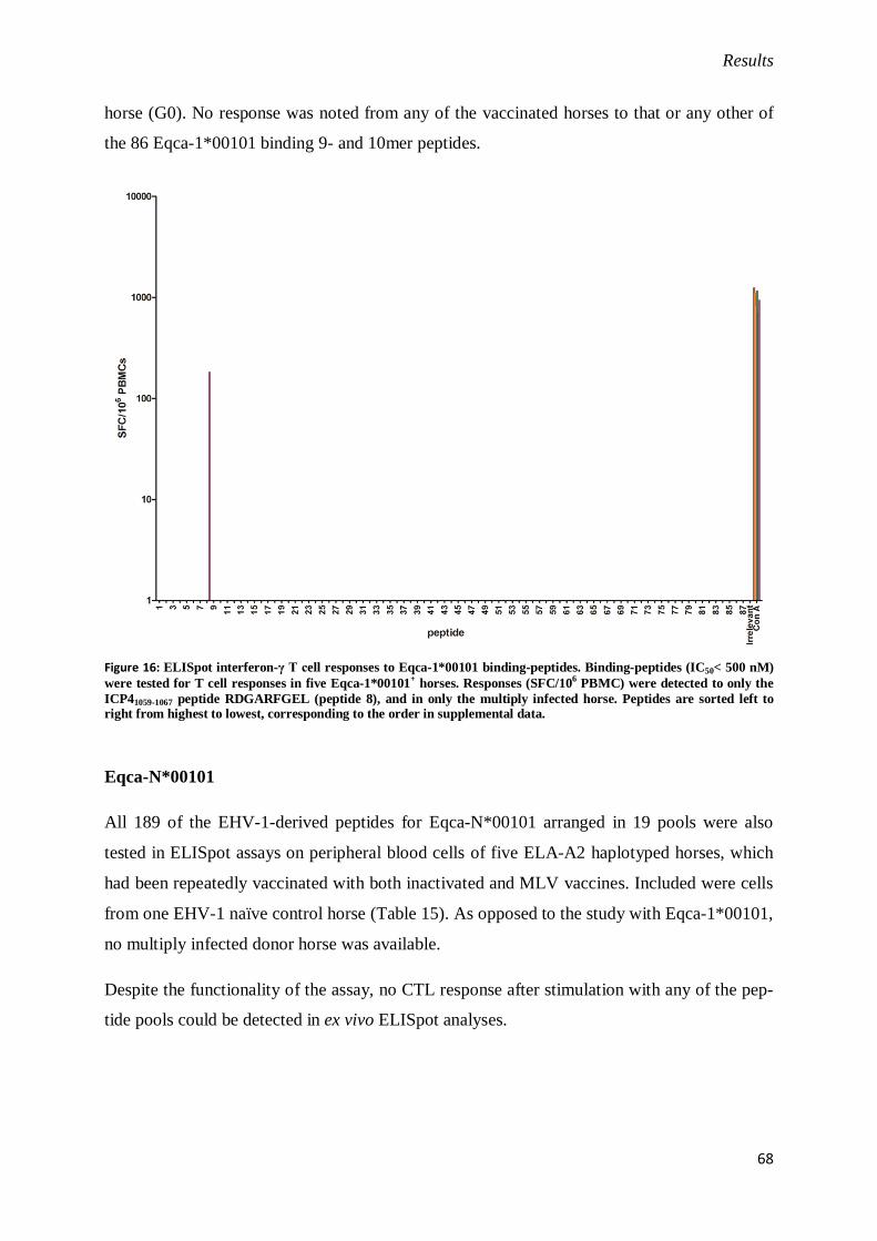
Results
68
horse (G0). No response was noted from any of the vaccinated horses to that or any other of
the 86 Eqca-1*00101 binding 9- and 10mer peptides.
Figure 16: ELISpot interferon-γ T cell responses to Eqca-1*00101 binding-peptides. Binding-peptides (IC50< 500 nM)
were tested for T cell responses in five Eqca-1*00101+ horses. Responses (SFC/10
6 PBMC) were detected to only the
ICP41059-1067 peptide RDGARFGEL (peptide 8), and in only the multiply infected horse. Peptides are sorted left to right from highest to lowest, corresponding to the order in supplemental data.
Eqca-N*00101
All 189 of the EHV-1-derived peptides for Eqca-N*00101 arranged in 19 pools were also
tested in ELISpot assays on peripheral blood cells of five ELA-A2 haplotyped horses, which
had been repeatedly vaccinated with both inactivated and MLV vaccines. Included were cells
from one EHV-1 naïve control horse (Table 15). As opposed to the study with Eqca-1*00101,
no multiply infected donor horse was available.
Despite the functionality of the assay, no CTL response after stimulation with any of the pep-
tide pools could be detected in ex vivo ELISpot analyses.

Results
69
Figure 17: ELISpot interferon-γ T cell responses to Eqca-N*00101 binding-peptides. Binding-peptides (IC50 < 500 nM)
were tested for T cell responses in five Eqca-N*00101+ horses. No T cell response could be observed.
6.2 Detection of IFN-γ-secreting cells upon virus stimulus
As outlined before, control of EHV-1 infections of horses is mainly attributed to the action of
virus-specific T cells. So far, only virulent field strain infections seem to induce protective T
cell frequencies after multiple infections and/or reactivations. But robust detection systems for
quantification of a cellular immune response are absent. IFN-γ is generally considered a sur-
rogate marker for T cell activity [179]. Thusly, in order to correlate frequencies of virus-
specific, IFN-γ-secreting T lymphocytes with immunity to acute and recurrent EHV-1 infec-
tions, classical ELISpot approaches were modified to detect responses of EHV-1-specific T
lymphocyte populations upon virus stimulus.
6.2.1 RacL11-infected PBMCs co-express MHC-I
As previously reported, equine PBMCs are susceptible to infections with EHV-1. PBMCs
were infected with the laboratory strain RacL11, which codes for the green fluorescent protein
(GFP) as infection marker at an MOI of 1. This strain was chosen owing to its diminished
capacity to downregulate MHC-I surface presentation [61, 190]. After 24 h, cells were incu-

Results
70
bated with MHC-I-specific antibody and subjected to flow-cytometric analyses. GFP and
MHC-I double positive lymphocytes are indicative of MHC-I-restricted peptide presentation.
Figure 18: Equine PBMCs infected with EHV-1 strain RacL11 expressing the gene for GFP, and immunostained for
MHC-I. Mock-infected and isotype-controlled cells (left) only show background fluorescence. Upon infection, around 30 % of the lymphocyte subpopulation expresses virally encoded GFP, 57 % MHC-I and around 18 % both (right)
Around 18 % of the PBMCs are infected as evident by the production of GFP, and express
detectable levels of MHC-I on the cell surface (Figure 18). Although that is no direct proof of
MHC-I-restricted presentation of viral antigens, ongoing viral protein production and pres-
ence of MHC-I are likely to result in epitope presentation.
6.2.2 PBMCs from a horse with unknown infection history respond to RacL11
infection
In a first pilot experiment, 2 X 10⁵equine PBMCs from a donor horse with unknown infection
history were infected with EHV-1 strain RacL11 at an MOI of 1. The cells responded to the
EHV-1 stimulus by secreting IFN-γ. Mock-infected control cells only showed background
signals.
Figure 19: ELISpot analysis of mitogen-treated, mock- or RacL11-infected PBMCs. ConA-treated (60 SFC) and
RacL11-infected (32 SFC) cells show significantly increased IFN-γ signals. Mock-infected cells are negative (3 SFC).

Results
71
Although no data on the infection history of the donor horse was available, it seems likely that
a horse not housed under quarantine conditions would be naturally infected with virulent
EHV-1.
6.2.3 PBMCs from EHV-1 primed but not naïve horses respond to RacL11 stimu-
lus
PBMCs from the ELA-A3-typed horses that had been used in the Eqca-1*00101 epitope ex-
periments, namely G0 and 4231, which were infected with virulent EHV-1 strains on several
occasions (primed) or did not come into contact (naïve) with EHV-1 before, respectively,
were utilized to detect EHV-1-specific T lymphocyte activity. Two hundred thousand cells
were treated with mitogen, mock-infected or infected with RacL11 at an MOI of 1. The pre-
viously mapped epitope RDGARFGEL (peptide) and a respective DMSO control were in-
cluded as additional controls.
Figure 20: ELISpot analysis of PBMCs from EHV-1 primed (G0, upper panel) and naive (4231, lower panel) horses.
ConA-treated cells are positive, mock-infected are negative for both horses (72 and 52 SFC, or 3 and 4 SFC, respec-
tively). RacL11-stimulated cells are only positive for the EHV-1-primed horse (24 SFC as opposed to 5 SFC). The
previously identified peptide RDGARFGEL (restricted by Eqca-1*00101) also only elicits a response in the primed
PBMCs (19 SFC as opposed to 5 SFC).

Results
72
Figure 21: SFC per 106 PBMCs for an EHV-1 experienced (G0) and naïve (4231) horse. G0 responds with 120 and 95
SFC/106 PBMCs to RacL11 or peptide stimulation, respectively. 4231 remained negative.
As expected, PBMCs only from the EHV-1–primed horse (G0) were reactive upon stimulus
with EHV-1 and peptide with 120 and 95 SFC/106 cells, respectively. PBMCs from the naïve
horse only reacted upon ConA treatment but remained otherwise negative.
To further confirm these results, cells from three additional, multiply infected donor horses
(G1-G3) were tested. Also included were negative control cells from horse 4231.
Figure 22: SFC per 106 PBMCs for three multiply infected (G1-G3) horses and one EHV-1 naïve horse (4231). As
previously seen only EHV-1 experienced horses were responsive to RacL11 and peptide stimulation.
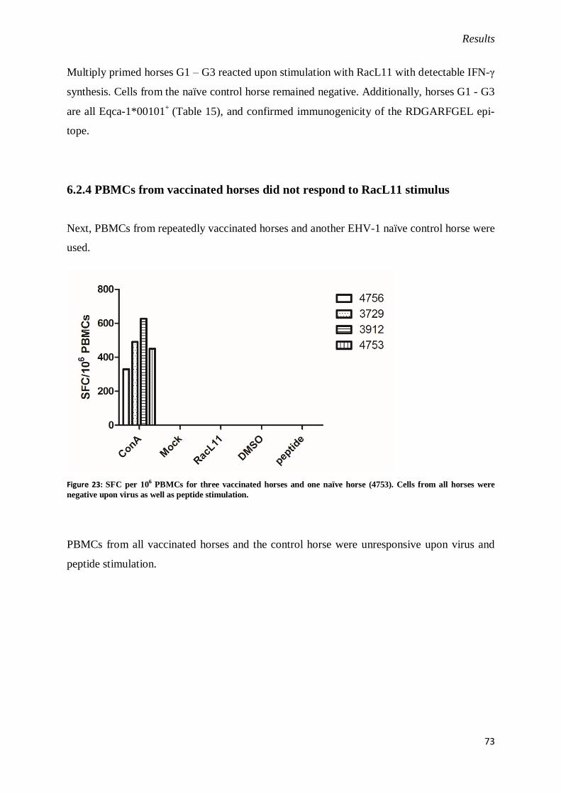
Results
73
Multiply primed horses G1 – G3 reacted upon stimulation with RacL11 with detectable IFN-γ
synthesis. Cells from the naïve control horse remained negative. Additionally, horses G1 - G3
are all Eqca-1*00101+
(Table 15), and confirmed immunogenicity of the RDGARFGEL epi-
tope.
6.2.4 PBMCs from vaccinated horses did not respond to RacL11 stimulus
Next, PBMCs from repeatedly vaccinated horses and another EHV-1 naïve control horse were
used.
Figure 23: SFC per 106 PBMCs for three vaccinated horses and one naïve horse (4753). Cells from all horses were
negative upon virus as well as peptide stimulation.
PBMCs from all vaccinated horses and the control horse were unresponsive upon virus and
peptide stimulation.
Discussion
74
7. Discussion
Cellular immunity against viral infections is of great importance for most viral diseases, albeit
some infections can be controlled or prevented by the action of neutralizing and non-
neutralizing antibodies alone, as observed for challenge infections after immunization with
inactivated equine influenza virus [209]. In general, virus-specific antibodies are thought to
play an important role in prevention of cell-to-cell spread and resistance to re-infection. How-
ever, the parasitic nature of viral pathogens most of the time deprives circulating antibodies of
their targets. Thusly, CD8+ T lymphocytes are key players in the adaptive immune response to
intracellular infections. In EHV-1 infections in particular, CTL responses have been found to
be superior to antibody-mediated immunity, probably due to the strict cell-association of the
virus during viraemic phases.
MHC class I restriction of CTL epitopes is an integral part in the recognition process of in-
fected cells. MHC-I allele-specific peptide presentation determines the T cell receptor reper-
toire of an individual. Therefore, determining binding motifs for MHC-I alleles can help de-
sign new MHC haplotype-specific peptide vaccines that directly stimulate T cell-mediated
immunity.
In the present study, detailed binding motifs for the four common horse MHC class I alleles
Eqca-1*00101, Eqca-N*00101, Eqca-16*00101 and Eqca-1*00201 were determined. Based
on those results, in in vitro binding assays high affinity binders derived from EHV-1 were
identified for Eqca-1*00101 and Eqca-N*00101.
At first, RMA-S stabilization assays for all alleles were established in our laboratory. As pre-
vious studies mapped CTL epitopes restricted by Eqca-1*00101 to the EHV-1 ICP4 protein
[77, 78], a peptide library consisting of 295 15mers spanning the entire ICP4 protein was
screened for MHC-I stabilizers on RMA-S cells expressing the desired ELA allele. For Eqca-
1*00101, Eqca-N*00101 and Eqca-1*00201, five, fourteen and nine peptides, respectively,
were identified as stabilizers (Figures 10, 11, and 12). Differences in the mean fluorescence
intensity (MFI) among the alleles can be attributed to different expression levels of the re-
spective MHC-I alleles, which could also be confirmed by immunoblotting (not shown). This
might be due to the locus of integration in the genome of the respective RMA-S cell clones,
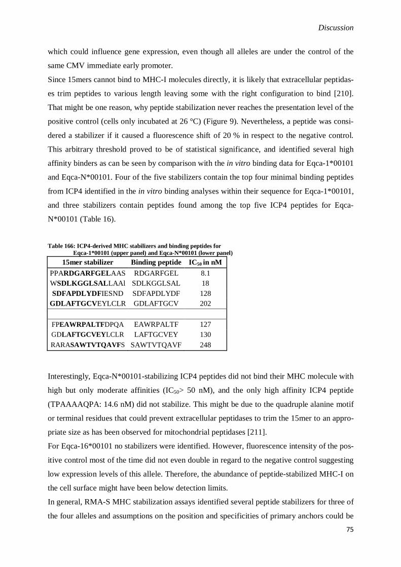
Discussion
75
which could influence gene expression, even though all alleles are under the control of the
same CMV immediate early promoter.
Since 15mers cannot bind to MHC-I molecules directly, it is likely that extracellular peptidas-
es trim peptides to various length leaving some with the right configuration to bind [210].
That might be one reason, why peptide stabilization never reaches the presentation level of the
positive control (cells only incubated at 26 °C) (Figure 9). Nevertheless, a peptide was consi-
dered a stabilizer if it caused a fluorescence shift of 20 % in respect to the negative control.
This arbitrary threshold proved to be of statistical significance, and identified several high
affinity binders as can be seen by comparison with the in vitro binding data for Eqca-1*00101
and Eqca-N*00101. Four of the five stabilizers contain the top four minimal binding peptides
from ICP4 identified in the in vitro binding analyses within their sequence for Eqca-1*00101,
and three stabilizers contain peptides found among the top five ICP4 peptides for Eqca-
N*00101 (Table 16).
Table 166: ICP4-derived MHC stabilizers and binding peptides for
Eqca-1*00101 (upper panel) and Eqca-N*00101 (lower panel)
15mer stabilizer Binding peptide IC50 in nM
PPARDGARFGELAAS RDGARFGEL 8.1
WSDLKGGLSALLAAl SDLKGGLSAL 18
SDFAPDLYDFIESND SDFAPDLYDF 128
GDLAFTGCVEYLCLR GDLAFTGCV 202
FPEAWRPALTFDPQA EAWRPALTF 127
GDLAFTGCVEYLCLR LAFTGCVEY 130
RARASAWTVTQAVFS SAWTVTQAVF 248
Interestingly, Eqca-N*00101-stabilizing ICP4 peptides did not bind their MHC molecule with
high but only moderate affinities (IC50> 50 nM), and the only high affinity ICP4 peptide
(TPAAAAQPA: 14.6 nM) did not stabilize. This might be due to the quadruple alanine motif
or terminal residues that could prevent extracellular peptidases to trim the 15mer to an appro-
priate size as has been observed for mitochondrial peptidases [211].
For Eqca-16*00101 no stabilizers were identified. However, fluorescence intensity of the pos-
itive control most of the time did not even double in regard to the negative control suggesting
low expression levels of this allele. Therefore, the abundance of peptide-stabilized MHC-I on
the cell surface might have been below detection limits.
In general, RMA-S MHC stabilization assays identified several peptide stabilizers for three of
the four alleles and assumptions on the position and specificities of primary anchors could be

Discussion
76
drawn. These premature motifs were confirmed later on. The assay provides a quick way to
confirm high affinity MHC binders, or to derive a very preliminary motif. For detailed in-
sights into binding specificities and large scale screening endeavors, however, it is not well
suited.
Therefore, we used a more sophisticated approach. A preliminary binding motif was derived
for all four alleles by eluting endogenously bound peptides from affinity purified MHC-I.
Between 20 and 40 peptides were recovered for each allele and all sets had a predominant
ligand size of nine amino acids. Only few peptides with ten, eleven or even thirteen residues
were observed, which conforms to the canonical ligand size for most human, non-human pri-
mate and mouse alleles known [115] (Figure 13). The elution reactions as well as peptide se-
quencing methods are not quantitative, so the limited number of sequenced ligands is not nec-
essarily indicative of the real binding repertoire, which was evaluated in in vitro binding stu-
dies. To establish high-throughput binding assays, tyrosinated endogenous peptides were ra-
diolabeled with iodine125
and their MHC binding capacities assessed. The assays in turn were
used to test a panel of positional scanning combinatorial libraries, resulting in detailed quan-
titative binding motifs for all four alleles (Figure 14). Briefly summarized, Eqca-1*00101
prefers aspartic acid in P2 and leucine or isoleucine at the C-terminus with secondary anchors
at P1, P2 and P7. For Eqca-N*00101, proline in P2 and phenylalanine at the C-terminus could
be identified as preferred primary anchors and P1, P3 and P8 as secondary anchor positions.
Eqca-16*00101 favors valine in P2 and F at the C-terminus, and has secondary anchor posi-
tions in P1, P3 and P6. Eqca-1*00201 has primary specificity for arginine in P2 and F at the
C-terminus, and secondary anchors in P1 and P3. These allele motifs can be classified under
known HLA supertypes (Eqca-1*00101: HLA-B44; -N*00101: HLA-B07; -16*00101: HLA-
A02; -1*00201: HLA-B27) underscoring the general validity of this concept and strengthen-
ing the hypothesis of convergent evolution of mammalian MHC [212].
With the motifs for Eqca-1*00101 and Eqca-N*00101, a complete scan of the EHV-1 prote-
ome was undertaken. In total, 87 and 26 EHV-1 peptides were identified that have the capaci-
ty to bind Eqca-1*00101 or Eqca-N*00101, respectively. Eqca-1*00101 peptides originated
from 40 different viral proteins, including 1, 12 and 27 from immediate early, early and late
proteins. Eqca-N*00101 bound peptides from 19 different EHV-1 proteins, including 1, 7 and
11 from immediate early, early and late proteins.
Finally, ELISpot assays to test Eqca-1*00101 and Eqca-N*00101 binding peptides for immu-
nogenicity revealed a narrow CTL epitope reservoir. In fact, only one epitope with the se-

Discussion
77
quence RDGARFGEL restricted by Eqca-1*00101 elicited a detectable IFN-γ response, whe-
reas PBMCs from all Eqca-N*00101 expressing A2 horses were unresponsive upon peptide
stimulus. However, from 13 donor horses only four had been exposed to virulent EHV-1
strains on multiple occasions in the past years. All other horses were either EHV-1 naïve or
had been vaccinated with inactivated vaccines and MLV in the months prior to sampling
(Table 15). Only multiply infected horses showed detectable frequencies of MHC-I-restricted
and IFN-γ-producing T cells upon peptide stimulation (Figures 16 and 22).
As outlined in the introduction, protection from disease and cell-associated viraemia can only
be achieved in multiply infected horses, whereas vaccinated animals only show reduced
symptoms. On the other hand, protection from abortion is clearly associated with elevated
virus-specific CTL frequencies in multiply infected and vaccinated horses [65]. However,
assays used in this study could only detect cytolytic activity of precursor CTLs and ignored
the noncytopathic effects of IFN-γ secreted by CD8+ and CD4
+ T cells. IFN-γ is likely to play
an important role in acute EHV-1 infections as high protein and mRNA levels can be detected
after challenge in multiply primed and protected horses. In young EHV-1 inexperienced foals,
however, IFN-γ levels are significantly lower, since these foals had not been primed repeated-
ly [67, 68, 213]. In this light, it seems possible that a broader T cells response than cytolytic
effector functions are necessary for protection, and that this broad response is not well stimu-
lated by commercial vaccines or rapidly wanes after vaccination. Our findings also point in
that direction, and it would explain why no CTL epitopes could be found for Eqca-N*00101
as no multiply infected donor horses expressing the allele were available.
MHC class I peptide-binding and T cell epitope repertoires should be regarded as fairly inde-
pendent. The peptide repertoire determined in in vitro studies does not depend on the source
of the proteins of provenance or their processing, but only on the binding motif. Hence, the
repertoire is not expected to change significantly when different pathogens are used to derive-
binding peptides. Differences in the amino acid frequencies in proteins of evolutionary distant
species could, in theory, influence the peptide repertoire. But these differences are mainly
thought to be insignificant [206].
T cell epitope repertoires, however, are not only determined by their ability to bind to MHC-I
molecules in in vitro settings but rely on several other factors. Abundance of the protein of
provenance, substrate specificities of the antigen processing pathway or MHC-peptide stabili-
ty significantly influence availability of a specific MHC-I-peptide complex at the cell surface
[214]. Finally, as TCR repertoires are the result of negative selection in the thymus, TCRs

Discussion
78
cross-reactive with self- and similar non-self peptide-MHC complexes are depleted by thymic
intolerance [215].
The peptide binding repertoires for both alleles Eqca-1*00101 and Eqca-N*00101 with 0.2 %
and 0.5 %, respectively, of all possible 9mer peptides are rather limited, compared to about
1.5 – 3 % on average for human, mouse and macaque class I alleles [206].
When compared to HLA peptide-binding motifs, Eqca-1*00101 ligands are very similar to
peptides restricted by HLA alleles in the B44 supertype [112, 216] and the non-human pri-
mate alleles Mamu-B*01, Mamu-A*11, and Patr-A*24. In the majority of these cases, how-
ever, the predominant specificity is for glutamic acid in position 2, with D only rarely tole-
rated. The notable exception is Mamu-B*01, which shares a strong preference for D in P2 as
well as for I at the C-terminus with Eqca-1*00101. Mamu-B*01 also has a very limited pep-
tide binding repertoire and only few T cell epitopes have been identified to date that are Ma-
mu-B*01-restricted [115].
Eqca-N*00101 also displays a rather limited peptide binding repertoire. However, ligands
with P at P2 are commonly produced in the human antigen processing pathway, and alleles
associated with the B07 supertype are also very common [154, 155]. Whether that is true for
equine antigen processing is not known. However, for epitopes from CMV it could be shown
that B07-restricted peptides are recognized more efficiently than those restricted by HLA-A02
pointing to a possibly general immunodominance associated with B07 alleles, which could
neutralize the constraints of a limited binding repertoire [217]. Further investigation of addi-
tional equine MHC-I alleles and the antigen processing specificities are required to draw more
detailed conclusions.
Another hypothesis to explain the identification of limited binding repertoires pertains the
various sources of β2-microglobulin (β2m) present in different contexts. The ELA molecules
analyzed with the elution studies are largely heterodimers associated with murine β2m or, to a
minor extent, with bovine β2m. Additionally, the binding assays are performed with human
β2m, and the entire peptide library was tested in ELISpot assays where equine β2m is predo-
minant. While the affinity of β2m may affect peptide binding, especially weak binders, in
general it is also accepted that the specificity of peptide selection would not change much.
Indeed, given the concordance between the elution (largely mouse β2m) and in vitro assays
(human β2m) in terms of specificity, and that the binding and T cell (equine β2m) repertoires
were addressed using a large set of peptides covering a broad range of predicted affinities, it
seems unlikely that the source of β2m has a significant influence.

Discussion
79
While no specific mechanism implicating D as CTL epitope repertoire limiting factor is ap-
parent, it is tempting to speculate that the presence of D near the N-terminus of potential li-
gands somehow influences the efficiency of ligand generation by processing. Whereas noth-
ing specific is known for horse TAP, rat TAP display a remarkably reduced affinity to pep-
tides carrying an aspartic acid or related residues in position 2 or 3 [218]. Also, human ERAP
have preferences for N-terminal hydrophobic residues, which also might influence peptide
repertoire for the peptide loading complex and hence CTL epitope repertoire [154, 219]. In
addition, EHV-1 inhibits TAP function by the action of the early protein pUL49.5, which
could prevent any other peptide than the one derived from the immediate early protein to be
shuttled into the ER and presented on Eqca-1*00101 [60]. Certain strains of EHV-1 also en-
code an additional early protein, pUL56, which downregulates MHC-I surface expression by
dynamin-dependent endocytosis [61]. Both proteins affect antigen presentation and therefore
could limit the antigen repertoire presented to CTLs.
In the sequences of 11 EHV-1 strains deposited in GenBank, no DNA sequence variation
within the ICP4 epitope could be found to implicate that escape variants have arisen. Several
interaction sites have been reported for the EHV-1 sole IE (ICP4) protein. It physically binds
to the viral proteins ICP0, ICP22, and ICP27, as well as several cellular transcription factors
[20-23]. Although none of these interaction sites include the sequence of the identified epi-
tope, a mutation leading to an escape variant might abolish binding or functional folding of
the ICP4 protein, which in turn might interrupt the viral replication cycle. However, the EHV-
1 closely related virus EHV-4 encodes for a substitution of aspartic acid with glutamic acid in
the identified CTL epitope (REGARFGEL). The ICP4 proteins of EHV-1 and EHV-4 share a
homology of 89 % on the amino acid level, which suggests that mutations in the epitope can
be tolerated provided no complementary mutations arose. On the other hand, it could imply
that the Eqca-1*00101 allele, while common (allele frequency estimated at 13.4 %), does not
contribute substantially to the control of EHV-1 field infections, and therefore does not exert a
strong selection pressure. That actually might not be necessary as the infection could be con-
trolled by CTL activity directed against other class I loci, or the Eqca-1*00101 allele confers
protection against other important equine infections.
It is tempting to speculate on a potential immunodominance of the ICP4 epitope. However,
that is only true for the Eqca-1*00101 allele so far. Whether the majority of the combined
CTL responses of all alleles expressed in all haplotypes is directed against this epitope re-
mains to be investigated. However, the fact that stimulation of IFN-γ+ T cells adjoined by a
reduction of cell-associated viraemia could be achieved in Eqca-1*00101+
horses after vacci-

Discussion
80
nation with a NYVAC-IE construct suggests [82, 213], paradoxically, that a narrow repertoire
might be beneficial with respect to control of, or protection against, EHV-1 infection, perhaps
allowing a more focused response, or avoiding immune diversion or decoy responses, which
have been described in the case of other herpesviruses [220]. A narrow epitope repertoire
might also be beneficial in context of immunity towards other infections. In patients seroposi-
tive for HCMV, for example, around 10 % of the memory CD4+ and CD8
+ T cell pool is
HCMV-specific, significantly reducing the T cell repertoire, which is thought to contribute to
immune senescence [221-223]. A limited but effective T cell response could ensure a suffi-
ciently diverse T cell repertoire against various infectious diseases.
In summary, we determined quantitative peptide-binding motifs for four common equine
MHC class I alleles to predict potential CTL epitope candidates from various pathogens of the
horse. All alleles have binding motifs similar to human MHC-I alleles organized in HLA su-
pertypes. Based on the motifs, we predicted and identified high affinity binders derived from
EHV-1 for two of these alleles in in vitro binding studies, and found one peptide (RDGARF-
GEL) from ICP4 to elicit IFN-γ secretion in PBMCs from multiply infected and Eqca-
1*00101+ horses in ELISpot assays. Cells from donor horses vaccinated with both inactivated
and modified-live vaccines (MLV) did not respond to any of the tested high affinity binding
peptides, which ties into the hypothesis that MLV-based vaccination does not sufficiently
induce T cell-mediated immunity.
To further elucidate this working hypothesis, ELISpot assays were adapted to detect IFN-γ
production in equine PBMCs after stimulation with replication-competent virus. PBMCs from
a horse with unknown infection history responded with detectable IFN-γ secretion to virus
stimulation but not to mock treatment (Figure 19). Since seroprevalence of EVH-1 in horse
populations ranges between 30 – 80 %, it seems likely that this horse also came into contact
with virulent EHV-1 strains in its life [31]. After these promising results it could be shown
that only cells from multiply infected donor horses (G0 – G4) but not from EHV-1 naïve
horses (4231, 4753) were responsive (Figures 21 and 22) excluding a potential NK cell in-
volvement in the IFN-γ response. Cells from vaccinated horses were not responsive (Figure
23) further strengthening the working hypothesis.
The reasons for the limited efficacy in T cell induction of the MLV are not yet known but
speculated upon. As shown for herpes viruses HSV-1, VZVand HCMV, CD8+ and CD4
+ T
cells play an important role in immunity, but their frequencies detectable after short-term an-
tigenic stimulation differ significantly. While no IFN-γ or TNF-α production could be de-

Discussion
81
tected in HSV and VZV primed PBMCs by intracellular staining, HCMV-specific T cell ac-
tivity was observed. The authors attribute these differences in part to the differing sites of
latency arguing that higher memory T cell frequencies are required to control reactivation
from cells of the monocyte/macrophage lineage as site of HCMV latency than from cells of
sensory ganglia for HSV and VZV [224]. Since EHV-1 like HCMV also latently infects lym-
phocytes and monocytes, a T cell boost after reactivation of virulent strains could be a conse-
quence. For the vaccine strain RacH, however, no latent infections have been described, so
that immunity might wane more quickly without frequent reactivations.
In addition, EHV-1 has some MHC-I-downregulating capacities. In the vaccine strain, how-
ever, one of the proteins responsible encoded by UL56 (ORF1) is deleted resulting in higher
MHC-I density on the cell surface [225]. Intuitively, that would suggest enhanced T cell prim-
ing and, hence, better stimulation of T cell mediated immunity. These assumptions are con-
trary to actual observations. However, MHC-I downregulation seems to be locus and allele
specific [172], which would alter the MHC-I composition on the surface of an infected cell.
As a consequence, priming after field-strain infections could result in a different TCR reper-
toire than after vaccination with RacH-based MLV. Epitopes evoking a T cell response in
infections with the vaccine strain but presented by downregulation-sensitive MHC-I could be
underrepresented under field conditions. A TCR repertoire primed by vaccination would be
less efficacious or even completely useless. On the other hand, priming of T cells with immu-
nodominant epitopes identified in multiply infected horses (like RDGARFGEL) and pre-
sented on downregulation-insensitive MHC-I could be less efficient after vaccination as the
presenting MHC-I is present in lower relative frequencies. Although unlikely, these effects
could contribute to the low CTL induction efficiency in horses after vaccination with MLV at
least for the haplotype A3.
Another explanation, of course, could be reduced replication and antigen production of the
vaccine strain leading to reduced availablitiy of epitope-containing proteins.
Interestingly, T cell frequencies after whole virus or peptide stimulation were comparable
(Figures 21 and 22) suggesting a strong immunodominance of the RDGARFGEL epitope
even in ELA-A heterozygous horses. However, peptide stimulation involves continously high
concentration (10 µg/mL) of peptide in the cell medium probably resulting in MHC-I satura-
tion on more or less any cell present, which might facilitate activation and detection of clonal
T cells [226]. In vitro infections done in this assay, however, used an MOI of 1, which results
in infection of only 63 % of the cell population provided all cells are equally susceptible.
Considering the heterogeneity of PBMCs and the cellular tropism of EHV-1, an even lower

Discussion
82
infection rate can be assumed restricting the number of MHC-peptide presenting cells and
thusly availability for T cell recognition. Epitope-specific responses might be overestimated
in peptide stimulation. Furthermore, infected cells present various epitopes on several differ-
ent MHC-I possibly activating different T cell clones in close proximity resulting in one larg-
er spot rather than in several distinct spots on the membrane, which could underestimate T
cell frequencies [227]. Additionally, the contribution of CD4+ T cells to the IFN-γ response in
this assay is not known. Further characterization of the T cell response after in vitro infection
with this and alternative approaches is clearly needed if a reliable correlate of IFN-γ+ T cell
frequencies with protection wants to be established.
However, the clear absence of responsive cells in all vaccinated horses indicates a diagnostic
and scientific potential of the modified ELISpot.
These preliminary results are encouraging but need further evaluation. In future experiments,
it should be possible to correlate protection from viraemia and consequently abortion and
EHM with a certain frequency of virus-specific, IFN-γ-producing T cells with this assay, and
to evaluate efficacy of T cell induction by vaccines.
In conclusion, several experimental findings and the current model of EHV-1 pathogenesis
suggest a predominantly T cell-mediated immunity against EHV-1 and an insufficient induc-
tion of this response by commercial vaccines. Our results strongly support this hypothesis.
However, robust assays to correlate T cell activity with protection are absent. The modified
ELISpot assay presented in this thesis could close that gap and prove to become a valuable
tool in research and diagnostics.

Outlook
83
8. Outlook
The immune system plays a central role in the defense against infections. A potent activation
of the adaptive immune response is a necessity not only for clearing the acute infection but
also in achieving immunity against re-infection. Defenses against viruses, and in particular
herpes viruses, rely on MHC-restricted cytopathic and noncytopathic actions of T lympho-
cytes [228]. Attenuated virus strains have proven to be valuable tools for inducing protective
B and T cell-mediated immunity, and passaging virulent viruses in animals or on cell cultures
to derive attenuated strains has been the gold standard in the development of live vaccines for
decades [3]. However, as seen in the case of EHV-1, these classical approaches can fail to
induce protective immunity in genetically diverse populations. Vaccine design of the future
will have to take into account the genetic and immunological background of vaccinees.
With increasing knowledge of MHC specificities and T cell epitopes, a new generation of
peptide vaccines that cover a broad range of MHC haplotypes is conceivable. To better under-
stand MHC motifs and repertoire sizes in horses, characterization of common MHC-I alleles
from various haplotypes has to be continued. It is also notable that equine immune responses
to EHV-1 are particularly short-lived, but there is very little information on whether there are
differences in immune responses that would correlate with the ELA haplotype. Methods to
approach this question have been introduced and established for equine MHC class I mole-
cules in this work, but also MHC-II specificities should be addressed in the near future.
The current problem in peptide-based vaccination approaches of efficiently targeting peptides
to MHC molecules for presentation could be tackled in different ways. Artificial sugar beads
containing synthetic proteins combining several immunodominant epitopes could be routed
through the endosomal pathway for cross-presentation on MHC-I [229, 230]. However, the
class I antigen processing pathway would be ignored in this approach. Alternatively, virus-
like particles as antigen carriers have been improved in the recent past. Particles carrying fu-
sion proteins with the translocation motif (TLM) of hepatitis B virus (HBV) at the surface are
cell-permeable and deliver their cargo directly into the cytoplasm of a DC for intracellular
processing [231-233]. To date, there are no peptide vaccines commercially available but in-
creasing understandings of MHC specificitites and antigen delivery systems will help effi-
ciently target cell-mediated immunity.

Outlook
84
For detection of virus-specific T cell frequencies after EHV-1 infections in the horse, the
modified ELISpot assay needs to be evaluated in further experiments involving more multiply
infected, vaccinated, and control animals. Classical approaches to enumerate virus-specific T
cell frequencies (e.g. Cr51
-release assays) could confirm the results yielded by ELISpot. If
consistency and reproducibility are given, subsequent challenge studies can hopefully help
define protective T cell frequencies against EHV-1 infections. Depletion experiments could
shed more light on the contributions of CD8+ and CD4
+ T cells to the IFN-γ response.

Summary
85
9. Summary
Infections of horses with the equine herpes virus type 1 (EHV-1) can result in rhinopneumoni-
tis, abortion in pregnant mares, and sometimes fatal neurological disorder. The virus primarily
replicates in the epithelium of the upper respiratory tract, and subsequently spreads via leuko-
cyte-associated viraemia to secondary sites of infection (endothelia of blood and lymphatic
vessels). Thusly, the infection is thought to be mainly controlled by the action of virus-
specific cytotoxic T lymphocytes (CTL) as precursor CTL frequencies clearly correlate with
protection. High neutralizing antibody titers can be induced by several commercial vaccines,
but complete protection from infection and disease is only achieved in horses multiply in-
fected with virulent strains.
CTL recognize an infected cell by binding with their polymorphic T cell receptors (TCR) to a
complex of pathogen-derived peptide and surface major histocompatibility complex class I
molecules (MHC-I). MHC-I molecules bind peptides of intracellular origin according to their
binding motifs mediated by polymorphic residues in the peptide-binding groove. These bind-
ing motifs can be summarized in terms of primary and secondary anchors in the peptide, and
used to predict putative CTL epitopes from a pathogen.
In this study, we identified the MHC-I binding motifs for four common equine MHC-I alleles,
namely Eqca-1*00101, Eqca-N*00101, Eqca-16*00101 and Eqca-1*00201 by eluting and
sequencing endogenous peptide binders, and by in vitro binding approaches using positional
scanning combinatorial libraries (PSCL). We found these binding motifs to be similar to those
of known human MHC-I alleles organized in HLA supertypes. The peptide binding reper-
toires of Eqca-1*00101 and Eqca-N*00101 with 0.2 % and 0.5 %, respectively, are rather
limited compared to 3 % on average for most human and mouse class I alleles.
Based on the motifs of the first two alleles, we predicted and identified several high affinity
peptides derived from EHV-1, and found peptide RDGARFGEL restricted by Eqca-1*00101
to be immunogenic in ex vivo ELISpot assays.
In addition, ELISpot assays were adapted to detect virus-specific T cell activity in peripheral
blood mononuclear cells (PBMC) after stimulation with replication-competent virus. This
method was used to evaluate induction of T cell activity after multiple infections with virulent
EHV-1 strains or vaccination with attenuated virus. Detectable T cell frequencies were found

Summary
86
only in multiply infected but not in vaccinated horses. The modified ELISpot assay can hope-
fully provide a reliable tool to correlate virus-specific T cell frequencies with protective im-
munity against EHV-1-associated diseases of the horse.
Zusammenfassung
87
10. Zusammenfassung
Infektionen des Pferds mit dem equinen Herpesvirus Typ 1 (EHV-1) führen zu Rhinopneu-
monitis, Aborten in trächtigen Stuten und neurologischen Erkrankungen. Das Virus repliziert
primär in Epithelzellen der oberen Atemwege, bevor es via Leukozyten-assoziierter Virämie
zu sekundären Infektionsorten (Endothel von Blut- und Lymphgefäßen) transportiert wird.
Frequenzen virusspezifischer zytotoxischer T Lymphozyten (CTL) korrelieren eindeutig mit
einem Immunschutz, weswegen davon ausgegangen wird, dass die Infektion größtenteils
durch CTL-Aktivität kontrolliert wird. Kommerzielle Impfstoffe können hohe virusneutrali-
sierende Antikörpertiter induzieren, ein kompletter Schutz vor Infektion und Erkrankung kann
allerdings nur in Pferden beobachtet werden, die mehrmals mit virulenten Stämmen infiziert
wurden.
CTL erkennen infizierte Zellen, indem sie mit ihrem polymorphen T-Zellrezeptor (TCR) ei-
nen Komplex aus Peptiden pathogenen Ursprungs und Haupthistokompatibilitätskomplex
Klasse I-Molekülen (MHC-I) auf der Oberfläche der Zelle binden. MHC-I-Moleküle assoziie-
ren mit Peptiden intrazellulären Ursprungs entsprechend ihres Bindemotivs, das durch poly-
morphe Aminosäurereste in der Bindespalte für Peptide bestimmt wird. Diese Bindemotive
können als primäre und sekundäre Ankerreste im Peptid zusammengefasst und zur Vorhersa-
ge vermeintlicher CTL-Epitope genutzt werden.
In dieser Arbeit wurden die Bindemotive für vier häufige, equine MHC-I-Allele namens Eq-
ca-1*00101, Eqca-N*00101, Eqca-16*00101 und Eqca-1*00201 durch Eluierung und Se-
quenzierung endogener Peptidliganden sowie in vitro Bindungsstudien mit Hilfe von positio-
nal scanning combinatorial libraries (PSCL) identifiziert. Die Bindemotive ähneln denen be-
kannter humaner MHC-I-Allele, die entsprechend ihrer Spezifitäten in Supertypen zusam-
mengefasst werden können. Die Peptidrepertoires von Eqca-1*00101 und Eqca-N*00101 sind
mit 0,2 % bzw. 0,6 % eher limitiert verglichen mit durchschnittlich 3 % für die meisten hu-
manen und murinen Klasse I-Allele.
Basierend auf den Motiven für die ersten zwei Allele wurden mehrere, hoch affine Bindepep-
tide aus dem EHV-1-Proteom vorhergesagt und identifiziert. Das Peptid RDGARFGEL wird
von Eqca-1*00101 präsentiert und ist in ex vivo ELISpot Assays immunogen.

Zusammenfassung
88
Zusätzlich wurde der ELISpot Assay adaptiert, um virusspezifische T-Zellaktivität in mono-
nuklearen Zellen aus peripherem Blut (PBMC) nach Stimulierung mit replikationskompeten-
tem Virus zu detektieren. Diese Methode wurde angewandt, um die Induzierung der T-
Zellantwort nach multiplen Infektionen mit virulenten EHV-1 Stämmen oder Immunisierung
mit attenuiertem Virus zu evaluieren. T-Zellfrequenzen waren nur in infizierten aber nicht in
geimpften Tieren nachweisbar. Der modifizierte ELISpot Assay wird hoffentlich ein verläss-
liches System darstellen, mit dem virusspezifische T-Zellfrequenzen mit protektiver Immuni-
tät gegen EHV-1-assoziierte Erkrankungen des Pferdes korreliert werden können.

References
89
11. References 1. Murphy, K., et al., Janeway's immunobiology. 8th ed2012, New York: Garland Science. xix,
868 p. 2. Davison, A.J., et al., The order Herpesvirales. Arch Virol, 2009. 154(1): p. 171-7. 3. Fields, B.N., et al., Fields virology, 2002, Lippincott Williams & Wilkins,: Philadelphia, PA. p. 1
computer laser optical disc. 4. Pellett, P.E., Trunkloads of viruses. J Virol, 2014. 88(23): p. 13520-2. 5. Boehmer, P.E., et al., Association of origin binding protein and single strand DNA-binding
protein, ICP8, during herpes simplex virus type 1 DNA replication in vivo. J Biol Chem, 1994. 269(46): p. 29329-34.
6. Browning, G.F. and M.J. Studdert, Genomic heterogeneity of equine betaherpesviruses. J Gen Virol, 1987. 68 ( Pt 5): p. 1441-7.
7. Browning, G.F., N. Ficorilli, and M.J. Studdert, Asinine herpesvirus genomes: comparison with those of the equine herpesviruses. Arch Virol, 1988. 101(3-4): p. 183-90.
8. Maramorsch, K., F.A. Murphy, and A.J. Shatkin, Advances in virus research. Volume 55, in Advances in virus research v 552000, Academic Press,: New York. p. 1 online resource (x, 638 p.).
9. Genbank. 10. Telford, E.A., et al., The DNA sequence of equine herpesvirus-1. Virology, 1992. 189(1): p. 304-
16. 11. Telford, E.A., et al., The DNA sequence of equine herpesvirus-4. J Gen Virol, 1998. 79 ( Pt 5): p.
1197-203. 12. Azab, W., et al., Glycoprotein C of equine herpesvirus 4 plays a role in viral binding to cell
surface heparan sulfate. Virus Res, 2010. 151(1): p. 1-9. 13. Osterrieder, N., Construction and characterization of an equine herpesvirus 1 glycoprotein C
negative mutant. Virus Res, 1999. 59(2): p. 165-77. 14. Spear, P.G., Herpes simplex virus: receptors and ligands for cell entry. Cell Microbiol, 2004.
6(5): p. 401-10. 15. Kurtz, B.M., et al., Equus caballus major histocompatibility complex class I is an entry receptor
for equine herpesvirus type 1. J Virol, 2010. 84(18): p. 9027-34. 16. Sasaki, M., et al., Equine major histocompatibility complex class I molecules act as entry
receptors that bind to equine herpesvirus-1 glycoprotein D. Genes Cells, 2011. 16(4): p. 343-57.
17. Sasaki, M., et al., Single amino acid residue in the A2 domain of major histocompatibility complex class I is involved in the efficiency of equine herpesvirus-1 entry. J Biol Chem, 2011. 286(45): p. 39370-8.
18. Azab, W., M.J. Lehmann, and N. Osterrieder, Glycoprotein H and alpha4beta1 integrins determine the entry pathway of alphaherpesviruses. J Virol, 2013. 87(10): p. 5937-48.
19. Sodeik, B., M.W. Ebersold, and A. Helenius, Microtubule-mediated transport of incoming herpes simplex virus 1 capsids to the nucleus. J Cell Biol, 1997. 136(5): p. 1007-21.
20. Albrecht, R.A., et al., Direct interaction of TFIIB and the IE protein of equine herpesvirus 1 is required for maximal trans-activation function. Virology, 2003. 316(2): p. 302-12.
21. Derbigny, W.A., et al., The EICP22 protein of equine herpesvirus 1 physically interacts with the immediate-early protein and with itself to form dimers and higher-order complexes. J Virol, 2000. 74(3): p. 1425-35.
22. Kim, S.K., et al., Interaction of the equine herpesvirus 1 EICP0 protein with the immediate-early (IE) protein, TFIIB, and TBP may mediate the antagonism between the IE and EICP0 proteins. J Virol, 2003. 77(4): p. 2675-85.

References
90
23. Zhao, Y., et al., Regulatory function of the equine herpesvirus 1 ICP27 gene product. J Virol, 1995. 69(5): p. 2786-93.
24. Taddeo, B. and B. Roizman, The virion host shutoff protein (UL41) of herpes simplex virus 1 is an endoribonuclease with a substrate specificity similar to that of RNase A. J Virol, 2006. 80(18): p. 9341-5.
25. Boehmer, P.E. and I.R. Lehman, Herpes simplex virus DNA replication. Annu Rev Biochem, 1997. 66: p. 347-84.
26. Boehmer, P.E. and A.V. Nimonkar, Herpes virus replication. IUBMB Life, 2003. 55(1): p. 13-22. 27. Morissette, G. and L. Flamand, Herpesviruses and chromosomal integration. J Virol, 2010.
84(23): p. 12100-9. 28. Smith, D.J., et al., In vitro reactivation of latent equid herpesvirus-1 from CD5+/CD8+
leukocytes indirectly by IL-2 or chorionic gonadotrophin. J Gen Virol, 1998. 79 ( Pt 12): p. 2997-3004.
29. Slater, J.D., et al., The trigeminal ganglion is a location for equine herpesvirus 1 latency and reactivation in the horse. J Gen Virol, 1994. 75 ( Pt 8): p. 2007-16.
30. Allen, G.P. and J.T. Bryans, Molecular epizootiology, pathogenesis, and prophylaxis of equine herpesvirus-1 infections. Prog Vet Microbiol Immunol, 1986. 2: p. 78-144.
31. Patel, J.R. and J. Heldens, Equine herpesviruses 1 (EHV-1) and 4 (EHV-4)--epidemiology, disease and immunoprophylaxis: a brief review. Vet J, 2005. 170(1): p. 14-23.
32. Vandekerckhove, A.P., et al., Equine alphaherpesviruses (EHV-1 and EHV-4) differ in their efficiency to infect mononuclear cells during early steps of infection in nasal mucosal explants. Vet Microbiol, 2011. 152(1-2): p. 21-8.
33. Baghi, H.B. and H.J. Nauwynck, Impact of equine herpesvirus type 1 (EHV-1) infection on the migration of monocytic cells through equine nasal mucosa. Comparative immunology, microbiology and infectious diseases, 2014. 37(5): p. 321-329.
34. Edington, N., B. Smyth, and L. Griffiths, The role of endothelial cell infection in the endometrium, placenta and foetus of equid herpesvirus 1 (EHV-1) abortions. J Comp Pathol, 1991. 104(4): p. 379-87.
35. Edington, N., C.G. Bridges, and J.R. Patel, Endothelial cell infection and thrombosis in paralysis caused by equid herpesvirus-1: equine stroke. Arch Virol, 1986. 90(1-2): p. 111-24.
36. Smith, K.C., et al., Abortion of virologically negative foetuses following experimental challenge of pregnant pony mares with equid herpesvirus 1. Equine Vet J, 1992. 24(4): p. 256-9.
37. Smith, K.C., et al., An immunohistological study of the uterus of mares following experimental infection by equid herpesvirus 1. Equine Vet J, 1993. 25(1): p. 36-40.
38. Patel, J.R., N. Edington, and J.A. Mumford, Variation in cellular tropism between isolates of equine herpesvirus-1 in foals. Arch Virol, 1982. 74(1): p. 41-51.
39. Kydd, J.H., et al., Distribution of equid herpesvirus-1 (EHV-1) in respiratory tract associated lymphoid tissue: implications for cellular immunity. Equine Vet J, 1994. 26(6): p. 470-3.
40. Kydd, J.H., et al., Distribution of equid herpesvirus-1 (EHV-1) in the respiratory tract of ponies: implications for vaccination strategies. Equine Vet J, 1994. 26(6): p. 466-9.
41. Scott, J.C., S.K. Dutta, and A.C. Myrup, In vivo harboring of equine herpesvirus-1 in leukocyte populations and subpopulations and their quantitation from experimentally infected ponies. Am J Vet Res, 1983. 44(7): p. 1344-8.
42. Allen, G.P. and C.C. Breathnach, Quantification by real-time PCR of the magnitude and duration of leucocyte-associated viraemia in horses infected with neuropathogenic vs. non-neuropathogenic strains of EHV-1. Equine Vet J, 2006. 38(3): p. 252-7.
43. Smith, D., A. Hamblin, and N. Edington, Equid herpesvirus 1 infection of endothelial cells requires activation of putative adhesion molecules: an in vitro model. Clin Exp Immunol, 2002. 129(2): p. 281-7.
44. Goehring, L.S., et al., Infection of central nervous system endothelial cells by cell-associated EHV-1. Vet Microbiol, 2011. 148(2-4): p. 389-95.

References
91
45. Smith, K.C., Herpesviral abortion in domestic animals. Vet J, 1997. 153(3): p. 253-68. 46. Smith, K. C., and K. Borchers. "A study of the pathogenesis of equid herpesvirus-1 (EHV-1)
abortion by DNA in-situ hybridization." Journal of comparative pathology 125.4 (2001): 304-310.
47. Perkins, G.A., et al., Investigation of the prevalence of neurologic equine herpes virus type 1 (EHV-1) in a 23-year retrospective analysis (1984-2007). Vet Microbiol, 2009. 139(3-4): p. 375-8.
48. Nugent, J., et al., Analysis of equid herpesvirus 1 strain variation reveals a point mutation of the DNA polymerase strongly associated with neuropathogenic versus nonneuropathogenic disease outbreaks. J Virol, 2006. 80(8): p. 4047-60.
49. Goodman, L.B., et al., A point mutation in a herpesvirus polymerase determines neuropathogenicity. PLoS Pathog, 2007. 3(11): p. e160.
50. Van de Walle, G.R., et al., A single-nucleotide polymorphism in a herpesvirus DNA polymerase is sufficient to cause lethal neurological disease. J Infect Dis, 2009. 200(1): p. 20-5.
51. van der Meulen, K.M., et al., Immune escape of equine herpesvirus 1 and other herpesviruses of veterinary importance. Vet Immunol Immunopathol, 2006. 111(1-2): p. 31-40.
52. van der Meulen, K.M., H.J. Nauwynck, and M.B. Pensaert, Absence of viral antigens on the surface of equine herpesvirus-1-infected peripheral blood mononuclear cells: a strategy to avoid complement-mediated lysis. J Gen Virol, 2003. 84(Pt 1): p. 93-7.
53. Huemer, H.P., et al., gp13 (EHV-gC): a complement receptor induced by equine herpesviruses. Virus Res, 1995. 37(2): p. 113-26.
54. Bryant, N.A., et al., Glycoprotein G isoforms from some alphaherpesviruses function as broad-spectrum chemokine binding proteins. EMBO J, 2003. 22(4): p. 833-46.
55. Sheoran, A.S., et al., Serum and mucosal antibody isotype responses to M-like protein (SeM) of Streptococcus equi in convalescent and vaccinated horses. Vet Immunol Immunopathol, 1997. 59(3-4): p. 239-51.
56. Hannant, D., R. Easeman, and J. Mumford. Equine mucosal immune system: intranasal vaccination with inactivated equine influenza virus protects from infection. in Proc. 8th int. Conf. equine inf. Dis. 1999.
57. Breathnach, C.C., et al., The mucosal humoral immune response of the horse to infective challenge and vaccination with equine herpesvirus-1 antigens. Equine Vet J, 2001. 33(7): p. 651-7.
58. Hannant, D., et al., Responses of ponies to equid herpesvirus-1 ISCOM vaccination and challenge with virus of the homologous strain. Res Vet Sci, 1993. 54(3): p. 299-305.
59. Kydd, J.H., H.G. Townsend, and D. Hannant, The equine immune response to equine herpesvirus-1: the virus and its vaccines. Vet Immunol Immunopathol, 2006. 111(1-2): p. 15-30.
60. Koppers-Lalic, D., et al., Varicellovirus UL 49.5 proteins differentially affect the function of the transporter associated with antigen processing, TAP. PLoS Pathog, 2008. 4(5): p. e1000080.
61. Huang, T., et al., Major histocompatibility complex class I downregulation induced by equine herpesvirus type 1 pUL56 is through dynamin-dependent endocytosis. J Virol, 2014. 88(21): p. 12802-15.
62. Heldens, J.G., et al., Clinical and virological evaluation of the efficacy of an inactivated EHV1 and EHV4 whole virus vaccine (Duvaxyn EHV1,4). Vaccination/challenge experiments in foals and pregnant mares. Vaccine, 2001. 19(30): p. 4307-17.
63. Lunn, D.P., et al., Equine herpesvirus-1 consensus statement. J Vet Intern Med, 2009. 23(3): p. 450-61.
64. O'Neill, T., et al., Determination of equid herpesvirus 1-specific, CD8+, cytotoxic T lymphocyte precursor frequencies in ponies. Vet Immunol Immunopathol, 1999. 70(1-2): p. 43-54.
65. Kydd, J.H., E. Wattrang, and D. Hannant, Pre-infection frequencies of equine herpesvirus-1 specific, cytotoxic T lymphocytes correlate with protection against abortion following

References
92
experimental infection of pregnant mares. Vet Immunol Immunopathol, 2003. 96(3-4): p. 207-17.
66. Tewari, D., et al., Characterization of immune responses to baculovirus-expressed equine herpesvirus type 1 glycoproteins D and H in a murine model. J Gen Virol, 1994. 75 ( Pt 7): p. 1735-41.
67. Paillot, R., et al., Equine interferon gamma synthesis in lymphocytes after in vivo infection and in vitro stimulation with EHV-1. Vaccine, 2005. 23(36): p. 4541-51.
68. Paillot, R., et al., Frequency and phenotype of EHV-1 specific, IFN-gamma synthesising lymphocytes in ponies: the effects of age, pregnancy and infection. Dev Comp Immunol, 2007. 31(2): p. 202-14.
69. Osterrieder, N., et al., A touchdown PCR for the differentiation of equine herpesvirus type 1 (EHV-1) field strains from the modified live vaccine strain RacH. J Virol Methods, 1994. 50(1): p. 129-136.
70. Soboll Hussey, G., et al., Evaluation of immune responses following infection of ponies with an EHV-1 ORF1/2 deletion mutant. Vet Res, 2011. 42: p. 23.
71. Goodman, L.B., et al., Comparison of the efficacy of inactivated combination and modified-live virus vaccines against challenge infection with neuropathogenic equine herpesvirus type 1 (EHV-1). Vaccine, 2006. 24(17): p. 3636-45.
72. Goehring, L.S., et al., Control of EHV-1 viremia and nasal shedding by commercial vaccines. Vaccine, 2010. 28(32): p. 5203-11.
73. Bresgen, C., et al., Serological responses and clinical outcome after vaccination of mares and foals with equine herpesvirus type 1 and 4 (EHV-1 and EHV-4) vaccines. Vet Microbiol, 2012. 160(1-2): p. 9-16.
74. McGuire, T.C., et al., Equine infectious anaemia virus proteins with epitopes most frequently recognized by cytotoxic T lymphocytes from infected horses. J Gen Virol, 2000. 81(Pt 11): p. 2735-9.
75. Ridgely, S.L. and T.C. McGuire, Lipopeptide stimulation of MHC class I-restricted memory cytotoxic T lymphocytes from equine infectious anemia virus-infected horses. Vaccine, 2002. 20(13-14): p. 1809-19.
76. Zhang, W., S.M. Lonning, and T.C. McGuire, Gag protein epitopes recognized by ELA-A-restricted cytotoxic T lymphocytes from horses with long-term equine infectious anemia virus infection. J Virol, 1998. 72(12): p. 9612-20.
77. Soboll, G., et al., Identification of equine herpesvirus-1 antigens recognized by cytotoxic T lymphocytes. J Gen Virol, 2003. 84(Pt 10): p. 2625-34.
78. Kydd, J.H., et al., A molecular approach to the identification of cytotoxic T-lymphocyte epitopes within equine herpesvirus 1. J Gen Virol, 2006. 87(Pt 9): p. 2507-15.
79. Kydd, J.H., et al., Immediate-early protein of equid herpesvirus type 1 as a target for cytotoxic T-lymphocytes in the Thoroughbred horse. J Gen Virol, 2014. 95(Pt 8): p. 1783-9.
80. Soboll, G., et al., Antibody and cellular immune responses following DNA vaccination and EHV-1 infection of ponies. Vet Immunol Immunopathol, 2006. 111(1-2): p. 81-95.
81. Paillot, R., et al., Antibody and IFN-gamma responses induced by a recombinant canarypox vaccine and challenge infection with equine influenza virus. Vet Immunol Immunopathol, 2006. 112(3-4): p. 225-33.
82. Soboll, G., et al., Vaccination of ponies with the IE gene of EHV-1 in a recombinant modified live vaccinia vector protects against clinical and virological disease. Vet Immunol Immunopathol, 2010. 135(1-2): p. 108-17.
83. Little, C.C. and E.E. Tyzzer, Further experimental studies on the inheritance of susceptibility to a Transplantable tumor, Carcinoma (J. W. A.) of the Japanese waltzing Mouse. J Med Res, 1916. 33(3): p. 393-453.
84. Little, C.C., A Possible Mendelian Explanation for a Type of Inheritance Apparently Non-Mendelian in Nature. Science, 1914. 40(1042): p. 904-6.

References
93
85. Gorer, P.A., The genetic and antigenic basis of tumour transplantation. The Journal of Pathology and Bacteriology, 1937. 44(3): p. 691-697.
86. Gorer, P., The antigenic basis of tumour transplantation. The Journal of Pathology and Bacteriology, 1938. 47(2): p. 231-252.
87. Zinkernagel, R.M. and P.C. Doherty, H-2 compatability requirement for T-cell-mediated lysis of target cells infected with lymphocytic choriomeningitis virus. Different cytotoxic T-cell specificities are associated with structures coded for in H-2K or H-2D. J Exp Med, 1975. 141(6): p. 1427-1436.
88. Danchin, E., et al., The major histocompatibility complex origin. Immunol Rev, 2004. 198(1): p. 216-232.
89. The, M., Complete sequence and gene map of a human major histocompatibility complex. Nature, 1999. 401(6756): p. 921-923.
90. Parham, P., et al., Nature of polymorphism in HLA-A, -B, and -C molecules. Proc Natl Acad Sci U S A, 1988. 85(11): p. 4005-9.
91. Braud, V.M., D.S. Allan, and A.J. McMichael, Functions of nonclassical MHC and non-MHC-encoded class I molecules. Curr Opin Immunol, 1999. 11(1): p. 100-8.
92. Carpenter, S., et al., Molecular and functional characterization of genes encoding horse MHC class I antigens. Immunogenetics, 2001. 53(9): p. 802-9.
93. Vaiman, M., P. Chardon, and D. Cohen, DNA polymorphism in the major histocompatibility complex of man and various farm animals. Anim Genet, 1986. 17(2): p. 113-33.
94. Tseng, C.T., et al., Identification of equine major histocompatibility complex haplotypes using polymorphic microsatellites. Anim Genet, 2010. 41 Suppl 2: p. 150-3.
95. Tallmadge, R.L., et al., Analysis of MHC class I genes across horse MHC haplotypes. Immunogenetics, 2010. 62(3): p. 159-72.
96. Kydd, J., et al., Report of the first international workshop on equine leucocyte antigens, Cambridge, UK, July 1991. Vet Immunol Immunopathol, 1994. 42(1): p. 3-60.
97. Azab, W., et al., Equid herpesvirus type 4 uses a restricted set of equine major histocompatibility complex class I proteins as entry receptors. J Gen Virol, 2014. 95(Pt 7): p. 1554-63.
98. Tallmadge, R.L., T.L. Lear, and D.F. Antczak, Genomic characterization of MHC class I genes of the horse. Immunogenetics, 2005. 57(10): p. 763-74.
99. Antczak, D.F., et al., Joint report of the Third International Workshop on Lymphocyte Alloantigens of the Horse, Kennett Square, Pennsylvania, 25-27 April 1984. Anim Genet, 1986. 17(4): p. 363-73.
100. Warmuth, V., et al., Reconstructing the origin and spread of horse domestication in the Eurasian steppe. Proc Natl Acad Sci U S A, 2012. 109(21): p. 8202-6.
101. Lindgren, G., et al., Limited number of patrilines in horse domestication. Nat Genet, 2004. 36(4): p. 335-6.
102. Cieslak, M., et al., Origin and history of mitochondrial DNA lineages in domestic horses. PLoS One, 2010. 5(12): p. e15311.
103. Ling, Y., et al., Identification of Y chromosome genetic variations in Chinese indigenous horse breeds. J Hered, 2010. 101(5): p. 639-43.
104. Orlando, L., et al., Recalibrating Equus evolution using the genome sequence of an early Middle Pleistocene horse. Nature, 2013. 499(7456): p. 74-78.
105. Niskanen, A.K., et al., MHC variability supports dog domestication from a large number of wolves: high diversity in Asia. Heredity (Edinb), 2013. 110(1): p. 80-5.
106. Sun, J., D.J. Leahy, and P.B. Kavathas, Interaction between CD8 and major histocompatibility complex (MHC) class I mediated by multiple contact surfaces that include the alpha 2 and alpha 3 domains of MHC class I. J Exp Med, 1995. 182(5): p. 1275-1280.
107. Guo, H.-C., et al., Different length peptides bind to HLA-Aw68 similarly at their ends but bulge out in the middle. 1992.

References
94
108. Collins, E.J., D.N. Garboczi, and D.C. Wiley, Three-dimensional structure of a peptide extending from one end of a class I MHC binding site. Nature, 1994. 371(6498): p. 626-9.
109. Engelhard, V.H., Structure of peptides associated with class I and class II MHC molecules. Annu Rev Immunol, 1994. 12: p. 181-207.
110. Saper, M.A., P.J. Bjorkman, and D.C. Wiley, Refined structure of the human histocompatibility antigen HLA-A2 at 2.6 A resolution. J Mol Biol, 1991. 219(2): p. 277-319.
111. Latron, F., et al., A critical role for conserved residues in the cleft of HLA-A2 in presentation of a nonapeptide to T cells. Science, 1992. 257(5072): p. 964-967.
112. Sidney, J., et al., HLA class I supertypes: a revised and updated classification. BMC Immunol, 2008. 9: p. 1.
113. Mealey, R.H., et al., A single amino acid difference within the alpha-2 domain of two naturally occurring equine MHC class I molecules alters the recognition of Gag and Rev epitopes by equine infectious anemia virus-specific CTL. J Immunol, 2006. 177(10): p. 7377-90.
114. Yao, S., et al., Structural Illumination of Equine MHC Class I Molecules Highlights Unconventional Epitope Presentation Manner That Is Evolved in Equine Leukocyte Antigen Alleles. J Immunol, 2016. 196(4): p. 1943-54.
115. Rammensee, H., et al., SYFPEITHI: database for MHC ligands and peptide motifs. Immunogenetics, 1999. 50(3-4): p. 213-9.
116. Khan, S., et al., Cutting edge: neosynthesis is required for the presentation of a T cell epitope from a long-lived viral protein. The Journal of Immunology, 2001. 167(9): p. 4801-4804.
117. Neefjes, J., et al., Towards a systems understanding of MHC class I and MHC class II antigen presentation. Nat Rev Immunol, 2011. 11(12): p. 823-36.
118. Wilkinson, K.D., Ubiquitin-dependent signaling: the role of ubiquitination in the response of cells to their environment. J Nutr, 1999. 129(11): p. 1933-6.
119. Peters, J.M., W.W. Franke, and J.A. Kleinschmidt, Distinct 19 S and 20 S subcomplexes of the 26 S proteasome and their distribution in the nucleus and the cytoplasm. J Biol Chem, 1994. 269(10): p. 7709-18.
120. Zwickl, P., D. Voges, and W. Baumeister, The proteasome: a macromolecular assembly designed for controlled proteolysis. Philos Trans R Soc Lond B Biol Sci, 1999. 354(1389): p. 1501-11.
121. Adams, J., The proteasome: structure, function, and role in the cell. Cancer Treat Rev, 2003. 29 Suppl 1: p. 3-9.
122. Kloetzel, P.M., The proteasome and MHC class I antigen processing. Biochim Biophys Acta, 2004. 1695(1-3): p. 225-33.
123. Sesma, L., et al., Species-specific differences in proteasomal processing and tapasin-mediated loading influence peptide presentation by HLA-B27 in murine cells. J Biol Chem, 2003. 278(47): p. 46461-72.
124. Cascio, P., et al., 26S proteasomes and immunoproteasomes produce mainly N-extended versions of an antigenic peptide. EMBO J, 2001. 20(10): p. 2357-66.
125. Wink, D.A., et al., DNA deaminating ability and genotoxicity of nitric oxide and its progenitors. Science, 1991. 254(5034): p. 1001-3.
126. Green, S.J., et al., Leishmania major amastigotes initiate the L-arginine-dependent killing mechanism in IFN-gamma-stimulated macrophages by induction of tumor necrosis factor-alpha. J Immunol, 1990. 145(12): p. 4290-7.
127. Frostell, C.G., et al., Inhaled nitric oxide selectively reverses human hypoxic pulmonary vasoconstriction without causing systemic vasodilation. Anesthesiology, 1993. 78(3): p. 427-35.
128. Murata, S., et al., Regulation of CD8+ T cell development by thymus-specific proteasomes. Science, 2007. 316(5829): p. 1349-53.
129. Begley, G.S., et al., Cytoplasmic domains of the transporter associated with antigen processing and P-glycoprotein interact with subunits of the proteasome. Mol Immunol, 2005. 42(1): p. 137-41.

References
95
130. Murray, R.J., et al., Identification of target antigens for the human cytotoxic T cell response to Epstein-Barr virus (EBV): implications for the immune control of EBV-positive malignancies. J Exp Med, 1992. 176(1): p. 157-68.
131. Levitskaya, J., et al., Inhibition of antigen processing by the internal repeat region of the Epstein-Barr virus nuclear antigen-1. Nature, 1995. 375(6533): p. 685-8.
132. Kwun, H.J., et al., Kaposi's sarcoma-associated herpesvirus latency-associated nuclear antigen 1 mimics Epstein-Barr virus EBNA1 immune evasion through central repeat domain effects on protein processing. J Virol, 2007. 81(15): p. 8225-35.
133. Tellam, J., et al., Endogenous presentation of CD8+ T cell epitopes from Epstein-Barr virus-encoded nuclear antigen 1. J Exp Med, 2004. 199(10): p. 1421-31.
134. Vinitsky, A., et al., The generation of MHC class I-associated peptides is only partially inhibited by proteasome inhibitors: involvement of nonproteasomal cytosolic proteases in antigen processing? J Immunol, 1997. 159(2): p. 554-64.
135. Reits, E., et al., Peptide diffusion, protection, and degradation in nuclear and cytoplasmic compartments before antigen presentation by MHC class I. Immunity, 2003. 18(1): p. 97-108.
136. Beninga, J., K.L. Rock, and A.L. Goldberg, Interferon-gamma can stimulate post-proteasomal trimming of the N terminus of an antigenic peptide by inducing leucine aminopeptidase. J Biol Chem, 1998. 273(30): p. 18734-42.
137. York, I.A., et al., Tripeptidyl peptidase II is the major peptidase needed to trim long antigenic precursors, but is not required for most MHC class I antigen presentation. J Immunol, 2006. 177(3): p. 1434-43.
138. Glas, R., et al., A proteolytic system that compensates for loss of proteasome function. Nature, 1998. 392(6676): p. 618-22.
139. Parcej, D. and R. Tampe, ABC proteins in antigen translocation and viral inhibition. Nat Chem Biol, 2010. 6(8): p. 572-80.
140. Russ, G., et al., Assembly, intracellular localization, and nucleotide binding properties of the human peptide transporters TAP1 and TAP2 expressed by recombinant vaccinia viruses. J Biol Chem, 1995. 270(36): p. 21312-8.
141. Trowsdale, J., et al., Sequences encoded in the class II region of the MHC related to the 'ABC' superfamily of transporters. Nature, 1990. 348(6303): p. 741-4.
142. Powis, S.J., et al., Effect of polymorphism of an MHC-linked transporter on the peptides assembled in a class I molecule. Nature, 1992. 357(6375): p. 211-5.
143. Neumann, L. and R. Tampe, Kinetic analysis of peptide binding to the TAP transport complex: evidence for structural rearrangements induced by substrate binding. J Mol Biol, 1999. 294(5): p. 1203-13.
144. Yan, G., L. Shi, and D. Faustman, Novel splicing of the human MHC-encoded peptide transporter confers unique properties. J Immunol, 1999. 162(2): p. 852-9.
145. Heemels, M.T., et al., Peptide translocation by variants of the transporter associated with antigen processing. Science, 1993. 262(5142): p. 2059-63.
146. Gorbulev, S., R. Abele, and R. Tampe, Allosteric crosstalk between peptide-binding, transport, and ATP hydrolysis of the ABC transporter TAP. Proc Natl Acad Sci U S A, 2001. 98(7): p. 3732-7.
147. Ma, W., et al., Interferon-gamma rapidly increases peptide transporter (TAP) subunit expression and peptide transport capacity in endothelial cells. J Biol Chem, 1997. 272(26): p. 16585-90.
148. Tomazin, R., et al., Stable binding of the herpes simplex virus ICP47 protein to the peptide binding site of TAP. EMBO J, 1996. 15(13): p. 3256-66.
149. Ahn, K., et al., Molecular mechanism and species specificity of TAP inhibition by herpes simplex virus ICP47. EMBO J, 1996. 15(13): p. 3247-55.
150. Hewitt, E.W., S.S. Gupta, and P.J. Lehner, The human cytomegalovirus gene product US6 inhibits ATP binding by TAP. EMBO J, 2001. 20(3): p. 387-396.

References
96
151. Koppers-Lalic, D., et al., Varicellovirus UL49. 5 proteins differentially affect the function of the transporter associated with antigen processing, TAP. PLoS Pathog, 2008. 4(5): p. e1000080.
152. Verweij, M.C., et al., The varicellovirus UL49.5 protein blocks the transporter associated with antigen processing (TAP) by inhibiting essential conformational transitions in the 6+6 transmembrane TAP core complex. J Immunol, 2008. 181(7): p. 4894-907.
153. Horst, D., et al., Specific targeting of the EBV lytic phase protein BNLF2a to the transporter associated with antigen processing results in impairment of HLA class I-restricted antigen presentation. The Journal of Immunology, 2009. 182(4): p. 2313-2324.
154. Serwold, T., S. Gaw, and N. Shastri, ER aminopeptidases generate a unique pool of peptides for MHC class I molecules. Nat Immunol, 2001. 2(7): p. 644-51.
155. Chang, S., T. Saric, and A. Goldberg. An IFN-gamma-induced aminopeptidase in the ER, ERAP1, trims precursors to MHC class I-presented peptides. in Second Charite Zeuthener See Workshop. 2003.
156. Saveanu, L., et al., Concerted peptide trimming by human ERAP1 and ERAP2 aminopeptidase complexes in the endoplasmic reticulum. Nat Immunol, 2005. 6(7): p. 689-697.
157. Hammer, G.E., et al., The aminopeptidase ERAAP shapes the peptide repertoire displayed by major histocompatibility complex class I molecules. Nat Immunol, 2006. 7(1): p. 103-112.
158. Wearsch, P.A. and P. Cresswell, The quality control of MHC class I peptide loading. Current opinion in cell biology, 2008. 20(6): p. 624-631.
159. Park, B., et al., Redox regulation facilitates optimal peptide selection by MHC class I during antigen processing. Cell, 2006. 127(2): p. 369-382.
160. Hughes, E.A. and P. Cresswell, The thiol oxidoreductase ERp57 is a component of the MHC class I peptide-loading complex. Current biology, 1998. 8(12): p. 709-713.
161. !!! INVALID CITATION !!! 162. Bangia, N., et al., The N-terminal region of tapasin is required to stabilize the MHC class I
loading complex. Eur J Immunol, 1999. 29(6): p. 1858-1870. 163. Barnden, M.J., et al., Tapasin-mediated retention and optimization of peptide ligands during
the assembly of class I molecules. The Journal of Immunology, 2000. 165(1): p. 322-330. 164. Peh, C.A., et al., HLA-B27–restricted antigen presentation in the absence of tapasin reveals
polymorphism in mechanisms of HLA class i peptide loading. Immunity, 1998. 8(5): p. 531-542.
165. Howarth, M., et al., Tapasin enhances MHC class I peptide presentation according to peptide half-life. Proc Natl Acad Sci U S A, 2004. 101(32): p. 11737-11742.
166. Lee, S., et al., Structural and functional dissection of human cytomegalovirus US3 in binding major histocompatibility complex class I molecules. J Virol, 2000. 74(23): p. 11262-11269.
167. Bennett, E.M., et al., Cutting Edge: Adenovirus E19 Has Two Mechanisms for Affecting Class I MHC Expression1. The Journal of Immunology, 1999. 162(9): p. 5049-5052.
168. Gewurz, B.E., et al., Antigen presentation subverted: Structure of the human cytomegalovirus protein US2 bound to the class I molecule HLA-A2. Proceedings of the National Academy of Sciences, 2001. 98(12): p. 6794-6799.
169. Boname, J.M. and P.G. Stevenson, MHC class I ubiquitination by a viral PHD/LAP finger protein. Immunity, 2001. 15(4): p. 627-636.
170. Jones, T.R., et al., Human cytomegalovirus US3 impairs transport and maturation of major histocompatibility complex class I heavy chains. Proceedings of the National Academy of Sciences, 1996. 93(21): p. 11327-11333.
171. Eisfeld, A.J., et al., Downregulation of class I major histocompatibility complex surface expression by varicella-zoster virus involves open reading frame 66 protein kinase-dependent and-independent mechanisms. J Virol, 2007. 81(17): p. 9034-9049.
172. Rappocciolo, G., J. Birch, and S.A. Ellis, Down-regulation of MHC class I expression by equine herpesvirus-1. J Gen Virol, 2003. 84(Pt 2): p. 293-300.
173. Ljunggren, H.-G., et al., Empty MHC class I molecules come out in the cold. 1990.

References
97
174. Moss, P., et al., Persistent high frequency of human immunodeficiency virus-specific cytotoxic T cells in peripheral blood of infected donors. Proceedings of the National Academy of Sciences, 1995. 92(13): p. 5773-5777.
175. Beverley, P., Lymphocytes: A Practical Approach, 1988, Elsevier. 176. Altman, J.D., et al., Phenotypic analysis of antigen-specific T lymphocytes. Science, 1996.
274(5284): p. 94-96. 177. Schmittel, A., U. Keilholz, and C. Scheibenbogen, Evaluation of the interferon-gamma
ELISPOT-assay for quantification of peptide specific T lymphocytes from peripheral blood. J Immunol Methods, 1997. 210(2): p. 167-74.
178. Sette, A., et al., Characterization of the peptide-binding specificity of Mamu-A* 11 results in the identification of SIV-derived epitopes and interspecies cross-reactivity. Immunogenetics, 2005. 57(1-2): p. 53-68.
179. Ghanekar, S.A., et al., Gamma interferon expression in CD8(+) T cells is a marker for circulating cytotoxic T lymphocytes that recognize an HLA A2-restricted epitope of human cytomegalovirus phosphoprotein pp65. Clin Diagn Lab Immunol, 2001. 8(3): p. 628-31.
180. Scheibenbogen, C., et al., Quantitation of antigen-reactive T cells in peripheral blood by IFNγ-ELISPOT assay and chromium-release assay: a four-centre comparative trial. J Immunol Methods, 2000. 244(1): p. 81-89.
181. Hines, S.A., et al., Clearance of virulent but not avirulent Rhodococcus equi from the lungs of adult horses is associated with intracytoplasmic gamma interferon production by CD4+ and CD8+ T lymphocytes. Clin Diagn Lab Immunol, 2003. 10(2): p. 208-215.
182. Komori, H., et al., Identification of HLA-A2-or HLA-A24-restricted CTL epitopes possibly useful for glypican-3-specific immunotherapy of hepatocellular carcinoma. Clinical Cancer Research, 2006. 12(9): p. 2689-2697.
183. Wang, M., et al., CTL epitopes for influenza A including the H5N1 bird flu; genome-, pathogen-, and HLA-wide screening. Vaccine, 2007. 25(15): p. 2823-2831.
184. Altfeld, M.A., et al., Identification of dominant optimal HLA-B60-and HLA-B61-restricted cytotoxic T-lymphocyte (CTL) epitopes: rapid characterization of CTL responses by enzyme-linked immunospot assay. J Virol, 2000. 74(18): p. 8541-8549.
185. Shrestha, R., et al., In-vitro assessment of cell-mediated immunity by demonstrating effector-t cells for diagnosis of tuberculosis in Nepalese subjects. Nepal Med Coll J, 2011. 13(4): p. 275-278.
186. Jin, C., et al., An enhanced ELISPOT assay for sensitive detection of antigen-specific T cell responses to Borrelia burgdorferi. Cells, 2013. 2(3): p. 607-620.
187. Guidotti, L.G. and F.V. Chisari, Noncytolytic control of viral infections by the innate and adaptive immune response. Annu Rev Immunol, 2001. 19: p. 65-91.
188. Breathnach, C., et al., Equine herpesvirus-1 infection induces IFN-γ production by equine T lymphocyte subsets. Vet Immunol Immunopathol, 2005. 103(3): p. 207-215.
189. Kärre, K., et al., Selective rejection of H-2-deficient lymphoma variants suggests alternative immune defence strategy. Nature, 1985. 319(6055): p. 675-678.
190. Rudolph, J., D. O'CALLAGHAN, and N. Osterrieder, Cloning of the Genomes of Equine Herpesvirus Type 1 (EHV‐1) Strains KyA and RacL11 as Bacterial Artificial Chromosomes (BAC). Journal of Veterinary Medicine, Series B, 2002. 49(1): p. 31-36.
191. Ljunggren, H.G., et al., Empty MHC class I molecules come out in the cold. Nature, 1990. 346(6283): p. 476-80.
192. Falk, K., et al., Allele-specific motifs revealed by sequencing of self-peptides eluted from MHC molecules. Nature, 1991. 351(6324): p. 290-6.
193. Lazetic, S., et al., Human natural killer cell receptors involved in MHC class I recognition are disulfide-linked heterodimers of CD94 and NKG2 subunits. J Immunol, 1996. 157(11): p. 4741-5.
References
98
194. Fu, T.M., et al., Dose dependence of CTL precursor frequency induced by a DNA vaccine and correlation with protective immunity against influenza virus challenge. J Immunol, 1999. 162(7): p. 4163-70.
195. Antczak, D.F., et al., Lymphocyte alloantigens of the horse. I. Serologic and genetic studies. Tissue Antigens, 1982. 20(3): p. 172-87.
196. Sidney, J., et al., Measurement of MHC/peptide interactions by gel filtration or monoclonal antibody capture. Curr Protoc Immunol, 2013. Chapter 18: p. Unit 18 3.
197. Ljunggren, H.G. and K. Karre, Host resistance directed selectively against H-2-deficient lymphoma variants. Analysis of the mechanism. J Exp Med, 1985. 162(6): p. 1745-59.
198. Rock, K.L., C. Gramm, and B. Benacerraf, Low temperature and peptides favor the formation of class I heterodimers on RMA-S cells at the cell surface. Proceedings of the National Academy of Sciences, 1991. 88(10): p. 4200-4204.
199. Tangri, S., et al., Structural features of peptide analogs of human histocompatibility leukocyte antigen class I epitopes that are more potent and immunogenic than wild-type peptide. J Exp Med, 2001. 194(6): p. 833-46.
200. Ross, P., et al., A cell-based MHC stabilization assay for the detection of peptide binding to the canine classical class I molecule, DLA-88. Vet Immunol Immunopathol, 2012. 150(3-4): p. 206-12.
201. Sette, A., et al., The relationship between class I binding affinity and immunogenicity of potential cytotoxic T cell epitopes. J Immunol, 1994. 153(12): p. 5586-92.
202. Udaka, K., et al., An automated prediction of MHC class I-binding peptides based on positional scanning with peptide libraries. Immunogenetics, 2000. 51(10): p. 816-28.
203. Udaka, K., et al., Decrypting the structure of major histocompatibility complex class I-restricted cytotoxic T lymphocyte epitopes with complex peptide libraries. J Exp Med, 1995. 181(6): p. 2097-2108.
204. Stryhn, A., et al., Peptide binding specificity of major histocompatibility complex class I resolved into an array of apparently independent subspecificities: quantitation by peptide libraries and improved prediction of binding. Eur J Immunol, 1996. 26(8): p. 1911-1918.
205. Kotturi, M.F., et al., The CD8+ T-cell response to lymphocytic choriomeningitis virus involves the L antigen: uncovering new tricks for an old virus. J Virol, 2007. 81(10): p. 4928-4940.
206. Paul, S., et al., HLA class I alleles are associated with peptide-binding repertoires of different size, affinity, and immunogenicity. The Journal of Immunology, 2013. 191(12): p. 5831-5839.
207. Assarsson, E., et al., A quantitative analysis of the variables affecting the repertoire of T cell specificities recognized after vaccinia virus infection. The Journal of Immunology, 2007. 178(12): p. 7890-7901.
208. Loffredo, J.T., et al., The high frequency Indian rhesus macaque MHC class I molecule, Mamu-B* 01, does not appear to be involved in CD8+ T lymphocyte responses to SIVmac239. The Journal of Immunology, 2005. 175(9): p. 5986-5997.
209. Newton, J., et al., Immunity to equine influenza: relationship of vaccine‐induced antibody in young Thoroughbred racehorses to protection against field infection with influenza A/equine‐2 viruses (H3N8). Equine Vet J, 2000. 32(1): p. 65-74.
210. Amoscato, A.A., D.A. Prenovitz, and M.T. Lotze, Rapid extracellular degradation of synthetic class I peptides by human dendritic cells. J Immunol, 1998. 161(8): p. 4023-32.
211. Song, M.-C., T. Ogishima, and A. Ito, Importance of residues carboxyl terminal relative to the cleavage site in substrates of mitochondrial processing peptidase for their specific recognition and cleavage. J Biochem, 1998. 124(5): p. 1045-1049.
212. Yeager, M. and A.L. Hughes, Evolution of the mammalian MHC: natural selection, recombination, and convergent evolution. Immunol Rev, 1999. 167(1): p. 45-58.
213. Paillot, R., et al., Characterisation of CTL and IFN-gamma synthesis in ponies following vaccination with a NYVAC-based construct coding for EHV-1 immediate early gene, followed by challenge infection. Vaccine, 2006. 24(10): p. 1490-500.

References
99
214. Dutoit, V., et al., Functional avidity of tumor antigen-specific CTL recognition directly correlates with the stability of MHC/peptide multimer binding to TCR. The Journal of Immunology, 2002. 168(3): p. 1167-1171.
215. Deng, Y., et al., MHC affinity, peptide liberation, T cell repertoire, and immunodominance all contribute to the paucity of MHC class I-restricted peptides recognized by antiviral CTL. The Journal of Immunology, 1997. 158(4): p. 1507-1515.
216. Lund, O., et al., Definition of supertypes for HLA molecules using clustering of specificity matrices. Immunogenetics, 2004. 55(12): p. 797-810.
217. Lacey, S.F., et al., Relative dominance of HLA-B* 07 restricted CD8+ T-lymphocyte immune responses to human cytomegalovirus pp65 in persons sharing HLA-A* 02 and HLA-B* 07 alleles. Hum Immunol, 2003. 64(4): p. 440-452.
218. Gubler, B., et al., Substrate selection by transporters associated with antigen processing occurs during peptide binding to TAP. Mol Immunol, 1998. 35(8): p. 427-33.
219. Blanchard, N. and N. Shastri, Coping with loss of perfection in the MHC class I peptide repertoire. Curr Opin Immunol, 2008. 20(1): p. 82-88.
220. Gillet, L., et al., The murine gammaherpesvirus-68 gp150 acts as an immunogenic decoy to limit virion neutralization. PLoS One, 2007. 2(8): p. e705.
221. Sylwester, A.W., et al., Broadly targeted human cytomegalovirus-specific CD4+ and CD8+ T cells dominate the memory compartments of exposed subjects. J Exp Med, 2005. 202(5): p. 673-685.
222. Khan, N., et al., Cytomegalovirus seropositivity drives the CD8 T cell repertoire toward greater clonality in healthy elderly individuals. The Journal of Immunology, 2002. 169(4): p. 1984-1992.
223. Pourgheysari, B., et al., The cytomegalovirus-specific CD4+ T-cell response expands with age and markedly alters the CD4+ T-cell repertoire. J Virol, 2007. 81(14): p. 7759-7765.
224. Asanuma, H., et al., Frequencies of memory T cells specific for varicella-zoster virus, herpes simplex virus, and cytomegalovirus by intracellular detection of cytokine expression. Journal of Infectious Diseases, 2000. 181(3): p. 859-866.
225. Hübert, P., et al., Alterations in the Equine Herpesvirus Type‐1 (EHV‐1) Strain RacH During Attenuation. Journal of Veterinary Medicine, Series B, 1996. 43(1‐10): p. 1-14.
226. Ishioka, G.Y., et al., Failure to demonstrate long-lived MHC saturation both in vitro and in vivo. Implications for therapeutic potential of MHC-blocking peptides. The Journal of Immunology, 1994. 152(9): p. 4310-4319.
227. Cella, M., et al., Ligation of CD40 on dendritic cells triggers production of high levels of interleukin-12 and enhances T cell stimulatory capacity: TT help via APC activation. J Exp Med, 1996. 184(2): p. 747-752.
228. Guidotti, L.G. and F.V. Chisari, Noncytolytic control of viral infections by the innate and adaptive immune response. Annual review of immunology, 2001. 19(1): p. 65-91.
229. Nair-Gupta, P., et al., TLR signals induce phagosomal MHC-I delivery from the endosomal recycling compartment to allow cross-presentation. Cell, 2014. 158(3): p. 506-521.
230. Blander, J.M., Coupling Toll-like receptor signaling with phagocytosis: potentiation of antigen presentation. Trends in immunology, 2007. 28(1): p. 19-25.
231. Oess, S. and E. Hildt, Novel cell permeable motif derived from the PreS2-domain of hepatitis-B virus surface antigens. Gene Ther, 2000. 7(9).
232. Bleifuss, E., et al., The translocation motif of hepatitis B virus improves protein vaccination. Cellular and molecular life sciences, 2006. 63(5): p. 627-635.
233. Koho, T., Virus-Like Particles for Vaccine Development. 2015.

Publications
100
12. Publications
The common equine class I molecule Eqca-1*00101 (ELA-A3.1) is characterized by narrow peptide
binding and T cell epitope repertoires (2015). Tobias Bergmann, Carrie Moore, John Sidney, Donald
Miller, Rebecca Tallmadge, Rebecca M. Harman, Carla Oseroff, Amanda Wriston, Jeffrey Shabano-
witz, Donald F. Hunt, Nikolaus Osterrieder, Bjoern Peters, Douglas F. Antczak, Alessandro Sette. Im-
munogenetics DOI 10.1007/s00251-015-0872-z
Peptide binding motifs of two common equine class I MHC molecules. Tobias Bergmann; Mikaela
Lindvall; Erin Moore; Eugene Moore; John Sidney; Donald Miller; Rebecca L. Tallmadge; Paisley T.
Myers; Stacy A. Malaker; Jeffrey Shabanowitz; Nikolaus Osterrieder; Bjoern Peters; Donald F. Hunt;
Douglas F. Antczak; Alessandro Sette. Under review

Acknowledgements
101
13. Acknowledgements
First of all, I want to thank Prof. Dr. Klaus Osterrieder for giving me the opportunity to work
on this project, enabling me to build a fruitful collaboration with our colleagues in California,
USA, and supervising my thesis. I would also like to thank Prof. Dr. Petra Knaus for evaluat-
ing my thesis as well as Prof. Dr. Doug Antczak and Prof. Dr. Dr. Alex Sette for supporting
this work and providing much needed supplies and expertise. My special thanks goes to Dr.
John Sidney, who was tremendously helpful in determining and analyzing MHC binding data,
and who made me feel really welcome in his group at the La Jolla Insitute for Allergy and
Immunology. Without our long discussions and his willingness to explain every foggy detail
this work would not have been possible. I also want to thank Carla Oseroff for training me in
and helping me with ELISpot assays, and Carrie Moore, Mikaela Lindvall and Erin Moore for
their help and kindness in everything pertaining MHC preparation and binding. Drs. Don Mil-
ler and Rebecca Talmadge are thanked for supplying RMA-S, P815, and CZ3-producing hy-
bridoma cell clones as well as for very helpful answers to many questions I raised over these
last years. I would also like to thank Prof. Don Hunt and his co-workers, particularly Amanda
Wriston and Paisley Myers, for peptide sequencing, and Prof. Gisela Soboll-Hussey for sup-
plying blood from multiply infected horses.
Of course, I want to thank all members of my lab for professional as well as moral support, in
particular Ann, Dusan, Walid, Armando, Ben, Netti, Kathrin, Timo, Darren, Dimi, Bart, An-
nachiara, Renato, Tereza, Basti, Jakob, Kia, and Rosi.

Selbstständigkeitserklärung
102
14. Selbstständigkeitserklärung
Hiermit erkläre ich, dass ich die vorliegende Arbeit selbstständig verfasst und keine anderen
als die angegebenen Quellen und Hilfsmittel verwendet habe. Ich versichere, dass diese Ar-
beit in dieser oder anderer Form noch keiner Prüfungsbehörde vorgelegt wurde. Der Inhalt der
Promotionsordung des Fachbereichs Biologie, Chemie, Pharmazie der Freien Universität Ber-
lin vom 10. Januar und 11. Juli 2007 ist mir bekannt.

Curriculum vitae
103
15. Curriculum vitae
Der Lebenslauf ist in der Online-Version aus Gründen des Datenschutzes nicht enthalten.

Curriculum vitae
104

Curriculum vitae
105

Supplemental material
106
16. Supplemental data
Supplemental Table 1: EHV-1-derived peptides tested for binding to Eqca-1*00101
Peptide Len Protein GI no. Locus Position IC50 nM
RDFAVGGGLL 10 nuclear egress membrane protein 50313267 YP_053071.1 7 1.1
LDLSVHPSL 9 envelope glycoprotein C 50313257 YP_053061.1 222 2.5
SDILWQASLI 10 envelope glycoprotein D 126801894 YP_053116.2 151 4.7
KSMRHIGDI 9 capsid portal protein 50313297 YP_053100.1 388 5.1
ADANHTMSI 9 single-stranded DNA-binding protein 50313272 YP_053076.1 655 6.1
SDILWQASL 9 envelope glycoprotein D 126801894 YP_053116.2 151 6.9
VDLRIQADV 9 helicase-primase primase subunit 50313248 YP_053052.1 319 7.9
RDGARFGEL 9 transcriptional regulator ICP4 50313321 YP_053124.1 1059 8.1
RDIPQDGML 9 tegument protein UL21 50313282 YP_053086.1 140 8.8
LDMLNGGFI 9 single-stranded DNA-binding protein 50313272 YP_053076.1 723 10
VDMNCTTSV 9 envelope glycoprotein C 50313257 YP_053061.1 89 11
VDISNVVSPL 10 nuclear protein UL55 50313245 YP_053049.1 39 13
ADIRAILNM 9 ribonucleotide reductase subunit 1 50313262 YP_053066.1 307 14
RDIALEVEI 9 helicase-primase subunit 126801893 YP_053098.2 198 14
ADMERLSRL 9 capsid portal protein 50313297 YP_053100.1 557 15
RDLSKTMSL 9 deoxyribonuclease 50313291 YP_053046.1 149 17
SDLKGGLSAL 10 transcriptional regulator ICP5 50313321 YP_053124.1 1122 18
TDLPISDDL 9 ribonucleotide reductase subunit 2 50313261 YP_053065.1 45 22
TDINRSLLL 9 large tegument protein 50313265 YP_053069.1 3403 22
RDVDGTPTL 9 virion protein US2 50313309 YP_053112.1 286 25
LDLTHLTSF 9 helicase-primase primase subunit 50313248 YP_053052.1 286 27
KDVPIGIGI 9 envelope protein UL43 50313258 YP_053062.1 180 31
KDILDHLDFI 10 ribonucleotide reductase subunit 1 50313262 YP_053066.1 105 38
IDMKLIATL 9 thymidine kinase 50313280 YP_053084.1 203 47
FDTNLAVDL 9 DNA packaging tegument protein UL25 50313278 YP_053082.1 151 48
KDMSMAPLT 9 tegument protein UL37 50313264 YP_053068.1 222 50
RDLEHHSTA 9 tegument protein UL11 50313289 YP_053093.1 230 58
RDQHGFMSV 9 helicase-primase helicase subunit 50313298 YP_053101.1 766 62
RDLSVSGYM 9 nuclear egress lamina protein 50313270 YP_053074.1 225 65
SDIYDSTSF 9 nuclear egress lamina protein 50313270 YP_053074.1 6 67
SQIEKIVSDI 10 envelope glycoprotein H 50313281 YP_053085.1 433 70
KDMLPVDVI 9 small capsid protein 50313266 YP_053070.1 28 72
SDMSTLHQSV 10 helicase-primase subunit 126801893 YP_053098.2 616 75
SDINVEAEL 9 envelope glycoprotein H 50313281 YP_053085.1 440 77
RDFAVGGGL 9 nuclear egress membrane protein 50313267 YP_053071.1 7 83
RNIDKGPVI 9 tegument protein VP13/14 50313255 YP_053059.1 235 87
SDMLKVPQL 9 large tegument protein 50313265 YP_053069.1 1384 87
RDLIPSSDV 9 DNA packaging protein UL33 126801892 YP_053072.2 36 91
SDLAVLPVI 9 single-stranded DNA-binding protein 50313272 YP_053076.1 41 101

Supplemental material
107
RDLVGEMEL 9 deoxyribonuclease 50313291 YP_053046.1 111 105
SDILINTLV 9 large tegument protein 50313265 YP_053069.1 2048 106
GDMARVSMEV 10 single-stranded DNA-binding protein 50313272 YP_053076.1 764 110
KDASLFLSL 9 envelope glycoprotein H 50313281 YP_053085.1 529 116
ADLMGAVSSL 10 capsid matruation protease 50313276 YP_053080.1 488 121
SDFAPDLYDF 10 transcriptional regulator ICP6 50313321 YP_053124.1 6 128
IDQSLMPSI 9 DNA packaging terminase subunit 2 50313273 YP_053077.1 589 129
TDAKVLRNI 9 tegument protein VP13/14 50313255 YP_053059.1 229 136
SDFVQVRLI 9 tegument protein UL22 50313282 YP_053086.1 246 137
VDMNLRLIV 9 envelope glycoprotein H 50313281 YP_053085.1 394 153
ADMERFRVL 9 large tegument protein 50313265 YP_053069.1 858 155
FDMPNVMPTV 10 major capsid protein 50313284 YP_053088.1 561 155
RDYTTTISTL 10 tegument protein UL37 50313264 YP_053068.1 452 161
GDMSMCALI 9 thymidine kinase 50313280 YP_053084.1 156 163
YDLQAVKDFI 10 helicase-primase primase subunit 50313248 YP_053052.1 263 164
QDVISRVDAI 10 ribonucleotide reductase subunit 1 50313262 YP_053066.1 16 181
RDLQLTRTT 9 helicase-primase primase subunit 50313248 YP_053052.1 607 189
HDVGLTIDL 9 tegument protein UL15 50313290 YP_053094.1 444 190
GDLAFTGCV 9 transcriptional regulator ICP4 50313321 YP_053124.1 1171 202
KDLRVTLAKF 10 nuclear protein UL3 50313301 YP_053104.1 96 204
RDLVERARLL 10 transcriptional regulator ICP8 50313321 YP_053124.1 722 204
SDLLAALDKI 10 large tegument protein 50313265 YP_053069.1 1243 207
TDLILMGCDI 10 tegument host shutoff protein 50313260 YP_053064.1 221 217
MDFKYSDTVI 10 tegument protein UL23 50313282 YP_053086.1 1 217
RDIVYYWEVL 10 helicase-primase helicase subunit 50313298 YP_053101.1 188 219
ADLNAYGYRL 10 tegument protein UL16 50313290 YP_053094.1 302 255
ADYNRFQSL 9 DNA replication origin-binding helicase 50313294 YP_053097.1 713 263
SDREFITYI 9 helicase-primase primase subunit 50313248 YP_053052.1 347 265
RQLSTTEVV 9 tegument protein UL51 50313249 YP_053053.1 212 273
RDLVERARL 9 transcriptional regulator ICP4 50313321 YP_053124.1 722 273
RDLAGKLYV 9 capsid portal protein 50313297 YP_053100.1 231 289
KDMRAQAEGL 10 transcriptional regulator ICP4 50313321 YP_053124.1 1312 293
SDDMLVALV 9 DNA packaging terminase subunit 10 50313286 YP_053090.1 705 312
RRAAWMHQI 9 transcriptional regulator ICP4 50313321 YP_053124.1 1075 319
HDYFRVNASV 10 envelope glycoprotein I 50313314 YP_053117.1 115 357
RDCPHFLEA 9 uracil-DNA glycosylase 50313302 YP_053105.1 287 377
HDFSVWFEDL 10 DNA packaging protein UL33 126801892 YP_053072.2 59 397
RDEHLAGSI 9 DNA packaging terminase subunit 19 50313287 YP_053091.1 425 409
SLFGQFREAL 10 large tegument protein 50313265 YP_053069.1 1007 412
CDHQRLSDV 9 multifunctional expression regulator 50313246 YP_053050.1 373 419
IDLWNVMYTL 10 tegument host shutoff protein 50313260 YP_053064.1 33 420
KILSVARGI 9 nuclear protein UL55 50313245 YP_053049.1 180 428
RDVKTENIFI 10 serine/threonine protein kinase US3 50313310 YP_053113.1 206 435
RDLLEHVQAL 10 major capsid protein 50313284 YP_053088.1 691 440
VDLLATVFV 9 DNA packaging tegument protein UL26 50313278 YP_053082.1 157 451
SDGRTHPVV 9 nuclear protein UL3 50313301 YP_053104.1 176 456
CDLSFDGEL 9 nuclear egress lamina protein 50313270 YP_053074.1 300 475

Supplemental material
108
FDLRRKGAI 9 ribonucleotide reductase subunit 1 50313262 YP_053066.1 424 496
ADLRADVVTL 10 tegument protein UL37 50313264 YP_053068.1 803 502
REMIFKMHTL 10 tegument protein UL12 50313289 YP_053093.1 20 512
RDTPEASPV 9 envelope glycoprotein E 50313315 YP_053118.1 518 513
RDFKIREGLA 10 single-stranded DNA-binding protein 50313272 YP_053076.1 646 524
RDNMLEDLEI 10 tegument protein VP13/14 50313254 YP_053058.1 776 526
YDIDSVVNV 9 envelope glycoprotein B 50313274 YP_053078.1 792 570
KSNSTFVGI 9 envelope glycoprotein D 126801894 YP_053116.2 344 571
LDADQTAYI 9 DNA packaging tegument protein UL27 50313278 YP_053082.1 345 630
RDLVVVDSV 9 myristylated tegument protein CIRC 50313244 YP_053048.1 122 636
KDILDHLDF 9 ribonucleotide reductase subunit 1 50313262 YP_053066.1 105 647
PDIASTSPL 9 ubiquitin E3 ligase ICP0 50313304 YP_053107.1 486 648
KDQRPKSAL 9 transcriptional regulator ICP12 50313321 YP_053124.1 336 649
ADQLLGLDVI 10 DNA packaging terminase subunit 3 50313273 YP_053077.1 372 658
VDHVKGENI 9 DNA packaging terminase subunit 11 50313286 YP_053090.1 315 668
ADLQNFTMTL 10 envelope glycoprotein H 50313281 YP_053085.1 206 671
YDMPFVQEGL 10 tegument protein UL37 50313264 YP_053068.1 192 703
RGYHKRRRF 9 capsid portal protein 50313297 YP_053100.1 709 704
TDVEAVPKI 9 envelope protein UL43 50313258 YP_053062.1 233 707
PDPEDVKVV 9 transcriptional regulator ICP13 50313321 YP_053124.1 1084 749
MDATANAQL 9 DNA replication origin-binding helicase 50313294 YP_053097.1 236 752
ADLRADVVT 9 tegument protein UL37 50313264 YP_053068.1 803 756
KQYNALVRM 9 transcriptional regulator ICP14 50313321 YP_053124.1 475 766
RYLDGESGV 9 ribonucleotide reductase subunit 1 50313262 YP_053066.1 346 768
MDYDRVNIF 9 capsid portal protein 50313297 YP_053100.1 264 768
HDILQLRFGL 10 large tegument protein 50313265 YP_053069.1 1906 799
EDIETITAI 9 single-stranded DNA-binding protein 50313272 YP_053076.1 866 839
IDFALTADSI 10 large tegument protein 50313265 YP_053069.1 55 849
GDIVYASPF 9 envelope glycoprotein B 50313274 YP_053078.1 317 861
KDIVVTRVW 9 envelope glycoprotein B 50313274 YP_053078.1 190 869
MDQRPMVVI 9 single-stranded DNA-binding protein 50313272 YP_053076.1 982 875
KDMRAQAEG 9 transcriptional regulator ICP15 50313321 YP_053124.1 1312 887
IDNSAFVPI 9 tegument protein UL37 50313264 YP_053068.1 267 903
LDLPAFKGF 9 tegument protein UL7 50313296 YP_053099.1 100 910
ADIWSAGIV 9 serine/threonine protein kinase US4 50313310 YP_053113.1 262 916
ADQLLGLDV 9 DNA packaging terminase subunit 4 50313273 YP_053077.1 372 927
SDNPNLNAV 9 tegument protein UL51 50313249 YP_053053.1 93 937
RDFSFIDTGV 10 helicase-primase primase subunit 50313248 YP_053052.1 843 941
RDAVMRGENL 10 multifunctional expression regulator 50313246 YP_053050.1 299 988
RDTVISEHI 9 helicase-primase helicase subunit 50313298 YP_053101.1 860 990
LHLYTVPTI 9 tegument protein UL24 50313282 YP_053086.1 148 1005
RDPIISSSV 9 multifunctional expression regulator 50313246 YP_053050.1 335 1008
RGFAICTYV 9 helicase-primase subunit 126801893 YP_053098.2 493 1009
KQFHMLVTF 9 tegument protein UL6 50313288 YP_053092.1 99 1016
RSLISALYV 9 deoxyribonuclease 50313291 YP_053046.1 233 1019
YHIYMRAAL 9 transcriptional regulator ICP16 50313321 YP_053124.1 1214 1026
VDLPGGDEV 9 myristylated tegument protein CIRC 50313244 YP_053048.1 34 1050
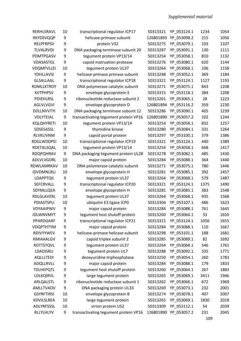
Supplemental material
109
RERHLSRAVL 10 transcriptional regulator ICP17 50313321 YP_053124.1 1234 1054
RKYDSVQQP 9 helicase-primase subunit 126801893 YP_053098.2 215 1056
RELPFRPSV 9 protein V32 50313275 YP_053079.1 103 1107
TLVALRVDI 9 DNA packaging terminase subunit 20 50313287 YP_053091.1 130 1111
PDMTPGASV 9 tegument protein VP13/14 50313254 YP_053058.1 810 1132
VDASASTGL 9 capsid matruation protease 50313276 YP_053080.1 620 1144
VDQMFVLLEI 10 tegument protein UL37 50313264 YP_053068.1 106 1158
YDHLLAVSI 9 helicase-primase primase subunit 50313248 YP_053052.1 369 1184
GLSALLAAL 9 transcriptional regulator ICP18 50313321 YP_053124.1 1127 1193
RDMLLKTRDY 10 DNA polymerase catalytic subunit 50313271 YP_053075.1 843 1208
KIITPHPSV 9 envelope glycoprotein E 50313315 YP_053118.1 384 1208
PDIEHLRSL 9 ribonucleotide reductase subunit 2 50313261 YP_053065.1 28 1223
AGLVLVGVI 9 envelope glycoprotein D 126801894 YP_053116.2 359 1230
DDLLNVVTYI 10 DNA packaging terminase subunit 12 50313286 YP_053090.1 405 1239
VDLYTEIAL 9 transactivating tegument protein VP16 126801890 YP_053057.2 102 1244
KQLQHYRETI 10 tegument protein VP13/14 50313254 YP_053058.1 832 1257
SDSESASSL 9 thymidine kinase 50313280 YP_053084.1 331 1264
RLVKLIVNM 9 capsid portal protein 50313297 YP_053100.1 379 1386
RDGLWDDPEI 10 transcriptional regulator ICP19 50313321 YP_053124.1 440 1389
RDETALSQAL 10 tegument protein VP13/14 50313254 YP_053058.1 668 1417
RDQPQHRAV 9 DNA packaging tegument protein UL28 50313278 YP_053082.1 485 1430
ADLVLVGDRL 10 major capsid protein 50313284 YP_053088.1 364 1440
RDWLAMRKAV 10 DNA polymerase catalytic subunit 50313271 YP_053075.1 780 1446
IDVDMNLRLI 10 envelope glycoprotein H 50313281 YP_053085.1 392 1457
LDAPPTQII 9 tegument protein UL37 50313264 YP_053068.1 579 1487
SEFCRVALL 9 transcriptional regulator ICP20 50313321 YP_053124.1 1375 1490
SDYMLLQEA 9 envelope glycoprotein H 50313281 YP_053085.1 383 1548
RDLGLAVERL 10 tegument protein UL37 50313264 YP_053068.1 935 1568
PDIASTSPLI 10 ubiquitin E3 ligase ICP0 50313304 YP_053107.1 486 1623
VDYAAIPWV 9 major capsid protein 50313284 YP_053088.1 761 1645
IDLWNVMYT 9 tegument host shutoff protein 50313260 YP_053064.1 33 1650
PPARDGARF 9 transcriptional regulator ICP21 50313321 YP_053124.1 1056 1655
VDQPTHTYM 9 major capsid protein 50313284 YP_053088.1 110 1667
RDIVYYWEV 9 helicase-primase helicase subunit 50313298 YP_053101.1 188 1681
KMIAAALGV 9 capsid triplex subunit 2 50313285 YP_053089.1 82 1692
RDTTSTQVL 9 tegument protein UL37 50313264 YP_053068.1 546 1761
LDADISRLI 9 tegument protein UL7 50313288 YP_053092.1 335 1771
AQLLLTEDI 9 deoxyuridine triphosphatase 50313250 YP_053054.1 260 1783
ADQLLRVLL 9 major capsid protein 50313284 YP_053088.1 179 1833
TDLHEPQTL 9 tegument host shutoff protein 50313260 YP_053064.1 267 1883
LDLKQIRVL 9 large tegument protein 50313265 YP_053069.1 3411 1946
ARLQALSTL 9 ribonucleotide reductase subunit 1 50313262 YP_053066.1 672 1969
AMLLTVADV 9 DNA packaging protein UL33 50313269 YP_053073.1 232 2001
GSYRFTIRSI 10 envelope glycoprotein B 50313274 YP_053078.1 407 2007
KDVILSLREA 10 large tegument protein 50313265 YP_053069.1 1830 2018
ADLYRFSSSL 10 virion protein US2 50313309 YP_053112.1 54 2039
RLLYLHLYV 9 transactivating tegument protein VP16 126801890 YP_053057.2 231 2045
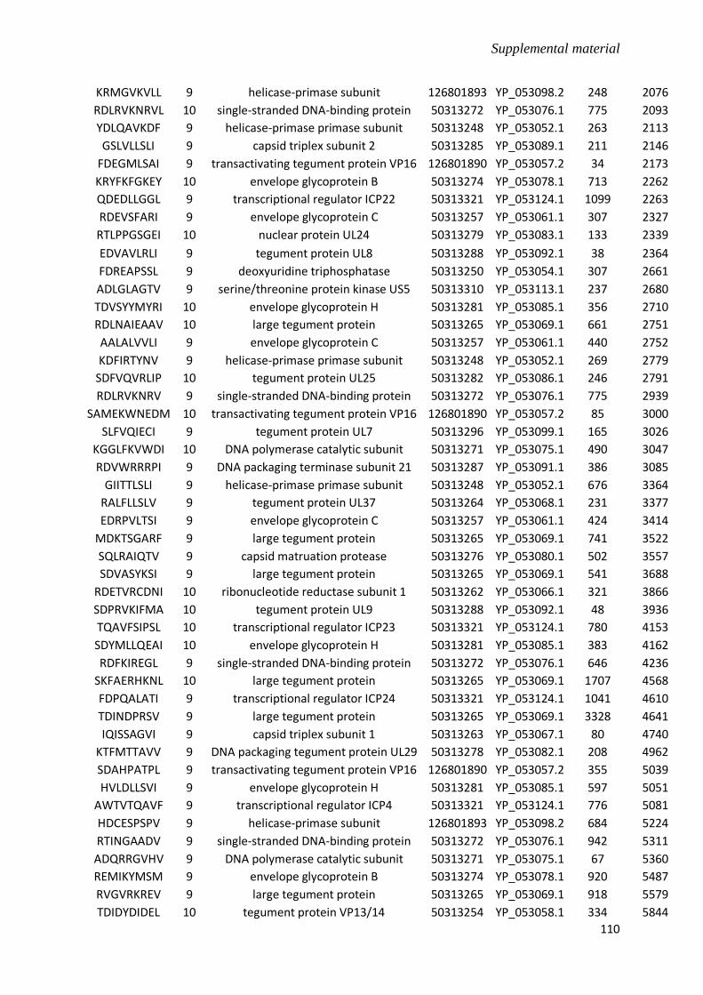
Supplemental material
110
KRMGVKVLL 9 helicase-primase subunit 126801893 YP_053098.2 248 2076
RDLRVKNRVL 10 single-stranded DNA-binding protein 50313272 YP_053076.1 775 2093
YDLQAVKDF 9 helicase-primase primase subunit 50313248 YP_053052.1 263 2113
GSLVLLSLI 9 capsid triplex subunit 2 50313285 YP_053089.1 211 2146
FDEGMLSAI 9 transactivating tegument protein VP16 126801890 YP_053057.2 34 2173
KRYFKFGKEY 10 envelope glycoprotein B 50313274 YP_053078.1 713 2262
QDEDLLGGL 9 transcriptional regulator ICP22 50313321 YP_053124.1 1099 2263
RDEVSFARI 9 envelope glycoprotein C 50313257 YP_053061.1 307 2327
RTLPPGSGEI 10 nuclear protein UL24 50313279 YP_053083.1 133 2339
EDVAVLRLI 9 tegument protein UL8 50313288 YP_053092.1 38 2364
FDREAPSSL 9 deoxyuridine triphosphatase 50313250 YP_053054.1 307 2661
ADLGLAGTV 9 serine/threonine protein kinase US5 50313310 YP_053113.1 237 2680
TDVSYYMYRI 10 envelope glycoprotein H 50313281 YP_053085.1 356 2710
RDLNAIEAAV 10 large tegument protein 50313265 YP_053069.1 661 2751
AALALVVLI 9 envelope glycoprotein C 50313257 YP_053061.1 440 2752
KDFIRTYNV 9 helicase-primase primase subunit 50313248 YP_053052.1 269 2779
SDFVQVRLIP 10 tegument protein UL25 50313282 YP_053086.1 246 2791
RDLRVKNRV 9 single-stranded DNA-binding protein 50313272 YP_053076.1 775 2939
SAMEKWNEDM 10 transactivating tegument protein VP16 126801890 YP_053057.2 85 3000
SLFVQIECI 9 tegument protein UL7 50313296 YP_053099.1 165 3026
KGGLFKVWDI 10 DNA polymerase catalytic subunit 50313271 YP_053075.1 490 3047
RDVWRRRPI 9 DNA packaging terminase subunit 21 50313287 YP_053091.1 386 3085
GIITTLSLI 9 helicase-primase primase subunit 50313248 YP_053052.1 676 3364
RALFLLSLV 9 tegument protein UL37 50313264 YP_053068.1 231 3377
EDRPVLTSI 9 envelope glycoprotein C 50313257 YP_053061.1 424 3414
MDKTSGARF 9 large tegument protein 50313265 YP_053069.1 741 3522
SQLRAIQTV 9 capsid matruation protease 50313276 YP_053080.1 502 3557
SDVASYKSI 9 large tegument protein 50313265 YP_053069.1 541 3688
RDETVRCDNI 10 ribonucleotide reductase subunit 1 50313262 YP_053066.1 321 3866
SDPRVKIFMA 10 tegument protein UL9 50313288 YP_053092.1 48 3936
TQAVFSIPSL 10 transcriptional regulator ICP23 50313321 YP_053124.1 780 4153
SDYMLLQEAI 10 envelope glycoprotein H 50313281 YP_053085.1 383 4162
RDFKIREGL 9 single-stranded DNA-binding protein 50313272 YP_053076.1 646 4236
SKFAERHKNL 10 large tegument protein 50313265 YP_053069.1 1707 4568
FDPQALATI 9 transcriptional regulator ICP24 50313321 YP_053124.1 1041 4610
TDINDPRSV 9 large tegument protein 50313265 YP_053069.1 3328 4641
IQISSAGVI 9 capsid triplex subunit 1 50313263 YP_053067.1 80 4740
KTFMTTAVV 9 DNA packaging tegument protein UL29 50313278 YP_053082.1 208 4962
SDAHPATPL 9 transactivating tegument protein VP16 126801890 YP_053057.2 355 5039
HVLDLLSVI 9 envelope glycoprotein H 50313281 YP_053085.1 597 5051
AWTVTQAVF 9 transcriptional regulator ICP4 50313321 YP_053124.1 776 5081
HDCESPSPV 9 helicase-primase subunit 126801893 YP_053098.2 684 5224
RTINGAADV 9 single-stranded DNA-binding protein 50313272 YP_053076.1 942 5311
ADQRRGVHV 9 DNA polymerase catalytic subunit 50313271 YP_053075.1 67 5360
REMIKYMSM 9 envelope glycoprotein B 50313274 YP_053078.1 920 5487
RVGVRKREV 9 large tegument protein 50313265 YP_053069.1 918 5579
TDIDYDIDEL 10 tegument protein VP13/14 50313254 YP_053058.1 334 5844
Supplemental material
111
RELGDACVL 9 transcriptional regulator ICP4 50313321 YP_053124.1 659 5847
ALLPATLTI 9 tegument protein UL51 50313249 YP_053053.1 45 5858
RFVTEVREV 9 tegument protein UL7 50313296 YP_053099.1 46 5899
SEFKKMMDL 9 membrane protein US8A 50313316 YP_053119.1 68 6338
RDIGSHGRN 9 nuclear protein UL55 50313245 YP_053049.1 16 6386
HHLQYIINTV 10 tegument protein VP13/14 50313255 YP_053059.1 323 6386
SDNETASMFI 10 membrane protein US9 50313317 YP_053120.1 165 6386
QAPRALGPM 9 transcriptional regulator ICP4 50313321 YP_053124.1 976 6386
IDILLPSDL 9 capsid triplex subunit 2 50313285 YP_053089.1 8 6410
RDFWHAACS 9 envelope glycoprotein N 50313251 YP_053055.1 44 6443
ADDPRTTEV 9 DNA packaging terminase subunit 22 50313287 YP_053091.1 139 6532
MILMHATYFI 10 DNA packaging terminase subunit 13 50313286 YP_053090.1 208 6787
HIFTGRVTL 9 envelope glycoprotein G 50313311 YP_053114.1 162 7087
RDEAGRPDSL 10 transcriptional regulator ICP4 50313321 YP_053124.1 584 7124
QDFKEKLLF 9 envelope glycoprotein L 155041723 YP_053106.2 118 7144
PEDVKVVVL 9 transcriptional regulator ICP4 50313321 YP_053124.1 1086 7338
REFVMTAEL 9 DNA polymerase catalytic subunit 50313271 YP_053075.1 1030 7430
SDKRLNPIF 9 deoxyribonuclease 50313291 YP_053046.1 96 7495
GDAHAEAAV 9 protein V32 50313275 YP_053079.1 12 7555
SDAGALESV 9 DNA replication origin-binding helicase 50313294 YP_053097.1 656 7563
TDYNTVLVNY 10 tegument host shutoff protein 50313260 YP_053064.1 477 7721
RDGTLLRTL 9 DNA replication origin-binding helicase 50313294 YP_053097.1 272 7762
YDLGDAVGV 9 large tegument protein 50313265 YP_053069.1 433 7878
ADFINNSGL 9 tegument protein UL17 50313290 YP_053094.1 427 8022
RDYVHSRWA 9 DNA polymerase catalytic subunit 50313271 YP_053075.1 850 8047
HEFNNYALM 9 DNA polymerase processivity subunit 50313259 YP_053063.1 175 8195
RFFERWHMLM 10 major capsid protein 50313284 YP_053088.1 504 8240
RELEAVIKEI 10 large tegument protein 50313265 YP_053069.1 834 8249
YYWEVLVDI 9 helicase-primase helicase subunit 50313298 YP_053101.1 192 8250
KDWQTHLDS 9 capsid portal protein 50313297 YP_053100.1 114 8327
KFFAQVRIQL 10 helicase-primase primase subunit 50313248 YP_053052.1 399 8371
HDFLETILM 9 nuclear protein UL4 50313299 YP_053102.1 192 8463
SDTVIHNGV 9 tegument protein UL26 50313282 YP_053086.1 6 8489
KQYNALVRMV 10 transcriptional regulator ICP4 50313321 YP_053124.1 475 8712
RHLPHALSV 9 DNA packaging terminase subunit 5 50313273 YP_053077.1 244 8737
SVFIKFVGL 9 DNA polymerase catalytic subunit 50313271 YP_053075.1 890 8895
VDPARLREI 9 virion protein US2 50313309 YP_053112.1 222 8932
TDCDSDSSV 9 myristylated tegument protein CIRC 50313244 YP_053048.1 244 9146
RDLLNINVR 9 large tegument protein 50313265 YP_053069.1 3278 9209
ADENTLTYAL 10 major capsid protein 50313284 YP_053088.1 1015 9242
ADVSMDGFV 9 large tegument protein 50313265 YP_053069.1 1661 9287
KHYHFDLRRK 10 ribonucleotide reductase subunit 1 50313262 YP_053066.1 420 9444
HDEMWEVVI 9 tegument protein VP13/14 50313255 YP_053059.1 106 9564
RVYRMFCDM 9 virion protein US10 50313319 YP_053122.1 83 9607
PELLSEFCRV 10 transcriptional regulator ICP4 50313321 YP_053124.1 1371 9626
CDAYSFTLI 9 envelope glycoprotein G 50313311 YP_053114.1 118 9716
SQRSDFAPDL 10 transcriptional regulator ICP4 50313321 YP_053124.1 3 9842

Supplemental material
112
FGPGLFARA 9 transcriptional regulator ICP4 50313321 YP_053124.1 1249 9952
RPLQAMMQI 9 nuclear egress membrane protein 50313267 YP_053071.1 141 9994
HDCCETAQI 9 DNA packaging terminase subunit 14 50313286 YP_053090.1 68 9994
PDYKLMCFDI 10 DNA polymerase catalytic subunit 50313271 YP_053075.1 355 10008
VDGVWMRQI 9 envelope glycoprotein M 50313293 YP_053096.1 79 10152
MAIAILSVV 9 envelope glycoprotein D 126801894 YP_053116.2 16 10393
QMYINRNEI 9 helicase-primase primase subunit 50313248 YP_053052.1 631 10461
TDYYFSHLL 9 DNA polymerase catalytic subunit 50313271 YP_053075.1 1136 10751
VDFWRARDNM 10 tegument protein VP13/14 50313254 YP_053058.1 770 10824
HFVGKLVPI 9 tegument protein VP13/14 50313255 YP_053059.1 361 10850
RRFLHPDFI 9 tegument protein UL13 50313289 YP_053093.1 95 10865
ADQLATALL 9 helicase-primase primase subunit 50313248 YP_053052.1 75 11164
HDAENFLKQL 10 DNA packaging terminase subunit 6 50313273 YP_053077.1 147 11198
KQLRGPGGI 9 DNA replication origin-binding helicase 50313294 YP_053097.1 564 11226
AAILAFVFV 9 nuclear egress membrane protein 50313267 YP_053071.1 254 11270
RRQVTIVRI 9 thymidine kinase 50313280 YP_053084.1 20 11278
FYLIAAPYV 9 single-stranded DNA-binding protein 50313272 YP_053076.1 411 11345
MILMHATYF 9 DNA packaging terminase subunit 15 50313286 YP_053090.1 208 11402
GKYFYCNQI 9 multifunctional expression regulator 50313246 YP_053050.1 461 11532
TMFVGRYSI 9 helicase-primase helicase subunit 50313298 YP_053101.1 670 11601
RTFNAEMGV 9 thymidine kinase 50313280 YP_053084.1 344 11604
APRVRSISI 9 transcriptional regulator ICP4 50313321 YP_053124.1 176 11618
AVEPSELLI 9 transcriptional regulator ICP4 50313321 YP_053124.1 1362 11660
DELRLAVRAV 10 transcriptional regulator ICP4 50313321 YP_053124.1 743 11667
IDYDIDELI 9 tegument protein VP13/14 50313254 YP_053058.1 336 11804
TRYYLFQAI 9 helicase-primase primase subunit 50313248 YP_053052.1 246 11837
QKSFILQSL 9 transcriptional regulator ICP4 50313321 YP_053124.1 565 11868
AALSLLSLL 9 envelope glycoprotein G 50313311 YP_053114.1 6 11971
LDPDSIPMI 9 DNA polymerase processivity subunit 50313259 YP_053063.1 308 12146
RHFFLHVCF 9 DNA packaging protein UL33 50313269 YP_053073.1 179 12250
VDATTDTMV 9 tegument protein UL51 50313249 YP_053053.1 130 12266
SDYGYANEL 9 ribonucleotide reductase subunit 1 50313262 YP_053066.1 26 12459
AKFVYLIFVY 10 capsid triplex subunit 1 50313263 YP_053067.1 305 12471
PEPDDELEI 9 transcriptional regulator ICP4 50313321 YP_053124.1 1431 12572
DDFTLAAPI 9 large tegument protein 50313265 YP_053069.1 1331 12633
TVTQAVFSI 9 transcriptional regulator ICP4 50313321 YP_053124.1 778 13177
AGERTRVPL 9 transcriptional regulator ICP4 50313321 YP_053124.1 1287 13356
SPAPGLAAM 9 transcriptional regulator ICP4 50313321 YP_053124.1 108 13381
ALPHAAASV 9 transcriptional regulator ICP4 50313321 YP_053124.1 547 13556
DWPQDGPAI 9 transcriptional regulator ICP4 50313321 YP_053124.1 1203 13831
ADFGAAQFPV 10 serine/threonine protein kinase US6 50313310 YP_053113.1 224 13853
GDFAAVVRAV 10 virion protein US11 50313319 YP_053122.1 53 14127
SRGTFVMREN 10 tegument protein VP22 50313252 YP_053056.1 36 14648
HCYWLYNRF 9 helicase-primase subunit 126801893 YP_053098.2 571 14972
RFFETRLGI 9 virion protein US12 50313319 YP_053122.1 162 15003
MDPAWRRRI 9 membrane rotein V1 50313243 YP_053047.1 1 15278
RIAATVTTI 9 DNA polymerase catalytic subunit 50313271 YP_053075.1 833 15321

Supplemental material
113
NDLILALAI 9 helicase-primase primase subunit 50313248 YP_053052.1 155 15369
SLFKFFKEM 9 envelope protein UL43 50313258 YP_053062.1 250 15534
HKIDLPYKEI 10 DNA polymerase catalytic subunit 50313271 YP_053075.1 546 15545
FDPDLFSPV 9 deoxyribonuclease 50313291 YP_053046.1 326 15566
RDYIAHNVN 9 helicase-primase primase subunit 50313248 YP_053052.1 411 15619
FAPDLYDFI 9 transcriptional regulator ICP4 50313321 YP_053124.1 8 15885
DDAPLSTPAV 10 transcriptional regulator ICP4 50313321 YP_053124.1 56 16096
ADAYLRRVV 9 major capsid protein 50313284 YP_053088.1 1143 16199
SDVSEFKKMM 10 membrane protein US8A 50313316 YP_053119.1 65 16407
AVLTSSTLF 9 transcriptional regulator ICP4 50313321 YP_053124.1 1241 16495
FTLPAYTFV 9 helicase-primase helicase subunit 50313298 YP_053101.1 428 16499
KVYLHRDNVI 10 nuclear protein UL3 50313301 YP_053104.1 196 16538
YDLLYFVCL 9 DNA packaging tegument protein UL30 50313278 YP_053082.1 569 17276
SVANPLPHI 9 transcriptional regulator ICP4 50313321 YP_053124.1 527 17366
SRLDIASTV 9 large tegument protein 50313265 YP_053069.1 2168 17427
KLIVNMKSM 9 capsid portal protein 50313297 YP_053100.1 382 17693
HYYIEQESI 9 ribonucleotide reductase subunit 2 50313261 YP_053065.1 99 17937
GDSDPTHRL 9 transcriptional regulator ICP4 50313321 YP_053124.1 732 18674
RDWLAMRKA 9 DNA polymerase catalytic subunit 50313271 YP_053075.1 780 18727
SDNETASMF 9 membrane protein US9 50313317 YP_053120.1 165 18890
RFERFHVYDI 10 DNA polymerase catalytic subunit 50313271 YP_053075.1 122 18962
QDLLALNSGV 10 tegument protein UL51 50313249 YP_053053.1 173 19068
RKFVKHVVSM 10 tegument host shutoff protein 50313260 YP_053064.1 412 19247
RIVAKKINWI 10 tegument protein UL37 50313264 YP_053068.1 202 19343
SSYDEVRNM 9 tegument protein UL27 50313282 YP_053086.1 443 19751
AQAPVPVFV 9 transcriptional regulator ICP4 50313321 YP_053124.1 459 19826
RDLLEHVQA 9 major capsid protein 50313284 YP_053088.1 691 20172
FDKFGRVLV 9 DNA polymerase catalytic subunit 50313271 YP_053075.1 1008 20620
RCLYAVSEL 9 DNA packaging terminase subunit 7 50313273 YP_053077.1 305 20847
SDLYVYVDP 9 DNA packaging terminase subunit 16 50313286 YP_053090.1 504 20968
MDPPSPSAV 9 transactivating tegument protein VP16 126801890 YP_053057.2 389 21216
SYYMYRIVAR 10 envelope glycoprotein H 50313281 YP_053085.1 359 21371
IDFTLCIIQM 10 tegument protein UL7 50313296 YP_053099.1 143 21570
KQLNDGTFV 9 virion protein V67 50313318 YP_053121.1 132 21787
SVLRDFSFI 9 helicase-primase primase subunit 50313248 YP_053052.1 840 22433
KHFFERCTL 9 DNA replication origin-binding helicase 50313294 YP_053097.1 502 22641
SDVWLFSEP 9 deoxyribonuclease 50313291 YP_053046.1 412 22642
HDFLKFKGLS 10 myristylated tegument protein CIRC 50313244 YP_053048.1 111 23054
LQDEDLLGGL 10 transcriptional regulator ICP4 50313321 YP_053124.1 1098 23173
RKVARNREAI 10 helicase-primase primase subunit 50313248 YP_053052.1 931 23256
HDFSVWFED 9 DNA packaging protein UL33 126801892 YP_053072.2 59 23385
CEYHVRVHL 9 DNA packaging terminase subunit 8 50313273 YP_053077.1 124 23523
LDASSDVWL 9 deoxyribonuclease 50313291 YP_053046.1 408 24783
SPFDTFVDNI 10 helicase-primase helicase subunit 50313298 YP_053101.1 690 25214
RDSEHIYET 9 tegument protein VP22 50313252 YP_053056.1 83 25445
IPIPSERYV 9 virion protein V67 50313318 YP_053121.1 204 26388
GILRSEVRI 9 envelope glycoprotein J 50313312 YP_053115.1 718 26457

Supplemental material
114
QDMAFIIVRS 10 ribonucleotide reductase subunit 1 50313262 YP_053066.1 394 26633
HDQASFIGL 9 single-stranded DNA-binding protein 50313272 YP_053076.1 615 26633
CDIILDIMPL 10 tegument host shutoff protein 50313260 YP_053064.1 228 27040
VDYTQKNHS 9 envelope glycoprotein M 50313293 YP_053096.1 69 27360
ADEDLYSDI 9 serine/threonine protein kinase US7 50313310 YP_053113.1 45 28012
RRFFERWHML 10 major capsid protein 50313284 YP_053088.1 503 28601
RDEGQQTDM 9 nuclear egress lamina protein 50313270 YP_053074.1 277 29740
YDLQIARAV 9 large tegument protein 50313265 YP_053069.1 3157 29966
RDCGQREIV 9 helicase-primase subunit 126801893 YP_053098.2 671 30257
ADVLQAVGV 9 large tegument protein 50313265 YP_053069.1 1418 30359
SDVSEFKKM 9 membrane protein US8A 50313316 YP_053119.1 65 31558
KGGLSALLA 9 transcriptional regulator ICP4 50313321 YP_053124.1 1125 31587
CCRATQTNA 9 transcriptional regulator ICP4 50313321 YP_053124.1 872 32359
AVLGAAVYV 9 envelope glycoprotein M 50313293 YP_053096.1 178 32444
AALPPASRL 9 capsid matruation protease 50313276 YP_053080.1 41 32907
AMIGLIVYI 9 envelope glycoprotein G 50313311 YP_053114.1 373 33107
AQAGASASF 9 transcriptional regulator ICP4 50313321 YP_053124.1 939 33148
FESREAMSWL 10 transcriptional regulator ICP4 50313321 YP_053124.1 485 33498
KIFMAVSVL 9 tegument protein UL10 50313288 YP_053092.1 53 34026
PDAEAEPLVL 10 transcriptional regulator ICP4 50313321 YP_053124.1 1385 34250
TQKSFILQSL 10 transcriptional regulator ICP4 50313321 YP_053124.1 564 34300
GGEPFSGSA 9 transcriptional regulator ICP4 50313321 YP_053124.1 816 34862
RRLRTRARI 9 thymidine kinase 50313280 YP_053084.1 191 34879
AEAEPLVLPI 10 transcriptional regulator ICP4 50313321 YP_053124.1 1387 34879
GDAMAAGNA 9 transcriptional regulator ICP4 50313321 YP_053124.1 536 34966
CDWGYRLIAV 10 nuclear protein UL4 50313299 YP_053102.1 36 35513
KNLVSEHEI 9 tegument host shutoff protein 50313260 YP_053064.1 453 35633
KTWFLVPLI 9 DNA packaging terminase subunit 17 50313286 YP_053090.1 265 35702
PSLVGGMLG 9 transcriptional regulator ICP4 50313321 YP_053124.1 787 35773
FPEAWRPAL 9 transcriptional regulator ICP4 50313321 YP_053124.1 1031 36037
RYMQAHSRV 9 tegument protein UL14 50313289 YP_053093.1 59 36096
EDMAADVPV 9 major capsid protein 50313284 YP_053088.1 295 36934
ITGSVANPL 9 transcriptional regulator ICP4 50313321 YP_053124.1 524 37987
KVYLHRDNV 9 nuclear protein UL3 50313301 YP_053104.1 196 38243
LDLSAVPTRV 10 large tegument protein 50313265 YP_053069.1 700 39443
STASSTTSI 9 envelope glycoprotein J 50313312 YP_053115.1 92 39925
VDYTQKNHSV 10 envelope glycoprotein M 50313293 YP_053096.1 69 40419
RKFAAINII 9 membrane rotein V1 50313243 YP_053047.1 175 41474
TDAYAESPAV 10 membrane rotein V1 50313243 YP_053047.1 116 42279
ADRDASANL 9 large tegument protein 50313265 YP_053069.1 1532 42526
YDRTQKSFIL 10 transcriptional regulator ICP4 50313321 YP_053124.1 561 43103
VDFWRARDN 9 tegument protein VP13/14 50313254 YP_053058.1 770 43326
APRRRAPRV 9 transcriptional regulator ICP4 50313321 YP_053124.1 1399 43384
FDIPPPPPL 9 myristylated tegument protein 50313292 YP_053095.1 38 43901
AQALRRAPI 9 transcriptional regulator ICP4 50313321 YP_053124.1 1459 44141
GDAMAAGNAL 10 transcriptional regulator ICP4 50313321 YP_053124.1 536 44916
GDLRQRRVL 9 envelope glycoprotein B 50313274 YP_053078.1 21 45029

Supplemental material
115
IDGAGDVELV 10 transcriptional regulator ICP4 50313321 YP_053124.1 1439 45828
RVIFDGQQI 9 DNA polymerase catalytic subunit 50313271 YP_053075.1 606 45895
NDLISRDEAI 10 ribonucleotide reductase subunit 2 50313261 YP_053065.1 198 46532
EDWPQDGPAI 10 transcriptional regulator ICP4 50313321 YP_053124.1 1202 46953
LNAQGVLLL 9 transcriptional regulator ICP4 50313321 YP_053124.1 1160 47514
YDFIESNDF 9 transcriptional regulator ICP4 50313321 YP_053124.1 13 47673
DSRDGLWDD 9 transcriptional regulator ICP4 50313321 YP_053124.1 438 48314
SPAPRVRSI 9 transcriptional regulator ICP4 50313321 YP_053124.1 174 48580
RERHLSRAV 9 transcriptional regulator ICP4 50313321 YP_053124.1 1234 49529
LDAWGVQQV 9 envelope glycoprotein E 50313315 YP_053118.1 19 50889
RINYRRREL 9 transactivating tegument protein VP16 126801890 YP_053057.2 301 51031
RDRRTEQRL 9 capsid triplex subunit 1 50313263 YP_053067.1 214 51111
APLPPGAFI 9 capsid triplex subunit 2 50313285 YP_053089.1 91 51717
RRYARGKEC 9 envelope glycoprotein M 50313293 YP_053096.1 128 52703
KFFAQVRIQ 9 helicase-primase primase subunit 50313248 YP_053052.1 399 53024
RHFRQRATV 9 DNA packaging terminase subunit 18 50313286 YP_053090.1 248 53838
RIVAKKINW 9 tegument protein UL37 50313264 YP_053068.1 202 54882
HDFLKFKGL 9 myristylated tegument protein CIRC 50313244 YP_053048.1 111 55550
RRYARKRRH 9 capsid matruation protease 50313276 YP_053080.1 444 55679
GELAASGPL 9 transcriptional regulator ICP4 50313321 YP_053124.1 1065 56183
GLYRRVIEI 9 envelope glycoprotein D 126801894 YP_053116.2 183 56467
EDLLAEIDAI 10 tegument protein UL37 50313264 YP_053068.1 1006 56574
AALPSEVYL 9 membrane rotein V1 50313243 YP_053047.1 139 56832
VEHPGPSPGV 10 transcriptional regulator ICP4 50313321 YP_053124.1 1449 57166
REANLAKTL 9 transcriptional regulator ICP4 50313321 YP_053124.1 306 57306
AISQYHIYM 9 transcriptional regulator ICP4 50313321 YP_053124.1 1210 58062
HQIPDPEDV 9 transcriptional regulator ICP4 50313321 YP_053124.1 1081 58621
PEAPAPSVRV 10 transcriptional regulator ICP4 50313321 YP_053124.1 613 58926
DDKEIMSLI 9 DNA packaging terminase subunit 23 50313287 YP_053091.1 242 59010
RKSIHVTSI 9 deoxyuridine triphosphatase 50313250 YP_053054.1 144 59528
QDGPAISQY 9 transcriptional regulator ICP4 50313321 YP_053124.1 1206 59591
KLFFVHAHI 9 DNA polymerase catalytic subunit 50313271 YP_053075.1 762 59942
ACVLACQAV 9 transcriptional regulator ICP4 50313321 YP_053124.1 664 60523
FEYDQRTLI 9 ribonucleotide reductase subunit 1 50313262 YP_053066.1 707 60881
WDDPEIVLA 9 transcriptional regulator ICP4 50313321 YP_053124.1 444 61443
EGARARASA 9 transcriptional regulator ICP4 50313321 YP_053124.1 768 61770
LTKSLLSDI 9 large tegument protein 50313265 YP_053069.1 2042 62520
AELIDNQLF 9 transcriptional regulator ICP4 50313321 YP_053124.1 1023 63215
LEGVREANL 9 transcriptional regulator ICP4 50313321 YP_053124.1 302 64550
SDLYTFLTK 9 serine/threonine protein kinase US8 50313310 YP_053113.1 166 64800
RLYVVCGEL 9 ribonucleotide reductase subunit 1 50313262 YP_053066.1 67 65841
TQPAAPDLL 9 transcriptional regulator ICP4 50313321 YP_053124.1 38 66157
YFWAQRRKF 9 transactivating tegument protein VP16 126801890 YP_053057.2 265 66296
GYFLSKRDI 9 helicase-primase subunit 126801893 YP_053098.2 192 67298
QQHQPQKAR 9 transcriptional regulator ICP4 50313321 YP_053124.1 885 68295
SRFAYVKFI 9 myristylated tegument protein CIRC 50313244 YP_053048.1 64 -
PDLTNFKCI 9 nuclear protein UL55 50313245 YP_053049.1 107 -

Supplemental material
116
RDAVMRGEN 9 multifunctional expression regulator 50313246 YP_053050.1 299 -
FVVAVTIVEI 10 envelopeenvelope glycoprotein K 50313247 YP_053051.1 259 -
FDYVCRENW 9 envelopeenvelope glycoprotein K 50313247 YP_053051.1 79 -
KELHDKVRI 9 envelopeenvelope glycoprotein K 50313247 YP_053051.1 100 -
SRTFKRDPI 9 envelopeenvelope glycoprotein K 50313247 YP_053051.1 198 -
LHYEQRIQI 9 envelopeenvelope glycoprotein K 50313247 YP_053051.1 326 -
LDVDFGIKRR 10 helicase-primase primase subunit 50313248 YP_053052.1 651 -
VDFGIKRRV 9 helicase-primase primase subunit 50313248 YP_053052.1 653 -
GDPGVKQRI 9 envelope glycoprotein N 50313251 YP_053055.1 27 -
GHVDLYTEI 9 transactivating tegument protein VP16 126801890 YP_053057.2 100 -
SLMHKFKQV 9 transactivating tegument protein VP16 126801890 YP_053057.2 209 -
HKFKQVVRD 9 transactivating tegument protein VP16 126801890 YP_053057.2 212 -
SEYHDHGEM 9 tegument protein VP13/14 50313254 YP_053058.1 169 -
SDVVTKQNA 9 tegument protein VP13/14 50313254 YP_053058.1 447 -
SSVGKRGLI 9 tegument protein VP13/14 50313254 YP_053058.1 848 -
KDVTENIVGQ 10 tegument protein VP13/14 50313255 YP_053059.1 42 -
HDGFIRRTM 9 tegument protein VP13/14 50313255 YP_053059.1 720 -
VDFTKYVTN 9 envelope glycoprotein C 50313257 YP_053061.1 261 -
RRGGQLGVI 9 envelope glycoprotein C 50313257 YP_053061.1 150 -
RWYKNAREV 9 envelope glycoprotein C 50313257 YP_053061.1 253 -
SKVVFEIENY 10 DNA polymerase processivity subunit 50313259 YP_053063.1 125 -
HDYDSSTED 9 tegument host shutoff protein 50313260 YP_053064.1 323 -
RKFVKHVVS 9 tegument host shutoff protein 50313260 YP_053064.1 412 -
KFVKHVVSM 9 tegument host shutoff protein 50313260 YP_053064.1 413 -
RIYELFSEA 9 ribonucleotide reductase subunit 2 50313261 YP_053065.1 230 -
KYWLNSTEI 9 ribonucleotide reductase subunit 1 50313262 YP_053066.1 89 -
THVFPHKHM 9 capsid triplex subunit 1 50313263 YP_053067.1 235 -
HVFPHKHMT 9 capsid triplex subunit 1 50313263 YP_053067.1 236 -
KRMESIEEF 9 tegument protein UL37 50313264 YP_053068.1 171 -
REYLKKEDL 9 tegument protein UL37 50313264 YP_053068.1 1000 -
REYLKKEDLL 10 tegument protein UL37 50313264 YP_053068.1 1000 -
GDGFQSTQI 9 large tegument protein 50313265 YP_053069.1 124 -
KLVARVEEL 9 large tegument protein 50313265 YP_053069.1 581 -
HDKFDAIEI 9 large tegument protein 50313265 YP_053069.1 674 -
SDTTNMDEL 9 large tegument protein 50313265 YP_053069.1 688 -
KVIKKVESM 9 large tegument protein 50313265 YP_053069.1 711 -
SLFGQFREA 9 large tegument protein 50313265 YP_053069.1 1007 -
REFAKRAEQ 9 large tegument protein 50313265 YP_053069.1 1278 -
SKFAERHKN 9 large tegument protein 50313265 YP_053069.1 1707 -
GLYDEYQKI 9 large tegument protein 50313265 YP_053069.1 1783 -
HDILQLRFG 9 large tegument protein 50313265 YP_053069.1 1906 -
RALEEVTYV 9 large tegument protein 50313265 YP_053069.1 1433 -
RTLQASMGV 9 large tegument protein 50313265 YP_053069.1 1676 -
WDQHLVPAV 9 large tegument protein 50313265 YP_053069.1 3302 -
LDYTKYTED 9 small capsid protein 50313266 YP_053070.1 46 -
MDSYNYRDF 9 nuclear egress membrane protein 50313267 YP_053071.1 1 -
RRVLTLVDI 9 DNA packaging protein UL33 50313269 YP_053073.1 169 -

Supplemental material
117
KSVHLHYRLI 10 nuclear egress lamina protein 50313270 YP_053074.1 241 -
KLYAVFDDF 9 nuclear egress lamina protein 50313270 YP_053074.1 314 -
RFHVYDIVET 10 DNA polymerase catalytic subunit 50313271 YP_053075.1 125 -
HVYGVRHYF 9 DNA polymerase catalytic subunit 50313271 YP_053075.1 172 -
RHYFYMAKA 9 DNA polymerase catalytic subunit 50313271 YP_053075.1 177 -
GDVLGEHKI 9 DNA polymerase catalytic subunit 50313271 YP_053075.1 540 -
HKPYSVRVI 9 DNA polymerase catalytic subunit 50313271 YP_053075.1 876 -
KKYIGVING 9 DNA polymerase catalytic subunit 50313271 YP_053075.1 940 -
ARITLTRVI 9 DNA polymerase catalytic subunit 50313271 YP_053075.1 600 -
HDAENFLKQ 9 DNA packaging terminase subunit 9 50313273 YP_053077.1 147 -
HKFFELING 9 DNA packaging terminase subunit 10 50313273 YP_053077.1 599 -
RANVYYKDI 9 envelope glycoprotein B 50313274 YP_053078.1 184 -
RVPVSVEEI 9 envelope glycoprotein B 50313274 YP_053078.1 213 -
TKWKEVDEL 9 envelope glycoprotein B 50313274 YP_053078.1 392 -
QDGVHREQI 9 envelope glycoprotein B 50313274 YP_053078.1 533 -
TLWNEMVKI 9 envelope glycoprotein B 50313274 YP_053078.1 611 -
KRYFKFGKE 9 envelope glycoprotein B 50313274 YP_053078.1 713 -
RYFKFGKEY 9 envelope glycoprotein B 50313274 YP_053078.1 714 -
KEYVYYENY 9 envelope glycoprotein B 50313274 YP_053078.1 720 -
TAWFMFRNI 9 envelope protein UL20 50313283 YP_053087.1 119 -
TLFDFFKQI 9 major capsid protein 50313284 YP_053088.1 32 -
THTYMMKHI 9 major capsid protein 50313284 YP_053088.1 114 -
KHERNRKEL 9 major capsid protein 50313284 YP_053088.1 234 -
RRFFERWHM 9 major capsid protein 50313284 YP_053088.1 503 -
RFFERWHML 9 major capsid protein 50313284 YP_053088.1 504 -
MDRRSEAFKI 10 major capsid protein 50313284 YP_053088.1 1 -
GDAHRRQCV 9 DNA packaging terminase subunit 24 50313287 YP_053091.1 273 -
SYFVHFHAM 9 envelope glycoprotein M 50313293 YP_053096.1 318 -
RRYANKVRG 9 envelope glycoprotein M 50313293 YP_053096.1 383 -
RPGVAHDEI 9 DNA replication origin-binding helicase 50313294 YP_053097.1 594 -
SFFGGLQHI 9 helicase-primase subunit 126801893 YP_053098.2 459 -
SRFEHKNLK 9 helicase-primase helicase subunit 50313298 YP_053101.1 305 -
RLYSSHKEV 9 helicase-primase helicase subunit 50313298 YP_053101.1 398 -
AKYNSKADI 9 serine/threonine protein kinase US9 50313310 YP_053113.1 256 -
KDGQPEPVV 9 serine/threonine protein kinase US10 50313310 YP_053113.1 112 -
GIVEFTIVNM 10 envelope glycoprotein G 50313311 YP_053114.1 130 -
HRNYQRLEY 9 envelope glycoprotein G 50313311 YP_053114.1 401 -
HDYFRVNAS 9 envelope glycoprotein I 50313314 YP_053117.1 115 -
TDFVVHGYHS 10 envelope glycoprotein E 50313315 YP_053118.1 198 -
RFYKQLNDG 9 virion protein V67 50313318 YP_053121.1 129 -
AEAQAPVPVF 10 transcriptional regulator ICP4 50313321 YP_053124.1 457 -
GDSTKQYNAL 10 transcriptional regulator ICP4 50313321 YP_053124.1 471 -
QQPEAPAPSV 10 transcriptional regulator ICP4 50313321 YP_053124.1 611 -
PQAAASPAHL 10 transcriptional regulator ICP4 50313321 YP_053124.1 895 -
AEGPDRRGGF 10 transcriptional regulator ICP4 50313321 YP_053124.1 986 -
PDRRGGFRRV 10 transcriptional regulator ICP4 50313321 YP_053124.1 989 -
PEAWRPALTF 10 transcriptional regulator ICP4 50313321 YP_053124.1 1032 -
Supplemental material
118
PDPEDVKVVV 10 transcriptional regulator ICP4 50313321 YP_053124.1 1084 -
LEPDAEAEPL 10 transcriptional regulator ICP4 50313321 YP_053124.1 1383 -
DEAGRPDSL 9 transcriptional regulator ICP4 50313321 YP_053124.1 585 -
SDDRAIHGR 9 transcriptional regulator ICP4 50313321 YP_053124.1 914 -
GDAADPVAL 9 transcriptional regulator ICP4 50313321 YP_053124.1 141 -
NDFGEDPLI 9 transcriptional regulator ICP4 50313321 YP_053124.1 19 -
YGSQNMFGV 9 transcriptional regulator ICP4 50313321 YP_053124.1 47 -
VDDAPLSTP 9 transcriptional regulator ICP4 50313321 YP_053124.1 55 -
PLSTPAVVI 9 transcriptional regulator ICP4 50313321 YP_053124.1 59 -
PGLAAMLKM 9 transcriptional regulator ICP4 50313321 YP_053124.1 111 -
SSSSSSSSM 9 transcriptional regulator ICP4 50313321 YP_053124.1 186 -
EDDQADGAG 9 transcriptional regulator ICP4 50313321 YP_053124.1 196 -
DDQADGAGA 9 transcriptional regulator ICP4 50313321 YP_053124.1 197 -
GDRPAAGAA 9 transcriptional regulator ICP4 50313321 YP_053124.1 251 -
APSRPGGGL 9 transcriptional regulator ICP4 50313321 YP_053124.1 283 -
SDPPPMGRV 9 transcriptional regulator ICP4 50313321 YP_053124.1 423 -
GDSRDGLWD 9 transcriptional regulator ICP4 50313321 YP_053124.1 437 -
DGLWDDPEI 9 transcriptional regulator ICP4 50313321 YP_053124.1 441 -
EAQAPVPVF 9 transcriptional regulator ICP4 50313321 YP_053124.1 458 -
STKQYNALV 9 transcriptional regulator ICP4 50313321 YP_053124.1 473 -
ESREAMSWL 9 transcriptional regulator ICP4 50313321 YP_053124.1 486 -
GQDQNLAQF 9 transcriptional regulator ICP4 50313321 YP_053124.1 501 -
HAPRGHGSF 9 transcriptional regulator ICP4 50313321 YP_053124.1 515 -
APRGHGSFI 9 transcriptional regulator ICP4 50313321 YP_053124.1 516 -
AAGNALWAL 9 transcriptional regulator ICP4 50313321 YP_053124.1 540 -
PHAAASVAM 9 transcriptional regulator ICP4 50313321 YP_053124.1 549 -
YDRTQKSFI 9 transcriptional regulator ICP4 50313321 YP_053124.1 561 -
GRPDSLAAV 9 transcriptional regulator ICP4 50313321 YP_053124.1 588 -
VPGLDPSEI 9 transcriptional regulator ICP4 50313321 YP_053124.1 686 -
GSLQTLPPL 9 transcriptional regulator ICP4 50313321 YP_053124.1 828 -
GSSSDDRAI 9 transcriptional regulator ICP4 50313321 YP_053124.1 911 -
EGPDRRGGF 9 transcriptional regulator ICP4 50313321 YP_053124.1 987 -
IDNQLFPEA 9 transcriptional regulator ICP4 50313321 YP_053124.1 1026 -
EAWRPALTF 9 transcriptional regulator ICP4 50313321 YP_053124.1 1033 -
TGAPDVSAL 9 transcriptional regulator ICP4 50313321 YP_053124.1 1152 -
PDAEAEPLV 9 transcriptional regulator ICP4 50313321 YP_053124.1 1385 -
EAEPLVLPI 9 transcriptional regulator ICP4 50313321 YP_053124.1 1388 -
FGSRSTSVL 9 transcriptional regulator ICP4 50313321 YP_053124.1 1413 -
IDGAGDVEL 9 transcriptional regulator ICP4 50313321 YP_053124.1 1439 -
ALRRAPIKI 9 transcriptional regulator ICP4 50313321 YP_053124.1 1461 -
GGDWCNPYL 9 transcriptional regulator ICP4 50313321 YP_053124.1 1478 -
PDLYDFIES 9 transcriptional regulator ICP4 50313321 YP_053124.1 10 -
PLIRAASAA 9 transcriptional regulator ICP4 50313321 YP_053124.1 25 -
GLLPPGARI 9 transcriptional regulator ICP4 50313321 YP_053124.1 290 -
DDPEIVLAA 9 transcriptional regulator ICP4 50313321 YP_053124.1 445 -
GGASAVPGL 9 transcriptional regulator ICP4 50313321 YP_053124.1 681 -
AAEGSLQTL 9 transcriptional regulator ICP4 50313321 YP_053124.1 825 -

Supplemental material
119
DPEDVKVVV 9 transcriptional regulator ICP4 50313321 YP_053124.1 1085 -
APDVSALNA 9 transcriptional regulator ICP4 50313321 YP_053124.1 1154 -
PSELLIPEL 9 transcriptional regulator ICP4 50313321 YP_053124.1 1365 -
PDSLAAVAG 9 transcriptional regulator ICP4 50313321 YP_053124.1 590 -
AASQQQPEA 9 transcriptional regulator ICP4 50313321 YP_053124.1 607 -
RVREAYTRV 9 transcriptional regulator ICP4 50313321 YP_053124.1 621 -
DELRLAVRA 9 transcriptional regulator ICP4 50313321 YP_053124.1 743 -
GEAVSLLAP 9 transcriptional regulator ICP4 50313321 YP_053124.1 795 -
GPDRRGGFR 9 transcriptional regulator ICP4 50313321 YP_053124.1 988 -
AGAVEPSEL 9 transcriptional regulator ICP4 50313321 YP_053124.1 1360 -
DMNSSQATG 9 transcriptional regulator ICP4 50313321 YP_053124.1 380 -
RPDSLAAVA 9 transcriptional regulator ICP4 50313321 YP_053124.1 589 -
GAGATTATC 9 transcriptional regulator ICP4 50313321 YP_053124.1 864 -
RSPQAAASP 9 transcriptional regulator ICP4 50313321 YP_053124.1 893 -
SPPAPKRRV 9 transcriptional regulator ICP4 50313321 YP_053124.1 962 -
APRALGPMP 9 transcriptional regulator ICP4 50313321 YP_053124.1 977 -
DEDLLGGLP 9 transcriptional regulator ICP4 50313321 YP_053124.1 1100 -
RHLSRAVLT 9 transcriptional regulator ICP4 50313321 YP_053124.1 1236 -
YGGTGDSRD 9 transcriptional regulator ICP4 50313321 YP_053124.1 433 -
AAAAAPGTR 9 transcriptional regulator ICP4 50313321 YP_053124.1 641 -
ARSPQAAAS 9 transcriptional regulator ICP4 50313321 YP_053124.1 892 -
FGELAASGP 9 transcriptional regulator ICP4 50313321 YP_053124.1 1064 -
ISQYHIYMR 9 transcriptional regulator ICP4 50313321 YP_053124.1 1211 -
HPGPSPGVA 9 transcriptional regulator ICP4 50313321 YP_053124.1 1451 -
QALRRAPIK 9 transcriptional regulator ICP4 50313321 YP_053124.1 1460 -
APDLYDFIE 9 transcriptional regulator ICP4 50313321 YP_053124.1 9 -
QNMFGVDDA 9 transcriptional regulator ICP4 50313321 YP_053124.1 50 -
AQPASPAPS 9 transcriptional regulator ICP4 50313321 YP_053124.1 100 -
RSPHRLPKD 9 transcriptional regulator ICP4 50313321 YP_053124.1 329 -
GASKRKRAN 9 transcriptional regulator ICP4 50313321 YP_053124.1 346 -
VRMVFESRE 9 transcriptional regulator ICP4 50313321 YP_053124.1 481 -
ALSLYELRD 9 transcriptional regulator ICP4 50313321 YP_053124.1 715 -
VSTEDWPQD 9 transcriptional regulator ICP4 50313321 YP_053124.1 1199 -
GARILEYLE 9 transcriptional regulator ICP4 50313321 YP_053124.1 295 -
LAASGPLRR 9 transcriptional regulator ICP4 50313321 YP_053124.1 1067 -
VLYSPLQDE 9 transcriptional regulator ICP4 50313321 YP_053124.1 1093 -
AAFARLYPD 9 transcriptional regulator ICP4 50313321 YP_053124.1 1259 -
DTRAGERTR 9 transcriptional regulator ICP4 50313321 YP_053124.1 1284 -
VLPDYDGCK 9 transcriptional regulator ICP4 50313321 YP_053124.1 1304 -
VEPSELLIP 9 transcriptional regulator ICP4 50313321 YP_053124.1 1363 -
LLEPDAEAE 9 transcriptional regulator ICP4 50313321 YP_053124.1 1382 -
WEPGFGSRS 9 transcriptional regulator ICP4 50313321 YP_053124.1 1409 -
CLPEPDDEL 9 transcriptional regulator ICP4 50313321 YP_053124.1 1429 -
AEEAPQTAV 9 transcriptional regulator ICP4 50313321 YP_053124.1 367 -
AEAQAPVPV 9 transcriptional regulator ICP4 50313321 YP_053124.1 457 -
CQAVFEALL 9 transcriptional regulator ICP4 50313321 YP_053124.1 669 -
PQTAVWDLL 9 transcriptional regulator ICP4 50313321 YP_053124.1 371 -

Supplemental material
120
SELLIPELL 9 transcriptional regulator ICP4 50313321 YP_053124.1 1366 -
PDYDGCKDM 9 transcriptional regulator ICP4 50313321 YP_053124.1 1306 -
GDVELVVEH 9 transcriptional regulator ICP4 50313321 YP_053124.1 1443 -
LEIDGAGDV 9 transcriptional regulator ICP4 50313321 YP_053124.1 1437
Supplemental Table2: EHV-1-derived peptides tested for binding to Eqca-N*00101
Peptide Len Protein GI no. Locus Position IC50 nM
NPIFYRLAY 9 deoxyribonuclease 50313291 YP_053046.1 101 2,9
TVWYGRVAF 9 membrane protein V1 50313243 YP_053047.1 55 8,4
TPAAAAQPA 9 transcriptional regulator ICP4 50313321 YP_053124.1 95 14,6
SAWNGPMAF 9 ubiquitin E3 ligase ICP0 50313304 YP_053107.1 100 22,8
NPFIGKRPF 9 DNA polymerase catalytic subunit 50313271 YP_053075.1 17 25,7
VPWLNVIPL 9 envelope glycoprotein J 50313312 YP_053115.1 621 30,4
GPFRNQNEF 9 envelope glycoprotein K 50313247 YP_053051.1 148 45,7
DPITFQCLF 9 DNA polymerase processivity subunit 50313259 YP_053063.1 212 53,7
YAASPMPVF 9 virion protein US10 50313319 YP_053122.1 95 57,5
SAFASYSSF 9 envelope glycoprotein H 50313281 YP_053085.1 796 77,7
GPWFAKFDF 9 helicase-primase subunit 126801893 YP_053098.2 412 91,1
DAFEMLTPI 9 DNA polymerase processivity subunit 50313259 YP_053063.1 45 93,3
FPHKHMTVF 9 capsid triplex subunit 1 50313263 YP_053067.1 238 101,3
LAFTGCVEY 9 transcriptional regulator ICP4 50313321 YP_053124.1 1173 126,9
EAWRPALTF 9 transcriptional regulator ICP4 50313321 YP_053124.1 1033 130,0
DPIGFLCEH 9 envelope glycoprotein K 50313247 YP_053051.1 204 133,2
DALRSSDAF 9 DNA packaging terminase subunit 1 50313286 YP_053090.1 146 150,1
TPLGASVFW 9 envelope glycoprotein H 50313281 YP_053085.1 81 200,4
WPALGMSAF 9 helicase-primase subunit 126801893 YP_053098.2 351 207,4
FPGPSRFAY 9 myristylated tegument protein CIRC 50313244 YP_053048.1 60 221,9
SPLPLDLTW 9 nuclear protein UL55 50313245 YP_053049.1 46 238,2
SAWTVTQAVF 10 transcriptional regulator ICP4 50313321 YP_053124.1 775 248
EARSCRNAY 9 DNA packaging terminase subunit 1 50313286 YP_053090.1 130 261,4
TCPGSPQPEF 10 transcriptional regulator ICP4 50313321 YP_053124.1 155 271
SPLEAEPLF 9 transactivating tegument protein VP16 126801890 YP_053057.2 324 292,4
NATALASPF 9 envelope glycoprotein D 126801894 YP_053116.2 61 393,5
NSMAMRAEF 9 tegument serine/threonine protein kinase 50313290 YP_053094.1 380 396,8
PPHTAGVPY 9 ribonucleotide reductase subunit 1 50313262 YP_053066.1 468 406,5
VLWKFTTDF 9 deoxyuridine triphosphatase 50313250 YP_053054.1 299 496,1
CPGSPQPEF 9 transcriptional regulator ICP4 50313321 YP_053124.1 156 558,2
CPGSPQPEF 9 transcriptional regulator ICP4 50313321 YP_053124.1 156 558
DGYKLLLSY 9 envelope glycoprotein H 50313281 YP_053085.1 839 559,0
DAYAESPAV 9 membrane protein V1 50313243 YP_053047.1 117 575,4
SPAPSPAPG 9 transcriptional regulator ICP4 50313321 YP_053124.1 104 615
RAFFGSLVY 9 tegument protein VP13/14 50313254 YP_053058.1 540 620,7
DTALLRTPH 9 major capsid protein 50313284 YP_053088.1 1339 723,0

Supplemental material
121
NSAVFRAFF 9 tegument protein VP13/14 50313254 YP_053058.1 535 907,1
SPAPSPAPGL 10 transcriptional regulator ICP4 50313321 YP_053124.1 104 945
QAPYKKSDF 9 envelope glycoprotein C 50313257 YP_053061.1 460 945,7
VAAPLSLAF 9 envelope protein UL43 50313258 YP_053062.1 344 1019,0
NPKSERPAF 9 multifunctional expression regulator 50313246 YP_053050.1 84 1100,4
DPDSAYQSW 9 tegument protein VP13/14 50313254 YP_053058.1 298 1132,1
TPPRPVEDF 9 helicase-primase helicase subunit 50313298 YP_053101.1 26 1221,8
SNYTYPTSF 9 DNA packaging tegument protein UL25 50313278 YP_053082.1 250 1230,4
CPRVANNAF 9 envelope glycoprotein I 50313314 YP_053117.1 95 1231,2
QVASFVARF 9 DNA packaging terminase subunit 1 50313286 YP_053090.1 156 1285,1
DTLSLMAAF 9 capsid triplex subunit 2 50313285 YP_053089.1 59 1665,8
TAGVPYFDF 9 ribonucleotide reductase subunit 1 50313262 YP_053066.1 471 1681,3
TRPDLTVVF 9 thymidine kinase 50313280 YP_053084.1 129 1820,7
GLFARAEAAF 10 transcriptional regulator ICP4 50313321 YP_053124.1 1252 1849
DIMPLFPPT 9 tegument host shutoff protein 50313260 YP_053064.1 233 1852,6
TPRVACAVRW 10 transcriptional regulator ICP4 50313321 YP_053124.1 1223 2037
SPAPGLAAML 10 transcriptional regulator ICP4 50313321 YP_053124.1 108 2104
FPNEVYLNF 9 helicase-primase helicase subunit 50313298 YP_053101.1 39 2132,4
TVAAALPPA 9 capsid maturation protease 50313276 YP_053080.1 38 2162,6
AGLFLTAVY 9 tegument protein UL37 50313264 YP_053068.1 711 2190,9
DPAAENIPT 9 tegument protein UL16 50313288 YP_053092.1 149 2351,9
NPPLEMIVV 9 envelope glycoprotein H 50313281 YP_053085.1 262 2385,8
DPPPMGRVRY 10 transcriptional regulator ICP4 50313321 YP_053124.1 424 2759
HPLAMNPSW 9 tegument protein VP13/14 50313254 YP_053058.1 312 2970,8
IAAASKLSF 9 tegument protein UL37 50313264 YP_053068.1 568 3617,1
EALRTRRVF 9 large tegument protein 50313265 YP_053069.1 415 3944,4
DPQALATIAA 10 transcriptional regulator ICP4 50313321 YP_053124.1 1042 3950
NEAWANVTF 9 envelope glycoprotein G 50313311 YP_053114.1 217 4145,3
LANTLRHVW 9 tegument protein UL37 50313264 YP_053068.1 908 4156,3
SAFRLRELG 9 transcriptional regulator ICP4 50313321 YP_053124.1 654 4185
DVDGHLWEF 9 virion protein US2 50313309 YP_053112.1 24 4259,5
TVLEFDSEY 9 DNA polymerase catalytic subunit 50313271 YP_053075.1 431 4307,2
NPAGLETAAL 10 transcriptional regulator ICP4 50313321 YP_053124.1 707 4816
QPERPPVIF 9 envelope glycoprotein G 50313311 YP_053114.1 41 4956,4
SADPLRRAV 9 virion protein US10 50313319 YP_053122.1 105 5027,8
DVPPVPHFF 9 major capsid protein 50313284 YP_053088.1 986 5400,0
EAARRRFGF 9 single-stranded DNA-binding protein 50313272 YP_053076.1 114 5409,7
IAPSLKNAF 9 DNA polymerase processivity subunit 50313259 YP_053063.1 53 5592,6
CPLAIIQDV 9 ribonucleotide reductase subunit 1 50313262 YP_053066.1 10 5598,3
DAFKAKTQT 9 DNA polymerase processivity subunit 50313259 YP_053063.1 114 5667,3
DAAPPRGPA 9 virion protein US2 50313309 YP_053112.1 268 5758,5
DALLLLQEV 9 major capsid protein 50313284 YP_053088.1 889 5766,8
PPGARILEY 9 transcriptional regulator ICP4 50313321 YP_053124.1 293 5870
LPFDSYPGV 9 ribonucleotide reductase subunit 1 50313262 YP_053066.1 583 6088,1
DPALLPPLI 9 major capsid protein 50313284 YP_053088.1 728 6466,7
LPPGARILEY 10 transcriptional regulator ICP4 50313321 YP_053124.1 292 6528
DPSYSGAAA 9 serine/threonine protein kinase US3 50313310 YP_053113.1 37 6585,4
Supplemental material
122
DSPPISPEV 9 tegument protein VP11/12 50313255 YP_053059.1 656 6889,2
DAPTPTMAH 9 tegument protein UL37 50313264 YP_053068.1 660 6892,2
GPLRRRAAW 9 transcriptional regulator ICP4 50313321 YP_053124.1 1071 6979,0
GPLRRRAAW 9 transcriptional regulator ICP4 50313321 YP_053124.1 1071 6979
DMAYPRDEA 9 transcriptional regulator ICP4 50313321 YP_053124.1 579 7272,1
DMAYPRDEA 9 transcriptional regulator ICP4 50313321 YP_053124.1 579 7272
LIPELLSEF 9 transcriptional regulator ICP4 50313321 YP_053124.1 1369 7423,6
LIPELLSEF 9 transcriptional regulator ICP4 50313321 YP_053124.1 1369 7424
AAMVPPLEW 9 helicase-primase helicase subunit 50313298 YP_053101.1 70 7564,1
RVRDLQRAF 9 ubiquitin E3 ligase ICP0 50313304 YP_053107.1 501 7592,0
GAAALPSEV 9 membrane protein V1 50313243 YP_053047.1 137 7653,9
DPPSTPEEY 9 serine/threonine protein kinase US3 50313310 YP_053113.1 284 7939,8
FVFGFSGTF 9 helicase-primase helicase subunit 50313298 YP_053101.1 514 8056,8
SPAHLSQEA 9 transcriptional regulator ICP4 50313321 YP_053124.1 900 8060
SGPLRRRAAW 10 transcriptional regulator ICP4 50313321 YP_053124.1 1070 8215
FPDTPTAPW 9 protein V32 50313275 YP_053079.1 138 8692,6
SPSDPRHPL 9 major capsid protein 50313284 YP_053088.1 857 8892,6
SPACPPEAL 9 transcriptional regulator ICP4 50313321 YP_053124.1 696 8902
TYDEFLTEF 9 tegument host shutoff protein 50313260 YP_053064.1 254 8908,3
NPLGLYTYL 9 large tegument protein 50313265 YP_053069.1 599 8929,6
AAASCAPGVY 10 transcriptional regulator ICP4 50313321 YP_053124.1 398 9097
DGRRIYTDF 9 envelope glycoprotein D 126801894 YP_053116.2 192 9804,9
TAPPSYSEA 9 membrane protein UL56 50313242 YP_053046.1 119 10028,7
NAPKPSPSF 9 ribonucleotide reductase subunit 2 50313261 YP_053065.1 284 10125,9
TAPVPINEV 9 DNA packaging tegument protein UL25 50313278 YP_053082.1 518 10496,1
FLLYLSRNF 9 myristylated tegument protein CIRC 50313244 YP_053048.1 84 10679,3
NTLDWLPGF 9 capsid triplex subunit 1 50313263 YP_053067.1 56 10688,7
NALFACPEH 9 major capsid protein 50313284 YP_053088.1 961 11467,8
QPASPAPSPA 10 transcriptional regulator ICP4 50313321 YP_053124.1 101 11685
RAASAAEEGF 10 transcriptional regulator ICP4 50313321 YP_053124.1 28 11840
DEFVYSRPF 9 single-stranded DNA-binding protein 50313272 YP_053076.1 220 11911,9
TPMLLNHVV 9 DNA replication origin-binding helicase 50313294 YP_053097.1 448 12148,7
SPGAPAPAPA 10 transcriptional regulator ICP4 50313321 YP_053124.1 270 12168
VSDSIREYF 9 large tegument protein 50313265 YP_053069.1 720 12631,6
EPPRTCPKF 9 envelope glycoprotein B 50313274 YP_053078.1 152 13366,0
SVAVYRPPT 9 nuclear protein UL4 50313299 YP_053102.1 138 13403,4
RVPLAPREY 9 transcriptional regulator ICP4 50313321 YP_053124.1 1292 13586
GPQGPRPIF 9 major capsid protein 50313284 YP_053088.1 1158 14521,5
DALVYAEEL 9 helicase-primase subunit 126801893 YP_053098.2 521 14939,8
TLPPLWPTV 9 transcriptional regulator ICP4 50313321 YP_053124.1 832 15222
DRYSLYLDY 9 nuclear egress lamina protein 50313270 YP_053074.1 68 15630,9
SANALCCVF 9 large tegument protein 50313265 YP_053069.1 102 15869,0
QAPRPRADF 9 tegument protein VP13/14 50313254 YP_053058.1 98 15948,9
SGLDMSEDF 9 DNA replication origin-binding helicase 50313294 YP_053097.1 698 17351,4
DPQALATIA 9 transcriptional regulator ICP4 50313321 YP_053124.1 1042 17389
LPSDLSPAD 9 capsid triplex subunit 2 50313285 YP_053089.1 12 18028,3
TPPPQEPPA 9 large tegument protein 50313265 YP_053069.1 3179 18356,2

Supplemental material
123
VPLDWWTET 9 large tegument protein 50313265 YP_053069.1 3245 18394,7
DAMAAGNALW 10 transcriptional regulator ICP4 50313321 YP_053124.1 537 18497
LAASRYAEA 9 transcriptional regulator ICP4 50313321 YP_053124.1 451 18500
FWGYLRRVF 9 tegument protein VP13/14 50313254 YP_053058.1 81 18577,9
VPNTLNILF 9 major capsid protein 50313284 YP_053088.1 871 19190,2
PPNALASLW 9 large tegument protein 50313265 YP_053069.1 2139 19702,4
FATGFHPGF 9 major capsid protein 50313284 YP_053088.1 1040 20099,3
LPLAGFVGL 9 large tegument protein 50313265 YP_053069.1 956 20601,4
TALVLDCGV 9 nuclear protein UL4 50313299 YP_053102.1 68 20889,9
VRMVFESRE 9 transcriptional regulator ICP4 50313321 YP_053124.1 481 21071,7
SAAAPATRV 9 membrane protein US9 50313317 YP_053120.1 130 23184,0
AAFARLYPD 9 transcriptional regulator ICP4 50313321 YP_053124.1 1259 23227,9
MAAAEAVAA 9 membrane protein US9 50313317 YP_053120.1 45 23880,1
LRADLRADV 9 tegument protein UL37 50313264 YP_053068.1 801 24012,8
DPTLLPTET 9 envelope glycoprotein B 50313274 YP_053078.1 109 24214,4
GASKRKRAN 9 transcriptional regulator ICP4 50313321 YP_053124.1 346 24310,8
TAASTSAET 9 envelope glycoprotein J 50313312 YP_053115.1 138 25096,3
AAAALRTPV 9 major capsid protein 50313284 YP_053088.1 1111 25296,3
SFLARRTVF 9 single-stranded DNA-binding protein 50313272 YP_053076.1 677 25326,7
ALMHLYLSV 9 tegument protein UL51 50313249 YP_053053.1 119 26503,6
LARDTAPRF 9 DNA packaging protein UL32 50313269 YP_053073.1 390 26693,5
EAQAPVPVF 9 transcriptional regulator ICP4 50313321 YP_053124.1 458 26784
SAMLRNSMH 9 large tegument protein 50313265 YP_053069.1 2010 27233,5
PAPPKTTPV 9 large tegument protein 50313265 YP_053069.1 2483 27554,2
SLQTLPPLW 9 transcriptional regulator ICP4 50313321 YP_053124.1 829 28129,6
SLQTLPPLW 9 transcriptional regulator ICP4 50313321 YP_053124.1 829 28130
PAISQYHIY 9 transcriptional regulator ICP4 50313321 YP_053124.1 1209 29173
TLATLMLEV 9 tegument protein VP13/14 50313254 YP_053058.1 602 30085,3
SPPAPKRRV 9 transcriptional regulator ICP4 50313321 YP_053124.1 962 30648
AWTVTQAVF 9 transcriptional regulator ICP4 50313321 YP_053124.1 776 30711,9
DLHEPQTLA 9 tegument host shutoff protein 50313260 YP_053064.1 268 31473,0
CAAYCPPELV 10 transcriptional regulator ICP4 50313321 YP_053124.1 1013 31572
NAPLLDLSV 9 envelope glycoprotein C 50313257 YP_053061.1 218 32081,1
PPPLPKPVF 9 myristylated tegument protein 50313292 YP_053095.1 43 32422,4
DAPSGRSGF 9 envelope glycoprotein E 50313315 YP_053118.1 505 32568,8
VALLCRQFV 9 DNA packaging tegument protein UL17 50313287 YP_053091.1 168 33633,5
RLPRLETPL 9 helicase-primase helicase subunit 50313298 YP_053101.1 81 34401,0
DAMAAGNAL 9 transcriptional regulator ICP4 50313321 YP_053124.1 537 34492
NPAGLETAA 9 transcriptional regulator ICP4 50313321 YP_053124.1 707 34656
RVLSWARDY 9 tegument protein UL37 50313264 YP_053068.1 623 34888,7
SVATSHPGI 9 large tegument protein 50313265 YP_053069.1 252 37098,2
HGRSLRLPF 9 helicase-primase primase subunit 50313248 YP_053052.1 855 37394,4
VLVEGRAWT 9 large tegument protein 50313265 YP_053069.1 67 37876,9
SAFVPITEV 9 tegument protein UL37 50313264 YP_053068.1 270 38531,3
SRLRLEPPG 9 envelope glycoprotein H 50313281 YP_053085.1 284 38682,2
MLLGGRTAY 9 envelope glycoprotein K 50313247 YP_053051.1 1 39597,0
PEFATGYNI 9 DNA polymerase catalytic subunit 50313271 YP_053075.1 454 39969,8
Supplemental material
124
CAPPWRPDV 9 regulatory protein ICP22 50313320 YP_053123.1 87 40654,8
AVLTSSTLF 9 transcriptional regulator ICP4 50313321 YP_053124.1 1241 40681
RPPGGRRDV 9 virion protein US2 50313309 YP_053112.1 280 41981,0
VPPDNHPGF 9 envelope glycoprotein D 126801894 YP_053116.2 279 42242,9
PPQDTCPPT 9 tegument host shutoff protein 50313260 YP_053064.1 139 43504,6
DAPLSTPAVV 10 transcriptional regulator ICP4 50313321 YP_053124.1 57 43524
FQPVLRRRF 9 single-stranded DNA-binding protein 50313272 YP_053076.1 714 43557,1
TRAPRPSAF 9 transcriptional regulator ICP4 50313321 YP_053124.1 648 43796
RLPFLYPEV 9 virion protein V67 50313318 YP_053121.1 141 44816,9
IVDDARGPF 9 capsid maturation protease 50313276 YP_053080.1 67 45282,7
PAAASCAPGV 10 transcriptional regulator ICP4 50313321 YP_053124.1 397 45520
DLLYGSQNMF 10 transcriptional regulator ICP4 50313321 YP_053124.1 44 45873
AATAAEREL 9 large tegument protein 50313265 YP_053069.1 1176 46217,1
PALFPTTPL 9 tegument serine/threonine protein kinase 50313290 YP_053094.1 506 46310,9
AAPILNTLF 9 large tegument protein 50313265 YP_053069.1 1336 47077,1
TAPRFARFV 9 DNA packaging protein UL32 50313269 YP_053073.1 394 47259,7
FALTTSVLF 9 envelope glycoprotein M 50313293 YP_053096.1 40 47329,9
TVPDPMPTT 9 envelope glycoprotein I 50313314 YP_053117.1 171 48596,4
GAPAPAPASA 10 transcriptional regulator ICP4 50313321 YP_053124.1 272 48690
IPPRLYTDL 9 envelope glycoprotein H 50313281 YP_053085.1 605 49188,9
TAATAGTEV 9 virion protein V67 50313318 YP_053121.1 7 49211,1
SVNDHLPGV 9 envelope glycoprotein C 50313257 YP_053061.1 355 51741,3
LMHAAGTEA 9 DNA packaging protein UL32 50313269 YP_053073.1 506 52086,2
EPADLGLAG 9 serine/threonine protein kinase US3 50313310 YP_053113.1 235 55815,1
FPPRFMQPI 9 small capsid protein 50313266 YP_053070.1 108 55890,8
AAPPPRVPT 9 tegument protein VP22 50313252 YP_053056.1 132 55970,8
CPPELVAEL 9 transcriptional regulator ICP4 50313321 YP_053124.1 1017 56440
KDAGLLLRR 9 DNA packaging protein UL32 50313269 YP_053073.1 596 57265,1
PAPSPAPGLA 10 transcriptional regulator ICP4 50313321 YP_053124.1 105 57559
EPLVLPITEA 10 transcriptional regulator ICP4 50313321 YP_053124.1 1390 57658
CAAYCPPEL 9 transcriptional regulator ICP4 50313321 YP_053124.1 1013 57735
FPEAWRPALT 10 transcriptional regulator ICP4 50313321 YP_053124.1 1031 58175
RLMGGYPEV 9 tegument protein UL37 50313264 YP_053068.1 943 59695,0
LPFDLNQTV 9 nuclear egress lamina protein 50313270 YP_053074.1 103 60598,3
HPLKLRAAG 9 virion protein US2 50313309 YP_053112.1 181 60991,7
THDILQLRF 9 large tegument protein 50313265 YP_053069.1 1905 60993,8
SRAVPRADV 9 large tegument protein 50313265 YP_053069.1 1655 61868,3
DVFSLLYAV 9 DNA packaging terminase subunit 2 50313273 YP_053077.1 756 61876,5
SLPSKQMVF 9 tegument serine/threonine protein kinase 50313290 YP_053094.1 289 62243,2
DMAYPRDEAG 10 transcriptional regulator ICP4 50313321 YP_053124.1 579 62520
HPGPSPGVA 9 transcriptional regulator ICP4 50313321 YP_053124.1 1451 63418,3
PKFDLGRNF 9 envelope glycoprotein B 50313274 YP_053078.1 158 64165,9
DYLALACGF 9 single-stranded DNA-binding protein 50313272 YP_053076.1 453 64271,8
VALDSDTETC 10 transcriptional regulator ICP4 50313321 YP_053124.1 147 65394
DDIDELAEY 9 large tegument protein 50313265 YP_053069.1 3149 67257,7
NVPAAPNPT 9 helicase-primase primase subunit 50313248 YP_053052.1 276 -
DINDPRSVV 9 large tegument protein 50313265 YP_053069.1 3329 -

Supplemental material
125
LAALDKIET 9 large tegument protein 50313265 YP_053069.1 1246 -
LPKRRRPPW 9 large tegument protein 50313265 YP_053069.1 319 -
RPNIGLKRT 9 small capsid protein 50313266 YP_053070.1 99 -
DLAGLQPSV 9 DNA polymerase catalytic subunit 50313271 YP_053075.1 743 -
DVATAAAEV 9 DNA polymerase catalytic subunit 50313271 YP_053075.1 983 -
DWLAMRKAV 9 DNA polymerase catalytic subunit 50313271 YP_053075.1 781 -
KVLDPVSGF 9 DNA polymerase catalytic subunit 50313271 YP_053075.1 701 -
DMARVSMEV 9 single-stranded DNA-binding protein 50313272 YP_053076.1 765 -
PCSAPTSVV 9 single-stranded DNA-binding protein 50313272 YP_053076.1 606 -
DVIDGMLVG 9 DNA packaging terminase subunit 2 50313273 YP_053077.1 379 -
TMALEYAEV 9 DNA packaging terminase subunit 2 50313273 YP_053077.1 85 -
AVSSLQQEV 9 capsid maturation protease 50313276 YP_053080.1 493 -
HPRNLPPET 9 DNA packaging tegument protein UL17 50313287 YP_053091.1 448 -
DAEEPIYDV 9 envelope glycoprotein M 50313293 YP_053096.1 416 -
LVTASLPQT 9 envelope glycoprotein M 50313293 YP_053096.1 51 -
VAFLYYTTL 9 envelope glycoprotein M 50313293 YP_053096.1 103 -
EVPGLPPSF 9 tegument protein UL7 50313296 YP_053099.1 53 -
DMMTAATAG 9 virion protein V67 50313318 YP_053121.1 4 -
DVNRLAGDV 9 regulatory protein ICP22 50313320 YP_053123.1 94 -
AGAVEPSEL 9 transcriptional regulator ICP4 50313321 YP_053124.1 1360 -
ALSLYELRD 9 transcriptional regulator ICP4 50313321 YP_053124.1 715 -
APDLYDFIE 9 transcriptional regulator ICP4 50313321 YP_053124.1 9 -
AQPASPAPS 9 transcriptional regulator ICP4 50313321 YP_053124.1 100 -
ARSPQAAAS 9 transcriptional regulator ICP4 50313321 YP_053124.1 892 -
CCRATQTNA 9 transcriptional regulator ICP4 50313321 YP_053124.1 872 -
DEAGRPDSL 9 transcriptional regulator ICP4 50313321 YP_053124.1 585 -
FGELAASGP 9 transcriptional regulator ICP4 50313321 YP_053124.1 1064 -
GEAVSLLAP 9 transcriptional regulator ICP4 50313321 YP_053124.1 795 -
GGEPFSGSA 9 transcriptional regulator ICP4 50313321 YP_053124.1 816 -
ISQYHIYMR 9 transcriptional regulator ICP4 50313321 YP_053124.1 1211 -
KGGLSALLA 9 transcriptional regulator ICP4 50313321 YP_053124.1 1125 -
QNMFGVDDA 9 transcriptional regulator ICP4 50313321 YP_053124.1 50 -
QQHQPQKAR 9 transcriptional regulator ICP4 50313321 YP_053124.1 885 -
RSPHRLPKD 9 transcriptional regulator ICP4 50313321 YP_053124.1 329 -
RSPQAAASP 9 transcriptional regulator ICP4 50313321 YP_053124.1 893 -
VLYSPLQDE 9 transcriptional regulator ICP4 50313321 YP_053124.1 1093 -
VSTEDWPQD 9 transcriptional regulator ICP4 50313321 YP_053124.1 1199 -
RPDSLAAVA 9 transcriptional regulator ICP4 50313321 YP_053124.1 589 -
TVPVFAPPL 9 membrane protein UL45 126801891 YP_053048.2 33 -
LLVTLRPAA 9 helicase-primase subunit 126801893 YP_053098.2 131 -
EAPRRRAPRV 10 transcriptional regulator ICP4 50313321 YP_053124.1 1398 -
DAPLSTPAV 9 transcriptional regulator ICP4 50313321 YP_053124.1 57 -
FAPDLYDFI 9 transcriptional regulator ICP4 50313321 YP_053124.1 8 -
DDAPLSTPAV 10 transcriptional regulator ICP4 50313321 YP_053124.1 56 -
VALDSDTET 9 transcriptional regulator ICP4 50313321 YP_053124.1 147 -
PPPMGRVRY 9 transcriptional regulator ICP4 50313321 YP_053124.1 425 -
FPEAWRPAL 9 transcriptional regulator ICP4 50313321 YP_053124.1 1031 -

Supplemental material
126
PAPSPAPGL 9 transcriptional regulator ICP4 50313321 YP_053124.1 105 -
EGPDRRGGF 9 transcriptional regulator ICP4 50313321 YP_053124.1 987 -
LGAWLRPVY 9 transcriptional regulator ICP4 50313321 YP_053124.1 1344 -
HAPRGHGSF 9 transcriptional regulator ICP4 50313321 YP_053124.1 515 -
PPARDGARF 9 transcriptional regulator ICP4 50313321 YP_053124.1 1056 -
CPPELVAELI 10 transcriptional regulator ICP4 50313321 YP_053124.1 1017 -
GPPARDGARF 10 transcriptional regulator ICP4 50313321 YP_053124.1 1055 -
DDAPLSTPA 9 transcriptional regulator ICP4 50313321 YP_053124.1 56 -
DGGDWCNPY 9 transcriptional regulator ICP4 50313321 YP_053124.1 1477 -
AANRWGLGAW 10 transcriptional regulator ICP4 50313321 YP_053124.1 1338 -
PLLTPSGDPW 10 transcriptional regulator ICP4 50313321 YP_053124.1 411 -
GTRAPRPSAF 10 transcriptional regulator ICP4 50313321 YP_053124.1 647 -
AFARLYPDSA 10 transcriptional regulator ICP4 50313321 YP_053124.1 1260 -
IDNQLFPEAW 10 transcriptional regulator ICP4 50313321 YP_053124.1 1026 -