Enzyme kinetics and associated reactor design: Determination of the kinetic parameters of

ANALYTICALBIOCHEMISTRY
Analytical Biochemistry 338 (2005) 71–82
www.elsevier.com/locate/yabio
Determination of pathogen-related enzyme actionby mass spectrometry analysis of pectin breakdown products
of plant cell walls
Hyun Joo Ana, Susan Lurieb, L. Carl Grevec, Danielle Rosenquista, Crystal Kirmiza, John M. Labavitchc, Carlito B. Lebrillaa,¤
a Department of Chemistry, University of California, Davis, CA 95616, USAb Department of Postharvest Science, The Volcani Center, Bet-Dagan, Israelc Department of Pomology, University of California, Davis, CA 95616, USA
Received 25 August 2004Available online 2 December 2004
Abstract
An analytical approach using matrix-assisted laser desorption/ionization mass spectrometry for the structural characterizationand assessment of the degree of polymerization of cell wall pectin-derived oligosaccharides (PDOs) in three regions of Botrytis cine-rea-infected tomato fruit tissue is described. The PDOs were isolated from lesion centers (extensively macerated tissue), the area justbeyond visible lesion margins, and healthy and intact tissue of an inoculated fruit, sampled at a distance from developing lesions.PDO mixtures were directly analyzed by mass spectrometry without chromatographic separation, after minimum cleanup by mem-brane drop dialysis. The structures identiWed implied the action of three diVerent pathogen pectin-modifying enzymes. ModiWcationssuch as methyl esteriWcation were identiWed by determination of exact PDO molecular masses and tandem mass spectrometry viacollision-induced dissociation. We have identiWed four PDO series that were generated through the breakdown of homogalacturo-nan pectins. The decayed and lesion edge areas had fewer and less diverse PDOs than healthy tissues, possibly due to metabolicby-products of the pathogen. This analytical technique provides a simple and rapid method to characterize the pectin-derived oligo-saccharides produced by in vivo digestion during pathogen infection. 2004 Elsevier Inc. All rights reserved.
Pectins are a family of complex and highly heteroge-neous polysaccharides found in plant primary cell wallsthat make important contributions to the texture offruits, vegetables, and their processed products [1,2]. Pec-tin backbones mainly consist of �-1,4-linked–D-galact-uronic acid residues, with various degrees of methylesteriWcation of the carboxyl groups on GalUA1 resi-dues.
0003-2697/$ - see front matter 2004 Elsevier Inc. All rights reserved.doi:10.1016/j.ab.2004.11.004
* Corresponding author.E-mail address: [email protected] (C.B. Lebrilla).
1 Abbreviations used: GalUA, galacturonic acid; PDOs, pectin-derivedTOF, time of Xight; FTMS, Fourier transform mass spectrometry; DP, degmatography; CE, capillary electrophoresis; PGC, porous graphitized carbotrile; CID, collision-induced dissociation; PG, polygalacturonase; PL, pectin
Plant biochemists and food scientists are highly inter-ested in methods for characterizing pectin polymers tounderstand their physiological and biochemical roles inplants. Plant pathogenic fungi produce extracellularenzymes which can degrade the cell wall components ofplants. These fungi not only degrade cell wall polymersto use their sugars as an important nutrient source butalso digest the cell wall to aid in penetrating cells and
oligosaccharides; MALDI, matrix-assisted laser desorption/ionization;ree of polymerization; HPAEC, high-performance anion exchange chro-n; SPE, solid-phase extraction; TFA, tri Xuoroacetic acid; ACN, acetoni- lyase; PME, pectin methylesterase.

72 Determination of pathogen-related enzyme action / H.J. An et al. / Anal. Biochem. 338 (2005) 71–82
spreading through the plant tissue. Cell wall pectin-derived oligosaccharides (PDOs) are generated as pec-tins are digested in fruit as they ripen [3] and as they aredigested by pathogen enzymes during tissue colonization[4–6]. It is thought that these oligosaccharides playimportant roles in the regulation of fruit developmentsuch as ripening [3] and are important factors for regu-lating fruit responses to infection by pathogens [7–9].Thus, the characterization of PDOs is important for (1)identifying the enzymes that act on cell wall pectins, (2)understanding pectolytic enzyme speciWcity as expressedin vivo, and (3) identifying structures of potentiallyimportant cellular signals.
Over the years, many techniques such as high-perfor-mance liquid chromatography (HPLC), high-perfor-mance anion exchange chromatography (HPAEC),capillary eletrophoresis (CE), and NMR spectroscopyhave been used for the analysis of enzymatic or chemicaldegradation products of pectin polymers [10–16]. Thesetechniques are very useful for the identiWcation andstructure characterization of degraded GalUA oligo-mers. However, chromatographic techniques are notstraightforward, particularly for modiWed pectin oligo-mers, which may be partially methylated or acetylated,because oligomers having similar molecular weight andcharge may give the same chromatographic properties.NMR is a useful technique and direct tool for analyzingPDOs but it requires relatively large amounts (mg scale)of material and often requires chromatographic separa-tion [14,16–18].
Mass spectrometry has been shown to be highly eVec-tive in the analyses of GalUA oligomers [19–26]. Electro-spray ionization with an ion trap mass analyzer wasemployed by Korner et al [27]. Tandem mass spectrome-try (MSn) was used to sequence partially methyl-esteri-Wed pectin or oligosaccharides produced by the in vitrodigestion of pectin polymers [21,28,29]. The groups ofVoragen and RoepstroV have used matrix-assisted laserdesorption/ionization–time of Xight (MALDI-TOF) toanalyze GalUA oligomers [22,27,30–34].
In this paper, we describe an analytical method forthe characterization of cell wall pectin-derived oligosac-charides extracted from tomato tissues infected by thegray mold pathogen, Botrytis cinerea. MALDI-Fouriertransform mass spectrometry (FTMS) has not been usedfor these types of studies. However, FTMS has theadvantages of mass accuracy, high sensitivity, and tan-dem MS (MSn), even for ions produced by MALDI.There is considerable interest in the types of enzymesthat the pathogen uses to infect fruit. This study illus-trates the determination of pathogen-speciWc endoglyco-sidases based on the oligogalacturonide products. ItdiVers from earlier studies of oligogalacturonides in thatit deals with the complexity and heterogeneity of directbiological samples. Previous studies have focused onmodel compounds and their in vitro enzymatic reactions
[33,35,36]. To facilitate the analysis various desaltingtechniques for acidic oligosaccharides were studied toincrease the MS sensitivity and speed. After minimumclean up by drop dialysis, we were able to identify impor-tant structural domains of PDOs and screen their speciesusing a combination of MALDI-FTMS and MALDI-TOF mass spectrometers. This technique provides asimple and rapid method to characterize the pectin olig-omers from plant cell walls. To our best knowledge, thisis the Wrst report of the structure analysis of cell-wall-derived oligosaccharides produced in fruit tissues byin vivo digestion during pathogen infection.
Experimental
Materials
2,5-Dihydroxybenzoic acid and polygalacturonic acid(sodium salt) were obtained from Sigma (St. Louis, MO).Solvents were of HPLC grade. Evaporation of smallamounts of solvent was done on a Centrivap Concentra-tor (Labconco, Kansas City, MO). Porous graphitizedcarbon (PGC) cartridges for desalting were purchasedfrom Alltech Associates (DeerWeld, IL). Membrane con-taining nitrocellulose for drop dialysis was obtainedfrom Millipore (Bedford, MA).
Preparation of pectin-derived oligosaccharides
Mature green tomatoes were harvested and held at20 °C for 2 days. Their surfaces were then sterilized with1% bleach, dried, and wounded in four places aroundthe stem end to a depth of 2 mm with a sterile needle.Ten microliters of solution containing 105 B. cinereaspores were placed in each of three wounds. In thefourth wound 10 �L of sterile water was placed as a con-trol. The spores had been frozen in sterile water at aconcentration of 107/mL. They were thawed, diluted in10 mM phosphate buVer (pH 7), 10 mM sucrose, andallowed to stand for 2 h at 20 °C before inoculation.Inoculated tomatoes were held for 5–7 days at 15 °C ina plastic container with moist paper towels to createhigh humidity, until the average lesion diameter was20 mm. Then, with a scalpel, three areas of the tomatowere excised: the macerated center of the lesion (desig-nated “decay”), a 3-mm-wide ring of tissue just outsideof the macerated area (designated “edge”), and an areaof tissue at the blossom end of the fruit where there wasno visible decay (designated “healthy-inoculated”). Theexcised tissue was frozen immediately in liquid nitrogen.In each experiment between 30 and 50 g of tissue of eachtype was collected. The frozen samples were cut intosmall sections with a scalpel and put into boiling 90%ethanol for 30 min. The solution was cooled and thenground in a Polytron (Beckman Instruments). The
Determination of pathogen-related enzyme action / H.J. An et al. / Anal. Biochem. 338 (2005) 71–82 73
slurry was vacuum Wltered through glass Wber Wlters(GF/C, Whatman). The residue on the Wlters was sus-pended in water with 0.01% thimersol to prevent micro-bial growth and stirred overnight to solubilize some cellwall pectic polymers and wall digestion products. Aftercentrifuging the slurry, the supernatant was collected,frozen, and lyophilized. The dried sample was dissolvedin a small volume of 0.2 M NH4CO2CH3 (pH 5.0) andassayed for uronic acid [37] and neutral sugar content[38]. A 1-mL fraction of this material was passedthrough a Bio-Gel P-4 column (25 cm length £ 2 cm I.D,Bio-Rad Laboratories) and 1.1-mL fractions were col-lected. These fractions were assayed for uronic acid andneutral sugar content. Following the large peak ofwater-soluble uronic acid representing polymeric pec-tin, which appeared in the void fractions of the column,the fractions representing early and late-eluted uronic-acid-containing oligosaccharides in the fractionatedvolume of the column were pooled (generally pooled asearly and late-eluting fractions, 22–30 and then 31–40,respectively). These two fractions of PDOs were frozen,lyophilized, and resuspended in water to give a concen-tration of uronic acid between 0.3 and 2 mg/mL. Thesemixtures were used for characterization of the PDOs byMALDI-MS. Aliquots of the P-4 fractionated andpooled fractions were also separated by HPLC using aDionex instrument Wtted with a Carbopac PA-1 columnand PAD detection [3].
Desalting PDOs
Drop dialysis. Fractions (5 �L) of PDOs were treatedwith Bio-Gel P-4 separation and loaded on a nitrocellu-ose membrane (0.025 �m pore size) for drop dialysisagainst nanopure water (1–2 h). For MALDI analysis, a1-�L sample desalted by drop dialysis was directlyapplied to the MALDI probe followed by a matrix.
On-probe cleanup by NH4 resin. The cation exchangeresin (ammonium form) was prepared by a previouslypublished procedure [39]. An H+-cation exchange resin(100–200 mesh; Bio-Rad) was stirred in a 1 M ammo-nium acetate solution for 12 h. The product was Wlteredand washed with 1 M ammonium acetate solution,deionized water, acetone, and hexane. The resin wasdried and stored for future use. After loading the sampleon the MALDI probe, a NH4 resin was added to removealkali metals.
Porous graphitized carbon cartridge. PDOs were puri-Wed by solid-phase extraction (SPE) using a porousgraphitized carbon cartridge. A PGC cartridge waswashed with H2O followed by 0.05% (v/v) TFA in 80%ACN/H2O (v/v). The solution of PDOs was applied tothe PGC cartridge. Subsequently the cartridge waswashed with nanopure water at a Xow rate of about1 mL/min to remove salts and buVer. PDOs were elutedwith 10% ACN in H2O and 40% ACN in 0.05% TFA in
H2O. Each fraction was collected and concentrated invacuo prior to MALDI analysis.
Mass spectrometry
Mass spectra were recorded on an external sourceHiResMALDI (IonSpec, Irvine, CA) equipped with a7.0-Tesla magnet. The HiResMALDI was equipped witha pulsed YAG laser (266 nm). MALDI-TOF mass spec-tra were recorded in linear mode using a ProXex III(Bruker Daltonics). 2,5-Dihydroxy-benzoic acid wasused as a matrix (5 mg/100 �L in ethanol). For MALDIanalysis, 1�L of the desalted sample was applied Wrst tothe MALDI probe followed by the matrix (1 �L, 1 nmol).The sample was dried under a stream of air and sub-jected to mass spectrometric analysis.
Collision-induced dissociation (CID)
A desired ion was readily selected in the analyzer withthe use of an arbitrary wave form generator and a fre-quency synthesizer. All CID experiments were per-formed at +1000 Hz oV resonance to the cyclotronfrequency of the isolated ion. The CID excitation timewas 1000–2000 ms. Two pulses of argon were introducedinto the analyzer chamber at 0 and 500 ms for collisiongas. The excitation voltages ranged from 8 to 13 Vdepending on the desired level of fragmentation and thesize of the oligosaccharide.
Results and discussion
Preseparation of PDOs on a Bio-Gel P-4 column
The PDO samples obtained from the B. cinerea-infected tomato fruit tissues were redissolved in a smallvolume of 0.2 M NH4CO2CH3 (pH 5.0) and assayed foruronic acid and neutral sugar content. The PDOs wereseparated from a large amount of higher molecularweight uronide (water-soluble pectins) and roughlydesalted by passage through the Bio-Gel P-4 column.The elution proWles of the three samples are shown inFig. 1. These P-4 fractions were assayed for uronic acid.All fractions eluted after the void volume (V0, approxi-mately fraction 18) contained polymeric uronide. Theextracts from the healthy-inoculated and the edge tissuesamples produced similar proWles with a maximum atthe void volume consistent with large, water-soluble uro-nide polymers in the healthy tissues. In contrast, the P-4uronic acid proWle for the extract from the decay tissueshad a maximum at fraction 38, in the middle of the col-umn’s fractionation range, indicating the extent of pectindepolymerization in the decayed tissue.
Fractions collected from the Bio-Gel P4 column con-tained substantial buVer and salts, which interfered with

74 Determination of pathogen-related enzyme action / H.J. An et al. / Anal. Biochem. 338 (2005) 71–82
the MS analyses. Various desalting methods for PDOswere studied to increase MS sensitivity. Solid phaseextraction using a PGC cartridge, on-probe cleanupusing NH4 resin, and membrane drop dialysis were com-pared. The PGC-SPE eVectively removes salts and buVerbut requires relatively more time than the other meth-ods. We obtained poor MS sensitivity with on-probecleanup using NH4 resin, although it is relatively fast.The use of cation-exchange resins in a resin-Wlled col-umn with a 5% ammonia solution for desalting wasreported by the RoepstorV group [22] for samples fromcommercial sources. In our hands, the best result forremoving salts was obtained with drop dialysis againstnanopure water through a 0.025-�m cut-oV membrane.This cleanup step takes only 1–2 h per sample, althoughmultiple samples can be treated easily and simulta-neously. This procedure allows rapid analysis of com-plex mixtures of PDOs, even without preseparation byHPLC, CE, or gel electrophoresis before MS analysis.All MS spectra reported were obtained after minimalsample cleanup by employing drop dialysis, alone.
Separation of PDOs using HPLC
A number of attempts to separate the PDO compo-nents into pure fractions for further analyses were madehowever, the oligosaccharide heterogeneity of the mix-tures made this diYcult. Fractions 22–40 from the Bio-Gel P-4 column were combined and separated byHPAEC-PAD. Fig. 2 shows HPLC separation of thePDO samples from the three tissue regions—decay (D),edge (E), and inoculated-healthy (H-I). The three sam-ples show very similar chromatograms with a number ofoverlapping peaks that were clearly not resolved in theHPLC. As a control, polygalacturonic acid from a com-mercial source was hydrolyzed with dilute HCl. Theresulting PDOs were subjected to the same HPLC sepa-ration and yielded the chromatogram in Fig. 3A. The
Fig. 1. ProWle of uronic acid distributions in fractions from Bio-Gel P-4 separations of water-soluble pectin from three areas of tomato fruitinfected with Botrytis cinerea: decay (�), edge (�), and healthy-inocu-lated (�).
chromatogram shows a very regular pattern of peakswith the larger hexuronic acid oligomers eluting at latertimes. It was possible to distinguish peaks correspondingto degrees of polymerization (DP) up to 12 in the HPLCchromatogram. Mass spectrometry of individual HPLCfractions yielded a single product corresponding to theoligomer DP (data not shown). It was not possible tocorrelate the HPLC peaks in the HCl-digested polygal-acturonic acid control with those from the tomato fruittissues. The peaks from the three fruit samples were gen-erally asymmetric and some had signiWcant tailing. Thelack of a simple correlation between the HPLC of thesamples and the control suggested that the in vivo gener-ated PDOs were not simply the result of the hydrolysisof unmethylated cell wall homogalacturonan. The MS ofthe samples supported this notion (see below). TheMALDI-FT mass spectrum of the control mixtureyielded a single oligomeric series consistent with theHPLC. Conversely, the samples from the fruit yielded anumber of overlapping progressions corresponding to anumber of oligomeric series. The MS analyses of the iso-lated HPLC peaks in the control produced single prod-ucts, while the analyses of individual HPLC-separatedpeaks from the fruit produced complex spectra varyingin relative abundances but generally showing the sameoligomers in each, suggesting that only partial separa-tion was accomplished by HPLC (see below). For thisreason, the HPLC was abandoned and the sample mix-tures from the pooled Bio-Gel P-4 column fractions wereanalyzed directly using MS. The individual componentswere isolated by tandem MS, and structure elucidationswere accomplished by CID.
Characterization of PDOs by mass spectrometry
Pectin oligomers produce intense signals ([M ¡ H]¡)in negative mode because of the negative charge fromthe deprotonated carboxyl group on galacturonic acid.
Fig. 2. HPLC separation of PDO mixture from three tomato tissuesthrough a Bio-Gel P4 column.

Determination of pathogen-related enzyme action / H.J. An et al. / Anal. Biochem. 338 (2005) 71–82 75
As the GalUA readily forms salts with alkali metals,especially Na([M + Na]+), multiple peaks ([M ¡ nH+ +(n + 1)Na]+, n D No. of GalUA) are observed as theresult of partial alkali–salt formation in the positivemode. Similar observations with regard to multiplesodium attachment have also been reported using elec-trospray ionization [40]. Fig. 3B shows the MALDI-FTmass spectrum of the acid-hydrolyzed pectin whoseHPLC is shown in Fig. 3A. There are two major oligo-meric series separated by one H2O unit. Both series areexpected products of acid hydrolysis of pectins. Theanalysis of each HPLC peak shows an oligomer corre-sponding to both acid hydrolysis products. Also the ionmarked with asterisks corresponds to the replacement ofone hydrogen with one sodium atom. In highly acidicsamples, this type of substitution is often observed evenin negative mode. Diligent removal of the salt is neces-sary to minimize this eVect, and the drop dialysistechnique is the best approach that we have found. Oftenmass spectra containing higher order substitution areproduced when the samples have not been properlydesalted. The additional peaks complicate the spectra;
drop dialysis minimizes the problem. Attempts to ana-lyze the individual fractions from the HPLC separationsof the three fruit samples were made. The MALDI-FTmass spectrum of the HPLC peak labeled 3 (retentiontime 13.23 min; Fig. 2) is shown in Fig. 4A. A later-elut-ing fraction (11, retention time 25.81 min) is shown inFig. 4B. The earlier fraction is richer in the oligomers oflower DP, while the later fraction is richer in larger olig-omers. The fraction also shows a number of Na-exchanged peaks, suggesting that the HPLC separationdid not suYciently remove the salt contaminants. How-ever, the MALDI-FTMS analysis indicated that bothfractions contained oligomers of a range of DPs, sug-gesting that the HPLC had not accomplished eVectivesize discrimination. Consequently, no further analyseswere performed with the HPLC fractions.
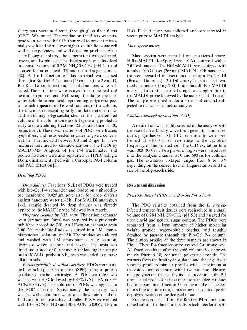
Direct PDO structural analysis of fruit-derived sam-ples was accomplished using drop dialysis of Bio-Gel P-4puriWed samples. The samples were analyzed byMALDI-FTMS and MALDI-TOF MS. Fig. 5 shows theMALDI-FT mass spectrum of PDOs from healthy-inoc-ulated tissue obtained from a B. cinerea-infected tomato.
Fig. 3. (A) HPLC separation of PDOs prepared by an acid digestion of deesteriWed polygalacturonic acid. (B) Negative MALDI-FTMS spectrum ofPDOs prepared by an acid digestion of deesteriWed polygalacturonic acid.

76 Determination of pathogen-related enzyme action / H.J. An et al. / Anal. Biochem. 338 (2005) 71–82
Several PDO series were observed with signal separationof 176 Da, corresponding to the GalUA residue. The fol-lowing series of peaks were identiWed: series A (Wlled cir-cles) with m/z 329.03, 505.06, 681.09, 857.12, 1033.15, and1209.18; series B (open circles) with m/z 351.05, 527.08,703.11, 879.14, 1055.17, 1253.18 (an H+ replaced by Na+),and 1451.20 (2H+ replaced by 2Na+); series C (opensquares) with m/z 383.06, 559.09, 735.12, 911.15, and1087.18; and series D (Wlled squares) with m/z 569.08,745.12, 921.15, 1097.17, and 1273.21. All major signalscorrespond to a deprotonated parent ion ([M ¡ H]¡).The GalUA readily forms salts with sodium as the num-ber of GalUA in an oligomer is increased. Although the
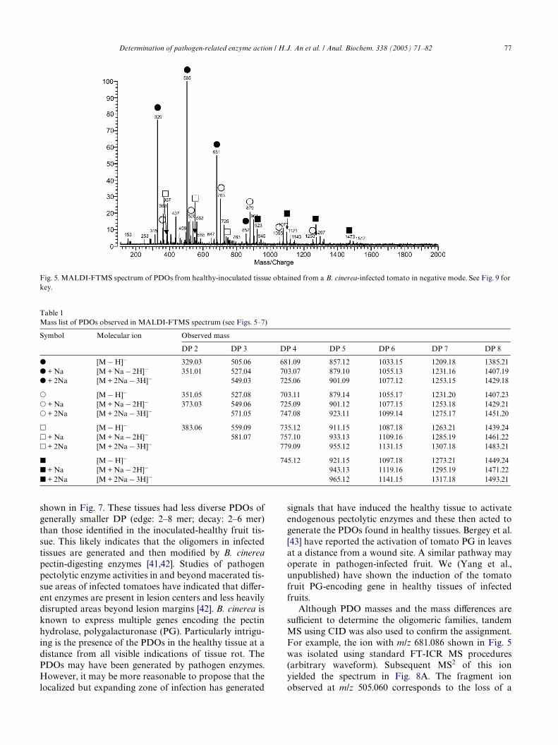
PDOs were not doped with Na, partial sodium adductformation was observed. PDOs, including Na-exchangedpeaks, are summarized in Table 1. The DP range of thesePDOs is 2–8. The DP was analyzed more eYciently withMALDI-TOF as metastable decomposition sometimesaVected MALDI-FT mass spectra. Fig. 6 shows theMALDI-TOF-MS spectrum of GalUA oligomers fromhealthy-inoculated fruit tissue with increasing DP (2–16mer). Several series of PDO peaks were observed. Thelabeled symbols correspond to the same series of PDOsas those deduced from the FTMS spectrum (Fig. 5). Thenegative mode MALDI-FTMS spectra of PDOs of edgeand decay fruit tissue obtained from infected tomato are
Fig. 4. Negative MALDI-FTMS spectrum of HPLC fraction of PDOs from healthyinoculated tissue with retention time at (A) 12.23 min (fraction 3)and (B) 25.51 min (fraction 11).
Determination of pathogen-related enzyme action / H.J. An et al. / Anal. Biochem. 338 (2005) 71–82 77
shown in Fig. 7. These tissues had less diverse PDOs ofgenerally smaller DP (edge: 2–8 mer; decay: 2–6 mer)than those identiWed in the inoculated-healthy fruit tis-sue. This likely indicates that the oligomers in infectedtissues are generated and then modiWed by B. cinereapectin-digesting enzymes [41,42]. Studies of pathogenpectolytic enzyme activities in and beyond macerated tis-sue areas of infected tomatoes have indicated that diVer-ent enzymes are present in lesion centers and less heavilydisrupted areas beyond lesion margins [42]. B. cinerea isknown to express multiple genes encoding the pectinhydrolase, polygalacturonase (PG). Particularly intrigu-ing is the presence of the PDOs in the healthy tissue at adistance from all visible indications of tissue rot. ThePDOs may have been generated by pathogen enzymes.However, it may be more reasonable to propose that thelocalized but expanding zone of infection has generated
signals that have induced the healthy tissue to activateendogenous pectolytic enzymes and these then acted togenerate the PDOs found in healthy tissues. Bergey et al.[43] have reported the activation of tomato PG in leavesat a distance from a wound site. A similar pathway mayoperate in pathogen-infected fruit. We (Yang et al.,unpublished) have shown the induction of the tomatofruit PG-encoding gene in healthy tissues of infectedfruits.
Although PDO masses and the mass diVerences aresuYcient to determine the oligomeric families, tandemMS using CID was also used to conWrm the assignment.For example, the ion with m/z 681.086 shown in Fig. 5was isolated using standard FT-ICR MS procedures(arbitrary waveform). Subsequent MS2 of this ionyielded the spectrum in Fig. 8A. The fragment ionobserved at m/z 505.060 corresponds to the loss of a
Fig. 5. MALDI-FTMS spectrum of PDOs from healthy-inoculated tissue obtained from a B. cinerea-infected tomato in negative mode. See Fig. 9 forkey.
Table 1Mass list of PDOs observed in MALDI-FTMS spectrum (see Figs. 5–7)
Symbol Molecular ion Observed mass
DP 2 DP 3 DP 4 DP 5 DP 6 DP 7 DP 8
� [M ¡ H]¡ 329.03 505.06 681.09 857.12 1033.15 1209.18 1385.21� + Na [M + Na ¡ 2H]¡ 351.01 527.04 703.07 879.10 1055.13 1231.16 1407.19� + 2Na [M + 2Na ¡ 3H]¡ 549.03 725.06 901.09 1077.12 1253.15 1429.18
� [M ¡ H]¡ 351.05 527.08 703.11 879.14 1055.17 1231.20 1407.23� + Na [M + Na ¡ 2H]¡ 373.03 549.06 725.09 901.12 1077.15 1253.18 1429.21� + 2Na [M + 2Na ¡ 3H]¡ 571.05 747.08 923.11 1099.14 1275.17 1451.20
� [M ¡ H]¡ 383.06 559.09 735.12 911.15 1087.18 1263.21 1439.24� + Na [M + Na ¡ 2H]¡ 581.07 757.10 933.13 1109.16 1285.19 1461.22� + 2Na [M + 2Na ¡ 3H]¡ 779.09 955.12 1131.15 1307.18 1483.21
� [M ¡ H]¡ 745.12 921.15 1097.18 1273.21 1449.24� + Na [M + Na ¡ 2H]¡ 943.13 1119.16 1295.19 1471.22� + 2Na [M + 2Na ¡ 3H]¡ 965.12 1141.15 1317.18 1493.21
78 Determination of pathogen-related enzyme action / H.J. An et al. / Anal. Biochem. 338 (2005) 71–82
galacturonic acid residue (¡176 mass units). A furtherfragmentation of this ion in an MS3 experiment yieldedthe ions with m/z 329.032, corresponding to the loss ofanother galaturonic acid residue (Fig. 8B). Using CID,the oligomer unit and the mass of the oligomer headgroup could be identiWed.
The PDO structures deduced from the combined MSanalysis are summarized in Fig. 9. The PDO head groupof series A (Wlled circles) is still unknown. MS4 of the ionwith m/z 329.032 was performed using various collisionenergies to identify the molecular ion but no fragmenta-tion was observed even at very high energy. As a control,the PDOs prepared by acid digestion of polygalactu-ronic acid were analyzed by MALDI-MS (Fig. 3B).However, we did not observe the ion with m/z 329.032 orPDOs of the class A series (Wlled circles). As shown inFig. 2, the intense peak at 9–10 min in the HPLC fractio-nations was observed only in three tomato tissuesextracts and not in the control. We found that this peakdid not contain hexuronic acid. This suggests that thispeak may be related to the unknown head group ofseries A. Elemental composition based on accurate mass(329.032 § 0.002) and restriction to C, H, N, and O gave10 hits with only one candidate, C7H11N3O12, resemblinga carbohydrate formula. We believe that this residuemay represent a modiWed or unknown sugar which isbound to a pectin polymer backbone and is digestedduring pathogen development.
Analysis of enzyme speciWcity
Determination of the DP and structures of PDOs isimportant for understanding the structure–function rela-
Fig. 6. MALDI-TOF-MS spectrum of PDOs from healthy-inoculatedtissue obtained from a B. cinerea-infected tomato in negative mode.See Fig. 9 for key.
tionship of pectin oligomers and the speciWcities of patho-gen and plant pectolytic enzymes. Enzymatic cleavage ofpectins by bacteria or fungi has been reported in theliterature and is considered to be an early importantaspect of pathogen colonization of plant tissues [4,6].Both pectin hydrolases (e.g., PG) and pectin lyases (PL)have been reported to be important pathogen enzymesinvolved in host plant pectin polymer degradation [44].PDOs that would be produced by both of these classes ofpectolytic enzymes are found in B. cinerea-infectedtomato fruits (Fig. 9). In classes B and D (open circles andWlled squares in Figs. 4 and 5), PDOs with a 4,5-unsatu-rated nonreducing end are shown. These PDOs will begenerated by PL action on homogalacturonan polymers.The PDOs with a saturated nonreducing end in class C(open squares in Figs. 4 and 6) would have been generatedby PG action on homogalacturonans, with -endo-, ratherthan exo-, PG being more likely. In fact, PDO “synthesis”could involve PG action on a homogalacturonan sub-strate and either PL action on the PG products that arestill suYciently polymeric to be substrates for the lyase orPL action followed by PG action. The PDO structure ofthe head group in class A (Wlled circles in Figs. 4 and 6) isnot yet completely characterized. It is likely that otherenzymes are also involved in the generation of infection-related PDOs. Some structures (classes C and D) containmethylester groups, suggesting that pectin methylesterase(PME) might have acted on other residues in the back-bone to open up sites for PL or PG action. A recentreport [45] indicates that tomato fruit express genesencoding PL when they ripen. Thus, the unsaturatedPDOs that have been identiWed in the healthy tissues ofinfected fruits may also be generated by the action ofendogenous, rather than pathogen, enzymes.
The FTMS spectra of PDOs obtained from the threetomato tissues were very similar, although the edge anddecay samples had fewer and less diverse PDOs. How-ever, a distinction between these and PDOs from healthytissues was identiWed by MALDI-TOF. The DP ofPDOs from healthy tissues (» 16 mer) is greater thanthat in edge and decay samples (» 8 mer). This suggeststhat pectolytic enzyme activities, including PG, PL, andPME, are more diverse and/or active in infected areas.This can be explained by the presence of pathogenenzymes acting, perhaps in addition to fruit pectolyticenzymes, while only fruit enzymes are responsible for thePDOs in healthy tissues from infected fruits. It also mayreXect the more intense conversion of pectin polymersinto metabilizable substrate to be used by the pathogenbiomass in lesion centers. We believe that PDOs are sig-nals regulating plant responses to pathogens, so struc-tural analysis will add to our understanding of factorsaVecting the expression of plant defenses and fruit sus-ceptibility. This will include the testing of the ability ofthe diVerent classes of infected fruit PDOs to activatedefense gene expression in infected tomato fruit tissues.

Determination of pathogen-related enzyme action / H.J. An et al. / Anal. Biochem. 338 (2005) 71–82 79
Fig. 8. CID spectrum of m/z 681 obtained from PDO oligomer mixtur
(A) MS/MS (MS2, 681/505) and (B) MS/MS/MS (MS3, 681/505/329).Fig. 7. MALDI-FTMS spectrum of PDOs from (A) decay and (B) edge tissue obtained from a B. cinerea-infected tomato in negative mode. See Fig.9 for key.
e

80 Determination of pathogen-related enzyme action / H.J. An et al. / Anal. Biochem. 338 (2005) 71–82
Conclusions
Pectin-derived oligosaccharides in B. cinerea-infectedtomato tissues were isolated and characterized byMALDI-MS after minimal sample cleanup by drop dial-ysis. The highest sensitivity in MS was obtained in thenegative mode. We were able to obtain more exact infor-mation about PDO degree of polymerization and adetailed structure analysis of PDOs using two types ofmass spectrometry, MALDI-FTMS and MALDI-TOF.This combination is a rapid, speciWc, sensitive, and repro-ducible technique for the characterization of PDOs with-out time-consuming sample preparation. The PDOsobtained from three diVerent portions of tissue frominfected fruits were apparently generated through thebreakdown of homogalacturonan pectins. The PDOstructures suggest that endo-polygalacturonase, pectinlyase, methylesterase activities were involved in the pectinbackbone scission. The PDOs from edge and decay areasof infected fruits, tissues with which the gray mold patho-gen interacted directly, had fewer and less diverse PDOs
than those from the healthy tissues remote from infectionsites. We believe that the characterization of these endog-enous pectin breakdown products will add to our under-standing of the regulation of processes that determine theoutcome of plant interactions with pathogens.
Acknowledgments
Financial support by the National Institute of Healthand the National Science Foundation is gratefullyacknowledged.
References
[1] P.J.H. Daas, K. Meyer-Hansen, H.A. Schols, G.A. De Ruiter,A.G.J. Voragen, Investigation of the non-esteriWed galacturonicacid distribution in pectin with endopolygalacturonase, Carbo-hydr. Res. 318 (1999) 135–145.
[2] P.J.H. Daas, B. Boxma, A.M.C.P. Hopman, A.G.J. Voragen, H.A.Schols, NonesteriWed galacturonic acid sequence homology ofpectins, Biopolymers 58 (2001) 1–8.
Fig. 9. The four classes of PDOs deduced from FTMS analysis.

Determination of pathogen-related enzyme action / H.J. An et al. / Anal. Biochem. 338 (2005) 71–82 81
[3] E. Melotto, L.C. Greve, J.M. Labavitch, Cell-wall metabolism inripening fruit. 7. Biologically-active pectin oligomers in ripeningtomato (Lycopersicon-Esculentum mill) fruits, Plant Physiol. 106(1994) 575–581.
[4] W.M. Wanjiru, Z.S. Kang, H. Buchenauer, Importance of cell walldegrading enzymes produced by Fusarium graminearum duringinfection of wheat heads, Eur. J. Plant Pathol. 108 (2002) 803–810.
[5] M.I. Isla, R.M. Ordonez, M.I.N. Moreno, A.R. Sampietro, M.A.Vattuone, Inhibition of hydrolytic enzyme activities and plantpathogen growth by invertase inhibitors, J. Enzyme Inhib. Med.Chem. 17 (2002) 37–43.
[6] N.J. Tonukari, J.S. Scott-Craig, J.D. Walton, The Cochlioboluscarbonum SNF1 gene is required for cell wall-degrading enzymeexpression and virulence on maize, Plant Cell 12 (2000) 237–247.
[7] C.A. Ryan, Oligosaccharides as recognition signals for the expres-sion of defensive genes in plants, Biochemistry 27 (1988) 8879–8883.
[8] E. Olano-Martin, G.R. Gibson, R.A. Rastall, Comparison of thein vitro biWdogenic properties of pectins and pectic-oligosaccha-rides, J. Appl. Microbiol. 93 (2002) 505–511.
[9] B.L. Ridley, M.A. O’Neill, D.A. Mohnen, Pectins: structure, bio-synthesis, and oligogalacturonide-related signaling, Phytochemis-try 57 (2001) 929–967.
[10] R.G. Cameron, A.T. Hotchkiss, S.W. KauVman, K. Grohmann,Utilization of an evaporative light scattering high-performancesize-exclusion chromatography acid oligomers, J. Chromatogr. A1011 (2003) 227–231.
[11] T.J.M. Deconinck, A. Ciza, G.M. Sinnaeve, J.T. Laloux, P. Thon-art, High-performance anion-exchange chromatography—DADas a tool for the identiWcation and quantiWcation of oligogalactu-ronic acids in pectin depolymerisation, Carbohydr. Res. 329(2000) 907–911.
[12] T. Ishii, J. Ichita, H. Matsue, H. Ono, I. Maeda, Fluorescent label-ing of pectic oligosaccharides with 2-aminobenzamide andenzyme assay for pectin, Carbohydr. Res. 337 (2002) 1023–1032.
[13] A.J. Mort, E.M.W. Chen, Separation of 8-aminonaphthalene-1,3,6-trisulfonate (ANTS)-labeled oligomers containing galact-uronic acid by capillary electrophoresis: application to determin-ing the substrate speciWcity of endopolygalacturonases,Electrophoresis 17 (1996) 379–383.
[14] C. Tokoh, K. Takabe, J. Sugiyama, M. Fujita, CP/MAS C-13NMR and electron diVraction study of bacterial cellulose struc-ture aVected by cell wall polysaccharides, Cellulose 9 (2002) 351–360.
[15] T.G. Neiss, H.N. Cheng, P.J.H. Daas, H.A. Schols, Compositionalheterogeneity in pectic polysaccharides: NMR studies and statisti-cal analysis, Macromol. Symp. 140 (1999) 165–178.
[16] H. Grasdalen, A.K. Andersen, B. Larsen, NMR spectroscopystudies of the action pattern of tomato pectinesterase: generationof block structure in pectin by a multiple-attack mechanism, Car-bohydr. Res. 289 (1996) 105.
[17] Z.K. Mukhiddinov, D.K. Khalikov, E.E. Grigoreva, V.P. Panov,Structure of pectin homogalacturonane, Khimiya PrirodnykhSoedinenii (1993) 91–96.
[18] C. Rosenbohm, I. Lundt, T.M.I.E. Christensen, N.W.G. Young,Chemically methylated and reduced pectins: preparation, charac-terisation by H-1 NMR spectroscopy, enzymatic degradation, andgelling properties, Carbohydr. Res. 338 (2003) 637–649.
[19] R.E. Aries, C.S. Gutteridge, W.A. Laurie, J.J. Boon, G.B. Eijkel, Apyrolysis mass-spectrometry investigation of pectin methylation,Anal. Chem. 60 (1988) 1498–1502.
[20] A. Jacobs, O. Dahlman, Enhancement of the quality of MALDImass spectra of highly acidic oligosaccharides by using a naWon-coated probe, Anal. Chem. 73 (2001) 405–410.
[21] H.C.M. Kester, J.A.E. Benen, J. Visser, M.E. Warren, R. Orlando,C. Bergmann, D. Magaud, D. Anker, A. Doutheau, Tandem massspectrometric analysis of Aspergillus niger pectin methylesterase:
mode of action on fully methyl-esteriWed oligogalacturonates, Bio-chem. J. 346 (2000) 469–474.
[22] R. Korner, G. Limberg, J.D. Mikkelsen, P. RoepstorV, Character-ization of enzymatic pectin digests by matrix-assisted laser desorp-tion/ionization mass spectrometry, J. Mass Spectrom. 33 (1998)836–842.
[23] A. Nakamura, H. Furuta, H. Maeda, T. Takao, Y. Nagamatsu,Analysis of the molecular construction of xylogalacturonan iso-lated from soluble soybean polysaccharides, Biosci. Biotechnol.Biochem. 66 (2002) 1155–1158.
[24] B.J. Savary, A. Nunez, Gas chromatography-mass spectrometrymethod for determining the methanol and acetic acid contents ofpectin using headspace solid-phase microextraction and stable iso-tope dilution, J. Chromatogr. A 1017 (2003) 151–159.
[25] G.J.W.M. van Alebeek, K. van Scherpenzeel, G. Beldman, H.A.Schols, A.G.J. Voragen, Partially esteriWed oligogalacturonides arethe preferred substrates for pectin methylesterase of Aspergillusniger, Biochem. J. 372 (2003) 211–218.
[26] T. Stoll, A. Schieber, R. Carle, Quantitative determination of satu-rated oligogalacturonic acids in enzymatic digests of polygalactu-ronic acid, pectin and carrot pomace by on-line LC-ESI-MS,Anal. Bioanal. Chem. 377 (2003) 655–659.
[27] R. Korner, G. Limberg, T.M.I.E. Christensen, J.D. Mikkelsen, P.RoepstorV, Sequencing of partially methyl-esteriWed oligogalac-turonates by tandem mass spectrometry and its use to determinepectinase speciWcities, Anal. Chem. 71 (1999) 1421–1427.
[28] M.H. Clausen, R. Madsen, Synthesis of hexasaccharide fragmentsof pectin, Chem. Eur. J. 9 (2003) 3821–3832.
[29] B. Quemener, C. Desire, M. Lahaye, L. Debrauwer, L. Negroni,Structural characterisation by both positive- and negative-ion elec-trospray mass spectrometry of partially methyl-esteriWed oligoga-lacturonides puriWed by semi-preparative high-performance anion-exchange chromatography, Eur. J. Mass. Spectrom. 9 (2003) 45–60.
[30] G. Limberg, R. Korner, H.C. Buchholt, T.M.I.E. Christensen, P.RoepstorV, J.D. Mikkelsen, Analysis of pectin structure part 1—analysis of diVerent de-esteriWcation mechanisms for pectin byenzymatic Wngerprinting using endopectin lyase and endopolygal-acturonase II from A-niger, Carbohydr. Res. 327 (2000) 293–307.
[31] G. Limberg, R. Korner, H.C. Buchholt, T.M.I.E. Christensen, P.RoepstorV, J.D. Mikkelsen, Analysis of pectin structure part 3—quantiWcation of the amount of galacturonic acid residues inblocksequences in pectin homogalacturonan by enzymatic Wnger-printing with exo- and endo-polygalacturonase II from Aspergillusniger, Carbohydr. Res. 327 (2000) 321–332.
[32] G.J.W.M. van Alebeek, H.A. Schols, A.G.J. Voragen, Amidationof methyl-esteriWed oligogalacturonides: examination of the reac-tion products using MALDI-TOF MS, Carbohydr. Polym. 46(2001) 311–321.
[33] G.J.W.M. van Alebeek, O. Zabotina, G. Beldman, H.A. Schols,A.G.J. Voragen, EsteriWcation and glycosydation of oligogalactu-ronides: examination of the reaction products using MALDI-TOF MS and HPAEC, Carbohydr. Polym. 43 (2000) 39–46.
[34] E. Vierhuis, W.S. York, V.S.K. Kolli, J.P. Vincken, H.A. Schols,G.J.W.M. Van Alebeek, A.G.J. Voragen, Structural analyses of twoarabinose containing oligosaccharides derived from olive fruit xylo-glucan: XXSG and XLSG, Carbohydr. Res. 332 (2001) 285–297.
[35] L.E.M. Fernandez, N. Obel, H.V. Scheller, P. RoepstorV, Charac-terization of plant oligosaccharides by matrix-assisted laserdesorption/ionization and electrospray mass spectrometry, J.Mass Spectrom. 38 (2003) 427–437.
[36] P.J.H. Daas, P.W. Arisz, H.A. Schols, G.A. De Ruiter, A.G.J. Vor-agen, Analysis of partially methyl-esteriWed galacturonic acid olig-omers by high-performance anion-exchange chromatography andmatrix-assisted laser desorption/ionization time-of-Xight massspectrometry, Anal. Biochem. 257 (1998) 195–202.
[37] N. Blumenkrantz, G. Asboe-Hansen, New method for quantitativedetermination of uronic acids, Anal. Biochem. 54 (1973) 484–489.

82 Determination of pathogen-related enzyme action / H.J. An et al. / Anal. Biochem. 338 (2005) 71–82
[38] Z. Dische, Color reactions of carbohydrates, in: R.L. Whistler,M.L. Wolfram (Eds.), Methods in Carbohydrate Chemistry, Aca-demic Press, New York, NY, 1962, pp. 475–514.
[39] B.H. Wang, K. Biemann, Matrix-assisted laser desorption/ioniza-tion time-of-Xight mass-spectrometry of chemically-modiWed oli-gonucleotides, Anal. Chem. 66 (1994) 1918–1924.
[40] A.V. Kuhn, H.H. Ruttinger, R.H.H. Neubert, K. Raith, IdentiW-
cation of hyaluronic acid oligosaccharides by direct couplingof capillary electrophoresis with electrospray ion trap massspectrometry, Rapid Commun. Mass Spectrom. 17 (2003) 576–582.
[41] A. ten Have, W.O. Breuil, J.P. Wubben, J. Visser, J.A.L. van Kan,Botrytis cinerea endopolygalacturonase genes are diVerentially
expressed in various plant tissues, Fungal Genet. Biol. 33 (2001)97–105.
[42] O. Kamoen, G. van der Cruyysen, Two secretions of Botrytis cine-rea important for pathogenesis, Meded. Fac. Landbouwwet.Rijksuniv. Genetics 61 (1996) 251–260.
[43] D.R. Bergey, M. Orozco-Cardenas, D.S. de Moura, C.A. Ryan, Awound- and systemin-inducible polygalacturonase in tomatoleaves, Proc. Natl. Acad. Sci. USA 96 (1999) 1756–1760.
[44] A. Collmer, N.T. Keen, The role of pectic enzymes in plant patho-genesis, Annu. Rev. Phytopathol. 24 (1986) 383–409.
[45] M.C. Saladie, S. Vendrell, M. Dominguez-Puijaner, E., 7th Inter-national Congress of Plant Molecular Biology (http://ispmb2003.com) Abstract (2003).