DETERMINATION OF AFLATOXINS iN TRADITIONAL …eprints.usm.my/31135/1/OSISI_IKENNA_ALPHONSE.pdf ·...
36
DETERMINATION OF AFLATOXINS iN TRADITIONAL MEDICINE by .' -: ,t.,". ! " . .:. ' 1 OSISI .·\LPHO?\SE submitted in paItial fulfillment of the requirements for the degree of \laster of Science April 2003
Transcript of DETERMINATION OF AFLATOXINS iN TRADITIONAL …eprints.usm.my/31135/1/OSISI_IKENNA_ALPHONSE.pdf ·...

DETERMINATION OF AFLATOXINS iN
TRADITIONAL MEDICINE PREPARAT'I()I~S
by
.' -: ,t.,".
! " . .:. .~. ' 1
OSISI [KE~\:A .·\LPHO?\SE
Th~sis submitted in paItial fulfillment
of the requirements for the degree of
\laster of Science
April 2003

q,\ I •
Saya isytiharkan bahawa kandungan yang dibentangkan di da/am tesis ini adalah hasil kerja saya sendiri dan telah dija/ankan di Universiti Sains Malaysia kecuali dimaklumkan
sebaliknya. Tesis ini juga tidak pemah diserahkan untuk ijazah yang lain sebelum ini.
Tandatangan Calon
Nama Calon:
Tandatangan Penyelia/Oekan
Cop Jabatan:
PUSAT PENG~HAN S~INS KIMI~ UNIVERSITI SAINS MAL'" 'iSlA 11800 USM PULAU PINANG.

ACKNO'VLEDGE~IENT
I would like to express my heartfelt gratitude to my parents Mr. and Mrs. Okamigbo
Osisi, for their financial support and encouragement throughout the course of my
studies. My sincere appreciation also goes to my supervisor Prof Madya Dr.
Bahruddin Saad who made it possible for me to settle down quickly to life in
Malaysia, and for his valuable guidance, and direction in the course of my research
and for his unwavering kindness. He would always be remembered. My thanks also
goes to Dr Norziah Hanni, for providing me with hands-on experience at the beginning
of this study, Norhayati Bte Ali. and Dr. Hiroshi Akiyama for pr?~!iding resource
materials at the start of the research. Special mention here must be made of Md Fazlul
Bari, for being a friend and counsellor from the first few days of arrival to the present
day, and Helal Uddin for his useful contributions and advice.
I would also like to sincerely thank i\,[r. Ong Chin Hwie, Mr. Vee Chin Leng, Mr
Aw Yeong, and iv'1r. Chow Cheng Por; some of the simplest yet most extraordinary
men I have ever met. Their kindness and willin!,1J1ess to. help even at short notice
would not be forgotten. My study would be incomplete without mentioning the
contributions of my class mates, Mr. Chang Seng Teong, Ms. Ng Lee Ping, Ms. Pang
Ming Meng and Ms. Tan Sin Ee. Their companionship and sincere friendship went a
long way to making me feel at home and I am .truly grateful. My gratitude also goes to
Ms. Teh Chin Loi and Ms. Lee Ee Lin for their kindness and true mendship.
Finally I would like to thank my siblings for their prayers and support
throughout my separation from them. My thanks also to all my mends and
acquaintances her~ in Malaysia, and all member of the FIA research group.
11

CONTENTS
Acknowledgement
Contents
List of Tables
List o~ Figures
Abstrak
Abstract
Chapter I: General Introduction
1.1 Mycotoxins
1.2 Aflatoxins
1.2.1 Sources and Occun'ences
1.2.2 Chemical Properties
1.2.3 Mode of Action
1.2.4 Economic and Regulatory Aspects
1.2.5 Incidences of Aflatoxin Poisoning
1.3 Analytical Metho}ls
1.3.1 Immunacbemical Methods
1.3.2 Thin LayerChromatography (TLC)
1.3.3 G~s C.hroffiato~hy (GC) ~
1.3.4 High Performance Liquid Chromatography (HPLC)
11l
Page
n
iii
VI
vii
3
3
4
8
8
10
13
13
14
16
17
-.. ~,..

1.4 Detection Methods 17
1.4.1 Ultravio let (UV) Absorption 18
1.4.2 Fluorescence Detection 18
1.4.3 Electrochemical Detection 18
l. 5 Derivatization 19
1.5. 1 Pre-column Derivatization 19
1.5.2 Post-column Derivatization 19
1.6 Isolation and Clean-up Methods 20
(i) Liquid-Liquid Extraction 20
(ii) Solid Phase Extraction (SPE) 21
1. 7 Objectives of the study 22
Chapter 2: Experimental
2.1 Materials and Methods 24
2.2 Chemicals and Aflatoxin Standards 24
2.2.1 Preparation and storage of aflatoxin standard and sample solutions 25
2.2.2 HPLC 29
2.2.3 Other Apparatus 30
2.2.4 Extraction, Clean-up and Derivatization 30
2.3 Akiyama Method 30
2.4 Modified Akiyama Method 31
2.5 Derivatization Procedure 33
Chapter 3: Results and Discussion
3.1 Modifica~on of the Akiyama Method
3.2 Linearity and Detection Limits
3.3 Repeatability
3.4 Mobile Phase Selection
IV
34
35
36
37

3.5 Recovery studies
3.6 Clean-up efficiency for single extract
3.7 Clean-up efficiency for multiple extracts
3.8 Analysis of Traditional Medicines
3.9 Conclusion
References
Appendices
Appendix A
Appendix B
v
41
43
45
47
53
54

List of Tables
Table 1.1
Table 1.2
Table 2. I
Table 2.2
Table 2.3
Table 2.4
Table 3.1
Table 3.2
Table 3.3
Conditions for production and the possible involvement of important mycotoxins in human disease
Chemical and physical properties of some aflatoxins and their metabolites
Infonnation on Thai traditional medicines studied
Infonnation on Malaysian traditional medicines studied
Infonnation on Indonesian traditional medicines studied
Infonnation on Nigerian traditional medicines studied
Relative Standard Deviation for Peak Area and Retention Time of Chromatograms for Aflatoxin Spiked Samples ·Over a 5 Day Period
Percent Recoveries of Aflatoxins Spiked to Traditional Medicine Extracts Using 2 mL of Extracts
Percent Recoveries of .-\flatoxins Spiked to Traditional ~fedicine Extracts Using 1 mL of Extracts
VI
2
6
26
27
28
29
37
42
42

List of Figures
Fig. 1.1 The structure of some aflatoxins and their metabolites 5
Fig. 1.2 Metabolic pathways of AFBI in the liver 7
Fig. 2.1 Schematic procedures for analysis of AF modified from the Akiyama Method 32
Fig. 3.1 Typical calibration graph for aflatoxins BI, G), B2, G2 using Varian ResElut column (150 x 4.60 mm); CH3CN-MeOH-H20 (10:20:70) 36
Fig. 3.2 Chromatograms of samples spiked with 10 ppb BI/Gl, and 20 ppb ofB2 and G2, using mobile phases of acetonitrile-methanol-water in the ratio (i) 8:27:55; (ii) 17: 17:70; (iii) 10:20:70 39
Fig: 3.3 HPLC chromato,gram sho\\ing separation of aflatoxin standards under optimum chromatographic conditions 40
Fig. 3.4 HPLC chromatogram of underivatised sample extracts (i) before passing through IMC (ii) after passing through the IMC 44
Fig. 3.5 HPLC chromatogram of (i) the first Iml eluate (ii) second I ml eluate and (iii) third I ml eluate passed through the same IMC 46
Fig. 3.6 HPLC chromatograms of (i) underivatised sample extract spiked with aflatoxins, and (ii) derivatised sample extract spiked with aflatoxins 48
Fig. 3.7 HPLC chromatograms of (i) underivatised and (ii) derivatised Thai sample extracts 49
Fi~. 3.8 HPLC chromatograms of (i) underivatised and (ii) derivatised Malaysian sample extract 50
Fig. 3.9 HPLC chromatograms of (i) underivatised and (ii) derivatised Indonesian sample extract 51
Fig. 3.10 HPLC chromatograms of (i) underivatised and (ii) derivatised Nigerian sample extract 52
Vll

Abstrak
Mikotoksin telah dikenalpasti sebagai satu masalah besar dahim makanan. Terdapat
lima jenis mikotoksin yang penting iaitu aflatoksin (AF), okratoksin (OTA),
deoksinivalenol (DON), danlatau nivalenol (NIV), zearelenon (ZEA) dan fumonisin
(FM). Campuran aflatoksin yang wujud secara semulajadi didapati karsinogenik
terhadap manusia. OT A dan FA mungkin adalah karsinogenik kepada manusia. Antara
aflatoksin yang menunjukkan tahap ketoksikan yang berbeza, aflatoksin BI, GI, B2, dan
G2 adalah paling banyak dikaji dengan aflatoksin BI dikenali sebagai paling toksik.
Beberapa kajian mengenai pencemaran aflatoksin dalam makanan semlllajadi telah
dijalankan dan didapati aflatoksin menyebabkan masalah yang ketara di seluruh dunia,
terutamanya di kawasan tropika seperti Asia Tenggara dan Afrika. TlIjllan kajian ini
adalall untuk menentukan tahap aflatoksin B1, GI, B2 , dan G2 dalam ubat tradisionaI dan
tiga negara Asia Tenggara dan satu negara Afrika. Kajian ini merupakan kajian Iengkap
pertama yang dijalankan ke atas pencemaran semulajadi aflatoksin sediaan ubat
tradisional. Tahap aflatoksin B 1, GJ, B2, dan G2 dalam sediaan ubat tradisional telah
ditentukan dengan menggunakan kaedah kromatografi cecair prestasi tinggi (HPLC)
fa sa terbalik. Terlebih dahulu, sampel diekstrak dengan asetonitril-air (9: 1) dan dibersih
dalam turns ISOLUTE™ MUL TIMODE (IMC) multifungsi pengektrakan fasa pepejal.
Sampel terbitkan pra-turus dengan menggunakan asid tl-ifluoroasetik (TF A) untuk
meningkatkan amatan pendaflour BI dan G1 sebelum pemisahan HPLC menggunakan
fasa bergerak asetonitril-metanol-air (10:20:70). Ciri utama pemisahan kromatografih
ini ialah kewujudan puncak kuat berpotensi mengganggu berdekatan dengan puncak G1
yang juga dikesall oleh ahli-ahli kajian dalam sampel-sampel tumbuhan lain.
Walaubagaimanapun, kehadiranlketidakhadiran afiatoksin bGIeh di-sahkan aengan
Vlll

membandingkan kromatogram ekstrak-ekstrak yang tidak terbitkan dan terbitkan.
Penggunaan turus IMC adalah menarik memandangkan ia melibatkan satu langkah yang
mana bahan. asing diperangkan sedangkan aflatoksin malalui turus tanpa ditahan.
Sampel yang dianalisis tennasuk jamu dan makjun. Alfatoxin tidak dikesan pada 35
sampel yang dianalisis. Rolehan semula untuk BI, GI, B2 dan G2 adalah 91.4, 92.9,
102.1 dan 90.8% masing-masing apabila 10 ppb (81 dan GI) dan 20 ppb (B2 dan G2)
pakukan kepada 3 jenis ubat-ubat tradisiOllal yang berlainan.
IX

Abstract
Mycotoxins have been recognized as a substantial problem in foods. There are five
important mycotoxins namely aflatoxins (AF), ochratoxins (OTA), deoxynivalenol
(DON) and/or nivalenol (NIV), zearelenone (ZEA), and fumonisins (FM). Naturally
occuning aflatoxins have been found to be human carcinogens. OT A and FM are
possibly carcinogenic to humans. Of the several known aflatoxins exhibiting different
levels of toxicity, aflatoxins 8 1• GI, 81, and G2 are the most studied, with aflatoxin 81
being the most toxic. Studies on aflatoxin contamination of natural foods have shown
that aflatoxins constitute a significant problem world wide, especially in tropical regions
of the world such as Southeast Asia and Africa. The present study is aimed at
determining levels of aflatoxins 8 1, 82, GI, and G2 in traditional medicine preparations
from 3 Southeast Asian countries and I African country, and is the first known
comprehensive study conducted on the natural contamination of aflatoxins in traditional
medicine preparations. Levels of aflatoxins 8 1, B1, GI, and G2 in the traditional
medicine preparations were determined using reverse-phase high performance liquid
chromatography (HPLC). The samples were first extracted with acetonitrile-water (9: I)
and cleaned-up on a multifunctional solid phase extraction ISOLUTE 1M MUL TIM ODE
column (IMC). The samples were pre-column derivatised using trifluoroacetic acid
(TF A) to enhance the fluorescent intensities ofBI and GI before their HPLC separation
using a mobile phase of acetonitrile-methanol-water (10:20:70). The main feature of the
. chromatographic separation of these extracts is the presence of an intense potentially
interfering peak near the G 1 peak that were also found by other researchers on other
plant samples. However, confirmation of the presence/absence of the atlat(}xms callid
x

be made by comparing chromatograms of the underivatised and derivatised extracts.
The use of the IMe column is attractive as it is a single step SPE where endogeneous
components are trapped, while the aflatoxins were unretained and passed through the
column. Samples analysed include the popular after-birth medications "jamu", and
"makjun". Aflato~s were not detected in any of the 35 samples that were analysed.
Recoveries for B I, GI, B2, and G2 of 91.4, 92.9, 102.1 and 90.8 % respectively were
obtained when to ppb (BI and GI) and 20 ppb (82 and G2) were spiked to 3 different
kinds of traditional medicines.
Xl

CHAPTER 1
INTRODUCTION
1.1 ~Iycotoxins
Mycotoxins are derived from the secondary metabolites of some filamentous fungi or
molds of the A ,\pergil/us , Penicillium, and FusariulJl genera. Under suitable temperature
and humidity conditions, they may develop on various foods and feeds, causing serious
health risks for humans and animals. Although currently more than 300 mycotoxins are
" knowTI, attention is focused .. mainly on those that have been proven to be ca~ci~?genic
and/or toxic. These include a metabolite of Aspergil1usjlavus and Aspergillus parasilicllS,
aflatoxin B! CAFB!), the most potent hepatocarcinogenic substance known, which has also
been recently proven to be genotoxic~ ochratoxin A, produced by Penicillium verrucasum
and Aspergillus ochracells, which is kno\\,TI to be carcinogenic in rodents and nephrotoxic
in humans; zearalenone, produced by various species of Fusarium, in particular F.
graminearum and F clilmarum, which has an estrogenous action and is significantly toxic
to the reproductive system of animals; the tricothecenes, a group of numerous metabolites
produced by Fusuriulll, Slac/z:vubo/ris, and Cephalosporium species, which cause mainly - . . dermotoxicity, immunotoxicity, and gastro-intestinal disturbances; and the fumonisins,
produced mainly by Fusarium monilijarme, which may induce as well as hepatotoxicity in
rats CPo~land & Wood,. 1987). More information on some of these mycotoxins are shown in
Table 1.1.
The incidence and extent of mycotoxin contamination are strictly related to
geographic and seasonal factors as well as cultivation, harvesting, stocking, and transport
I

been estimated that 25% of foodstuffs currently produced in the world are contaminated by
mycotoxins, with serious repercussions on human health and significant financial loss for
the various producing sectors. The real impact of these toxins on human health has only
recently been recognized. As a consequence, maximum tolerable limits for all potential
risky foodstuffs and consistent official control strategies have only rarely been defined on a
national level in several countries. (Brera el al., 1998).
Table 1.1 Conditions for production and the possible involvement of important t . . h d' (FAO 1997) myco ox inS In uman lsease ,
Mycotoxin Commodities Fungal Conditions I Effect Of Ingestion Damaged Source(s) for Toxin.
Production Deoxynivalenol Wheat, maIze, Fusarium High Human toxicosis. Toxic to
and barley, Kaminearum humidity animals, especiaUypigs. Nivalenol rice, Fusarium -20-25°C
rye, crookwellense for growth. oats, -15°C for walnuts. toxin
production
Zearalenone Maize. wheat. Fusari 11111 High Identitled by International Agency gramillearllm humidity for Research on Cancer (IARC) as Fllsarium -20-25°C a possible human carcinogen. crookll'ellense for growth. Affects reproductive system In
-l5°C for female pigs. toxin production
Ochratoxin A Barley, wheat, Aspergillus Suspected by IARC as human coffee beans. ochracells Relatively carcinogen. Carcinogenic in ham. dairy Penicillium non-specific laboratory animals and pIgS. products and ' l'e,.r/lCOSIlI11 Balkan nepropathy, renal tumors. many' other commodities.
Fumonisin BI Maize. Fusarium. Heat and Suspected by IARC as human mOlli/ijorme humidity carcinogen. Toxic to pIgS and plus several similar to poultry. Cause of equme less common that for AF eucoencephalomalacia (HEM), a species production fatal disease of horses.
Aflatoxins Maize, Aspergillus Warmth and Aflatoxin BI, and naturally .B\, B2, G I & G2 pe"!1uts, - f1avlI.'> moisture, occurring mixtures of atlatoxins,
edible nuts, High rainfall, identified as potent human cottonseed, Aspergillus High carcinogens, by !ARC Liver cereals. spices, parasiticlIs humidity, cancer, acute hepatitis, reye Fruits, Feeds, High syndrome. Adverse effect in Animal temperature_ various animals, especially pJ"Dducts. (25-30oq chickens.
2

1.2 Aflatoxins
1.2.1 Sources and Occurrence
Aflatoxins are highly toxic metabolites (mycotoxins) produced by the fungi Aspergillus
jlavllS and A,\pergillus parasilicus. They can be found in a wide range of food items and are
potentially hazardous to humans and animals (Stroka el al., 2000). The occurrence and
magnitude of aflatoxin contamination varies with geographical and seasonal factors, and
also with the conditions under which a crop is grown, harvested, and stored. Crops in
tropical and subtropical areas are more subject to contamination than those in temperate
regions, since optimal conditions for toxin formation are prevalent in areas with high
humidity and temperature. Toxin-producing fungi can infect growing crops as a
consequence of insect or other damage, and may produce toxins prior to harvest, or during
harvesting and storage.
Aflatoxins have been found in various agricultural commodities, such as peanuts,
pistachios, figs; grains, such as corn, rice and wheat; and spices such as paprika (Stroka, el
al., 2000). Other food products in which aflatoxins have been found include beans, coffee,
eggs, and beer (Jaimez et aI., 2000). Four compounds produced by these moulds are
aflatoxins BI (AFBI), B2 (AFB:), GI (AFGd, G2 (AFG2), (Fig.I.l). Together, these four
compounds occur as the major highly active constituents of the Aspergillus jlavus species.
Although 17 aflatoxins have been isolated, the term aflatoxin usually refers to these four
compounds. 'B' and 'G' refer to the blue and green fluorescent colours produced by these
compounds under ultraviolet light illumination on thin layer chromatography plates, while
the subscript numbers 1 and 2 indicate major and minor compounds, respectively. Residues
of AFB I can occur in animal products, including egg, milk and milk products. AFM! is also
found in human mIlk as a function of the dietary exposure of the mother to AFB!. High
3

levels of atlatoxin contamination' have been found especially in tropical and sub-tropical
countries, particularly in Africa and Southeast Asia regions (WHO, 1999).
Aflatoxin BI is the most carcinogenic and most commonly occurring variety
(Kussak et a/., 1995a). It is metabolized in the liver to 2,3-epoxide, which is responsible for
alk),lation of cellular nucleic acid (DNA) and subsequent carcinogenic and mutagenic
activity. Under certain conditions, the LDsll (lethal dose to cause 50% of mortality after
administration in animals) for aflatoxin BI was found to be 28 llg in day-old White Peking
ducklings (UNEP/WHO, 1979). The LDso for atlatoxin B2 is 85 /lg and for atlatoxins G I
and G2 they are estimated to be 60-90 /lg (UNEPfWHO, 1979).
1.2.2 Chemical Properties
The structures of a number of aflatoxins and of aflatoxin BI related metabolites are
illustrated in Fig, 1.2. The structure of atlatoxins BI and GI were determined by Asao et ai.,
(1965) and that of B2 by Chang el al. (1963), Atlatoxins B2 and Gz are dihydro derivatives
of the parent compounds, while atlatoxins MI and M2 are the hydroxylated metabolites of
B I and B2, respectively. Properties of some naturally occurring aflatoxins and metabolites
are summarized in Table 1.2.
Atlatoxins are freely soluble in moderately polar solvents (e.g., chloroform and'
methan?!) and especially in dimethylsulfoxide (the solvent usually used as a medium in the
administration of atlatoxins to experimental animals). As pure substances, the atlatoxins are
very stable at high temperatures, when heated in air. However, they are relatively unstable,
\"Hen exposed to Jrght, and particularly to UV radiation, and air on a TLC plate and
especially when dissolved in highly polar solvents. Chloroform and benzene solutions are
stable for years if kept in the dark and cold. Atlatoxins are converted into one or more
4

stable for years if kept in the ctark and cold Aflatoxins are converted into one or more
biologically active metabolites, such as aflatoxicol (AFL) and aflatoxin (AFPd in the liver
(Fig.I.1 ).
o 0
Aflatoxin B1 Aflatoxin 82
o
Aflatoxin G1 Aflatoxin G2
o o
Aflatoxin M1 Aflatoxin M2
o OH
Aflatoxin P1 Aflatoxicot', ..
Fig. 1.1 The structure of some aflatoxins and their metabolites
5
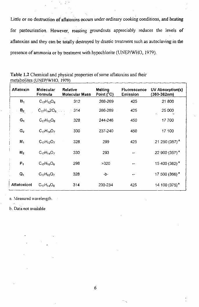
Little or no destruction ofaflato~ins occurs under ordinary cooki~g conditions, and heating·
for pasteurization. However, roasting groundnuts appreciably reduces the levels of
aflatoxins and they can be totally destroyed by drastic treatment such as autocIaving in the
presence of ammonia or by treatment with hypochlorite (UNEPIWHO, 1979).
Table 1.2 Chemical and physical properties of some aflatoxins and their metabolites (UNEPIWHO, 1979)
Aflatoxin Molecular Relative Melting Fluorescence UV Absorption(E) Formula Molecular Mass Point (0C) Emission (360-362nm)
8 1 C17H120 6 312 268-269 425 21800
8 2 C17Hl~06 314 286-289 425 25000
G1 C17H120 6 328 244-246 450 17700
G2 C17H140 7 330 237-240 450 17100
M1 C17H120 7 328 299 425 21 250 (357) a
M2 C17H140 7 330 293 22 900 (357) a
P1 C17H1OO6 298 >320 15 400 (362) a
01 C17H120 7 328 -b- 17 500 (366) a
I
! Aflatoxicol C17H140 6 314 230-234 425 14100 (375) a
a. \leasured wavelength.
b. Data not available
6

o 0
AFLATOXJCOl Ml
o 0
AFLftJ 0 XIN M 1
o 0 OAn Hr. HOo:xJ OCHs
AflATOXIN 62 . a
o 0
-------.... AfLATOXICOl
o 0
*"1 ~ OCH . 3
AfLATOXHIi B, \
o 0
~nA.A
I o I I
'0 0 OCH3
AFLATOXIN 8 2
13 EPOXIDE 1
ft.FLA TOXIN PI
Fig 1.2 Metabolic pathways of AFBI in the liver (UNEPIWHO, 1979)
7
AFLA TOXICOL HI
o 0
OH
.A.FL,a, TOXIN Q 1

1.2.3. i\tlode of Action
Aflatoxin BI is a liver carcinogen in at least 8 species including nonhuman primates. It acts
by causing chromosomal aberrations and DNA breakage in plant and animal cells. After·
microsomal activation, gene mutations in several bacterial test systems have been reported.
In high doses, it may be teratogenic (causing embryo malformation). Dose-response
relationships have been established in studies on rats and rainbow trout, with a 10% tumour
incidence estimated to occur at feed levels of AFBI of I /lglkg, and O. I/lglkg, respectively
(UNEPIWHO, 1979). Evidence suggests that aflatoxins are one hundred times more likely
to induce cancer than polychlorinated biphenyls (PCBs), and also inhibit the body's
immune system and reduce the effectiveness of vaccines (FAO, 1997).
Liver cancer is more common in some regions of Africa and Southeastern Asia
than in other parts of the world. When local epidemiological information is considered
together with experimental animal data, it appears that increased exposure to aflatoxins
may increase the risk of primary liver cancer. (UNEP/WHO, 1979).
1.2.4. Economic and Regulatory Aspects
International trade in agricultural commodities such as wheat, rice, barley, corn, sorghum, '
soybeans, groundnuts and oilseeds amounts to hundreds of millions of tonnes each y~ar
(F AO, 1988). Many of these commodities run a high risk of mycotoxin contamination .
... -Regulations ()n mycotoxins ilave been set and are strictly enforced by most importing
countries: For some- developing countries, where agricultural commodities account for as
much as 50 percent of the total national exports, the economic importance of mycotoxin
infection is considerable. There is a notable length of time between the purchase of the
8

agricultural commodity at the viliage market of the exporting country and its arrival at the·
distribution centre of the importing country. For these reasons, there is considerable
opportunity for mould and mycotoxin contamination of agricultural commodities to take
place throughout the food system - from production to distribution and transport - and this
may lead to economic losses (Bhat, 1988). The direct cost of aflatoxin contamination on
maize and groundnuts has recently been estimated to be greater than $A470 million per
annum, for South East Asian countries, including Australia. Maize was the more important
commodity amounting to 66% of the total amount. Indonesia was the worst affected,
bearing 48% of the estimated loss. The losses resulting form spoilage accounted for 24%
($A108 million) per year. (Bhat & Vasanthi, 1999).
Regulations have been set in more than 70 countries in order to restrict the intake of
mycotoxins. However, the legal limits vary significantly both from country to country and
by mycotoxin type and matrix. For example, limits for aflatoxins in foodstuffs range from 0
to 50 ng/g. The frequent occurrence, particularly of aflatoxins, has already led to temporary
bans of certain "high-risk" foods imported into Europe (for example, Egyptian peanuts
and Iranian pistachios), where limits have been established at relatively low levels. As a
result of the establishment of the EU and its aim of harmonization of the internal market, ..
the European Commission (EC) drafted regulations concerning certain contaminants. The
current maximum levels for aflatoxins set by the Ee are 2 ng/g for AFBI and 4 nglg for
total aflatoxins in groundnuts, nuts, dried fruits and cereals. These are to be extended to
cover spices with -limits of 5 ng/g and 10 ng/g for aflatoxin BI and total aflatoxins,
respectively. These leyels are about five times lower than those established in the USA
(EU, 1998). In Germany, the limits are 4 ppb (w/w) for the sum of aflatoxins BI, B2, G1 and
9

G2 and 2ppb for aflatoxin B1• For human dietary products, such as infant nutriment, there
are stronger legal limits: 0.05 ppb for aflatoxin BI and the sum of all aflatoxins. Indonesia
at present proposes a maximum ·level of 50 nglg for total aflatoxin in feedstuff. The
Philippines and Thailand have an authorized action level of 20 nglg, while Malaysia has
regulated a maximum level of35 nglg for total AF in foods (Ali el al., 1998).
1.2.5 Incidences of Aflatoxin Poisoning
The carcinogenicity of aflatoxins have been implied in reports which suggest that some
outbreaks of sub-acute poisonings resulting from ingestion of large amounts of·aflatoxins
over a period of time, with some of the poisonings resulting in fatalities.
Malaysia ( 1988)
An outbreak of food poisoning resulting in 13 deaths in children occurred in Malaysia
during the Chinese Festival of the Nine-emperor gods in 1988. The offending food was a
Chinese noodle called 'Loh See Fun' (LSF). The source was traced to a factory where a
banned food preservative (boric acid) was added to make the LSF. In addition, high levels
of aflatoxins up to 3465 pg/g of tissue for B I , 9116 pglg of tissue for GI, and 18,521 pg/g
of tissue for MI were found in the liver of some of the patients when autopsies were
conducted:> (Chao et. ai, 1991).
10

India (1974)
In the fall of 1974, an epidemic occurred in more than 150 villages in adjacent districts of
two neighboring states in a rural area of Northwest India. The disease was characterized by
onset \'v'ith high fever, rapidly progressive jaundice and ascites. According to one report of
the outbreak, 397 persons were affected and 108 people died. One notable feature of the
epidemic was that it was heralded by the appearance of similar symptoms in the village
dogs. Analysis of food samples revealed that the disease outbreak was probably due to the
consumption of maize heavily infested with the fungus A.~pergilllls jlavus. Unreasonable
rains prior to the harvest, chronic drought conditions, poor storage facilities and ignorance
of dangers of consuming fungal contaminated food all seem to have contributed to the
outbreak. The levels of aflatoxin in food samples consumed during the outbreak ranged
between 2.5 and 15.6 llg/g. Anywhere between 2 and 6 mg of aflatoxin seems to have been·
consumed daily by the affected people for many weeks. In contrast, analysis of com
samples from the same areas the following year (1975) revealed very low levels of
aflatoxins (i.e. less than 0.1 llg!g ), and this may have explained the absence of any
reoccurrence of the outbreak in 1975 (Krishnamachari, 1975).
Kenya (1981)
Between March and June 1981,20 patients (8 women and 12 men aged 2.5 to 45 years old)
were admitted to hospital in the Machakos district of Kenya with severe jaundice. The
patients reported that they had first exhibited symptoms of abdominal discomfort, anorexia,
general malaise and low-grade fever. After about 7 days, jaundice and dark urine appeared,
11

and the patients sought admissl()n' to hospital. Interestingly, the relatives and friends of one
family told that many of the local doves had died, then the local dogs, and finally the
people had become sick. The dogs \vere known to be consuming essentially the same diet
as the local people. On admission to hospital, all patients were jaundiced, some with low
grade fever, and extremely weak, Tachycardia and oedema (of the legs and to a lesser
extent the face and trunks) were seen. Eight of the 20 patients improved with a return of
appetite, and were discharged from hospital in 20 days. However, hepatic failure developed
in the remaining 12 patients and they died between 1 and 12 days foI1owing the admission.
An extensive investigation of the outbreak was performed. Aflatoxin levels in foods were
measured and showed high levels of aflatoxin Bl and B2. For example, maize grains from
the two homes where severe and fatal illness had occurred contained 12 mg/kg and 3.2
mg,'kg of aflatoxin B l , while maize from unaffected homes had a maximum of O.5-mglkg
aflatoxin B 1. The cumulative evidence suggests that aflatoxin poisoning was the cause of
the acute liver disease in this incident (Ngindu et.aI1982).
Germanv
A case has been reported of a 45-year-old man, who died shof!ly after an apparent gastric
illness after eating an unusually large amount of nuts, which apparently were quite mouldy.
Analysis_of his liver r~vealed the presence of a blue fluorescing material, which was
suspected' to be AFBj, suggesting acute aflatoxin poisoning (Wilson, 2001).
12

1.3 Analytical Methods
Several analytical methods for the detection of aflatosxins are reported in literature. A
discussion of some of the important ones are discussed below.
1.3.1 Immunochemical methods
Aflatoxins can be detennined usmg enzyme-linked immunosorbent assay (ELISA)
techniques. Immunoassay has been an important tool for aflatoxin testing since ELISA kits
for recognizing different mycotoxins were commercially available. The ELISA format for
aflatoxins contains typically three specific reagents: the mono- or. poly-clonal antibodies,
which recognize and bind with a specific aflatoxin, an aflatoxin-enzyme conjugate and an
enzyme substrate. The bonded enzyme catalyzes the oxidation of a substrate to form a
coloured complex for further qualitative or quantitative evaluation (Trucksess, et al., 1989;
Kussak, 1995b)
The major advantages of the ELISA and affinity column methods include speed,
ease of sample preparation, ease of use, and a potentially low cost per analysis. The
disadvantages include different antibody specificities for BJ and cross reactivity with other
aflatoxins. ELISA procedures are qualitative or semi-quantitative at best and are
temperature sensitive. The major application for ELISA procedures at present is screening
for aflatoxin BI below a predetermined concentration. The colour developed by the
enzyme-mediated reaction gives an indication of the amount of BJ present. With further
development, immunochemical methods will probably become more versatile and suited to
a wider variety of applications. (Trucksess, 1989).
13

1.3.2 Thin layer chromatography (TLC)
Many of the older methods adopted by scientific groups and government agencies are based
on TLC detection and quantification procedures that have been evaluated in collaborative
studies. The AOAC (1984) recommends the contamination branch (CB) and the best foods
(BF) methods for aflatoxin analysis in .!:,Tfoundnuts.
The CB method (AOAC 1984) is the standard by which other methods are judged.
In the CB method, samples are extracted \\ith a mixture of water and chloroform by
shaking in a 500 ml Erlenmeyer flask on a wrist-action shaker. The lipids and aflatoxins are
transferred to a silica-gel column where the lipids are selectively eluted vvith hexane and the
pigments and other interfering material eluted with absolute diethyl ether. Finally the
aflatoxins are eluted from the column with 3 % methanol in chloroform. Though the CB
method is an excellent TLC method, it has two major disadvantages: (i) it is expensive
because it uses large amounts of solvents, which creates a disposal problem, and (ii) the
major solvent used is chloroform, \vhich may be hazardous to workers (Wilson, 200 1).
Because the CB method is time-consuming, attempts have been made to simplifY it. Thus
the Best Foods (BF) method was developed, which is faster and more economical in terms
of the amounts of solvents used but provides poorer clean-up. The sample is extracted and
defatted with a two-phase aqueous methanol-hexane mixture; the aflatoxins are then
partitioned from the aqueous phase into chloroform, leaving lipids and pigments in the
hexane and aqueous methanol. In both the CB and BF methods, the aflatoxins are
concentrated by evaporation of the chloroform, and then separated by thin-layer
chromatography (TLC) (AOAC, 1984). To visualize the mycotoxin spots on thin-layer
plates, two kinds of techniques have been frequently applied: (i) examination under UV
14

light of long or short wavelength for naturally fluorescent mycotoxins like aflatoxins,
citrinin, ochratoxin A, or (ii) spraying the plates with a chemical reagent (e.g.
trifluoroacetic acid, hydrochloric acid, or sodium hydroxide) that reacts with the
mycotoxins to produce a coloured or a fluorescent product (Lin et aI., 1998). The amount
of aflatoxins is then determined by visually matching the intensity of fluorescence of the
sample spots with that of standard spots. More accurate quantification of mycotoxins are
popularly performed by fluorescence absorbency densitometry with quantification limits
ranging from 10 llg!kg down to 0.005 llg/kg levels for various mycotoxins (Kamimura et
aI., 1985; Trucksess eta/., 1984, 1990; Linelal., 1998)
Kamimura el al. (1985) described a simple rapid high performance TLC (HPTLC)
method, which compared favorably with the CB method. Trucksess et al. (1984) published
a rapid TLC method using a disposable silica gel column for clean-up and confirmation by
gas chromatography-mass spectrometry (GC/MS). However, no matter which TLC
methods are used, the aflatoxin identified needs to be confirmed, USing other
complementary anal}1ical techniques.
Overpressured-layer chromatography (OPLe) developed by Tyihak el al., (1979)
unites the advantages of the HPLC and HPTLC methods. OPLC operates in the same way··
as regular TLC, but operates in a closed system. Three edges of the plates are closed and
the stationary phase is covered with a membrane. A pump is used to supply the mobile
phase, which is forced to flow through the stationary phase, located in a closed
compartment. Therefore, a constant flow velocity is achieved, optimizing flow velocity and
improving efficiency. However, before and after the separation process, OPLC operates in
the same way as regular TLC. Aflatoxin analysis based on OPLC separation and
15

densitometric quantitative evaluation was carried out by Gulyas (1985), and by Otta el al.
(2000) in the analysis of atlatoxins in agricultural and food samples.
TLC and immunochemical methods may not always be cheaper than HPLC in the
long run because HPLC requires a single large initial investment, and TLC and ELISA both
use expensive disposable plates. HPLC is possibly more suitable for large analytical
laboratories while TLC is more suitable for laboratories with only a few samples to be
analysed.
1.3.3 Gas Chromatography (GC)
Historically, gas chromatography (GC) was introduced for mycotoxins in the early 1970's.
Gas chromatographic (GC) methods have been used, to a limited extent for determination
of some mycotoxins like T-2 toxin in grains and animal feeds. In GC, mycotoxins or their
derivatives are mostly detected with flame ionization or electron-capture. Goto et aI. (1988)
also reported a method for the determination of aflatoxins in food by capillary column gas
chromatography. However, in comparison with TLC, GC method requires that the
compounds be of sufficient volatility and relatively non-polar. Mycotoxins, which are not
sufficiently volatile, require pre-column derivatization such as silylation or
polyfluoroacylation to obtain a more volatile derivative. Apparently, such steps add to the
complexi!y of the proc~dure (Lin et at., 1998). GC can also be effectively coupled to a
mass spectrometer (GC-MS) to obtain qualitative data concerning the identity of the
components being analy~ed (Betina, 1989).
16

1.3.4 High Performance Liquid Chromatography (HPLC)
Aflatoxin analysis using HPLC for separation and detection is quite similar to TLC because
similar sampling and extraction procedures are used. However, due to its higher separation
power, ease of automation, improved accuracy, and shorter analysis time, HPLC techniques
using fluorescence detection has today become the most accepted method for the
determination of aflatoxins. Use of HPLC in the analysis of foods for at1atoxins has greatly
increased in recent years and several reviews have been made (Jaimez et al., 2000).
Both normal- and reversed-phase HPLC can be utilized in the separation of
aflatoxins using both UV and t1 uorescence detection. Aflatoxins can be resolved by reverse
phase HPLC columns with methanol-water or acetonitrile-water, but in these solvents, the
fluorescence of AFBI and AFGI is rather weak, and in order to overcome this problem
derivatization is carried out using strong acids such as trifluoroacetic acid (TF A) and
oxidants such as chloramines T, iodine, and bromine (Jaimez et aI., 2000). Several reverse
phase methods have been published (Cohen & Lapointe 1981) including comparisons to the
CB method (Trater et a/., 1984). In general, reverse phase HPLC systems are used more
frequently than those of normal-phase due to the easier manipulation as well as the lesser
toxicity of the mobile phases used.
1.4 Detection .Methods
Reverse phase HPLC has been used in combination with many types of detectors; the major
ones are discussed next.
17

1.4.1 Ultraviolet (UV) Absorption
Aflatoxins Br, B2, GI, and G2, which are usually determined in foodstuffs, possess strong
UV absorption in the region of 360 nm (in methanol) with extinction coefficients ranging
from 17,700 (for GI) to 24,000 (for B2) (Lin et aI., 1998).
1.4.2 Fluorescence Detection
Aflatoxins are intensely fluorescent \vhen they are irradiated under long-wave UV light,
thereby facilitating foodstuff analysis. Fluorescence detection, under optimum conditions,
is about 30-40 times more sensitive than UV detection for aflatoxins (Elizade-Gonzalez et
at., 1998), with quantification limits ranging from 10 mg! kg down to 0.005 mg! kg (Lin et
at., 1998). Currently, reverse phased HPLC in conjunction with fluorescence detection is
the most popular analytical method for aflatoxin analysis.
1.4.3 Electrochemical Detection
HPLC with electrochemical detection has become popular for trace analysis due to its high
sensitivity and excellent selectivity. Pulsed amperometric detection is carried out using
various types of electrodes such as platinum, gold, and glassy carbon. A reverse-phase
HPLC method with'amperometric detection using a glassy carbon electrode has recently
been reported for the separation and identification of AFB I, AFB2, AFG}, and AFG2 with
18
sensitivity of between 7 and lOng for the different aflatoxins (Elizade-Gonzalez el .
al.,1998).
1.5 Derivatization
1.5.1 Pre-column Derivatization
Fluorescent detection of aflatoxins can be enhanced considerably with the formation of
derivatives; and the most common method of derivatization is by pre-column
derivatization, leading to the formation of derivatives, and consequent enhancement in
signals. A comparison of pre- and post-column derivatization methods using HPLC show
that the detection limit of almost three times lower (0.3 Jlglkg for aflatoxin B2) was reached
by pre-column derivatization (Elizade-Gonzalez el aI., 1998).
1.5.2 Post-column Derivatization
Different. methods of post-column.deri.yatization have ,been developed. Davis and Di~ner
(1980) reported that AFB1 and AFG1 give some intens.elyfluorescent derivatives with
iodine. These authors developed chromatographic analysis of these AFB1 derivatives with
iodine and bromine. Although good results have been reported, halogenated systems
present several drawbacks, namely: (i) extended time to stabilize the mobile phase of at
least 1 hour, (ii) thermostating the column at 75°C, (iii) dilution caused by reagent addition;
(iv) the iodine solution must be prepared daily for suitability reasons, and (v) an additional
19

pump IS needed. Use of very saturated solutions contributes to the great physical and =
mechanical deterioration of the connection tubing and the post-column pumping device
suffers, owing to its prolonged contact with iodine (Jaimez et aI., 2000).
1.6 Isolation and Clean-Up Methods
Generally three steps are involved in mycotoxin analysis, namely extraction, purification
and determination. Test extracts have to be cleaned up before instrumental analysis to
remove co-extracted JTlaterials that ofte!! interfere with their determination. Since
mycotoxins are such a diverse group of chemical compounds, and are present in a wide
variety of food items it is difficult to find a simple procedure, which specifically removes
all "interfering" components whilst leaving the mycotoxins in the extract. Common clean
up techniques, which have been used, are:
(i) Liquid-liquid extraction: This is commonly used, often in conjunction with one of the
other clean-up procedures, to provide additional clean~upand also to transfer toxins from
one solvent system to another whilst at the same time considerably pre-concentrating the
toxins. The extraction is carried out in a separating funnel, which contains two immiscible
solvents. The solvents are selected so that the mycotoxins are preferentially partitioned into
one of t~e solvents. Proper attention to the choice of solvents is necessary in order to
minimize the risk of emulsion formation.
20

(ii) Solid-Phase Extraction (SPE):· Recent developments due to environmental hazards and
toxicity associated with use of organic solvents such as chloroform, methanol, and
acetonitrile in large quantities in conventional clean-up techniques, has seen an increase in
the use of disposable columns. Normal SPE columns act by retaining the analyte on the
absorbent, eluting the co- extracted non-aflatoxin material, and finally eluting the
aflatoxins. Recent developments have seen the use of affinity columns and multifunctional
SPE columns such as Multifunctional columns (MFC), and Isolute Multimode columns
(!MC). The dense solid phase packing material in these columns not only retains large
molecules, but also more importantly contains several types of active lipophilic (non-polar)
and charged (polar) sites to adsorb various classes of fats, xanthophylls· pigments,
chemicals, carbohydrates and proteins. Interfering compounds are removed in less than 30
seconds and at the same time allow compounds of interest to pass through, in a I -step
purification process. (Wilson and Romer, 1991). The MFC column differs from the affinity
columns and normal solid phase extraction (SPE) columns that have been used extensively
for mycotoxin extract purification. Both the affinity column and the SPE column clean-ups
require 3 steps for extract purification: retention of the mycotoxin on packing material of
the column, washing away undesirable compounds, and eluting compounds of interest. An
MFC column cleanup procedure requires only one-step with no wash or elution solvents.
Recovery of total mycotoxins through Multifunctional columns is typically between 85-
100% (Wilson and Romer, 1991; Ali et at., 1999; Akiyama et aI., 2001).
21
1.7 Objtttives of the sfudy
Mycotoxins are such a diverse group of chemical compounds that it is extremely diffi~ult to
find a simple procedure, which specifically removes all non- mycotoxin "interfering"
compounds whilst leaving the mycotoxins in the extract. Recently there has been a
substantial increase in the use of Multifunctional clean-up columns due to the advantages of
speed, ease of use and cost. The Multifunctional cleanup column (MFC) has recently been
developed for liquid chromatographic detennination of aflatoxins in agricultural products
(Wilson & Romer, 1991), and in spices (Akiyama el aI., 2001). Akiyama et al. (1996) have
recently reported. the analy~is of.atlatoxins in corn and peanut products using the Isolute
Multimode Column (IMC). In a comparative study conducted by Ali et al. (1999), the
performance of the IMC, and MFC with the official AOAC HPLC method in the analysis
of aflatoxins in commercial foods from Malaysia and the Philippines was evaluated. The
IMC was fouhd to be the most rapid, and cost-effective and the most efficient. It should be
pointed out that Akiyama el a1. (1996) developed the method for the analysis of peanuts
and com then later applied it to spices.
The objective of this project is to investigate the suitability of the method described
by Akiyama et al. (1996) for the analysis of aflatoxins in traditional medicine preparations.
The feasibility of the Isolute MuItimode Column (IMC) for the analysis of traditional
medicine preparations will also be investigated. Medicinal plants are the oldest known
health-care products and are also important for phannacological research and drug
development. In South-east Asia and Africa, there is a significant use of traditional
medicines in the fonn of preparations packaged in different fonns. It would therefore be
pertinent at this point to study levels of contamination of some of these pmduets by
22
-'-.

aflatoxins, as very limited and scattered reports have been published on these items (Hitoko
et aI., 1978~ Llewellyn et aI., 1981).
In this study, a total of 35 different samples have been taken from three Southeast
Asian countries namely Malaysia, Indonesia and Thailand (in the form of packaged
traditional medicine preparations) and one African country namely Nigeria. They were
analysed using a slight modification of the Akiyama method incorporating the fMC.
23
2.1 'Materials and Methods
. CHAPTER 2
EXPERIMENTAL
For this study, a total of35 traditional medicine samples were analysed, comprising of12
samples from Malaysia, 10 samples from Indonesia, 8 samples from Thailand, and 5
samples from Nigeria. The samples from Malaysia, Indonesia, and Thailand were in the
fonn of pre-packaged traditional medicine preparations, while those from Nigeria were in
the fonn of un-packaged traditional medicines. Samples were purchased at pharmacy
shops. Samples obtained from Nigeria were carried via person and stored immediately in a
freezer on arrival, along with other samples. A list of samples studied is shown in Tables
1.1- 2.4.
2.2 Chemicals and Aflatoxin standards
Methanol and acetonitrile used (Fischer Scientific Co; UK) were HPLC' grade, and ultra .
pure water was from an SG Water Treatment System, SG Wasserrautbereitung GmbH,
Germany. Aflatoxin standards and trifluoroacetic acid (TF A) were purchased from Sigma
Chemical Company, St. Louis, MO, U.S.A. Isolute Multimode ™ Multimode (3 ml
capacity, 100 mg mass) columns were purchased from International Sorbent Technology,
Mid Glamorgan, UK. -
24


![persona nasciturus (per-soh-na or [fr. Latin nascor "to be born"] Roman law. An unborn child. Sometimes shortened to nasci- turus. persona non grata. See PERSONA NON GRATA. persona](https://static.fdocuments.in/doc/165x107/5e654d46c8aa5716c61f8f2d/persona-nasciturus-per-soh-na-or-fr-latin-nascor-to-be-born-roman.jpg)