Circulating intercellular adhesion molecule-1 (ICAM-1) as an early ...
Detection of Vascular Adhesion Molecule-1 ExpressionUsing a Novel Multimodal Nanoparticle
Kimberly A. Kelly,* Jennifer R. Allport,* Andrew Tsourkas, Vivek R. Shinde-Patil,Lee Josephson, Ralph Weissleder
Abstract—Endothelial vascular adhesion molecule-1 (VCAM-1) is a critical component of the leukocyte–endothelialadhesion cascade, and its strict temporal and spatial regulation make it an ideal target for imaging and therapy. The goalof this study was to develop novel VCAM-1–targeted imaging agents detectable by MRI and fluorescence imaging usingphage display–derived peptide sequences and multimodal nanoparticles (NPs). We hypothesized that VCAM-1–mediated cell internalization of phage display–selected peptides could be harnessed as an amplification strategy tochaperone and trap imaging agents inside VCAM-1–expressing cells, thus improving target-to-background ratios. Toaccomplish our goal, iterative phage display was performed on murine endothelium under physiological flow conditionsto identify a family of VCAM-1–mediated cell-internalizing peptides. One specific sequence, containing the VHSPNKKmotif that has homology to the �-chain of very late antigen (a known ligand for VCAM-1), was shown to bind VCAM-1and block leukocyte–endothelial interactions. Compared with VCAM-1 monoclonal antibody, the peptide showed12-fold higher target-to-background ratios. A VHSPNKK-modified magnetofluorescent NP (VNP) showed high affinityfor endothelial cells expressing VCAM-1 but surprisingly low affinity for macrophages. In contrast, a control NPwithout VCAM-1–targeting sequences showed no affinity for endothelial cells. In vivo, VNP successfully identifiedVCAM-1–expressing endothelial cells in a murine tumor necrosis factor-�–induced inflammatory model andcolocalized with VCAM-1–expressing cells in atherosclerotic lesions present in cholesterol-fed apolipoprotein EapoE�/� mice. These results indicate that: (1) small peptide sequences can significantly alter targeting of NPs, (2) theused amplification strategy of internalization results in high target-to-background ratios, and (3) this technology is usefulfor in vivo imaging of endothelial markers. (Circ Res. 2005;96:327-336.)
Key Words: cardiovascular diseases � cell adhesion molecules � imaging � inflammation � vasculature
Cardiovascular diseases are the leading cause of death inthe developed world and represent an immense clinical
burden. Atherosclerosis is the major contributor to the patho-genesis of acute myocardial and cerebral thrombosis,1 with�500 000 deaths annually in the United States alone. Al-though significant advances have been made in the primaryprevention of cardiovascular disease, risk stratification re-mains suboptimal in large segments of the population. It isnow widely accepted that the degree of coronary arterystenosis is not predictive of the risk of plaque rupture andsubsequent thrombosis at that location. Clinical risk scores,such as that derived from the Framingham Heart Study, areuseful but may lose predictive value in the large segments ofthe population at intermediate risk.2 Despite the addition ofnew measurements to stratify clinical risk in individuals atintermediate risk, such as serum C-reactive protein or thedetection of coronary calcification by computed tomography,there remains a need for novel molecular imaging probes to
better define cardiovascular risk and guide therapy morerationally.
Atherosclerosis is a chronic, progressive inflammatorydisease characterized by a specific series of cellular andmolecular events,3 from early inflammatory lesions (“fattystreaks”), comprised of monocyte-derived macrophages andT lymphocytes,4 to development of mature plaque and poten-tially rupture and thrombosis. The earliest molecular changesoccur on endothelial surfaces of the aorta and vasa vasorumand are key contributors to the initiation, progression, andthrombotic complications of atherosclerosis.1 Although lipid-lowering therapies have demonstrated direct clinical benefit,there remains a dramatic need for novel diagnostic tools tononinvasively assess the benefit of therapies and identifyearly molecular changes associated with atherosclerosis, andfor new targeted and specific therapies.
The strict temporal and spatial regulation of inducibleendothelial adhesion molecules and their critical function in
Original received October 21, 2004; revision received December 28, 2004; accepted January 3, 2005.From the Center for Molecular Imaging Research, Department of Radiology, Massachusetts General Hospital and Harvard Medical School,
Charlestown.*These authors contributed equally to this work.Correspondence to Ralph Weissleder, MD, PhD, Professor, Harvard Medical School Director, Center for Molecular Imaging Research, Massachusetts
General Hospital, Harvard Medical School, 149 13th St, Room 5404, Charlestown, MA 02129. E-mail [email protected]© 2005 American Heart Association, Inc.
Circulation Research is available at http://www.circresaha.org DOI: 10.1161/01.RES.0000155722.17881.dd
327
by guest on June 22, 2018http://circres.ahajournals.org/
Dow
nloaded from
by guest on June 22, 2018http://circres.ahajournals.org/
Dow
nloaded from
by guest on June 22, 2018http://circres.ahajournals.org/
Dow
nloaded from
atherosclerosis makes them ideal targets for the next gener-ation of diagnostics and therapeutics. Vascular adhesionmolecule-1 (VCAM-1) is upregulated on the endotheliumunder inflammatory conditions including atherosclerosis andcardiac allograft rejection5–8 and appears on the endothelialcell surface of atheroprone areas before the onset of visibledisease.6 Its importance in the initiation of atherogenesis isdemonstrated by delayed lesion development in mice carry-ing a mutation that hinders VCAM-1 function.9 Comparableto mouse atherosclerosis, VCAM-1 expression is inducedearly in human atheroma and is an important element in theinflammatory component of atherosclerosis, contributing tomonocyte and lymphocyte recruitment from adventitial ves-sels and the arterial lumen.3,10,11 Although several approacheshave been described to image VCAM-1 expression usingradiolabeled antibodies (Abs),12,13 these agents usually resultin modest target-to-background ratios, limiting their use for invivo cardiovascular imaging.
We determined that additional amplification steps wouldbe required to image VCAM-1 expression directly in theendothelium. Thus, we developed a modified phage displayapproach to screen for specific peptide sequences that wouldbe internalized by VCAM-1–expressing endothelial cellsunder physiological flow conditions. One specific sequence(VHSPNKK), with homology to the �-chain of very lateantigen (VLA4; a known ligand for VCAM-1), was shown tobind VCAM-1 and to block leukocyte–endothelial interac-tions. A derived multivalent nanoparticle (NP) carrying theVHSPNKK peptide successfully identified VCAM-1–ex-pressing endothelial cells in a murine tumor necrosis factor-�(TNF-�)–induced inflammatory model via confocal micros-copy, and furthermore, it allowed in vivo detection ofatherosclerotic lesions in apolipoprotein E�/� (apoE�/�) micevia MRI.
Materials and Methods
AntibodiesAnti-mouse CD31 (clone MEC 13.3), anti-CD16/CD32 (Fc�Rblock), anti-mouse CD106 (VCAM-1, clone 429), and anti-mouseCD106-fluorescein isothiocyanate (FITC) VCAM-1, clone 429)were from BD Pharmingen (San Diego, Calif), and anti-mouseVCAM-1 (clone M/K 2.7) was from American Type CultureCollection (Manassas, Va). Biotinylated anti-rat and anti-rabbit IgG(heavy and light chain [H�L]) Texas Red or Cy3 anti-rat IgG (H�L)were from Jackson ImmunoResearch (West Grove, Pa), and anti-FITC/Oregon Green–horseradish peroxidase (HRP) was from Mo-lecular Probes (Eugene, Ore). Biotin–anti-M13 rabbit IgG was fromAbCam, and goat F(ab�)2 anti-rat IgG (H�L)-phycoerythrein andTexas Red were from Caltag Laboratories (Burlingame, Calif).
MiceC57BL/6 wild-type mice (7 to 9 weeks of age) from Taconic andapoE�/� (The Jackson Laboratory) were maintained in approvedpathogen-free institutional housing facilities. All experiments wereperformed according to institutional guidelines. Animals were anes-thetized for imaging procedures using isoflurane inhalation (1% to2% isoflurane; 1 L O2). Animals were euthanized by CO2 asphyxi-ation as approved by the panel on euthanasia at the AmericanVeterinary Association.
Endothelial Cell CultureMurine cardiac endothelial cells (MCECs) or murine lung endothe-lial cells (MLECs) were isolated using previously published meth-ods14 and used at passages 1 to 3.
Phage-Display Library SelectionThe parallel plate-flow chamber has been described in detail.15 Smithand Petrenko produced a detailed rationale of phage display selectionand negative selection or depletion procedures.16 Phage selection andnegative depletion were performed using strain-matched MCECs andMLECs. 1010 PFU of phage, displaying random disulfide-constrained7-aa peptides (C7C-PhD; New England Biolabs), were drawn acrossMCEC monolayers at 37°C for 10 minutes at 0.52 mL/min (esti-mated wall shear stress 1.0 dyne/cm2). MCECs were then incubatedat 37°C for an additional 15 minutes to allow time for VCAM-1–mediated internalization (t1/2 11.2 minutes).17 Extracellular-restrictedphage were removed with 0.2 mol/L glycine, pH 2.2 (3�8 minutes).Internalized phage were recovered by lysis with 0.1% triethanol-amine (Sigma) in PBS, pH 7.4, (4 minutes; room temperature).Extracts were neutralized with 500 �L of 0.5 mol/L Tris-HCl, pH9.0. To deplete phage that bound to identical markers present onMCECs and MLECs, the phage pool isolated after one round ofselection was subtracted by three rounds of successive incubation at37°C for 30 minutes with confluent monolayers of strain-matchedMLECs that do not express VCAM-1. The phage that were internal-ized by MCECs but not MLECs were amplified by Escherichia coli,titered, subjected to three additional rounds of positive selection, andindividual clones selected for ELISA assay and sequencing.
ELISA Assay of VCAM-1 BindingProtein A (20 �g/mL) on Nunc Maxisorp plates (Fisher; 4°Covernight) was incubated sequentially at room temperature withFc–VCAM-1 (10 �g/mL; 1 hour) and phage clones (1010 PFU; 1hour), washed with PBS containing 0.1% Tween-20, incubated withbiotinylated anti-M13 Ab (1:40; 1 hour), detected with streptavidin-HRP (1:500), and developed with tetra methyl benzidine, andabsorbance650 was determined (Emax; Molecular Devices).
Synthesis of FITC Peptides and FluorescentMagnetic NP ConjugatesThe identified peptide sequence “CVHSPNKKC” and correspondingmagnetofluorescent NPs were synthesized as described in the onlinedata supplement (available at http://circres.ahajournals.org). Thefollowing compounds were synthesized in bulk: VCAM-1 peptide(VP): CVHSPNKKCGGSK(FITC)GK; control peptide (CP): CP-KNVSKHCGGSK(FITC)GK; VCAM-1 NP (VNP): CVHSPNKK-CGGSK(FITC)GK(CLIO-Cy5.5); control NP (CNP): CPKNVSK-HCGGSK(FITC)GK(CLIO-Cy5.5); and CLIO-Cy5.5 (NP): CLIO-Cy5.5 backbone (no peptide).
Fluorescence Microscopy and Flow CytometryMCECs were incubated with 1 �mol/L of either VP or CP (1 hour;37°C), washed 3� with PBS, and visualized by fluorescencemicroscopy (Nikon Eclipse TE2000-S; Insight QE; �40 objective).For confocal microscopy, MCECs were incubated with 1 �mol/L ofVP as above, fixed with 2% paraformaldehyde, stained with anti–VCAM-1 (10 �g/mL; 1 hour), and detected with Texas Red–conjugated secondary Ab (1:100; 30 minutes). Stained cells wereanalyzed on an LSM 5 PASCAL confocal microscope (Zeiss). Forcompetition experiments, peptides were preincubated (30 minutes)with 5� murine Fc–VCAM-1 or Fc–ICAM-1 (R & D Systems).MCECs were then detached, stained for cell surface VCAM-1, andanalyzed via flow cytometry (10 000 cells/sample) on a BectonDickinson FACSCalibur. For comparison of VP with anti–VCAM-1–FITC, cells were incubated with equimolar concentrations(0.5 �mol/L) of VP or anti–VCAM-1–FITC for 2 hours at 37°C andanalyzed by flow cytometry.
328 Circulation Research February 18, 2005
by guest on June 22, 2018http://circres.ahajournals.org/
Dow
nloaded from
In Vitro Blocking Studies Under Flow ConditionsMCECs were activated for 5 hours with murine TNF-� (mTNF-�; R& D Systems) and incubated for 2 hours with either 25 �g/mL M/K2.7 anti–VCAM-1 Ab, 100 �mol/L VP, or vehicle control. Strain-matched mononuclear cells (1�106 cells/mL in DPBS�–0.1% BSA)were drawn across MCECs for 5 minutes at 0.52 mL/min. Leuko-cyte–endothelial interactions were determined from 6 to 8 high-power fields at 5 minutes.
Targeting of VNP to Endothelial CellsThe isolation of primary MCECs was performed as describedpreviously. Primary mouse macrophages were isolated as presentedin the online data supplement. Cells were incubated with either VNPor NP (4.5 �mol/L Cy5.5; 4 hours; 37°C) and analyzed by flowcytometry as described previously.
Immunohistochemistry and IntravitalConfocal MicroscopyC57BL/6 mice (n�10) were injected subcutaneously in the right earwith 5 ng/50 �L mTNF-� in normal saline. After 24 hours, animalswere anesthetized via inhaled isofluorane and injected intravenously(tail vein) with 10 nmol/L fluorochrome of VP (n�2), VNP (n�5),or CNP (n�3). Perfused vessels were located using bright-fieldillumination and fluorescence signal because of circulating peptide/conjugate. Intravital confocal microscopy was performed using aNikon EF600N equipped with a BioRad Radiance 2100 confocal and�40 water immersion objective at 0 hours, 4 hours, and 24 hoursafter agent administration. Image acquisition was performed simul-taneously in two fluorescence channels (photo multiplier tube[PMT]1: peptide-FITC; PMT3: peptide-NP) in several fields fromboth ears of each animal using built-in compensation routines toexclude channel bleed-through (Laser Sharp software; BioRad), and3D reconstructions were performed using Amira (TGS). Subse-quently, ears were removed for histological analysis. Adjacent serialfrozen sections were stained for the presence of CD31, VCAM-1, orinjected agent. Digital images were taken using a Nikon EclipseE400 upright microscope (�40 objective) equipped with an Insightcolor camera.
MRI of VCAM-1 ExpressionWe performed in vivo MRI of 1-year-old apoE�/� mice withextensive lesion burden that had been cholesterol-fed for 3 months(n�3). T1- (repetition time [TR] 50; echo time [TE] 2; 20 number ofexcitations [NEX] 10 minutes) and T2-weighted (TR 2000; TE 20 to200; NEX 6 minutes) spin echo images and gradient echo sequenceswere acquired before and 24 hours after intravenous administrationof VNP or NP to apoE�/� mice or VNP to C57BL/6 mice (5 mgFe/kg body weight). In addition, we obtained one bright-bloodangiographic sequence (TR 50; TE 2; 20 NEX 10 minutes) at the endof the imaging session using gadolinium-protected graph copolymer(Gd-PGC)18 (0.02 mmol/L Gd/kg body weight) to better outline thevascular lumen. The blood pressure and heart rate of animals beforeinjection and after injection were monitored. Subsequently, aortaswere removed for histological analysis. Serial frozen sections werestained for the presence of VCAM-1 (green), nuclei (4�,6-diamidino-2-phenylindole [DAPI]; blue), or injected agent (red).
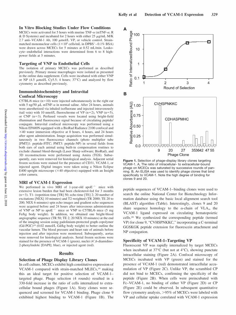
ResultsSelection of Phage Display Library ClonesIn cell culture, MCECs exhibit high constitutive expression ofVCAM-1 compared with strain-matched MLECs,19 makingthis an ideal target for positive selection of VCAM-1–targeted phage. Phage selection (4 rounds) resulted in a330-fold increase in the ratio of cells internalized to extra-cellular bound phages (Figure 1A). Sixty clones were se-quenced and screened for VCAM-1 binding. Phage clone 9exhibited highest binding to VCAM-1 (Figure 1B). The
peptide sequences of VCAM-1–binding clones were used tosearch the online National Center for Biotechnology Infor-mation database using the basic local alignment search tool(BLAST) algorithm (Table). Interestingly, clones 9 and 20share sequence homology to the �-chain of VLA4, theVCAM-1 ligand expressed on circulating hematopoieticcells.20 We synthesized the corresponding peptide (termedVP) for clone 9, “CVHSPNKKC,” incorporating a C-terminalGGSKGK peptide extension for fluorescein attachment andNP conjugation.
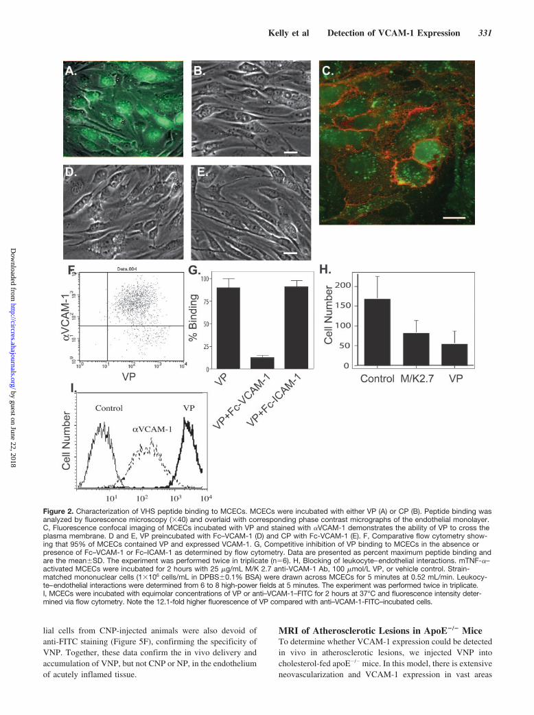
Specificity of VCAM-1–Targeting VPFluorescent VP was rapidly internalized by target MCECswhen incubated at 37°C (but not at 4°C), showing punctateintracellular staining (Figure 2A). Confocal microscopy ofMCECs incubated with VP (green) and stained for thepresence of VCAM-1 (red) demonstrated intracellular accu-mulation of VP (Figure 2C). Unlike VP, the scrambled CPdid not bind to MCECs, confirming the specificity of thepeptide (Figure 2B). When cells were preincubated withFc–VCAM-1, no binding of either VP (Figure 2D) or CP(Figure 2E) could be observed. In subsequent quantitativeflow cytometry analysis, �95% of MCECs were labeled withVP and cellular uptake correlated with VCAM-1 expression
Figure 1. Selection of phage-display library clones targetingVCAM-1. A, The ratio of intracellular- to extracellular-boundphage on MCECs was saturated by successive rounds of pan-ning. B, An ELISA was used to identify phage clones that bindspecifically to VCAM-1. Note the high degree of binding forclones 9 and 20.
Kelly et al Detection of VCAM-1 Expression 329
by guest on June 22, 2018http://circres.ahajournals.org/
Dow
nloaded from

(Figure 2F). Finally, preincubation of VP with Fc–VCAM-1(Figure 2G) significantly inhibited VP binding and uptake. Incontrast, preincubation of VP with Fc–ICAM-1 did not affectVP binding to MCECs (Figure 2G.) Together, these dataindicate that VP is specifically internalized by MCECs viaVCAM-1.
In functional analysis, preincubation of TNF-�–activatedMCECs with either function-blocking monoclonal Ab (mAb)M/K 2.7 or VP significantly inhibited mononuclear cellrecruitment under flow conditions. VP reduced mononuclearcell accumulation by 68%, compared with 52%, via mAbblocking, suggesting that the VP is as effective at preventingVCAM-1–mediated leukocyte–endothelial interactions (Fig-ure 2H). These data suggest that VP is effective at blockingleukocyte–endothelial interactions and the mechanism maybe attributable to binding of the peptide to the VLA4 adhesiondomain of VCAM-1 or may be the result of cell surfacedepletion of VCAM-1 by peptide binding.
Targeting of Magnetofluorescent NPs to VCAM-1Whereas Abs have been used to image VCAM-1 expression,the modest target-to-background ratios obtained using theseagents often limit their use in vivo for cardiovascular imag-ing. To test the comparative amplification potentially af-forded by cell-internalizing affinity ligands versus cell sur-face labeling with specific mAb, we directly comparedFITC-labeled �VCAM-1 and VP by FACS analysis usingequimolar concentrations. VP demonstrated a �12-foldhigher uptake compared with the Ab (Figure 2I).
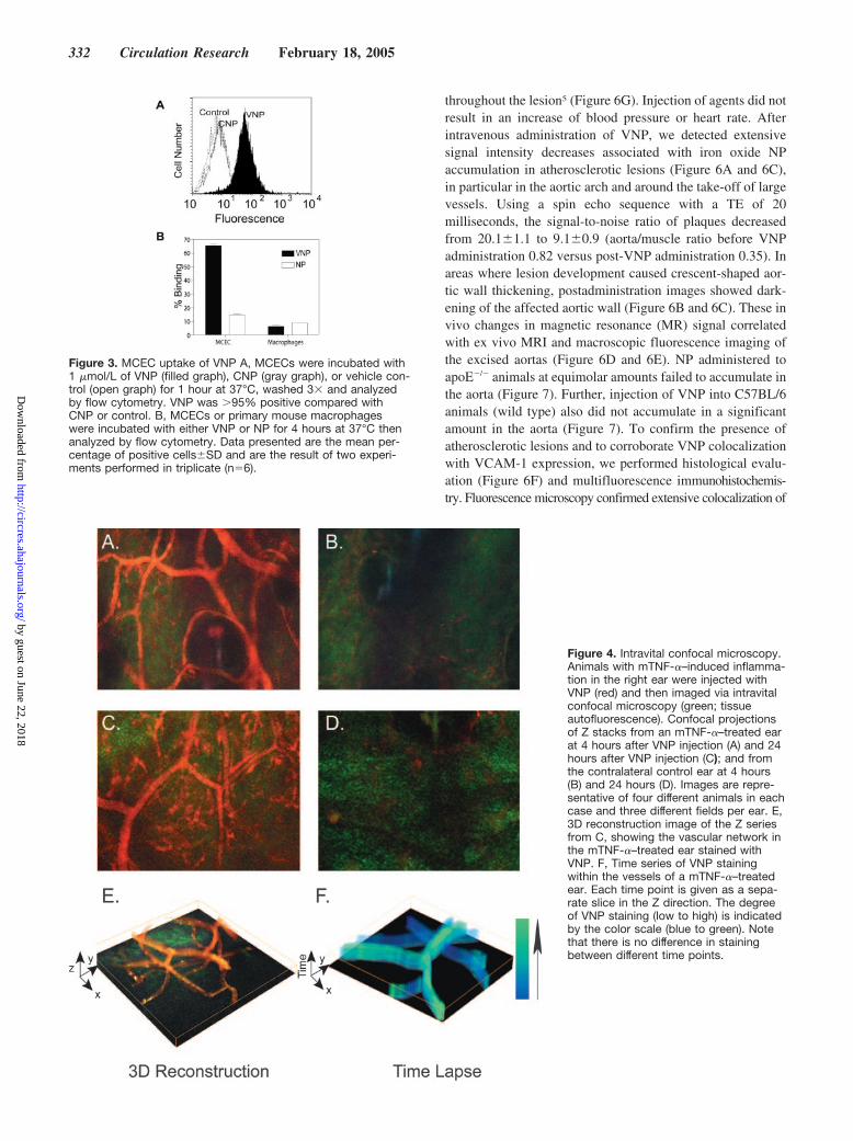
To permit evaluation of VCAM-1 expression via fluores-cence or MRI and to develop a VCAM-1–targeted imagingagent, we coupled VP multivalently to a superparamagneticfluorescent NP termed VNP. In vitro flow cytometric analysisdemonstrated that VNP retained the ability of the peptide tobind specifically to MCECs (Figure 3A) and in fact showedan �10-fold higher uptake by target cells compared withuptake of the negative control particle (CNP).
Because an NP-based in vivo imaging agent will contactmultiple cell types, we also sought to determine the specific-ity of VNP for endothelial cells as opposed to macrophages,which have been shown to accumulate iron oxide NPs. Invitro flow cytometric analysis demonstrated that VNP pref-erentially accumulated in endothelial cells (Figure 3B). Fur-ther, VNP had an 11-fold higher accumulation in endothelialcells compared with its accumulation in macrophages, con-
sistent with the known pattern of VCAM-1 expression (Fig-ure 3B). In contrast, endothelial cells lacked NP accumula-tion, suggesting VHS peptide labeling of NP changes thecellular specificity of the NP.
Intravital Confocal Microscopy ofVCAM-1–Expressing MicrovasculatureTo create a biologically monovariant in vivo mouse model,we next induced acute inflammation (24 hours) in mouse earsby injecting TNF-� to upregulate VCAM-1.12 When VP wasinjected intravenously into this mouse model of inflamma-tion, it exhibited very fast kinetics (as determined via intra-vital confocal microscopy), with a vascular T1/2 of 110seconds, rapid extravasation into the interstitium, and near-complete renal clearance within 10 minutes. In contrast,injection of VNP, CNP, or NP resulted in no apparentextravasation of the agents within 20 minutes after injection,as observed via intravital confocal microscopy. VNP, CNP,and NP were cleared from the circulation within 4 hours afterinjection, with only VNP remaining bound to the vasculature.At 4 hours after VNP injection, near-infrared fluorescencewas highly elevated in the vasculature of the inflamed ear,whereas no significant background fluorescence was ob-served (Figure 4A). In the control ear of the same animal,there was little detectable signal (Figure 4B). In controlanimals injected with CNP, an initial fluorescence signalthroughout the circulation immediately after administrationwas observed, but no significant fluorescence in either earafter 4 hours was determined. Control animals that receivedNP alone had cleared the agent from the circulation by 4hours after injection (data not shown). At 24 hours, signifi-cant amounts of VNP could still be visualized in the vesselwalls of mTNF-�–treated ears (Figure 4C and 4E), whereas itwas absent in the untreated ears (Figure 4D). In furthertime-lapse analysis (Figure 4F), the fluorescence profile ofVNP did not change during a 5-minute observation, confirm-ing that the probe was indeed retained at specific locationswithin the vessel wall and not free flowing in the circulation.
Histological analysis of mTNF-�–treated ears (Figure 5Athrough 5F) revealed the upregulation of VCAM-1 in endo-thelial cells (Figure 5B and 5E). In contrast, endothelial cellsfrom untreated ears did not show appreciable VCAM-1staining (Figure 5H). Anti-FITC Ab staining revealed thepresence of the VNP agent in endothelial cells from inflamedears (Figure 5C) but not in control ears (Figure 5I). Endothe-
Homology of Selected VCAM-1–Binding Peptides to Known Proteins
(Clone) Peptides Homology Sequence Accession No.
(9) VHSPNKK VLA-4 83-VNPGAIY-89 (Mus musculus) NP_034706
(20) DHASPMH 82-VSNPGAI-88 (Xenopus laevis) Q91687
88-VINPGAI-95 (Homo sapiens) NP_000876.1
(27) PTRIGQMC Glucagon-like 332-RAHQMC-337 (Mus musculus) AAH44746
(47) MHRAHQMC Peptide 2
(55) ISHQMPA Receptor
(35) PGHHTSQ No matches
(36) NNAHHKN
330 Circulation Research February 18, 2005
by guest on June 22, 2018http://circres.ahajournals.org/
Dow
nloaded from
lial cells from CNP-injected animals were also devoid ofanti-FITC staining (Figure 5F), confirming the specificity ofVNP. Together, these data confirm the in vivo delivery andaccumulation of VNP, but not CNP or NP, in the endotheliumof acutely inflamed tissue.
MRI of Atherosclerotic Lesions in ApoE�/� MiceTo determine whether VCAM-1 expression could be detectedin vivo in atherosclerotic lesions, we injected VNP intocholesterol-fed apoE�/� mice. In this model, there is extensiveneovascularization and VCAM-1 expression in vast areas
Figure 2. Characterization of VHS peptide binding to MCECs. MCECs were incubated with either VP (A) or CP (B). Peptide binding wasanalyzed by fluorescence microscopy (�40) and overlaid with corresponding phase contrast micrographs of the endothelial monolayer.C, Fluorescence confocal imaging of MCECs incubated with VP and stained with �VCAM-1 demonstrates the ability of VP to cross theplasma membrane. D and E, VP preincubated with Fc–VCAM-1 (D) and CP with Fc-VCAM-1 (E). F, Comparative flow cytometry show-ing that 95% of MCECs contained VP and expressed VCAM-1. G, Competitive inhibition of VP binding to MCECs in the absence orpresence of Fc–VCAM-1 or Fc–ICAM-1 as determined by flow cytometry. Data are presented as percent maximum peptide binding andare the mean�SD. The experiment was performed twice in triplicate (n�6). H, Blocking of leukocyte–endothelial interactions. mTNF-�–activated MCECs were incubated for 2 hours with 25 �g/mL M/K 2.7 anti-VCAM-1 Ab, 100 �mol/L VP, or vehicle control. Strain-matched mononuclear cells (1�106 cells/mL in DPBS�0.1% BSA) were drawn across MCECs for 5 minutes at 0.52 mL/min. Leukocy-te–endothelial interactions were determined from 6 to 8 high-power fields at 5 minutes. The experiment was performed twice in triplicate.I, MCECs were incubated with equimolar concentrations of VP or anti–VCAM-1–FITC for 2 hours at 37°C and fluorescence intensity deter-mined via flow cytometry. Note the 12.1-fold higher fluorescence of VP compared with anti–VCAM-1-FITC–incubated cells.
Kelly et al Detection of VCAM-1 Expression 331
by guest on June 22, 2018http://circres.ahajournals.org/
Dow
nloaded from
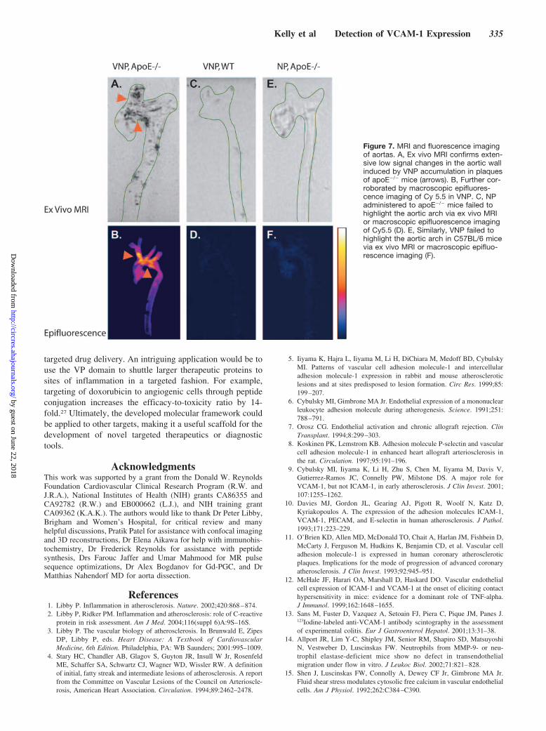
throughout the lesion5 (Figure 6G). Injection of agents did notresult in an increase of blood pressure or heart rate. Afterintravenous administration of VNP, we detected extensivesignal intensity decreases associated with iron oxide NPaccumulation in atherosclerotic lesions (Figure 6A and 6C),in particular in the aortic arch and around the take-off of largevessels. Using a spin echo sequence with a TE of 20milliseconds, the signal-to-noise ratio of plaques decreasedfrom 20.1�1.1 to 9.1�0.9 (aorta/muscle ratio before VNPadministration 0.82 versus post-VNP administration 0.35). Inareas where lesion development caused crescent-shaped aor-tic wall thickening, postadministration images showed dark-ening of the affected aortic wall (Figure 6B and 6C). These invivo changes in magnetic resonance (MR) signal correlatedwith ex vivo MRI and macroscopic fluorescence imaging ofthe excised aortas (Figure 6D and 6E). NP administered toapoE�/� animals at equimolar amounts failed to accumulate inthe aorta (Figure 7). Further, injection of VNP into C57BL/6animals (wild type) also did not accumulate in a significantamount in the aorta (Figure 7). To confirm the presence ofatherosclerotic lesions and to corroborate VNP colocalizationwith VCAM-1 expression, we performed histological evalu-ation (Figure 6F) and multifluorescence immunohistochemis-try. Fluorescence microscopy confirmed extensive colocalization of
Figure 3. MCEC uptake of VNP A, MCECs were incubated with1 �mol/L of VNP (filled graph), CNP (gray graph), or vehicle con-trol (open graph) for 1 hour at 37°C, washed 3� and analyzedby flow cytometry. VNP was �95% positive compared withCNP or control. B, MCECs or primary mouse macrophageswere incubated with either VNP or NP for 4 hours at 37°C thenanalyzed by flow cytometry. Data presented are the mean per-centage of positive cells�SD and are the result of two experi-ments performed in triplicate (n�6).
Figure 4. Intravital confocal microscopy.Animals with mTNF-�–induced inflamma-tion in the right ear were injected withVNP (red) and then imaged via intravitalconfocal microscopy (green; tissueautofluorescence). Confocal projectionsof Z stacks from an mTNF-�–treated earat 4 hours after VNP injection (A) and 24hours after VNP injection (C); and fromthe contralateral control ear at 4 hours(B) and 24 hours (D). Images are repre-sentative of four different animals in eachcase and three different fields per ear. E,3D reconstruction image of the Z seriesfrom C, showing the vascular network inthe mTNF-�–treated ear stained withVNP. F, Time series of VNP stainingwithin the vessels of a mTNF-�–treatedear. Each time point is given as a sepa-rate slice in the Z direction. The degreeof VNP staining (low to high) is indicatedby the color scale (blue to green). Notethat there is no difference in stainingbetween different time points.
332 Circulation Research February 18, 2005
by guest on June 22, 2018http://circres.ahajournals.org/
Dow
nloaded from

VNP (Cy.5.5 channel; Figure 6H) and VCAM-1 expression (anti–VCAM-1–FITC channel (Figure 6G). In contrast, there was littleaccumulation of VNP in macrophages, reminiscent of the in vitrodata shown in Figure 3B.
DiscussionPathophysiologically inducible endothelial targets provide anattractive paradigm for the development of targeted anddisease-specific molecular imaging agents, attributablelargely to their immediate accessibility via the circulation.The strict temporal and spatial regulation of VCAM-1 in thedevelopment and progression of atherosclerosis5–9 makes it aprime candidate for development of the next generation oftargeted diagnostics and therapeutics.
Several approaches have been described previously usingradiolabeled Abs to detect VCAM-1 expression;12,13 how-ever, this approach has resulted in very modest target-to-background ratios, limiting its use for in vivo cardiovascularimaging. To overcome these limitations, we commenceddevelopment of an agent that not only decorated the endo-thelial surface but was internalized by endothelial cells(“biological amplification through intracellular trapping”).21
We used phage display to identify peptide sequences that
bound under physiological flow conditions and were capableof VCAM-1–mediated endothelial cell internalization. Usingthis approach, we identified a novel VCAM-1–specific cellinternalizing peptide that results in higher target-to-background ratios than monoclonal Abs and allows in vivoimaging of inflammation and cardiovascular disease.
Our unique approach resulted in the identification of asmall panel of peptides forming 42 consensus groups, with asubset that bound specifically to the extracellular domain ofVCAM-1 (Table). One of these peptides, VHSPNKK, withthe highest binding to VCAM-1, was homologous to VLA4,the ligand for VCAM-1. Direct intravenous injection of thefluorescently labeled selected peptide (VP), resulted in rapidleakage into the interstitium, and the resulting high back-ground fluorescence in the surrounding tissue eliminated thepossibility of using the ligand for intravital work, although itremains a highly specific and valuable agent for in vitroassays. The derived multivalent (chemical amplification)magnetofluorescent imaging agent (VNP) exhibited im-proved in vivo pharmacokinetics, retained specificity forVCAM-1, and in vivo accumulated in the vessel wall,remaining detectable for at least 24 hours by noninvasiveoptical and MRI modalities, confirming the ability of the
Figure 5. Immunohistochemical analysisof in vivo delivery of VNP to endothelialcells. Adjacent serial sections of earstaken from the in vivo intravital experi-ments were stained for CD31 (A, D, andG), VCAM-1 (B, E, and H), or FITC (C, F,and I). Results are representative of sixsections in each case, taken from atleast three animals.
Kelly et al Detection of VCAM-1 Expression 333
by guest on June 22, 2018http://circres.ahajournals.org/
Dow
nloaded from

peptide to shuttle large molecules across the plasmamembrane.
A number of previous publications have identified peptidesequences that bind to atherosclerotic plaque or are poten-tially relevant to targeting cell surface receptors.22–25 Inparticular, Liu et al25 used phage display to interrogate thesurface of atherosclerotic lesions in apoE�/� mice and iden-tified consensus sequences that bound to endothelial cellsurface glucose-regulated protein 78 and a homologue oftissue inhibitor of metalloproteinase-2 (TIMP-2) that boundto mature lesions in vivo. In the present study, we identifiedthree peptide sequences with similarity to TIMP-2; however,no other similar sequences were isolated. The difference inthese results could be explained by the fact that Liu et alexamined the binding of phage in vivo in apoE�/� mice withadvanced lesion development,25 where proteins other thanVCAM-1 may be more abundant on the endothelial cellsurface.
The data presented here demonstrate for the first time thatendothelial targets can be visualized by in vivo MRI without
the use of monoclonal Abs. Our approach has far-reachingimplications in the design of future imaging agents byallowing the visualization of nonabundant targets throughintracellular accumulation. The first generation of nontar-geted magnetic NPs are in clinical use,26 and thus, it isconceivable that targeted NPs could provide significant ben-efits in specificity for imaging and therapeutics. Our in vitroand in vivo results suggest that conjugation of the magnet-ofluorescent NP with phage display–derived VCAM-1–spe-cific peptides changes the cellular specificity of the parent NPand allows the targeted imaging of activated endothelium.Indeed, we demonstrate that VCAM-1 expression can bevisualized by high-resolution intravital confocal microscopyand MRI. This could have a number of important clinicalimplications. For example, VNP could be used to noninva-sively monitor the efficacy of anti-inflammatory or lipid-lowering drugs by examining the level of VCAM-1 invessels. Furthermore, the agent or similarly developed probescould be used for combined or simultaneous imaging and
Figure 6. MRI of atherosclerotic lesions with VNP. A, Cholesterol-fed apoE�/� mice were imaged via MRI using Gd-PGC to delineatevascular lumen and structural aortic abnormalities such as narrowing (arrows). Axial images (at the level of the green line) were thenobtained before (B) and 24 hours after (C) administration of the magnetic VNP. Note the decrease in signal intensity of the eccentricallythickened aortic wall between the two arrows. D, Ex vivo MRI confirms extensive low signal changes in the aortic wall induced by VNPaccumulation (arrows), further corroborated by macroscopic epifluorescence imaging of Cy 5.5 in VNP (E). F through H, Histologicalvalidation of MR findings. F, Hematoxylin/eosin section of the aorta image in B and C confirms eccentric wall thickening (�2). G and H,Comparative immunofluorescence of VCAM-1 expression (G; green) and VNP accumulation (H; red). Nuclei in G and H are counter-stained with DAPI (blue). Note that there is extensive colocalization of VCAM-1 expression and VNP. Bars�10 �mol/L.
334 Circulation Research February 18, 2005
by guest on June 22, 2018http://circres.ahajournals.org/
Dow
nloaded from
targeted drug delivery. An intriguing application would be touse the VP domain to shuttle larger therapeutic proteins tosites of inflammation in a targeted fashion. For example,targeting of doxorubicin to angiogenic cells through peptideconjugation increases the efficacy-to-toxicity ratio by 14-fold.27 Ultimately, the developed molecular framework couldbe applied to other targets, making it a useful scaffold for thedevelopment of novel targeted therapeutics or diagnostictools.
AcknowledgmentsThis work was supported by a grant from the Donald W. ReynoldsFoundation Cardiovascular Clinical Research Program (R.W. andJ.R.A.), National Institutes of Health (NIH) grants CA86355 andCA92782 (R.W.) and EB000662 (L.J.), and NIH training grantCA09362 (K.A.K.). The authors would like to thank Dr Peter Libby,Brigham and Women’s Hospital, for critical review and manyhelpful discussions, Pratik Patel for assistance with confocal imagingand 3D reconstructions, Dr Elena Aikawa for help with immunohis-tochemistry, Dr Frederick Reynolds for assistance with peptidesynthesis, Drs Farouc Jaffer and Umar Mahmood for MR pulsesequence optimizations, Dr Alex Bogdanov for Gd-PGC, and DrMatthias Nahendorf MD for aorta dissection.
References1. Libby P. Inflammation in atherosclerosis. Nature. 2002;420:868–874.2. Libby P, Ridker PM. Inflammation and atherosclerosis: role of C-reactive
protein in risk assessment. Am J Med. 2004;116(suppl 6)A:9S–16S.3. Libby P. The vascular biology of atherosclerosis. In Brunwald E, Zipes
DP, Libby P, eds. Heart Disease: A Textbook of CardiovascularMedicine, 6th Edition. Philadelphia, PA: WB Saunders; 2001:995–1009.
4. Stary HC, Chandler AB, Glagov S, Guyton JR, Insull W Jr, RosenfeldME, Schaffer SA, Schwartz CJ, Wagner WD, Wissler RW. A definitionof initial, fatty streak and intermediate lesions of atherosclerosis. A reportfrom the Committee on Vascular Lesions of the Council on Arterioscle-rosis, American Heart Association. Circulation. 1994;89:2462–2478.
5. Iiyama K, Hajra L, Iiyama M, Li H, DiChiara M, Medoff BD, CybulskyMI. Patterns of vascular cell adhesion molecule-1 and intercellularadhesion molecule-1 expression in rabbit and mouse atheroscleroticlesions and at sites predisposed to lesion formation. Circ Res. 1999;85:199–207.
6. Cybulsky MI, Gimbrone MA Jr. Endothelial expression of a mononuclearleukocyte adhesion molecule during atherogenesis. Science. 1991;251:788–791.
7. Orosz CG. Endothelial activation and chronic allograft rejection. ClinTransplant. 1994;8:299–303.
8. Koskinen PK, Lemstrom KB. Adhesion molecule P-selectin and vascularcell adhesion molecule-1 in enhanced heart allograft arteriosclerosis inthe rat. Circulation. 1997;95:191–196.
9. Cybulsky MI, Iiyama K, Li H, Zhu S, Chen M, Iiyama M, Davis V,Gutierrez-Ramos JC, Connelly PW, Milstone DS. A major role forVCAM-1, but not ICAM-1, in early atherosclerosis. J Clin Invest. 2001;107:1255–1262.
10. Davies MJ, Gordon JL, Gearing AJ, Pigott R, Woolf N, Katz D,Kyriakopoulos A. The expression of the adhesion molecules ICAM-1,VCAM-1, PECAM, and E-selectin in human atherosclerosis. J Pathol.1993;171:223–229.
11. O’Brien KD, Allen MD, McDonald TO, Chait A, Harlan JM, Fishbein D,McCarty J, Ferguson M, Hudkins K, Benjamin CD, et al. Vascular celladhesion molecule-1 is expressed in human coronary atheroscleroticplaques. Implications for the mode of progression of advanced coronaryatherosclerosis. J Clin Invest. 1993;92:945–951.
12. McHale JF, Harari OA, Marshall D, Haskard DO. Vascular endothelialcell expression of ICAM-1 and VCAM-1 at the onset of eliciting contacthypersensitivity in mice: evidence for a dominant role of TNF-alpha.J Immunol. 1999;162:1648–1655.
13. Sans M, Fuster D, Vazquez A, Setoain FJ, Piera C, Pique JM, Panes J.123Iodine-labeled anti-VCAM-1 antibody scintography in the assessmentof experimental colitis. Eur J Gastroenterol Hepatol. 2001;13:31–38.
14. Allport JR, Lim Y-C, Shipley JM, Senior RM, Shapiro SD, MatsuyoshiN, Vestweber D, Luscinskas FW. Neutrophils from MMP-9- or neu-trophil elastase-deficient mice show no defect in transendothelialmigration under flow in vitro. J Leukoc Biol. 2002;71:821–828.
15. Shen J, Luscinskas FW, Connolly A, Dewey CF Jr, Gimbrone MA Jr.Fluid shear stress modulates cytosolic free calcium in vascular endothelialcells. Am J Physiol. 1992;262:C384–C390.
Figure 7. MRI and fluorescence imagingof aortas. A, Ex vivo MRI confirms exten-sive low signal changes in the aortic wallinduced by VNP accumulation in plaquesof apoE�/� mice (arrows). B, Further cor-roborated by macroscopic epifluores-cence imaging of Cy 5.5 in VNP. C, NPadministered to apoE�/� mice failed tohighlight the aortic arch via ex vivo MRIor macroscopic epifluorescence imagingof Cy5.5 (D). E, Similarly, VNP failed tohighlight the aortic arch in C57BL/6 micevia ex vivo MRI or macroscopic epifluo-rescence imaging (F).
Kelly et al Detection of VCAM-1 Expression 335
by guest on June 22, 2018http://circres.ahajournals.org/
Dow
nloaded from
16. Smith GP, Petrenko VA. Phage display. Chem Rev. 1997;97:391–410.17. Ricard I, Payet MD, Dupuis G. VCAM-1 is internalized by a clathrin-
related pathway in human endothelial cells but its alpha 4 beta 1 integrincounter-receptor remains associated with the plasma membrane in humanT lymphocytes. Eur J Immunol. 1998;28:1708–1718.
18. Lewin M, Bredow S, Sergeyev N, Marecos E, Bogdanov A Jr, WeisslederR. In vivo assessment of vascular endothelial growth factor-inducedangiogenesis. Int J Cancer. 1999;83:798–802.
19. Lim Y-C, Garcia-Cardena G, Allport JR, Zervoglos M, Connolly AJ,Gimbrone MA Jr, Luscinskas FW. Heterogeneity of endothelial cellsfrom different organ sites in T-cell subset recruitment. Am J Pathol.2003;162:1591–1601.
20. Holzmann B, Weissman IL. Integrin molecules involved in lymphocytehoming to Peyer’s patches. Immunol Rev. 1989;108:45–61.
21. Weissleder R, Ntziachristos V. Shedding light onto live molecular targets.Nat Med. 2003;9:123–128.
22. Molenaar TJ, Twisk J, de Haas SA, Peterse N, Vogelaar BJ, van LeeuwenSH, Michon IN, van Berketl TJ, Kuiper J, Biessen EA. P-selectin as acandidate target in atherosclerosis. Biochem Pharmacol. 2003;66:859–866.
23. Houston P, Goodman J, Lewis A, Campbell CJ, Braddock M. Homingmarkers for atherosclerosis: applications for drug delivery, gene deliveryand vascular imaging. FEBS Lett. 2001;492:73–77.
24. Winter PM, Morawski AM, Caruthers SD, Fuhrhop RW, Zhang H,Williams TA, Allen JS, Lacy EK, Robertson JD, Lanza GM, WicklineSA. Molecular imaging of angiogenesis in early-stage atherosclerosiswith alpha(v)beta3-integrin-targeted nanoparticles. Circulation. 2003;108:2270–2274.
25. Liu C, Bhattacharjee G, Boisvert W, Dilley R, Edgington T. In vivointerrogation of the molecular display of atherosclerotic lesion surfaces.Am J Pathol. 2003;163:1859–1871.
26. Harisinghani MG, Barentsz J, Hahn PF, Deserno WM, Tabatabaei S, vande Kaa CH, de la Rosette J, Weissleder R. Noninvasive detection ofclinically occult lymph-node metastases in prostate cancer. N Engl J Med.2003;348:2491–2499.
27. Arap W, Pasqualini R, Ruoslahti E. Cancer treatment by targeted drugdelivery to tumor vasculature in a mouse model. Science. 1998;279:377–380.
336 Circulation Research February 18, 2005
by guest on June 22, 2018http://circres.ahajournals.org/
Dow
nloaded from
Josephson and Ralph WeisslederKimberly A. Kelly, Jennifer R. Allport, Andrew Tsourkas, Vivek R. Shinde-Patil, Lee
NanoparticleDetection of Vascular Adhesion Molecule-1 Expression Using a Novel Multimodal
Print ISSN: 0009-7330. Online ISSN: 1524-4571 Copyright © 2005 American Heart Association, Inc. All rights reserved.is published by the American Heart Association, 7272 Greenville Avenue, Dallas, TX 75231Circulation Research
doi: 10.1161/01.RES.0000155722.17881.dd2005;96:327-336; originally published online January 13, 2005;Circ Res.
http://circres.ahajournals.org/content/96/3/327World Wide Web at:
The online version of this article, along with updated information and services, is located on the
http://circres.ahajournals.org/content/suppl/2005/02/17/96.3.327.DC1Data Supplement (unedited) at:
http://circres.ahajournals.org//subscriptions/
is online at: Circulation Research Information about subscribing to Subscriptions:
http://www.lww.com/reprints Information about reprints can be found online at: Reprints:
document. Permissions and Rights Question and Answer about this process is available in the
located, click Request Permissions in the middle column of the Web page under Services. Further informationEditorial Office. Once the online version of the published article for which permission is being requested is
can be obtained via RightsLink, a service of the Copyright Clearance Center, not theCirculation Researchin Requests for permissions to reproduce figures, tables, or portions of articles originally publishedPermissions:
by guest on June 22, 2018http://circres.ahajournals.org/
Dow
nloaded from
Synthesis of FITC peptides and fluorescent magnetic nanoparticle conjugates The identified peptide sequence ‘CVHSPNKKC’ was synthesized on an ACT APEX 396 peptide synthesizer using standard FMOC chemistry and extended at the C-terminus by the sequence ‘GGSKGK’ for attachment to CLIO-Cy5.5, a cross-linked iron oxide nanoparticle 1,2. This nanoparticle is similar in physicochemical and biological properties to monocrystalline iron oxide nanoparticles (MION)1,2. Lysines in the binding sequence were orthogonally protected. Peptides were cleaved with reagent L, purified by HPLC, cyclized via S-S linkage, lyophilized, and verified by mass spectral analysis (MALDI). CLIO-Cy5.5 was synthesized in bulk using established procedures and aliquots used for the synthesis of the various nanoparticle conjugates. CLIO-Cy5.5 had the following physical properties: a) size 38.7 nm, b) relaxation time constants R1-21.1 and R2-62.6 and c) an average of 2.3 Cy5.5 per CLIO particle based on 2600 Fe/CLIO. Suberic acid bis (N-hydroxy-succinimide ester) derivatized CLIO-Cy5.5 were reacted with 100 uL of 1mM FITC-labeled VCAM-1 peptide (VNP) or control peptide (CNP). VNP was purified on a Sephadex G-25 M gel chromatography column equilibrated with PBS, and concentrated on a Microcon YM-50 ultracentrifugation filter device (Millipore). A typical experiment would result in the attachment of 6 peptides per NP. To ensure no cross-linking of NPs occurred during the bioconjugation procedure, light scattering was performed following DSS labeling and peptide conjugation. Isolation of Primary Mouse Macrophages Primary mouse macrophages were isolated as follows: peripheral blood was obtained by cardiac puncture, (15 mice) mixed with sodium citrate (100 mM), dextrose (pH 6.5, 136 mM), and 100 mL of H2O and centrifuged for 4 minutes at 1000 xg. Pellet was resuspended in HBSS, layered onto Ficoll (Histopaque-1083, Sigma, St. Louis, MO) then centrifuged at 1600 RPM for 30 minutes at room temperature. After centrifugation, two distinct bands were present. The top band was removed and plated onto gelatin coated wells in RPMI containing 10% FBS, 40 nM phorbol myristate acetate (Sigma), and 1% pen/strep. Non-adherent cells were removed 24 hours after plating. 1. Wunderbaldinger P, Josephson L, Weissleder R. Tat peptide directs enhanced
clearance and hepatic permeability of magnetic nanoparticles. Bioconjug Chem. 2002;13:264-8.
2. Weissleder R, Stark D, Engelstad B, Bacon B, Compton C, White D, Jacobs P, Lewis J. Superparamagnetic Iron Oxide: Pharmacokinetics and Toxicicty. American Journal of Roentgenology. 1989;152:167-173.