Detection of swine hev modified
10
Detection of swine hepatitis E virus in the porcine hepatic lesion in Jeju Island
-
Upload
kpiller -
Category
Health & Medicine
-
view
192 -
download
1
Transcript of Detection of swine hev modified

Detection of swine hepatitis E virus in
the porcine hepatic lesion in Jeju Island

Nonenveloped Fecal-oral route Clinical signs: jaundice, anorexia, nausea,
hepatomegaly Exerts severe effects in pregnant women
Hepatitis E Virus

Zoonotic pigs not obviously ill, display very mild liver
lesions on histopathologic examination widespread throughout the world Antibodies to HEV in pigs from countries
where HEV is endemic and many industrialized countries
Pig as animal reservoir host for HEV
HEV in pigs

19 farms
40 pigs(10-70 days old)
33 pigs(<2 months old)
7 pigs(> 2 months old)
Animal samples

All liver samples were collected from pigs at necropsy
One-third frozen at -70℃ remaining part fixed in 10% neutral buffered
formalin embedded in paraffin wax cut into 3 µm sections stained with hematoxylin and eosin
Tissue processing
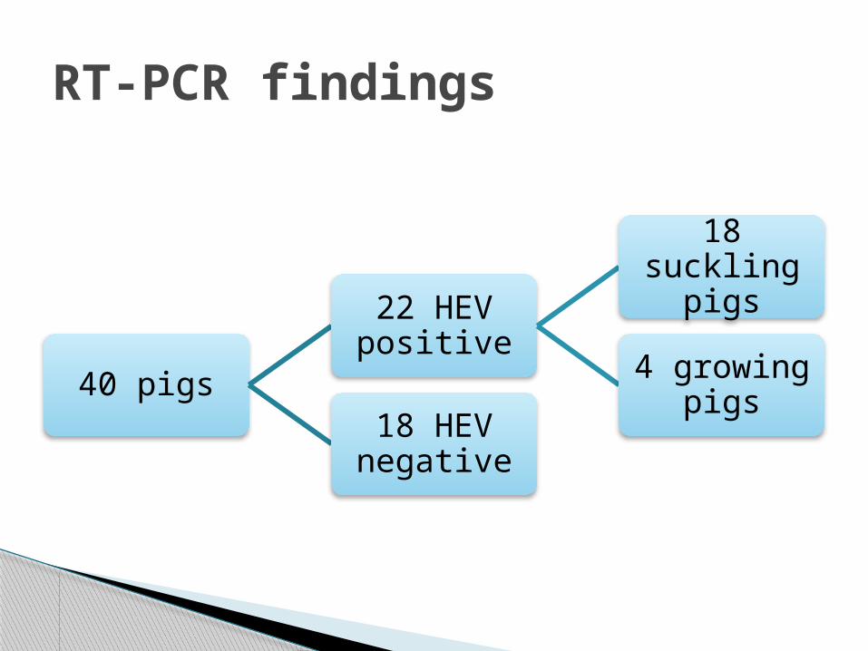
40 pigs
22 HEV positive
18 suckling pigs
4 growing pigs
18 HEV negative
RT-PCR findings

Moderate to severe multifocal lymphoplasmacytic and histiocytic hepatitis or
portal inflammation

mild hepatic enlargement and scattered yellowish discoloration foci

focal infiltration of lymphocytes, plasma cells, and macrophages in hepatic sinusoids and the
portal triad

swine HEV infection occurs at a very young age (under 2 months) in Jeju pigs
various hepatitis virus antigens detected in liver tissue
viral markers may not be present during active disease
immunohistochemistry not particularly useful PCR method more sensitive and applicable as a
specific assay for the diagnosis of various infectious diseases
individuals with swine-associated occupations at possible risk of zoonotic infection.
Conclusions