DETECTION OF APOPTOTIC CELLS IN INTESTINE FROM HORSES …
114
DETECTION OF APOPTOTIC CELLS IN INTESTINE FROM HORSES WITH AND WITHOUT GASTROINTESTINAL DISEASE by Emma Lucy Rowe BSc.BVMS (Hons.) Thesis submitted to the Faculty of the Virginia Polytechnic Institute and State University in partial fulfillment of the requirements for the degree of Master of Science In Veterinary Medical Science APPROVED: ___________________________________ Nathaniel A. White II, Chairman ____________________________ _____________________________ Virginia Buechner-Maxwell John L. Robertson April, 2003 Leesburg, Virginia Key words: horse, intestine, gastrointestinal disease, apoptosis Copyright 2003, Emma Lucy Rowe
Transcript of DETECTION OF APOPTOTIC CELLS IN INTESTINE FROM HORSES …

DETECTION OF APOPTOTIC CELLS IN INTESTINE FROM HORSES WITH AND WITHOUT
GASTROINTESTINAL DISEASE by
Emma Lucy Rowe BSc.BVMS (Hons.) Thesis submitted to the Faculty of the Virginia Polytechnic Institute and State University in partial fulfillment of the requirements for the degree of Master of Science In Veterinary Medical Science APPROVED: ___________________________________ Nathaniel A. White II, Chairman ____________________________ _____________________________ Virginia Buechner-Maxwell John L. Robertson April, 2003 Leesburg, Virginia Key words: horse, intestine, gastrointestinal disease, apoptosis Copyright 2003, Emma Lucy Rowe

DETECTION OF APOPTOTIS CELLS IN INTESTINE FROM HORSES WITH AND WITHOUT GASTROINTESTINAL DISEASE by Emma L. Rowe BSc.BVMS (Hons.) Committee Chairman: Nathaniel A. White II, DVM, MS Diplomate American College of Veterinary Surgeons Theodora Ayer Randolph Professor of Surgery Veterinary Medical Sciences (ABSTRACT) A study was performed to identify apoptotic cells in the equine intestine and to determine
if the occurrence of apoptosis is affected by gastrointestinal disease and tissue layer of intestine.
Samples of intestine were collected from 38 horses that underwent surgery or were humanely
destroyed for small or large bowel obstruction, strangulation or distension. Samples were also
taken from 9 horses which were humanely euthanized for reasons other than gastrointestinal
disease or systemic disease. Specimens were collected at surgery from intestine involved in the
primary lesion, distant to the primary lesion, or at necropsy from several sites including the
primary lesion. Tissues were fixed, serially sectioned and stained with hematoxylin and eosin
(H&E) and for apoptosis by the terminal deoxynucleotidyl transferase mediated dUTP nick end
labeling (TUNEL) technique. The number of apoptotic cells per high power field were counted
in the mucosa, circular muscle, longitudinal muscle and serosa for each sample of intestine.
Apoptotic staining nuclei were seen in all layers of intestine. An increased number of apoptotic
cells were found in the circular muscle of the intestine from horses with simple obstruction.

Intestine distant from the primary strangulating lesion had higher numbers of apoptotic cells than
intestine distant from a simple obstruction lesion or intestine taken at the site of a strangulating
or simple obstructive lesion. Intestine from horses with obstructing or strangulating lesions in
the small intestine and large colon has increased numbers of apoptotic cells. Further
investigation is required to determine whether increased apoptosis affects intestinal function.

iv
GRANT INFORMATION
This research was funded by the Patricia Bonsall Stuart Equine Research Award

v
DEDICATED TO
Christopher and Anne Rowe
My mother and father who have provided never-ending support and friendship throughout my
life and who gave me the opportunity to pursue my love of veterinary science.

vi
ACKNOWLEDGEMENTS
Nathaniel A. White- my research advisor and mentor, for his support, patience and enthusiasm
not only involving this project but in all aspects of my training.
Virginia Buechner-Maxwell- for her enthusiasm, guidance, knowledge and friendship.
Daniel L. Ward- who performed the statistical analysis of data for his skills, knowledge and
patience.
John L. Roberston- for his knowledge and insight into the pathology of gastrointestinal disease.
Patsy Dillon-Long- who provided my technical support in the laboratory.

vii
TABLE OF CONTENTS
1. LIST OF FIGURES…………………………………………………………ix
2. LIST OF TABLES…………………………………………………….........xiii
3. LIST OF ABBREVIATIONS………………………………………………xiv
4. INTRODUCTION…………………………………………………….........1
5. LITERATURE REVIEW
A. Gastrointestinal Disease in the equine
1. Importance………………………………………….3
2. Pathophysiology of gastrointestinal disease………..4
3. Ischemia……………………………………….........9
4. Reperfusion…………………………………………11
5. Intestinal Distension………………………………..18
6. Inflammation of the peritoneum..…………………..20
B. APOPTOSIS
1. Apoptosis-programmed cell death..…………………..21
2. Immunohistochemical staining to detect apoptosis…...27
3. Apoptosis and the intestine……………………………29
4. Apoptosis and ischemia/reperfusion injury…………...33
5. Role of nitric oxide in apoptosis………………………35
6. Apoptosis and inflammation...…………………………37
7. Apoptosis and non-steroidal anti- inflammatory drugs...38
C. LITERATURE CITED…………………………………………………41

viii
6. DETECTION OF APOPTOTIC CELLS IN HORSES WITH AND WITHOUT
GASTROINTESTINAL DISEASE………………………………………………….66
A. Abstract…………………………………………………….67
B. Introduction………………………………………………...68
C. Materials and methods……………………………………..69
D. Results……………………………………………………..72
E. Discussion………………………………………………….74
F. References………………………………………………….81
G. List of figures………………………………………………86
7. CONCLUSION..…………………………………………………………………98
8. VITA……………………………………………………………………………..99

ix
LIST OF FIGURES Page
Figure legends: 88
Figure 1: Photomicrograph of the mucosa in the small intestine from
a horse with gastrointestinal disease. All dark staining nuclei represent
apoptotic cells. Healthy or necrotic cells did not stain. Diaminobenzidine
(DAB) stain and methyl green counterstain; bar equals 50µ.
Figure 2: Photomicrograph of the mucosa in the small intestine 88
from a horse in the control group. No apoptotic nuclei are present.
Diaminobenzidine (DAB) stain and methyl green counter stain;
bar equals 50µ.
Figure 3: Photomicrograph of the circular muscle in the small 89
intestine from a horse with gastrointestinal disease. All dark
staining nuclei represent apoptotic cells. Healthy or necrotic cells
did not stain. Diaminobenzidine (DAB) stain and methyl green
counterstain; bar equals 50µ.
Figure 4: Photomicrograph of the circular muscle in the small 89
intestine from a horse in the control group. No apoptotic nuclei
are present. Diaminobenzidine (DAB) stain and methyl green counter
stain; bar equals 50µ.

x
Page
Figure 5: Photomicrograph of a myenteric plexus in the large colon. 90
All dark staining nuclei represent apoptotic cells. Healthy cells or
necrotic cells did not stain. Neurons, glial cells and Schwann cells
were apoptotic in this myenteric plexus. Diaminobenzidine (DAB) stain
and methyl green counterstain; bar equals 50µ.
Figure 6: Photomicrograph of the serosa from the large colon. 90
All dark staining nuclei are apoptotic mesothelial cells on the
surface of the serosa. Healthy cells or necrotic cells did not stain.
Diaminobenzidine (DAB) stain and methyl green counterstain;
bar equals 50µ.
Figure 7: Graph of the mean numbers of apoptotic cells in the intestine 91
from horses in all 4 categories- simple obstruction, strangulation,
musculoskeletal, control. A significant increase in apoptotic cells
was present in the simple obstruction category (P< 0.0088) which
is indicated by the star (*). Columns represent the geometric mean
number of apoptotic cells while error bars represent the 95% confidence
intervals.

xi
Page
Figure 8: Graph of the mean number of apoptotic cells in intestine 92
taken from the primary lesion or at a distant site in horses with simple
obstruction and strangulation. A significant increase in apoptotic cells
was present at a distant site to a strangulation lesion (p< 0.0186)
which is indicated by the star (*). Columns represent the geometric
mean number of apoptotic cells while error bars represent the 95%
confidence intervals.
Figure 9: Graph of the mean number of apoptotic cells in the four 93
tissue layers from horse with simple obstruction or strangulation.
Columns represent the geometric mean number of apoptotic cells
while error bars represent the 95% confidence intervals.
Figure 10: Photomicrograph of thymus from mouse not treated with 94
dexamethasone. All dark staining nuclei are apoptotic thymic cells.
Healthy cells or necrotic cells did not stain. Diaminobenzidine (DAB)
stain and methyl green counterstain; bar equals 50µ.
Figure 11: Photomicrograph of thymus from a mouse treated with 94
Dexamethasone. All dark staining nuclei are apoptotic thymic cells.
Healthy cells or necrotic cells did not stain. Diaminobenzidine (DAB)
stain and methyl green counterstain; bar equals 50µ.

xii
Page
Figure 12: Photomicrographs of serial sections of cir cular muscle from 95
the jejunum of a horse with a strangulating lesion. The one on the left
is stained with Diaminobenzidine (DAB) and methyl green counterstain
and the one on the right with hematoxylin and eosin (H&E).

xiii
LIST OF TABLES
Page
Table 1: The ANOVA for split-plot with colic, intestine, and tissue as factors. 96
Results of Type 3 Tests of Fixed Effects.
Table 2: The ANOVA for split-plot with colic, intestine, and tissue as factors. 96
Results of The Mixed Procedure Tests of Effect Slices.
Table 3: The ANOVA for split-plot with affected (code for intestine at primary 97
site of the lesion compared with intestine at a distant site), colic, intestine, and
tissue as factors. Results of the Mixed Procedure Tests of Effect Slices.

xiv
LIST OF ABBREVIATIONS
ATP = adenosine triphosphate
O2-. = superoxide free radicals
OH-. = hydroxyl radical
NADPH = nicotinamide adenine dinucleotide phosphate (NADPH) oxidase system
TNF-a = tumor necrosis factor alpha
IL-1ß = interleukin-1-beta
IL-6 = interleukin-6
Ca++ = calcium ion
FADD = fas associated death domain
TNFR1 = tumor necrosis factor receptor
TRADD = TNFR1 associated death domain
RIP = receptor interacting protein
Apaf-1 = apoptosis protease activating factor
ADP = adenosine diphosphate
DNA = deoxyribonucleic acid
PT = mitochondrial permeability transition
NO = nitric oxide
H202 = hydrogen peroxide
iNOS = inducible NO synthase
TUNEL = terminal deoxynucleotide transferase mediated dUTP nick end labeling
3-OH = 3-hydroxy
PI = propidium iodide

xv
LPS = lipopolysaccharide
IGF-1 = insulin like growth factor
EMAP-11 = endothelial monocyte-activating polypeptide II
Hg2+ = mercury ion
NSAID = non-steroidal anti- inflammatory drugs
PG = prostaglandin
PGE2 = prostaglandin E2
ISNT = in situ nick translation
H & E = hematoxylin and eosin
PBS = phosphate buffered saline
TdT = terminal deoxynucleotidyl transferase
DAB = diaminobenzidine

1
INTRODUCTION
Gastrointestinal disease is the cause of high morbidity and mortality in the horse.
Experimental evidence suggests that one reason for morbidity and mortality in horses with
obstructive diseases of the gastrointestinal tract is reperfusion injury. 1,2 In the horse
experimental ischemia has been shown to cause mucosal and serosal damage, which is
exacerbated during reperfusion 2,3. These studies have been performed with and without
treatments directed at interrupting the inflammatory pathways associated with reperfusion injury
and have yielded conflicting results. 4 5 Intestinal ischemia-reperfusion injury has been largely
attributed to cellular necrosis. Apoptosis, a distinct form of cell death, has recently been
recognized as having an important role in both ischemia and reperfusion injury. 6,7 Apoptosis is
a natural physiologic mechanism to allow death of unwanted cells with minimal tissue
inflammation. Apoptosis has an important role in embryogenesis, tissue homeostasis,
lymphocyte development and function and tumor regression. 8
In the gastrointestinal tract apoptosis can be associated with the maintenance of
homeostasis and shedding of cells from the villous tips in the small intestine, or with
pathological states such as the development of intestinal adenocarcinomas, the pathogenesis of
inflammatory bowel disease and radiation induced gut damage. 9 The apoptotic response appears
to be activated by membrane changes in the mitochondria with subsequent transcription of
specific genes which affect individual cells of a specific cell type 10. Apoptosis has been
observed following experimental ischemia-reperfusion injury to the brain, heart, adrenals, liver,
and kidneys of other species. 7 Apoptosis has found to be important an important source of
inflammation in the kidney 11,12 and muscle injury 6. In human medicine non-steroidal anti-

2
inflammatory drugs have been found to induce apoptosis in the colon with sufficient magnitude
to disrupt the epithelial barrier causing inflammation. 13
Apoptosis is thus part of the homeostatic processes of the body but if it is present in
excess in certain cell types or if it is delayed in inflammatory cells, this form of cell death can
create inflammation. This inflammation has been documented with increased apoptosis in
ischemia reperfusion injury of the muscle and kidney and therefore may be important in
ischemia-reperfusion injury in the equine intestine. To date experimental and clinical
investigations into therapeutic intervention in reperfusion injury have been at time conflicting
and unrewarding. Apoptosis can be controlled and thus may have important therapeutic
interventions in certain disease states including ischemia reperfusion. To our knowledge
apoptosis has not been investigated following ischemia-reperfusion of the equine intestine.
Apoptosis can be examined using the TUNEL ( terminal deoxynucleotide transferase
mediated dUTP nick end labeling ) technique. This technique distinguishes apoptotic from
necrotic cells by specifically detecting DNA cleavage associated with apoptosis. This technique
has been shown to be effective in the detection of apoptosis in the formalin fixed samples of
human large intestine. 14
The objectives of this study were to 1) identify apoptosis in the equine intestine using the
TUNEL method, 2) to identify the type(s) of cells undergoing apoptosis, 3) determine whether
the occurrence of apoptotic cells was affected by gastroint estinal disease and tissue layer of the
intestine. We hypothesized that equine intestine directly or indirectly affected by naturally
occurring simple or strangulation obstruction would have increased numbers of apoptotic cells.

3
LITERATURE REVIEW
GASTROINTESTINAL DISEASE IN THE EQUINE IMPORTANCE
Acute abdominal pain (colic) in the horses is one of the most frequent problems
encountered by veterinarians in large animal practice 15 and these diseases causing colic have a
great economic impact due to the high case fatality rate 16. The annual national incidence of
colic has been estimated to be 4.2 colic events/100 horses per year with a case fatality rate 11%.
The annual cost of colic in the United States has been estimated to be $115,300,000 16.
Strangulating obstruction of the small intestine and large colon is a frequent cause of this acute
abdominal disease requiring surgery. 17-19 In a study with 2,385 horses referred to 14 veterinary
teaching hospitals with signs of abdominal pain, 18.4% had strangulating intestinal obstruction,
with overall mortality of 79.9% 19. Recent reports have documented improved survival with
surgery for strangulation of the large and small intestine. However, intestinal strangulation still
has the highest case fatality of diseases that cause colic, and it is associated with the highest rate
of post-operative complications. In a recent study the long-term survival rates of horses
recovered from anesthesia after surgery for a small intestinal lesion at 7 months and greater than
12 months following surgery, were 75% and 68% respectively. 20 Original reports suggested
survival rates for horses with a strangulating obstructing lesion of the large colon are lower than
for small intestinal strangulation with recovery rates expected between 21-42% after surgery.
21,22 However, recent short term survival rates of 83% and 93% have been reported for survival
after surgery for large colon volvulus. 23,24

4
PATHOPHYSIOLOGY OF GASTROINTESTINAL DISEASE
Any interference of the aboral flow of ingesta through the gastrointestinal tract has the
potential to cause bowel distention and severe pain. 25 There are many conditions which can
result in an acute abdomen and these can be grouped into categories: 1. primary gastrointestinal
tympany, 2. simple obstruction, 3. strangulation obstruction, 4. nonstrangulating infarction, and
5. inflammatory (enteritis and peritonitis). Primary gastrointestinal tympany is the result of
accumulation of gas within the lumen of the intestine or stomach. 25 The gas accumulation
causes distention and abdominal pain due to excessive stretch of enteric nerves. 25
Simple obstruction occurs commonly in both the small and large intestine. It occurs
when there is occlusion of the intestinal lumen by intraluminal obturation, an intramural mass, or
by external compression. 25 Simple obstruction of the small intestine can be caused by lesions
within the bowel wall that constrict the lumen, obstruction of the lumen by an intraluminal food
mass or foreign body, or by external compression by a mass such as an adhesion, abscess or
neoplasm. 26, 27, 28, 29 Failure of the small intestinal musculature to contract which may occur
with a physical blockage, surgical manipulation of the bowel, or which may be secondary to
peritonitis will also result in an obstruction. 30, 31,32 Functional obstructions may also be
produced by spastic contractions which can occur during spasmodic colic. 33
When small intestinal occlusion then prevents the aboral movement of ingesta, fluid and
gas, there is subsequent increase in osmolality of the intestinal content and water is then drawn
into the lumen from the mucosal vasculature. 34 If the distention worsens the increased pressure
compromises venous drainage and the mucosa will become edematous and congested. If the
obstruction is present for a prolonged period of time blood is shunted away from the mucosa and
serosa resulting in ischemia. 25, 35

5
The two most common cause of simple obstruction in the large colon are impactions and
nonstrangulating displacements. 26,33 Large colon impactions cause approximately 30% of all
colics diagnosed at referral centers. 36, 37 Large colon impactions will often occur at sites where
the lumen diameter is narrowed, for example at the pelvic flexure or just proximal to the
transverse colon in the right dorsal colon. 38 The mechanism of these impactions is not
completely understood, but poor quality feed, old age, debilitation, poor dentition, parasites,
overeating, inadequate water intake, motility disorders, and limited exercise have all been
reported as possible causes of this condition. 38 Impactions develop slowly over time (several
days to weeks) unless there is an enterolith or foreign body causing an acute obstruction. There
is progressive distention of the large colon proximal to the obstruction, but in the early stages
some ingesta and gas can still pass the mass. 38 If the obstruction becomes complete, ingesta
and gas accumulate more rapidly and marked distention occurs oral to the obstruction. The
distention may become so great that it exerts pressure against the diaphragm and vena cava
which results in impaired pulmonary function and venous return. This will ultimately cause
hypovolemic and hypoxemic shock. 38 Impactions also occur in the cecum. Prolonged
distention of the cecum or large colon may impair mucosal perfusion resulting in the mucosa
becoming devitalized and eventually rupturing, which is usually fatal. 38 Total blockages of the
large intestinal lumen with an enterolith or foreign body are usually located at the pelvic flexure
or transverse colon. Stretching of the mass over the bowel wall will cause pain and impairment
of venous drainage. If there is severe ischemia the mucosa will degenerate from pressure
necrosis. 38 The left ventral and dorsal colons are freely movable in the abdomen and can
become displaced or twisted. The cause of the abnormal positioning is not known but
predisposing factors may include the horse rolling from abdominal pain, motility changes with

6
stasis and hypermotility of segments of the large colon, and excessive and rapid gas production
with rotation of the colon due to lifting of the gas-filled segments. 38 The large colon may also
rotate 900 or 1800 without causing vascular occlusion but obstructing the passage of gas and
ingesta to cause a simple obstruction. 39 Lymphatic drainage distal to a non-strangulating
displacement results in edema of the bowel wall and mesentery eventually leading to intestinal
compromise. 39
Strangulating obstruction of the intestines involves simultaneous vascular occlusion and
luminal obstruction. 40 This type of obstruction can be the result of incarceration of bowel in an
intra-abdominal (mesenteric rent, epiploic foramen) or extra-abdominal (scrotum, diaphragm)
location or from the twisting of the mesentery associated with volvulus. 40 Strangulating
obstruction of the colon is usually the result of torsion along its long axis. The rotation has to be
greater than 1800 to produce venous occlusion and more than 2700 to obstruct arterial flow. 33
There are two types strangulating obstruction; hemorrhagic strangulating obstruction and
ischemic strangulating obstruction. In the horse most cases of strangulating obstruction are
hemorrhagic. 40 During hemorrhagic strangulating obstruction there is venous occlusion with
continued arterial flow resulting in congestion, hemorrhage, and edema of the bowel wall. 3
With the venous occlusion there is an increase in the hydrostatic pressure and the microvascular
permeability also increases in response to inflammatory mediators. 41 The other type of
strangulation, ischemic strangulation involves simultaneous arterial and venous occlusion. 3 In
this case there is no increase in hydrostatic pressure because the arterial supply is also constricted
and there is less hemorrhage and edema compared with the hemorrhagic form. During this form
of ischemia the serosa becomes blanched and cyanotic. 41

7
During hemorrhagic or ischemic strangulating obstruction in the small intestine fluid
sequesters in the subepithelial space causing the epithelium to loosen from the underlying
basement membrane at the villus tip. 42 The epithelial cells slough in sheets with the cells
attached to each other. This separation continues along the villus into the crypts. Loss of the
entire villus epithelium occurs after 3 hours of experimentally induced total arteriovenous
occlusion. 40 After 4-5 hours of occlusion there is complete necrosis of the mucosal epithelium
extending to the base of the crypts. 42 The degeneration progresses so that after 6-7 hours
necrosis has extended beyond the muscular layers. 40
Complete ischemia of the large colon induces cellular necrosis of groups of surface
epithelial cells which loosen from their basilar attachments and from the neighboring cells. 43
Compared to the small intestinal mucosa, cell degeneration becomes irreversible before epithelial
sloughing occurs. 44 Arteriovenous occlusion and transmural compression causing complete
ischemia for a period of 3-4 hours results in irreversible damage to the mucosa of the large colon.
43 Sloughing of 97% of the surface epithelium and at least 50% of the glandular epithelium is
associated with death in the naturally acquired large colon volvulus. 22
A nonstrangulating infarction results from reduced blood flow to a segment of bowel
caused by an intravascular occlusion. 45-47 In the horse the occlusion is usually due to a thrombus
or embolus and occurs most frequently in the ileocecocolic branch of the cranial mesenteric
artery. 25 Damage and thrombosis of these vessels is caused by migration of the Strongylus
vulgaris larvae. 33,48,49 Reduced blood flow and tissue perfusion result from verminous
thrombosis which leads to focal or multifocal infarction of the ileum, cecum or colon. Emboli
can break from the thrombus and can lodge in the peripheral branches of the ileocecocolic artery.
39,50 In some cases the emboli or thrombi are small and the resulting ischemia is only transient as

8
the circulation is re-established by collateral circulation. 27, 51 However, when the ischemic lesion
is extensive and cannot be reached by the collateral circulation an infarct develops, and the
affected intestinal segment becomes necrotic. 25
Inflammatory conditions of the bowel or enteritis can be caused by bacterial or viral
infections, or may result from parasites, trauma (surgical) or toxins. The mucosa is the most
susceptible layer of the intestine to this type of injury as it is the most metabolically active. 25 In
the small intestine inflammation extending to the submucosa and into the muscularis causes ileus
with the accompanying intestinal distention and gastric reflux. 52 Conversely in the large colon
any increase in water delivered by the small intestine, or any alteration in the absorptive
capabilities of their mucosa due to enteritis, results in the accumulation of large amounts of water
in the lumen and subsequent diarrhea. 53 Very large quantities of water and electrolytes may be
lost from the body when oral or aboral loss occurs, resulting in dehydration and hypovolemia.
Endotoxemia and/or septicemia may also occur when damage to the mucosa is severe
enough to allow bacteria and endotoxin to pass through the lamina propria into the circulating
blood. The development of the endotoxemia is enhanced when the endotoxin and bacteria leaks
into the peritoneal cavity through the bowel wall. 25 Inflammation of the peritoneum is usually
caused by ischemia or bacterial contamination through part of the gastrointestinal tract which is
devitalized, perforated or ruptured. Surgical procedures that involve entry into the abdomen or
accidental perforation of the abdominal wall may also cause peritonitis. 54 Extension of enteric
infections into the peritoneal cavity is a common cause of peritonitis in the horse where as blood-
borne infections are rare. 54

9
ISCHEMIA
Ischemia is a deficiency in blood flow to an organ or tissue. 55 A functional constric tion
or mechanical obstruction of blood vessels supplying intestines can lead to ischemia causing
inadequate tissue perfusion and oxygenation. 5 Distention of the intestines also causes ischemia-
this occurs in obstruction of the small intestine, resulting in severe distention proximal to the
obstruction, or distention of the cecum due to dysfunction. As the intraluminal pressure
increases, the capillaries and venules are compressed and the subsequent decrease in perfusion
and increased accumulation of interstitial fluid causes further vascular compromise. 2
Cell viability is dependant on the maintenance of an adequate blood flow for the delivery
of oxygen and the removal of waste products. 5 Cells have the capability to increase oxygen
extraction if there is a substantial decrease in blood flow and they continue to function with
energy reserves. 5 However, if the blood flow is decreased below the amount necessary to
maintain cellular viability or if the cellular metabolic rate and oxygen consumption increase, the
cell metabolism is compromised and there is change in both cellular structure and function. 5
Different cells in the intestine have different metabolic demands and susceptibility to ischemia.
As an example mucosal epithelial cells of the intestine are highly energy dependant and if there
is a reduction in blood supply and decreased oxygenation of the tissues, rapid injury and death
occur. 5
When ischemia is continuous, the cell metabolism goes through several alterations which
eventually lead to cell necrosis. In the cell, oxygen is consumed in the mitochondria where it is
reduced to water by the electron transport chain 56,57 which is coupled with the synthesis of
adenosine triphosphate (ATP). When the oxygen supply to a tissue decreases, both cellular
oxygen stores and stored energy are decreased 5 initiating anaerobic glycolysis. Anaerobic

10
gycolysis results in an accumulation of lactic acid within the cell causing an intracellular
acidosis, which inhibits the ATP production. 58 The synthesis of ATP is relatively inefficient
during anaerobic glycolysis and can result in increases in lactate and proton concentrations in the
bloodstream which have been associated with a poor prognosis in horses with ischemia-
reperfusion injury of the large colon. 59 The cell membrane and cytosol remain intact until the
energy dependant pumps, which maintain normal ionic balance within the cell, fail causing an
influx of calcium into the cell. 57 The accumulation of calcium within the cell activates proteases
which cause cell membrane damage and nuclear clumping. Calcium uptake within the
mitochondria is increased and this inhibits oxidative phosphorylation. 60 As the cell membranes
deteriorate intracellular homeostasis fails, resulting in swelling of the cell and the leakage of
cellular elements such as enzymes and electrolytes into the interstitium. 5 If the ischemia
continues hydrogen ions increase and the cellular proteins and structures are permanently altered.
57 Eventually proteases, lipases, and other degradative enzymes initiate autolytic destruction of
organelles resulting in irreversible injury and death.
Cell structure may change within 5 minutes of ischemia, with changes in the
mitochondria including swelling and disorganization of the cristae. 61 These mitochondrial
changes occur before the cytoplasmic and membrane changes which occur in the first 30 minutes
by activation of phospholipases, cytokine production and accumulation of arachidonic acid.
Microscopic changes are evident following 30 minutes of ischemia when the mucosal epithelial
cells and serosal mesothelial cells separate from their basement membranes creating a space at
the tip of the villus in the small intestine called Grunehagan’s space. 42 The separation appears to
be caused by water accumulation in the subepithelial space. Alterations in the basement are

11
caused by activation of metalloproteinases. 55 Concurrent with the sloughing of the mucosal cells
toward the crypts, cell degeneration progresses in the capillary tuft and the lamina propria.
The change in the large colon is similar, but the sloughing of epithelial cells off the
surface of the mucosa is slower as it proceeds to the crypts. The serosa also reacts with the
mesothelial cells lifting off the basement membrane before there is visible cell membrane or
cytoplasmic change. 55 There is minimal change in the architecture of the supporting tissues in
the mucosa or the serosa for the first 60 minutes of total ischemia. 55 After 180 minutes of
ischemia the lamina propria and mucosal vascular tuft has lost its architecture and the tissue is
necrotic with lack of definition and cell structure. 55
REPERFUSION
If oxygen and energy return to the tissue just prior to membrane and organelle failure the
cells can still survive. However if the blood flow returns when the cells are still viable, a cascade
of events is set in motion by the delivery of oxygen to the previously ischemic tissue. 5 62 This
resuscitation can be destructive and the resultant injury is called reperfusion injury. Reperfusion
injury relies on renewed oxygen in the tissue with participation of endothelial cells and afferent
receptors to create the inflammatory response which ensues. 55 If cells have been damaged the
reperfusion injury can result in inflammation, apoptosis and necrosis, thereby delaying healing.
Reperfusion injury is observed more commonly following partial or low flow ischemia 63 but can
be seen following complete ischemia unless severe cell injury does not allow the cells to respond
to reperfusion. 63, 64
Initially during reperfusion there is a reactive hyperemia due to increased bloodflow. 65-68
The response of the blood vessels’ smooth muscle to these substances depends on the particular
agent, the smooth muscle condition and whether there is an intact or functional endothelium. 69

12
The bloodflow and tissue perfusion is regulated by the endothelium which alters the smooth
muscle tone by releasing a balance of endothelium derived vasoconstrictors and vasodilators. 70
Vasodilatory mechanisms include the stimulation of the muscarinic receptors on intact
endothelial cells by acetylcholine which results in the release of nitric oxide from the
endothelium which causes vasodilation. 71 The endothelial cells synthesize nitric oxide from L-
arginine via NO synthase and this synthesis can be blocked by NO synthase inhibitors. 72-74
Prostacyclin is released from the endothelium and contributes a little to the endothelium
dependent relaxation in blood vessels and also inhibits platelet aggregation. 75 The endothelium
also releases vasoconstrictive substances including endothelin and eicosanoids. 76 During
physiologic and pathologic conditions the large colon can release other agents that cause arterial
constriction including angiotensin, histamine, serotonin and norepinephrine. Serotonin and
histamine stimulate smooth muscle contraction via the release of endothelium-derived substances
such as endothelin and therefore require a functional endothelium to work. 77
Damage to the endothelium of the arteries and veins has been thought to result in loss of
endothelium derived vasodilatory agents such as nitric oxide and thus an increased sensitivity to
vasoconstrictive agents. The contractile force in the vessels of the large colon are the strongest
with intact endothelium, less strong with a nitric oxide synthase inhibitor (Nw-Nitro-L-Arginine
methyl esther, L-NAME) and the least strong with a denuded endothelium. Contractile agents
such as thromboxane and vasopressin usually cause a minor response in the endothelium when
the vessel is damaged but their effect is enhanced if the endothelium has been damaged. 78 If the
vessels of the large colon are damaged endothelium derived contractile agents may be lost which
may contribute to the decreased colonic bloodflow and the pooling of blood in the venous
circulation. 77

13
The hyperemic response during immediate reperfusion in the large colon is equally
distributed from the mucosa to the seromuscular layer. 79,80 Mechanical or biochemical response
to reperfusion exacerbates the mucosal edema, hemorrhage and necrosis. 81 Enteric
neuropeptides, which increases during reperfusion of the large colon including calcitonin gene-
related peptide and substance P may be responsible for the vascular smooth muscle relaxation. 82
At the cellular level reperfusion initiates immediate formation of oxygen metabolites,
which occurs immediately on reperfusion. 83 Free radicals have an unpaired electron in their
outer orbit and are formed during chain reactions when electrons are released as part of
oxidation-reduction reactions within. 84 When there is a decrease in blood flow severe enough to
cause cellular hypoxia, the oxidative phosphorylation is uncoupled and there is a decrease in
ATP. 84 The electrons formed during the uncoupled reactions leak from the mitochondria and
react with oxygen when blood flow returns during reperfusion. There is a rapid generation of
oxygen free radicals during reperfusion, such as the superoxide free radicals (02-). 84, 5 Oxygen
free radicals are produced during normal cellular metabolism and can damage cells, but anti-
oxidants normally present in cells protect against injury. 85 The endogenous anti-oxidants are
both enzymatic (superoxide dismutase, catalase and glutathione peroxidase) and nonenzymatic
(alpha tocopherol, ascorbate and betacarotene). 85 These protective anti-oxidants are
overwhelmed during reperfusion injury allowing the free radicals and their metabolites to cause
injury to the cells. 5
Free radicals are also produced from intracellular reactions- the most frequently reported
reaction involves xanthine oxidase, an enzyme present in the cytosol of most animal cells.
Xanthine oxidase is retained as xanthine dehydrogenase which is linked to nicotinamide
dinucleotide. During ischemia the ATP decreases causing the cell to metabolize existing stores

14
which results in the accumulation of hypoxanthine. As the Na+K+ATPase pump fails calcium
accumulates within the cell and the high intracellular calcium concentration and calpain converts
xanthine dehydrogenase to xanthine oxidase. 86,87 The xanthine oxidase catalyzes the conversion
of hypoxanthine to xanthine which when oxygen is present results in superoxide production. 5
The superoxide radical is normally converted into hydrogen peroxide by endogenous superoxide
dismutase, which is then further degraded to water. 85 However, during reperfusion, the
endogenous mechanisms are overwhelmed and hydroxyl radicals (OH-) are formed through the
iron-dependant Haber-Weis reaction. Hydroxyl radicals are active against cell membranes and
cause further perturbations in cell metabolism. 5 The importance of the xanthine oxidase in
postischemic injury has been demonstrated with pretreatment of animals with superoxide
dismutase or allopurinol. 88-90 Detectable levels of xanthine oxidase/xanthine dehydrogenase
have been measured in the horses following one hour of small intestinal ischemia causing
conversion of xanthine dehydrogenase to xanthine oxidase. 91 However, Vatistas et al. did not
find conversion of this enzyme system during reperfusion after low-flow ischemia. 92 The large
colon of horses or other species has low levels of xanthine oxidase and increased levels of
aldehyde oxidase another enzyme which generates free radicals. 93 The cells in the lamina
propria of the feline large colon have been shown to have xanthine oxidase activity 94 and the
subepithelial cells can also produce free radicals. 95 Oxygen free radicals damage the tissue by
causing lipid peroxidation of cell membranes and intracellular organelles with subsequent
production of phospholipid mediators due to the activation of phospholipaseA2. 5
Malondialdehyde and conjugated dienes, which are byproducts of cell membrane lipid
peroxidation, have been detected in intestinal reperfusion injury in cats, rats and dogs. 96, 97,98

15
One of the primary initiators of reperfusion is the endothelial cell. 55 Endothelial cells are
metabolically active and they have specialized functions including the regulation of blood
pressure, vascular permeability, vascular tone, inflammatory cell adhesion, coagulation and
platelet aggregation. 99 During reperfusion injury when the endothelial cells are activated or
injured they swell, and there is disruption of the tight cell junctions which results in extravasation
of erythrocytes and neutrophils. 1 Increased permeability allows fluid and protein move into the
interstitium causing an increased interstitial pressure with capillary collapse. 1, 55 The vascular
collapse and endothelial swelling decreases the blood flow to the tissue causing further ischemia.
When stimulated by oxygen radicals or platelets, endothelial cells generate cytokines, which
attract and activate neutrophils during reperfusion injury. 55, 5 Studies in the feline intestine
have demonstrated that the neutrophil infiltration during reperfusion can be prevented with
pretreatment with allopurinol (inhibits xanthine oxidase) 100, superoxide dismutase (promotes the
dismutation of 02- to H2O2) 100, catalase (catalyzes conversion of H2O2 to H2O) 101 and
deferoxamine (inhibits conversion of 02- to OH-) 101. Neutrophil migration and accumulation in
the interstitium can cause a second phase of reperfusion injury with a decrease in reduced
glutathione and superoxide dismutase in the feline intestine. 100 Pretreatment with allopurinol or
administration of superoxide dismutase prevents the neutrophil influx and the decrease in
reduced glutathione levels 100 which suggests a relationship between the oxygen free radicals and
neutrophil migration as a cause of mucosal damage. This neutrophil response is seen in the
intestine affected by ischemia and also in the bowel adjacent or at a distant site to it. 102,103
When neutrophils are activated they degranulate and release free radicals, proteases and
hypochlorous acid which cause the vascular and mucosal injury seen in reperfusion injury. 5,94,96
Neutrophils also produce damaging superoxide free radicals via the nicotinamide adenine
16
dinucleotide phosphate (NADPH) oxidase system 85,87, which catalyzes the reduction of oxygen
to the superoxide anion and hydrogen peroxide. The production of these oxidative compounds is
the main mechanism by which the neutrophils phagocytize and subsequently destroy micro-
organisms and this process is called the “respiratory burst”. 104
White blood cells contain an enzyme called myeloperoxidase which is inactive in the
dormant neutrophil. 87 Various infectious or inflammatory conditions trigger the priming
response which activates neutrophils 105 and subsequent release of myeloperoxidase into
extracellular fluids 95. A significant increase in myeloperoxidase activity in the large colon has
been demonstrated during reperfusion following a period of ischemia. 106 Myeloperoxidase
catalyzes the enzymatic conversion of hydrogen peroxides and choride ions to hypochlorous
acid. 104 Hypochlorous acid is a potent oxidant and reacts with other compounds to form other
potent oxidants, such as the lipophilic N-chloramines. 95,107 Hypochlorous acid can cause tissue
damage by inactivating alpha-antiproteases and activating gelatinase and collagenases 95 and has
the ability to disrupt cell membranes 95,108. When applied in vitro to the equine right ventral
colon hypochlorous acid can affect the permeability of the mucosa and can cause cellular
damage 109 which suggests that neutrophils cause significant damage to the large colon during
reperfusion injury. Neutrophils have been shown to increase in the large colon lamina propria
and mucosa during low-flow ischemia with a subsequent increase during reperfusion. 110
Neutrophils increase significantly in the serosa of small intestine during reperfusion an event
associated with subsequent scarring. In a recent study the role of the neutrophil seemed less
clear when a filter in an extracorporeal circuit, which depleted leukocytes, failed to attenuate
reperfusion injury in the small intestine. 111 It is speculated that resident rather than circulatory

17
neutrophils mediate reperfusion injury 94 and/or that the production of free radicals alone may be
as important as a cause of tissue damage in the small intestine.
Proteases are enzymes that cleave peptide bonds and they contribute to reperfusion injury
by causing tissue damage. Granulocytes contain at least three serine proteases including
elastase, neutral proteases, and chymotrypsin- like proteases (cathepsin G), which are release
during phagocytosis. 5 These serine proteases catalyze the degradation of proteins or
polypeptides 112. For example elastase released from activated neutrophils, degrades the
intracellular barriers between the endothelial cells which allows the migration of the neutrophils
from the microvasculature to the interstitium. 5 Lysosomes are also a source of proteases and are
small intracellular organisms found in the cytoplasm of most cells. They degrade or digest
cellular debris. 112 Normally endogenous inhibitors protect against this type of injury 112 and
mucosal injury during reperfusion has been prevented experimentally with protease inhibitors in
the feline intestine 113. Granulocytes and lysosomes appear to be the most important source of
proteases in the large intestine. 5 In addition to these sources the pancreas is also a very
important protease source in the small intestine 112. The pancreatic endoproteases can maintain
some enzymatic activity when they bind to the intestinal mucosa and thus may cause injury by
proteolytic destruction of the mucosal cells. 5
Intestinal ischemia-reperfusion injury has been largely attributed to cellular necrosis,
however recent research indicates another form of cell death known as apoptosis may be
involved. Cellular necrosis is characterized by changes resulting from lack of oxygen, where as
apoptosis results from an intracellular signaling which initiates the transcription of specific
genes. 10 Cellular necrosis normally affects groups of cells or entire organs, while apoptosis
affects individual cells of a highly specific cell type. 10 Apoptosis has been recognized in several

18
species as having an important role in ischemia-reperfusion injury. Apoptosis has been observed
following ischemia-reperfusion injury to the brain, heart, adrenals, liver and kidneys in
experimental studies 7 and has been recognized in several species as having a role in
exacerbating reperfusion injury.
INTESTINAL DISTENTION
Distended intestine often appears to have a normal color and motility after
decompression, however the intestine has sustained damage which may not be evident grossly.
During intestinal distention the intraluminal pressure increases causing collapse of the veins and
capillaries, even when the blood pressure is normal. It has been demonstrated in clinical disease
when the small intestinal pressure is increased at 18cm of water there is a decrease in mesenteric
blood flow to the intestine by 50% resulting in low flow ischemia. 114 There is an increase in
capillary back pressure but the arterial pressure is maintained which alters the Starling forces 114
within the intestinal wall and subsequent secretion of fluid from the vasculature. Water and
protein escape into the interstitium causing submucosal and serosal edema. 55 At the higher
intraluminal pressures there is more secretion than absorption of water and thus the intraluminal
pressure increases further after the bowel compliance has reached its limit. 115 Eventually fluid
and protein leak into the peritoneal cavity and if the pressure is maintained the intestinal
compliance allows blood flow to gradually increase. 55 The serosa and submucosal edema
progresses, mucosal and serosal lymphatics dilate and red blood cells and white blood cells
migrate into the serosa and submucosa. 55 However in clinical cases with simple obstruction of
the small intestine or colon there is minimal mucosal injury. 55
If the bowel is decompressed there is a hyperemic response as is seen after low flow or
total ischemia. 55 The blood flow can initially double but then will decrease below normal. 114

19
The mucosa is relatively resistant to short term distention, however in the serosa the edema
causes capillary closure and increased vascular permeability. 55 Neutrophils migrate into the
serosa and cause destruction of collagen and ground substance. 103, 116, 117 The intestinal smooth
muscle is also affected by edema and neutrophil migration into the fascial planes around the
myenteric plexi. 55 The bowel can heal after this type of insult, however the serosa is frequently
thickened by fibrous tissue with the possibility of adhesion formation and permanent mesenteric
constriction. 116,118 It is rare that prolonged distention causes bowel necrosis. Bowel necrosis is
more common when focal distention is caused by foreign bodies or impactions. 55
The small intestine is more susceptible to distention injury compared to the large colon. 55
Ileus is common in horses with previously distended small intestine and the severity can be
predicted by the bowel pressure measured at surgery. 119,120 Clinical measurement of the
intraluminal pressure of the large colon during obstruction indicates that the colon can withstand
at least twice the distention pressure with no adverse effects. 119,120 The correlation of increased
intraluminal pressures and survival indicates that intestinal distention is an important cause of the
injury that is seen with those conditions of the acute abdomen causing distention, in addition to
the peritoneal changes and failure of the cardiovascular system. 35,114,116,120,121
Intestinal distention also effects the enteric nervous system. Impaction of the colon or
cecum resulting in chronic obstruction has been associated with a significant decrease in the
number of neurons in the myenteric plexi but with similar numbers of myenteric plexi as normal
horses. 122,123 The change in neuron number was associated with increased thickness of the
longitudinal muscle in the pelvic flexure or both circular and longitudinal muscle hypertrophy in
the cecum. 123 This may be similar to the pseudo-obstruction in humans and to the experimental
denervation of intestinal segments in rats. 55 The lack of nervous inhibition has been

20
hypothesized to allow constant and uncoordinated muscular contractions which result in the
hypertrophy and eventually the poor transit of ingesta. 55 Affected myenteric plexuses have also
been shown to have an increase in the number of glial cells. 55 This may be an inflammatory
response by the enteric nervous system to the intestinal distention, or the inflammation and
decrease in neurons may be responsible for the colon dysfunction in the affected horses due to
colon obstruction.
INFLAMMATION OF THE PERITONEUM
The serosal layer of the intestine has the important function of maintaining a lubricated barrier
at the bowel surface which is vital for normal intestinal motility. 55 The serosa is made up of a
single layer of mesothelial cells that attaches to a basement membrane which is adjacent to an
elastic layer. Some of the mesothelial cells are short with channels linking the peritoneal surface
to the serosa and others have long microvilli which help trap fluid on the surface of the
peritoneum, thus maintaining lubrication. The mesothelial cells react to circulating or
intraperitoneal lipopolysaccharide, infection and surgery by releasing TNF-a, Il-1ß, Il-6 and
macrophage inflammatory protein 124 resulting in the attraction and migration of neutrophils into
the serosal connective tissue. 55 During an ischemic process the mesothelium is rapidly lost with
serosal swelling and edema. With reperfusion the serosal vascular permeability increases and
white blood cells migrate through capillaries or venules and infiltrate into the serosa connective
tissue layer. 55 Neutrophils accumulate at the basement membrane around vessels and within
lymphatics and fibrin accumulates within the serosa and at the surface. The neutrophils release
lysozymes with apparent disruption of the collagen and the denuded serosal surface becomes
covered with a fibrin clot. 55 There is a massive accumulation of cells, predominately
neutrophils, in the serosa and at the surface within 24-48 hours. 55 The larger the inflammatory

21
reaction within the serosa and on the sur face, the more fibroplasia which delays the resurfacing
of the mesothelium and increases the chance of scarring and adhesion formation. The severity of
the adhesions has been experimentally correlated with increasing concentrations of TNF-a in the
peritoneal fluid and TNF- a antibodies have been shown to decrease adhesion formation. 125,126
Following small intestinal ischemia there is an initial hyperemia in most of the bowel but
actually a reduction of perfusion to the serosa. 55 The same process occurs during bowel
distention and is greater when the distention is relieved following decompression. There is
immediately edema formation in distended bowel which hypothetically increases serosal
pressure. 55 The increased serosal pressure exerts extravascular pressure and closes capillaries
and venules. 114,127 This process continues after decompression and a reduction in the distention
and thus during reperfusion there is ischemic injury, serosal endothelial cell swelling, and
capillary plugging. 55 This may explain adhesions involving bowel which was distended and was
proximal to an obstruction or strangulating lesion but not actually involved in the primary
ischemic lesion. 55 Adhesion formation due to septic peritonitis also occurs in response to the
massive inflammatory response in the serosa with neutrophil migration and fibrin deposition in
and on the serosa. 128 In some cases the inflammatory response is so great that it is possible that
lysozymes prevent adhesions by breaking down the adhesions. 55
APOPTOSIS
APOPTOSIS-PROGRAMMED CELL DEATH
Cellular death can occur in two biochemically and morphologically distinct ways-
cellular necrosis and apoptosis. Cellular necrosis refers to the light microscopic changes
resulting from enzymatic degradation of the nucleus and cytoplasm and is characterized by
22
biomechanical and morphologic alterations resulting from anoxia. 10 Necrosis typically occurs in
response to acute cellular dysfunction and is a passive and destructive process which results in
the release of intracellular contents into the extracellular environment. There is a subsequent
activation of a marked inflammatory response which exacerbates the condition. 129 Apoptosis is,
in contrast, a highly regulated process characterized by cell shrinkage. Cells’ contents are
subsequently packaged into apoptotic bodies that are subsequently engulfed by macrophages
thereby prevent ing an inflammatory response. 129 Apoptosis results from activation and
transcription of specific genes and affects individual cells of a highly specific cell type in
contrast to cellular necrosis in which there are large numbers of all cell types affected in tissue or
entire organs. 10 There is evidence that protein molecules, which are involved in the induction of
apoptosis, are constitutively expressed in mammalian cells and are normally inactivated by
antiapoptotic proteins. 10 These protein molecules are synthesized in the same cell in response to
certain survival signals including growth factors, extracellular matrix or fluid or a variety of
hormones. 10 Loss of these protective factors10 or activation of the harmful proteins induced by
genotoxic stimuli 130 can initiate apoptotic pathways which can ultimately result in the demise of
the cell. 131
Apoptosis is characterized by cellular shrinking, condensation and margination of the
chromatin and ruffling of the plasma membrane which is called budding. 137 The cell eventually
becomes divided into the apoptotic bodies which consist of cell organelles and/or nuclear
material surrounded by an intact plasma membrane. 137 These apoptotic cells are mostly
engulfed by neighboring cells, particularly macrophages. 132 The macrophage recognizes the
apoptotic cell fragments by their expression of phosphatidylserin on the outside of the plasma
membrane. 138 Other mechanisms of phagocytosis include expression vitronectin receptors or

23
stimulation by certain carbohydrates. 139 140 Apoptotic cells are not always recognized by
phagocytes and then they may undergo secondary141 or apoptotic necrosis142 Necrosis can be
thought of as the end stage of any cell death process. 142
Apoptosis is a physiological cell death that has a critical role in the development and
tissue homeostasis. 132, 8, 133 Apoptosis eliminates superfluous or potentially harmful cells. 134 It
is crucial for an organism to remove apoptotic cells before they become secondarily necrotic and
release intracellular contents, causing tissue damage or an inflammatory reaction. 135
Macrophages are the key cells in this process, using a number of receptors to recognize surface
changes appearing as cells die. 135 In addition to removing potentially inflammatory and
immunogenic cell debris, macrophages also inhibit inflammation by engulfing apoptotic cells
through the release of transforming growth factor ß and other inflammatory mediators. 135
Therefore as apoptosis is part of normal cellular turnover, the clearance by macrophages and
other phagocytes plays a critical role in tissue homeostasis. 135 Macrophages have also been
found to induce apoptosis. 135 Apoptotic cell engulfment has an important role in various
diseases such as cancer, virus- induced pathologic conditions, autoimmune diseases, and other
inflammatory states. 135
Apoptosis is involved with embryogenesis, tissue homeostasis, lymphocyte development
and function, and tumor regression, resulting from DNA damage induced by various factors. 8
Apoptosis is an energy-dependant, active form of cell death, which not only occurs normally
during development and in the immune system but also in response to injury. 132 Normal cells
will typically die by undergoing apoptosis or by entering a state of irreversible growth arrest,
termed replicative senescence, at the end of the lifespan. 129 The regulation of apoptosis has a
direct influence on the ability of a cell to survive cellular insult from both external and internal

24
sources and thus the genes involved in apoptosis influence cellular longevity. 129 A variety of
molecules have the ability to regulate apoptosis. Inhibitory molecules include heat shock
proteins, anti-apoptotic members of the Bcl2 family and anti-oxidant molecules. Molecules
which can promote apoptosis include pro-apoptotic members of the Bcl2 family and p53.
Almost every tissue can undergo apoptosis when provided with an appropriate stimulus and thus
many apoptotic genes are present in all tissues. However, the exact compositions of the
apoptosis regulators vary in tissues and this variation contributes to different responses of
different cells and tissues to different apoptotic stimuli. 136
Mitochondria play a central role in both apoptosis and necrosis. 143 Mitochondria provide
a key amplification step in the apoptotic pathway by releasing apoptogenic proteins in the
cytosol of the cells. 144 Mitochondria have a multiprotein complex formed at the contact site
between the inner and outer mitochondria membranes which is called the mitochondria
permeability transition (PT) pore. 145 The PT pore is involved in the regulation of matrix Ca++,
voltage, pH, transmembrane potential and volume and functions as a Ca++, voltage, pH and
redox-gated channel with several levels of conductance and little or no ion selectively. 145
Certain proteins in the cell, when activated, move from the cytoplasm or nucleus to the
mitochondrial membranes, where they interact with mitochondrial receptors such as hsp70,
BclXL, PTPC, Bax and Bcl2. 131 When these proteins are translocated to the mitochondrial
membranes they promote permeabilization of the membranes with the consequent release of
caspases or nuclease activators from the mitochondrial intermembrane space. 131 One of the
most critical proteins in this process is cytochrome c which in the presence of ATP and dATP,
forms a complex with apoptosis activating factor 1 and procaspase 9. 143 This induces cleavage
of procaspase 9 resulting in the release of active caspase 9 which then cleaves and activates

25
procaspase 3. 143 Active caspase 3 then induces proteolytic cleavage of a range of target
proteins. 143 These target proteins then cause changes in the cytosol, nucleus and plasma
membrane that are the characteristic of apoptosis. 146-148, 149
There are three major mechanisms through which apoptosis can be induced by the
activation of caspases. These include a receptor- ligand binding with activation of caspase-8; a
mitochondrial mechanism with activation of caspase-9; a process involving the endoplasmic
reticulum with activation of caspase-12. 150 The first mechanism involves the binding of a ligand
on a specific receptor which is located on the cell membrane. This binding activates the
conversion of procaspases to caspases. Two well known ligands are the Fas ligand, which has
Fas as the receptor, and TNF-a which binds to TNFR1. 137 Both of these receptors contain an
analogous cytoplasmic domain called the ‘death domain’, which is responsible for the signal
transduction in apoptosis. 151 There are two molecules which are linked to the death domain- the
Fas associated death domain (FADD) for Fas 152, and the TNFR1 associated death domain
(TRADD) for TNFR1 153. TRADD binds to FADD and thus both receptors use FADD to
transducer the death signal. 137 Procaspase 8 binds with its death domain to FADD and becomes
activated to caspase 8. 137 Activated caspase 8 results in the conversion of procaspase-3 into the
effector caspase 3 which seems essential for apoptosis. 154 TNF- alpha can induce apoptosis by
the binding of receptor interacting protein (RIP) to the TRADD which leads to the activation of
caspase 2. 155 There is also a highly specialized form of Fas mediated apoptosis caused by
cytotoxic T lymphocytes. 137 Other receptor- ligand mediated forms of apoptosis are known but
their exact mechanisms are not yet understood. 137 The second mechanism involves the
mitochondria. Certain cytotoxic agents such as nitrogen monoxide and radiation can cause
apoptosis which involves the mitochondrial protein cytochrome c. 156, 157 Cytochrome c is

26
localized on the outside of the inner mitochondrial membrane and in the intermembrane space 158
and has an important function in the intracellular electron transport chain for the production of
ATP 137. Cytochrome c is released into the cytosol during apoptosis and then it binds to the
apoptosis protease activating factor (Apaf-1). The cytochrome c and the Apaf-1 form a complex
together with dATP which then activates procaspase 9 to caspase 9. 148 This results in the
activation of procaspase-3 into caspase-3 which leads to the known characteristic morphological
consequences of apoptosis. 137 Most agree that the release of cytochrome c into the cytosol is
regulated by the proteins coded by the genes of the Bcl2 family. 137 Bcl2 and BclxL are both
apoptosis inhibitors 137 and Bid and Bax are two pro-apoptotic proteins 144. The proteins of the
Bcl2 family are mainly localized in the outer mitochondrial membrane 137 and the ratio of Bax
transcripts to the bcl2/bcl-xL transcripts will determine whether cytochrome c is release or not.
The mechanism of release of cytochrome c by Bax is not understood as yet. Some authors
believe that these proteins regulate the mitochondrial permeability via the PT pore. 159 The
opening of this channel results in the influx of ions which results in swelling of the
mitochondrion and breaks in the outer membrane while the inner membrane remains intact due
to its larger surface. 137 Cytochrome c then breaks through the outer membrane and enters the
cytosol. 159 This ion influx explains the decrease in transmembrane potential of the inner
mitochondrial membrane which has been documented in apoptotic cells. 160 Recent studies
suggest that the mitochondrial mechanism and the receptor- ligand mechanism are not completely
independent. 137 Caspase 8 can activate the protein Bid. Activation of Bid leads to the release
of cytochrome c and activation of caspase 9. 161 Another recent study showed that the outer
membrane permeabilization by Bcl2 family proteins does not require the mitochondrial matrix,
the inner mitochondrial membrane or other proteins. 162 Bid, or its BH3-domain peptide,

27
activated monomeric Bax to produce membrane openings that allowed passage of very large (2
megadalton) dextran molecules which explains the translocation of large mitochondrial proteins
during apoptosis. 162 This process requires cardiolipin and is inhibited by antiapoptotic BclxL. A
third mechanism in the apoptotic process has also been demonstrated in transgenic mice. Agents
that stress the endoplasmic reticulum such as tunicamycin and thapsigargin cause much
apoptosis in embryonic fibroblasts of normal mice but not caspase-12-/- knock-out mice. 150
This process appears to be independent of both former pathways described above.
In vascular smooth muscle cells it has been shown that cytochrome c activates K+
channels before inducing apoptosis. In vascular smooth muscle cells, K+ is the dominant cation
and Cl- is the dominant anion in the cytoplasm. Cytoplasmic K+ at a normal concentration
(~140mM) suppresses apoptosis by inhibiting caspase activation. 163 In a cell- free system with
only isolated nuclei, a decrease in K+ from 140-180mM caused a 1.6-fold increase of apoptosis
induced by the apoptosis-inducing factor. 164 Efflux of K+ through K+ channels, therefore, plays
an important role in initiating the apoptotic volume decrease and apoptosis. The cytochrome c-
mediated decrease in cytoplasmic K+ resulting from opened Kv channels, in addition to the
volume decrease which occurs during apoptosis, may contribute to induction of DNA
fragmentation and apoptosis by reducing the inhibitory effect of cytoplasmic K+ on the
cytoplasmic caspases and the internucleosomal DNA cleavage nuclease. 163 It is possible that
apoptosis inducers such as staurosporine, may also open K+ and Cl- channels through cyt-c
independent mechanisms. 165
IMMUNOHISTOCHEMICAL STAINING TO DETECT APOPTOSIS
In many of the pathways that are initiated in apoptosis, permeabilization of the
mitochondrial membrane is a critical event that results in release of various molecules from the

28
mitochondrial intermembrane space. 131 These molecules include procaspases (enzymes),
cytochrome c (a caspase activator), Smac/Diablo ( a caspase coactivator) and an apoptosis-
inducing factor. 131 The apoptosis inducing factor activates the nucleases that cleave the DNA
into small fragments. 131 The DNA strand breaks that result from the endonuclease(s) activation
can be labeled in situ, in individual fixed, permeabilized cells or tissues in sections, by the
terminal deoxynucleotide transferase mediated dUTP nick end labeling (TUNEL) technique
187,188. In this technique, the 3’-OH termini which are generated as a result of the DNA
fragmentation during apoptosis, are labeled with modified nucleotides by terminyl
deoxynucleotide transferase. 189 This enzyme selectively detects apoptotic rather than necrotic
cells and is more specific than DNA polymerase. 189 The new ends that are generated upon DNA
fragmentation are typically localized in the morphologically identifiable nuclei and apoptotic
bodies.
There is a high correlation between TUNEL staining and apoptosis. 189 190 When
compared to another technique, the ISNT (in situ nick translation), the TUNEL technique was
more sensitive. 191 A major advantage of the TUNEL technique is the ability to reveal early
DNA breaks during apoptosis which occur before histological changes characteristic of apoptosis
can be detected. The TUNEL technique is very successful in detecting apoptotic lymphocytes
and neutrophils in the lamina propria of fixed human intestinal samples. 14 Dexamethasone is
known to induce apoptosis in the murine thymus 192, 193,194 ApopTag® Peroxidase kits are used
to successfully stain apoptotic rat thymus lymphocytes treated in vitro with dexamethasone.
195,187
A potential concern of detecting apoptosis in formalin fixed tissue was that of random
DNA damage with long term fixation, resulting in nonspecific labeling. This effect has been

29
investigated in rat testicular tissue. Results indicated that formaldehyde is able to replace
protons in the purine and pyrimidine bases but does not react with sugar hydroxyls or phosphate
ester groups in nucleic acids. 196 It is possible that with prolonged fixation any single-stranded
regions of DNA, or ‘free ends’ are locked up and they are thus inaccessible to the polymerase. 196
Any damage resulting from fixation or cytopathological mechanisms such as necrosis would thus
not be detectable with this technique. 196 Therefore the test has a risk of a false negative result
rather than a false positive result.
APOPTOSIS AND THE INTESTINE
Apoptosis has been suggested to play a complementary and opposite role to mitosis in the
regulation of animal cell populations. 132 In the gastrointestinal tract apoptosis is important in
physiologic cell renewal. 197,198 Apoptosis in the gastrointestinal tract is associated with the
shedding of cells from the villous tips in the small intestine, and with the development of
intestinal adenocarcinomas, the pathogenesis of inflammatory bowel disease, and radiation
induced gut damage. 9 The apoptotic targeting of damaged stem cell populations, early response
for apoptotic removal of DNA-damaged cells, and early repair of DNA-damaged cells are
considered major indicators of differences in levels of tumorigenesis in the intestine. 199
Enterocyte apoptosis is increased in patients with untreated celiac disease which results in the
formation of villous atrophy. 200 Thus apoptosis plays an important role in the maintenance of
normal gastrointestinal homeostasis and in the induction of gastrointestinal disease. 201 202
Endothelial and epithelial barrier functions protect against potential pathogenic invasion.
9 An intact intestinal barrier prevents invasion of potential pathogens 203 and macromolecules
which are not normally permeable through the barrier may pass to variable degrees under
pathologic conditions. 204 The number of apoptotic cells in the colon in human patients with

30
diseases such as untreated inflammatory bowel disease, non-steroidal anti- inflammatory drug-
related colitis, and active celiac disease. 13, 200 In one study a model of intestinal epithelial
apoptosis was induced by injecting doxyrubicin into the peritoneum of rats. When the
endothelial and epithelial permeability and the amount of apoptotic cells were measured there
was a significant increase in gut water content, albumin flux, and bidirectional clearance of
albumin with a significantly higher apoptotic index. 9 This data indicates that occasional
apoptotic cells which appear in normal intestine may not cause alterations in intestinal barrier
integrity, whereas increased intestinal apoptosis causes increased endothelial and epithelial cell
permeability.
Both the glycolytic and protein synthetic pathways may play a crucial role in the
formation of mucosal endothelial apoptosis and dysfunction. 9 Chemotherapeutic agents can
induce apoptosis in the intestinal epithelium and cause an increase in barrier permeability. 205, 9
An increase in barrier permeability enables pathogenic bacteria and/or their toxins to pass
through the intestinal barrier which can result in gut-origin sepsis and other complications. 9 The
inflammatory response associated with the non-steroidal anti- inflammatory induced apoptosis in
the human colon indicates that apoptosis achieves sufficient magnitude to disrupt the epithelial
barrier and cause inflammation. 13
Morphological and biochemical evidence suggests that glutamine deprivation induces
apoptosis in rat small intestinal epithelial cells. 206 Glutamine is rapidly absorbed and
metabolized by enterocytes from intraluminal or arterial sources 207 and is the most abundant
amino acid in the blood 208. In human patients treated with sustained parenteral nutrition which
does not contain glutamine, have decreased mucosal weight and DNA and protein content. 209
Gut atrophy induced by glutamine starvation is caused in part by an increased rate of apoptosis in

31
intestinal epithelial cells which affects the homeostatic balance between cell proliferation and
cell death. Glutamine prevents apoptosis in the gut by supplying an essential energy source to
maintain cellular adenoside triphosphate (ATP) concentrations. 206 Administration of
antioxidants such as dimethyl sulfoxide prevents glutamine deprivation-induced apoptosis in
HuH-7 cells which may indicating that in some cells glutamine prevents apoptosis by
maintaining glutathione levels rather than ATP. 206
Recently apoptosis has been found to be a major mode of cell death in the destruction of
rat small intestinal epithelial cells induced by ischemia and ischemia/reperfusion injury. 210 The
intestinal mucosa is one of the most sensitive tissues to ischemia/reperfusion injury. 210
Apoptosis has been characterized in rat jejunum and ileum following occlusion of the superior
mesenteric artery for 15 or 60 minutes followed by a period of reperfusion. 211 Apoptosis does
not occur in the duodenum as blood flow is not decreased after occlusion of the artery. 211 The
percentage of fragmented DNA increases during ischemia and is maximal after 1hour of
reperfusion. 211 The magnitude of DNA fragmentation is directly proportional to the duration of
the ischemic insult with a significantly higher percentage of fragmented DNA after 60 minutes of
ischemia compared with 15 minutes of ischemia. 211 Because maximal DNA fragmentation
occurrs at 1 hr postreperfusion followed by a return to baseline values by 6hours, induction of
intestinal apoptosis and mucosal recovery appear are rapid processes. 211 There may be a time-
dependant increase in apoptosis-promoting factors during ischemia and early phases of
reperfusion which rapidly decreased with prolonged reperfusion. 211 There may also be a
simultaneous induction of inhibitors of apoptosis as well as promoters of tissue repair during the
later phase of reperfusion. 212,213 The exact mechanism for the induction of apoptosis is not
known. However studies have shown that free radicals can cause apoptosis 212,213 which is

32
consistent with the increase in free radical production during ischemia/reperfusion injury. 212,213
Certain authors have reported alterations in cell adhesion molecules or substratum along the
crypt-villous axis, and used this to support the idea of cell loss by shedding. 215,210 However, the
regulation of intestinal cell death and proliferation is highly complex and is most likely
controlled by a variety of factors. 214 It has been proposed that altered expression of ß1 integrins
along the crypt-villous axis facilitates cell loss. 215 Recently the interaction between extracellular
matrix materials and cells has been proposed as a critical role in causing apoptosis, with the cell
death occurring when the contacts are disrupted; a phenomenon is known as anoikis. 216
Affected epithelial cells lose their contact with the villous stroma indicating apoptosis is induced
by ischemia and ischemia/reperfusion could represent anoikis, however in this study not all the
detached cells demonstrated positive staining with the TUNEL method and some cells had
evidence of necrotic cell death when examined by DNA binding fluorochromes PI (propidium
iodide). 210
The intestine is susceptible to stress during surgery and surgical trauma induces oxidative
stress and damage through increased production of reactive oxygen species. 217 Surgical stress
increases intestinal permeability and induces bacterial translocation in the systemic circulation.
218 There are profound metabolic changes, organ dysfunction, and immunosuppression during
gastrointestinal surgery which form part of the surgical stress response to trauma. 219 The
intestine is highly susceptible even to surgery at remote locations. Mild handling of the intestine
following laparotomy caused oxidative stress in the enterocyte, accompanied by functional
alterations in the intestine. 220 Furthermore during surgical stress there is increased activity of
xanthine oxidase and increased oxygen free radicals which activate proteases in the enterocyte
mitochondria. 221 This protease activation was not seen in xanthine oxidase deficient animals.

33
Increased mitochondrial protease activity may be responsible for the induction of the
mitochondrial permeability transition and mitochondrial dysfunction seen during surgical stress.
221 Surgical stress also activates cytosolic proteases, which are modulated by thiol redox status
and are maximally activated by 60 min after surgical stress and return to normal by 24hours.
APOPTOSIS AND ISCHEMIA/REPERFUSION INJURY
Pro-apoptotic stimuli will stimulate cells to undergo apoptosis. However intracellular
ATP concentration is a determining factor in the reactions causing apoptosis. Decreased ATP
levels will favor necrosis over apoptosis and an increased ATP provided by glycolytic substrates
has the opposite effect. 166 The concept that the mitochondria can trigger apoptosis when
caspases are activated and necrosis when such proteases fail to act is compatible with the finding
that many drugs induce necrosis at high doses and apoptosis at lower doses. 145 Induction of the
mitochondrial PT pore in a smooth protracted fashion allows for the activation and action of
proteases before ATP depletion causes cell death. Alternatively when the PT pore is induced in
a massive, rapid fashion, heavily compromising cellular metabolism then necrosis will occur
before apoptogenic proteases come into action. 145 Several apoptosis- inducing agents such as
pro-oxidants and nitric oxide can act on the critical cysteine residues of caspases provoking their
inactivation. 167 Low doses of these compounds induce the PT pore and cause apoptosis, where
as high doses induce the PT pore but inhibit caspases causing necrosis. Therefore the intensity
of the PT-inducing stimulus may determine which among the two major consequences of PT will
dominate over the other resulting in either a bioenergetic catastrophe culminating in cytolysis, or
the activation/action of apoptogenic caspases. 145
The susceptibility to either apoptosis or necrosis is also influenced by the ratio between
glycolytic and respiratory ATP generation, which is differentially affected by the disruption of

34
mitochondrial function. This explains why lymphocytes in which glycolytic ATP generation
dominates tend to undergo apoptosis, whereas neurons in which oxidative ATP generation
dominates tend to undergo necrosis in response to stress. 145 The existence of an alternative
ATP source to oxidative phosphorylation may be a critical factor allowing cells to progress to
apoptosis. 168 For example if the onset of the mitochondrial permeability transition occurs in a
subpopulation of mitochondria as would occur during the early stages of TNFa-induced
apoptosis in hepatocytes, then the remaining functional mitochondria may produce sufficient
ATP to permit apoptosis to proceed. 168 In ischemic tissue pH decreases rapidly due to anerobic
glycolysis and the hydrolysis of ATP. During reperfusion normal pH is restored. While the
naturally occurring acidosis is protective against loss of cell viability during ischemia, the
restoration of a normal pH during reperfusion can accelerate cell death. A pH below 7 inhibits
conductance through the PT pore of the mitochondria so it is inhibited during ischemia.
However an increasing pH has been shown to trigger the onset of the mitochondrial permeability
transition which depolarizes mitochondria with uncoupler-stimulated mitochondrial ATPase
activation. 169 170 If the rise in pH is prevented after reperfusion the mitochondrial permeability
transition is blocked and there is improved ATP recovery and inhibition of cell death. 171
Cyclosporin A has also been shown to prevent cell death by blocking the mitochondrial
permeability transition without blocking recovery of pH. 171 Because acidosis and cyclosporin A
both act to block onset of the mitochondrial permeability transition, accelerated ATP hydrolysis
caused by onset of the mitochondrial permeability transition is likely to be the basis for
continued ATP depletion and subsequent cell death. 171

35
ROLE OF NITRIC OXIDE IN APOPTOSIS
Nitric oxide (NO) is an important molecule with diverse roles including the modulation
of apoptosis. Nitric oxide can either induce or inhibit apoptosis depending on the cell type and
coexisting metabolic or experimental conditions. Nitric oxide can inhibit caspases by S-
nitrosylation, by inhibiting Fas induced apoptosis, or by increasing anti-apoptotic proteins such
as Bcl2. 172 Some studies indicate that NO may be an important protective molecule against
ischemia/reperfusion injury, 173,174 while other studies demonstrate that NO is involved in the
breakdown of the intestinal mucosa after an ischemia/reperfusion insult. 175 Further tissue NO
generation increases during ischemia/reperfusion of the rat small intestine when adenosine is
added to the preservation solution and decreases when the adenosine receptors are blocked with
theophylline, even in the presence of exogenous xanthine. 176 The adenosine administration
increases NO generation that is concomitant with an increase in caspase-3- like activity and DNA
fragmentation. 176 These findings indicated that adenosine is related to NO production in
ischemia/reperfusion injury and there is a direct role of NO in the activation of apoptosis during
the ischemia/reperfusion of the rat small intestine. 176 This finding contraindicates the NO
inhibition of apoptosis previously found in rat gastric mucosa cells177, but is in agreement with
previous results demonstrating that NO is required for the activation of apoptosis173,178. DNA
fragmentation and morphologically recognizable apoptotic injury in small intestinal tissue
occurrs with the addition of exogenous xanthine which increases caspase-3-like activity. 176 The
protective effects of blocking adenosine function by theophylline are reversed by further addition
of xanthine. 176 The administration of NO donors to the xanthine-treated group induces the same
effect on apoptotic parameters as that produced by the administration of theophylline. This
indicates a NO-independent effect of xanthine on apoptosis which involves reactive oxygen

36
species. 176 The role of xanthine in the development of apoptosis during ischemia/reperfusion is
supported by the production of apoptotic tissue injury with the addition of exogenous xanthine
even when adenosine receptors are blocked.
The effect of NO on mitochondria has been examined in other studies. Short-term
exposure to low levels of NO results in a completely reversible inhibition of respiration (at
cytochrome oxidase), but longer-term exposure to high levels of NO, or exposure to NO in the
presence of reactive oxygen species, leads to irreversible inhibition of respiration. 179 The
conversion from the reversible to the irreversible phase of inhibition is mediated by the following
factors a) accumulating inhibition by peroxynitrite or NO, b) increased reactive-oxygen-species
production by mitochondria, c) increased H2O2 levels due to inhibition of catalase, d) reaction of
H2O2 and NO with superoxide dismutase to produce peroxynitrite, and e) secondary effects of
cytochrome oxidase inhibition, for example glutamate release from nerve terminals. 179 Nitric
oxide has specific interactions with mitochondria at the level of both cytochrome c oxidase and
complex III. 180 One study demonstrated NO restoration of the mitochondrial PT pore at high
release rates in excess of those to be encountered in vivo, even in a pathologic situation with
maximal stimulation of inducible NO synthase. 180 This study identified inhibition of
mitochondrial PT pore opening as a novel site of action for NO signaling in apoptosis. 180 The
mechanism involves the depolarization of the mitochondrial membrane and inhibition of calcium
accumulation and favors partial inhibition of mitochondria. 180
Nitric oxide can induce necrotic or apoptotic cell death. 181 Nitric oxide- induced necrosis
in the absence of glucose is due to the inhibition of mitochondrial respiration and subsequent
ATP depletion. 181 However when glucose, nitric oxide donors and mitochondrial inhibitors are
available, apoptosis is induced. This demonstrates that necrosis versus apoptosis induced by

37
either NO or mitochondrial inhibitors depends critically on the glycolytic capacity of the cell. 181
Nitric oxide- induced apoptosis has been shown to be mediated by caspase activation and
specifically the caspase-3 and caspase-3-processing-like proteases. 181 Nitric oxide also induces
cell death in several other cell types including cardiac myocytes and smooth muscle cells. 182,183
Inducible NO synthase (iNOS) expression and NO production is associated with an increase in
caspase-3 activity and apoptotic cells death in adult cardiac fibroblasts. 184 The cardiac fibroblast
apoptosis in response to IL-1ß is attenuated by the caspase-3 inhibitor. 184 In the smooth muscle
cells and cardiac myocytes , the proapoptotic effect of cytokines is usually mediated by the
induction of iNOS protein which only occurs in pathological conditions when there are high
levels of NO production. 185, 182,183 Nitric oxide as discussed before can have a proapoptotic or
antiapoptotic effects- low levels of NO generated by endothelial NOS may have a protective
effect, whereas the higher levels produced by iNOS may exert a proapoptotic effect. 184
APOPTOSIS AND INFLAMMATION
Apoptosis has been shown to be an important source of inflammation in the kidney which
is an important documentation of the fact that apoptosis is not only a benign process causing
minimal tissue damage. Apoptosis of glomerular endothelial cells may contribute to the
development of glomerulosclerosis during severe forms of glomerulonephritis. 11 Tumor
necrosis factor-a and lipopolysaccaride (LPS) induce apoptosis of bovine glomerular endothelial
cells which is characterized by early mitochondrial cytochrome c release, mitochondrial
permeability transition, Bak protein upregulation, BclXL protein downregulation and caspase-3
activation. 222 Co-treatment of bovine glomerular endothelial cells with 10nM dexamethasone
and TNF- a or LPS has been demonstrated to block approximately 90% of apoptotic cell death.
222 Using a murine model of ischemia and reperfusion Daeman demonstrated administration of

38
the anti-apoptotic agents IGF-1 and ZVAD-fmk (a caspase inactivator) prevented the early onset
of not only renal apoptosis and also inflammation and tissue injury. 12 When these agents were
added after the onset of apoptosis the effects were completely attenuated. 12 The presence of
apoptosis in this study was directly correlated with posttranslational processing of the endothelial
monocyte-activating polypeptideII (EMAP-II) which could explain apoptosis- induced influx and
sequestration of leukocytes in the reperfused kidney. 12 These results indicate that apoptosis is a
crucial event that initiate the inflammation observed in ischemia/reperfusion injury of the bovine
kidney and subsequent tissue injury. The potent nephrotoxin Hg2+ at cellular concentrations
does not compromise cellular energy production, yet impairs activation of NF-kB. This
increases the sensitivity of the kidney cells to the apoptosis- inducing effects of other toxicants
and infectious agents to which kidney cells are otherwise resistant. 223 Apoptosis is thought to
play an important role in the pathogenesis of renal failure associated with toxicant injury to
tubular epithelial cells. 224,225
Recently muscle injury (measured by creatinine kinase) caused by aortic clamping was
reduced with a pancaspase inhibitor. 6 In this study the muscle injury resulting from 90-120
minutes of ischemia was reduced by the pancaspase inhibitor z-VAD. 6 The pancaspase inhibitor
leads to a blockade of apoptosis which reduced inflammation and subsequent injury. 6 Thus in
this study apoptosis was associated with inflammation in the muscle which could be reduced
with a caspase inhibitor.
APOPTOSIS AND NON-STEROIDAL ANTI-INFLAMMATORY DRUGS Nonsteroidal anti- inflammatory drugs (NSAIDs) are associated with
gastrointestinal inflammatory damage and aggravation of gut inflammatory conditions in human
medicine and veterinary science. In human medicine NSAIDs have been shown to exert a

39
protective effect against colon cancer due to an increased colon cell apoptosis. 226 NSAIDs have
also been shown to modulate the release of colony stimulating factors in some cells. In one
study reduced secretion of granulocyte-colony stimulating factor and macrophage-colony
stimulating factor and increased HT-29 cell apoptosis was observed with the highest
concentrations of nonselective NSAIDs. 226 NSAID administration in rats induces systemic
release of TNF-a and drives gastric epithelial cells to apoptosis activating the pro-apoptotic
cascade of caspases. 227 In response neutrophils are recruited into the gastric microcirculation
through a process which involves activation of the adhesion molecules. 227 In the gastric mucosa
in vivo PGE2 is one of the most important protective factors and NSAID’s are thought to damage
the gastric mucosa by inhibiting cyclooxygenase and decreasing the amount of PGs at the level
of the gastric mucosa. It has been demonstrated that PGE2 shows cytoprotective effects on
cultured gastric mucosal cells in relation to apoptosis but not with respect to necrosis. 228 PGE2
has also been shown to inhibit spontaneous apoptosis in vitro. 229 It appears that the target of
PGE2 as an inhibitor of apoptosis is a protein such as caspase 3 which is common to both
apoptotic pathways. 228 The antiapoptotic effect of PGE2 in human colon cancer cells is due to
an increase in the expression of Bcl2 230 and therefore PGE2 may inhibit the cytochrome c-
dependent activation of caspase-9 through an increase in the expression of Bcl2 to inhibit gastric
irritant-induced apoptosis. 228 An increase in apoptotic bodies in equine right dorsal colon tissue
incubated with an NSAID compared with controls has been recently documented. 231 Possible
causes for this increased apoptosis in the presence of NSAID could be reactive oxygen
metabolites and proteases in addition to inhibition of prostaglandin synthesis. 231 In this study it
was postulated that even after extensive cell loss through apoptosis barrier function may be
preserved by rearrangement of tight junctions and desmosomes and by flattening and spreading

40
neighboring cells over the defect. 232 The inflammatory response associated with NSAID-
induced apoptosis in human colon suggests that apoptosis can achieve sufficient magnitude to
disrupt the epithelial barrier and cause inflammation so it is reasonable to suggest that the same
pathology may occur in the equine intestine and that intestinal function could be altered. 13

41
REFERENCES CITED
1. Dabareiner RM, Snyder JR, White NA, et al. Microvascular permeability and
endothelial cell morphology associated with low-flow ischemia/reperfusion injury in the equine
jejunum. Am J Vet Res 1995;56:639-48.
2. Dabareiner RM, Sullins KE, White NA, et al. Serosal Injury in the Equine
Jejunum and Ascending Colon After Ischemia-Reperfusion or Intraluminal Distention and
Decompression. Vet Surg 2001;30:114-125.
3. White NA. Pathophysiology and principles of therapy: Pathophysiology of
obstruction, strangulation and strangulation/obstruction ischemia. In: P. T. Colahan, I. G.
Mayhew, A. J. Merritt and J. N. Moore, eds. Equine Medicine and Surgery. Goleta, CA:
American Veterinary Publications, 1990.
4. Home MH, Pascoe PJ, Ducharme NGea. Attempts to modify reperfusion of
equine jejunal mucosa using DMSO, allopurinol and intraluminal oxygentaion. Vet Surg
1994;23:241-249.
5. Moore RM, Muir WW, Granger DN. Mechanisms of Gastrointestinal Ischemia-
Reperfusion Injury and Potential Therapeutic Interventions: A review and its implications in the
horse. Journal of Veterinary Internal Medicine 1995;9:115-132.
6. Iwata A, Harlan JM, Vedder NB, et al. The caspase inhibitor z-VAD is more
effective than CD18 adhesion blockade in reducing muscle ischemia-reperfusion injury:
implication for clinical trials. Blood 2002;100:2077-80.
7. Shah KA, Shurey S, Green CJ. Characterization of apoptosis in intestinal
ischaemia-reperfusion injury--a light and electron microscopic study. Int J Exp Pathol
1997;78:355-63.

42
8. Thompson CB. Apoptosis in the pathogenesis and treatment of disease. Science
1995;267:1456-62.
9. Sun Z, Wang X, Wallen R, et al. The Influence of Apoptosis on Intestinal Barrier
Integrity in Rats. Scand J Gastroenterol. 1998;33:415-422.
10. Jones TC. Introduction: cell death and tissues. Veterinary Pathology. 6th ed.
Baltimore: Williams and Wilkins, 1997;11-13.
11. Kitamura H, Shimizu A, Masuda Y, et al. Apoptosis in glomerular endothelial
cells during the development of glomeruloscleorosis in the remnant kidney model. Exp Nephrol.
1998;6:328-336.
12. Daeman MARC, Veer C, Denecker G, et al. Inhibition of apoptosis induced by
ischemia-reperfusion prevents inflammation. J Clin Invest 1999;104:541-549.
13. Lee FD. Importance of apoptosis in the histopathology of drug related lesions in
the large intestine. J. Clin. Pathol. 1993;46:118-122.
14. Ina K, Itoh J, Fukushima K, et al. Resistance of Crohn's Disease T cells to
Multiple Apoptotic Signals Is Associated with a Bcl-2/Bax Mucosal Imbalance. The Journal of
Immunology 1999;163:1081-1090.
15. Spier SJ, Snyder JR. Disorders causing colic In: B. Smith, ed. Large Animal
Internal Medicine. Second ed. St. Louis: Mosby-Year Book, Inc, 1996;749.
16. Traub-Dargatz JL, Kopral CA, Seitzinger AH, et al. National estimate of the
incidence of colic in U.S. horses and operation level risk factors for colic in 1998-1999. JAVMA
2001;219:67-71.
17. MacDonald MH, Pascoe JR, Stover SM, et al. Survival after Small Intestine
Resection and Anastomosis in Horses. Vet Surg. 1989;18:415-423.

43
18. Pascoe PJ, McDonnell WN, Trim CM. Mortality rates and associated factors in
equine colic operations-a retrospective study of 341 operations. Can. Vet. J 1983;1983:76-85.
19. White NA, Moore JN, Courgi Lea. The epizootiology and risk factors of equine
colic at university hospitals. Journal of the Americal Veterinary Medical Association
1989;195:575-764.
20. Freeman DE, Hammock PG, Baker GJ, et al. Short and long term survival and
prevalence of postoperative ileus after small intestinal surgery in the horse. Equine Vet J.
Supplement 2000;32:42-51.
21. Barclay WP, Foerner JJ, Phillips TN. Volvulus of the large colon in the horse.
Journal of the Americal Veterinary Medical Association 1980;177:629-630.
22. Snyder JR, Pascoe JR, Olander HJea. Strangulating volvulus of the ascending
colon in horses. Journal of the Americal Veterinary Medical Association 1989;195:757-764.
23. Embertson RM, Cook G, Hance SR, et al. Large colon volvulus: Surgical
treatment of 203 horses (1986-1995). Proc AAEP 42nd Ann. Conv. 1996;254-255.
24. Hunt RJ, Spirito M. Ventral Midline Coloplexy as a Prevention of Large Colon
Volvulus. Proc 41st AAEP Ann. Conv. 1995:202-203.
25. Allen D, Tyler DE. Pathophysiology of Acute Abdominal Disease In: N. A.
White, ed. The Equine Acute Abdomen. First ed. Malvern: Lea & Febiger, 1990;66-85.
26. Barker IK, Van Dremmel AA. The alimentary system In: W. Jubb, P. C. Kennedy
and N. Palmer, eds. Pathology of Domestic Animals. 3rd ed. New York: Academic Press,
1985;60-66.
27. Pierce GE, Brockenbrough E. The spectrum of mesenteric infarction. Am J Surg
1970;119:233-239.
44
28. Rooney JR, Jeffcott LB. Muscular hypertrophy in the ileum of a horse. Vet
Record 1968;83:217-219.
29. Watson DW, Sodeman WA. The small intestine In: W. A. Sodeman and W. A. J.
Sodemann, eds. Pathologic Physiology: Mechanisms of Disease. Philadelphia: W.B.Saunders
Co., 1974;734-766.
30. Condon RE, Sarna SK. Motility after abdominal surgery. Clin Gastroeneterol.
1975;11:609-620.
31. Hightower NAJ. Handbook of Physiology, Section 6: Alimentary Canal, Vol IV.
Washington, DC: Amer. Phys. Soc, 1968.
32. Lenz H, Elomer A, Dux A. Analysis of the propulsive movements of the small
intestine. Cineradiographic and experimental studies. Dig. Dis. 1971;16:1107-1115.
33. Robertson JT. Obstructive diseases In: R. A. Mansmann and E. S. McAllister,
eds. Equine Medicine and Surgery. 3rd ed. Santa Barbara: Veterinary Publications, 1982;559-
579.
34. Freeman DE. The stomach In: R. A. Mansmann and E. S. McAllister, eds. Equine
Medicine and Surgery. 3rd ed. Santa Barbara: Veterinary Publications, 1982;497-516.
35. Dabareiner RM, Snyder JR, Sullins KE, et al. Evaluation of the microcirculation
of the equine jejunum and ascending colon after ischemia and reperfusion. Am J Vet Res
1993;54:1683-92.
36. White NA. Epidemiology and eitiology of colic In: N. A. White, ed. The equine
acute abdomen. Philadelphia: Lea & Febiger, 1990;49-64.
37. Tennant BC, Wheat JD, Meagher DM. Observations on the causes and incidence
of acute intestinal obstruction in the horse. 18th Am Assoc Equine Pract 1972;251-257.

45
38. Sullins KE. Diseases of the large colon In: N. A. White, ed. The equine acute
abdomen. 1st ed. Philadelphia: Lea & Febiger, 1990;375-391.
39. Argenzio RA, Whitlock RH. The colon, rectum and anus. Part 1. Diseases of the
colon, rectum, and anus in the horse, cow and pig. In: N. V. Anderson, ed. Veterinary
Gastroenterology. Philadelphia: Lea & Febiger, 1980;523-552.
40. Moore RM. Principles of Intestinal Injury and Determining Intestinal Viability In:
J. A. Auer and J. A. Stick, eds. Equine Surgery. Philadelphia: W.B.Saunders Co., 1999;216-223.
41. Snyder JR. The pathophysiology of intestinal damage: effects of luminal
distention and ischemia. Vet Clin North Am Equine Pract 1989;5:247-70.
42. White NA, Moore JN, Trim CM. Mucosal alterations in experimentally induced
small intestine strangulation obstruction in ponies. American Journal of Veterinary Research
1980;41:193-198.
43. Snyder JR, Olander HJ, Pascoe JR, et al. Morphologic alterations observed during
experimental ischemia of the equine large colon. Am J Vet Res 1988;49:801-9.
44. Snyder JR. Ultrastructural mucosal injury after experimental ischemia of the
ascending colon in horses. American Journal of Veterinary Research 1992;53:1917-1924.
45. White NA. Intestinal infarction associated with mesenteric vascular thrombotic
disease in the horse. J. Am. Vet. Med. Assoc 1981;178:259-262.
46. Ottinger LW. Acute mesenteric ischemia. Med. Intell. 1982;307:535-537.
47. Bergen JJ, Yao JST. Visceral ischemic syndromes: Obstruction of the superior
mesenteric artery, celiac axis and inferior mesenteric artery. In: V. C. Sabaston, ed. Textbook of
Surgery. Philadelphia: W.B.Saunders Co., 1977;1987-1994.

46
48. Slocumbe JOD, al. e. Arteriographic prsentation of the early development of
vascular lesions on ponies infected with Strongylus vulgaris. Am. Assoc. Equine Pract.
1977;305-311.
49. Rous RC. Mesenteric thrombosis. J. S. Afr. Vet. Assoc. 1975;46:79-80.
50. Bennett DG. Predsiposition to abdominal crisis in the horse. J. Am. Vet. Med.
Assoc 1972;161:1189-1194.
51. Sellars AF, al. e. Retropulsion propulsion in equine large colon. Am J Vet Res
1982;43:390-396.
52. White NA, Tyler DE, Blackwell RB, et al. Hemorrhagic fibrinonecrotic
duodenitis-proximal jejunitis in horses: 20 cases (1977-1984). J. Am. Vet. Assoc. 1987;190:311-
315.
53. Merritt AM. Pathophysiology of nonobstructive GI disease In: R. A. Mansmann
and V. C. McAlister, eds. Equine Medicine and Surgery. 3rd ed. Santa Barbara: Veterinary
Publications, 1982;521-524.
54. Julian RJ. The peritoneum, retroperitoneum, and mesentery. In: W. Jubb, P. C.
Kennedy and N. Palmer, eds. Pathology of Domestic Animals. 3rd ed. New York: Academic
Press, 1985;335.
55. White NA. Intestinal Injury in the Horse In: B. Smith, ed. Large Animal Internal
Medicine. St. Louis: Mosby, 2001;624-633.
56. Fantone JC. Pathogenesis of the ischemia-reperfusion injury: an overview. In: G.
B. Zelenock, ed. Clinical ischemic Syndromes:Mechanisms and Consequences of Tissue Injury.
St. Louis: C.V Mosby Company, 1990;137-145.

47
57. Grum CM. Cellular energetics In: G. B. Zelenock, ed. Clinical ischemic
Syndromes: Mechanisms and Consequences of Tissue Injury. St. Louis: C.V. Mosby Company,
1990;47-62.
58. McAnulty JF, Stone WC, Darien BJ. The Effects of Ischemia and Reperfusion on
Mucosal Respiratory Function, Adenosine Triphosphate, Electrolyte, and Water Content in the
Ascending Colon of Ponies. Veterinary Surgery 1997;26:172-181.
59. Parry BW. Use of clinical pathology in evaluation of horses with colic. Vet Clin
North Am: Equine Pract 1987;3:529-542.
60. Paller MS, Greene EL. Role of calcium in reperfusion injury of the kidney.
Annals of the New York Academy of Sciences 1994;723:59-70.
61. Brown R, Chu-Jeng C, Scott HJ, et al. Ultrastructural changes in the canine ileal
mucosal cell after mesenteric arterial occlusion. Arch Surg 1970;101:290-297.
62. Kurose I, Granger DN. Evidence implicating xanthine oxidase and neutrophils in
reperfusion- induced microvascular dysfunction. Ann N Y Acad Sci 1994;723:158-79.
63. Parks DA, Grogaard B, Granger DN. Comparison of partial and complete arterial
occlusion models for studying intestinal ischemia. Surgery 1982;92:896-901.
64. Laws EG, Freeman DE. Significance of reperfusion injury after venous
strangulation obstruction of equine jejunum. J Invest Surg 1995;8:263-70.
65. Granger DN, Kvietys PR, Korthius RJea. Microcirculation of the intestinal
mucosa. In: S. G. Shultz, J. B. Wood and B. B. Rauner, eds. Handbook of physiology. Section 6:
The gastrointestinal system. Bethesda: American Physiology Society, 1989;1405-1474.
66. Mortillaro NA, Granger DN. Reactive hyperemia and oxygen extraction in the
feline small intestine. Circ Res 1977;41:859-865.

48
67. Selkurt EE, Rothe CF, Richardson D. Characteristics of reactive hyperemia in the
canine intestine. Circ Res 1964;15:532-544.
68. Shepherd AP. Metabolic control of intestinal oxygenation and blood flow. Fed
Proc 1982;41:2084-2089.
69. Luscher TF, Richard V, Tschudi Mea. Endothelial control of vascular tone in
large and small coronary arteries. J Am Coll Cardiol 1990;15:519-527.
70. Sedrish SA, Venugopalan CS, Holmes EP, et al. In vitro response of large colon
arterial and venous rings to vasodilating drugs in horses. American Journal of Veterinary
Research 1999;60:204-210.
71. Shepherd JT, Katusic ZS. Endothelium-dependant relaxation. Hypertension
1991;18:76-85.
72. Palmer RMH, Ferrige AG, Monacada S. Nitric oxide release accounts for the
bilogical acitivity of endothelial-derived relaxing factor. Nature 1987;327:524-526.
73. Monacada S, Higgs EA. Endogenous nitric oxide: Physiology, pathology and
clinical relevance. Eur J Clin Invest 1991;21:361-374.
74. Monacada S, Higgs EA. The L-arginine oxide pathway. N Eng J Med
1993;329:2002-2012.
75. Monacada S, Grygelewski R, Bunting Sea. An enzyme isolated from arteries
transforms prostaglandin endoperoxidase to an unstable substance that inhibitis platelet
aggregation. Nature 1976;263:663-665.
76. Katusic ZS, Shepherd JT. Endothelium-derived vasoactive factors: II.
Endothelium dependent contraction. Hypertension 1991;18(Suppl III):86-92.
49
77. Venguopalan CS, Moore RM, Sedrish SA. Biphasic responses of equine colonic
vessel rings to vasoactive inflammatory mediators. J Autonom Pharmacol 1998;18:1-7.
78. Venguopalan CS, Moore RM, Holmes EP, et al. Role of Endothelium and Nitric
Oxide in the Response of Equine Colonic Arterial Rings to Vasoconstrictor Agents. Vet Surg
1997;26:182-188.
79. Moore RM, Muir WW, Bertone AL, et al. Characterization of the hemodynamic
and metabolic alterations in the large colon of horses during low-flow ischemia and reperfusion.
Am J Vet Res 1994;55:1444-53.
80. Moore RM, Hardy J, Muir WW. Mural blood flow distribution in the large colon
of horses during low-flow ischemia and reperfusion. American Journal of Veterinary Research
1995;56:812-818.
81. Zikria B, King T, Stanford J. A biophysical approach to the capillary
permeability. Surgery 1989;105:625-631.
82. Moore RM, Charalambous AC, Masty J. Alterations in colonic arterial and
venous plasma neuropeptide concentrations in horses during low-flow ischemia and reperfusion
of the large colon. American Journal of Veterinary Research 1996;57:1200-1205.
83. Moore RM. Clinical relevance of intestinal reperfusion injury in horses. Journal
of the Americal Veterinary Medical Association 1997;211:1362-1366.
84. Hitt ME. Oxygen-derived free radicals: Pathophysiology and implications. Comp.
Continu. Educ Pract Vet 1988;10:939-945.
85. Flaherty JT, Weisfeldt ML. Reperfusion injury. Free Radical Biology and
Medicine 1988;5:409-419.
50
86. Cohen MV. Free radicals in ischemic and reperfusion myocardial injury: A
selective review. Ann Int Med 1989;111:918-931.
87. Ernster L. Biochemistry of reoxygenation injury. Crit Care Med 1988;16:947-
953.
88. Granger DN, McCord JM, Parks DA, et al. Xanthine oxidase inhibitors attenuate
ischemia- induced vascular permeability changes in the cat intestine. Gastroenterology
1986;90:80-4.
89. Parks DA, Granger DN. Ischemia- induced vascular changes:role of xanthine
oxidase and hydroxyl radicals. American Journal of Physiology 1983;245:G285-G289.
90. Parks DA, Bulkley GB, Granger DNea. Ischemic injury in the cat small intestine:
role of of superoxide radicals. Gastroenterology 1982;82:9-15.
91. Prichard M, Ducharme NG, Wilkins PA, et al. Xanthine oxidase formation during
experimental ischemia of the equine small intestine. Can J Vet Res 1991;55:310-4.
92. Vatistas NJ, Snyder JR, Nieto J, et al. Morphologic changes and xanthine oxidase
activity in the equine jejunum during low flow ischemia and reperfusion. Am J Vet Res
1998;59:772-6.
93. Kvietys PR, Granger DN. Physiology, pharmacology, and pathology of the
colonic circulation In: A. P. Shepard and D. N. Granger, eds. Physiology of the intestinal
circulation. New York: Raven Press, 1984.
94. Kubes P, Hunter J, Granger DN. Ischemia/reperfusion- induced feline intestinal
dysfunction: importance of granulocyte recruitment. Gastroenterology 1992;103:807-812.
95. Weiss SJ. Tissue destruction by neutrophils. New England Journal Of Medicine
1989;320:365-376.

51
96. Granger DN, Hollwarth ME, Parks DA. Ischemia-reperfusion injury: role of
oxygen-derived free radicals. Acta Physiol Scand Suppl 1986;548:47-63.
97. Lantz GC, Badylak SF, Hiles MCea. Treatment of reperfusion injury in dogs with
experimentally induced gastric dilation-volvulus. American Journal of Veterinary Research
1992;53:1594-1598.
98. Udea S, Yoshikawa T, Takahashi Sea. Role of ree radicals and lipid peroxidation
in gastric mucosal injury induced by ischemia-reperfusion in rats. Scand J Gastroenterol.
1989;24:55-58.
99. Palombo JD, Blackburn GL, Forse RA. Endothelial cell factors and response to
injury. Surg Gynecol Obstet 1991;173:505-518.
100. Grisham MB, Hernandez LA, Granger DN. Xanthine oxidase and neutrophil
infiltration in intestinal ischemia. American Journal of Physiology 1986;251:G567-74.
101. Zimmerman BJ, Grisham MB, Granger DN. Role of oxidants in
ischemia/reperfusion- induced granulocyte infiltration. American Journal of Physiology
1990;258:G185-G190.
102. Meschter CL, Tyler DE, White NA, et al. Histologic findings in the
gastrointestinal tract of horses with colic. Am J Vet Res 1986;47:598-606.
103. Gerard MP, Blikslager AT, Roberts MC, et al. The characteristics of intestinal
injury peripheral to strangulating obstruction lesions in the equine small intestine. Equine Vet J
1999;31:331-5.
104. Klebanoff SS. Products of oxygen metabolism. In: J. I. Gallin, I. M. Goldstein and
R. Snyder, eds. Basic Principles and Clinical Correlates. New York: Raven Press, 1988;391-
444.

52
105. Smith JA. Nuetrophils, host defense, and inflammation: a double-edged sword.
Journal of Leukocyte BioIogy 1994;56:672-686.
106. Yarbrough B, Snyder JR, Harmon FA, et al. Evaluation of myeloperoxidase
concentrations in experimentally induced equine colonic ischaemia and reperfusion. Equine Vet J
1994;26:67-9.
107. Tamai H, Kachur JF, Baron DAea. Monochloramine, a neutrophil-derived oxidant
stimulates rat coloinc secretions. J Pharmacol Exp Ther 1991;257:887-894.
108. Guo Y, Schneider LA, Wangenstein D. HOCL effects on tracheal epithelium:
conductance and permeability measurements. J Appl Physiol 1995;78:1330-1338.
109. Inoue OJ, Freeman DE, Wallig M. Effects of hypochlorous acid and ascorbic acid
on conductance, permeability, and structure of equine colonic mucosa in vitro. Am J Vet Res
1998;59:82-7.
110. Moore RM, Bertone AL, Bailey MQ, et al. Neutrophil accumulation in the large
colon of horses during low-flow ischemia and reperfusion. Am J Vet Res 1994;55:1454-63.
111. Van Hoogmoed LM, Snyder JR, Nieto JE, et al. Use of an extracorporeal circuit
to evaluate effects of ischemia and reperfusion of the equine large colon. Am J Vet Res
2000;61:1042-51.
112. Borgstrom A, Haglund UH. Proteases: role in mucosal injury. In: A. Marston, G.
B. A. Bulkey, R. G. Fiddan-Green and U. H. Haglund, eds. Splanchic Ischemia and Multiple
Organ Failure. St. Louis: Mosby, 1989;159-163.
113. Parks DA, Granger DN, Bulkey GBea. Soybean trypsin inhibitor attenuates
ischemic injury to the feline small intestine. Gastroenterology 1985;89:6-12.

53
114. Dabareiner RM, White NA, Donaldson LL. Effects of intraluminal distention and
decompression on microvascular permeability and hemodynamics of the equine jejunum. Am J
Vet Res 2001;62:225-36.
115. Donawick WJ, Ramberg CFJ, Tokpkis GA. Absorption and secretion of water and
electrolytes by normal and obstructed ileum of ponies. Equine colic research symposium 1982.
116. Lundin C, Sullins KE, White NA, et al. Induction of peritoneal adhesions with
small intestinal ischaemia and distention in the foal. Equine Vet J 1989;21:451-8.
117. Schmall LM, Argenzio RA, Whipp SC. Effects of intravenous Escherichia coli
endotoxin on gastrointestinal function in the pony. Proceedings of the equine colic research
symposium 1982.
118. Henninger DD, Snyder JR, Pascoe JR, et al. Microvascular permeability changes
in ischemia/reperfusion injury in the ascending colon of horses. J Am Vet Med Assoc
1992;201:1191-6.
119. Allen DJ, White NA, Tyler DE. Factors for prognostic use in equine obstructive
small intestinal disease. Journal of the Americal Veterinary Medical Association 1986;189:777-
780.
120. Moore RM, Hance SR, Hardy J, et al. Colonic luminal pressure in horses with
strangulating and nonstrangulating obstruction of the large colon. Vet Surg 1996;25:134-41.
121. Allen DJ, White NA, Tyler DE. Morphological effects of experimental distention
of equine small intestine. Veterinary Surgery 1988;17:10-14.
122. Schusser GE, White NA. Morphologic and quantitative evaluation of the
myenteric plexuses and neurons in the large colon of horses. Journal of the Americal Veterinary
Medical Association 1997;210:928-34.

54
123. Schusser GE, Scheidemann W, Huskamp B. Muscle layer thickness and neuron
density in the cecum of horses with chronic recurrent cecal impaction. 6th Equine Research
Colic Symposium 1998.
124. Tsukada K, Katoh H, Shiojima M, et al. Concentrations of cytokines in peritoneal
fluid after abdominal surgery. Eur J Surg 1993;159:475-9.
125. Kaidi AA, Nazzal M, Gurchumelidze T, et al. Preoperative administration of
antibodies against tumor necrosis factor-alpha (TNF-alpha) and interleukin-1 (IL-1) and their
impact on peritoneal adhesion formation. Am Surg 1995;61:569-72.
126. Kaidi AA, Gurchumelidze T, Nazzal M, et al. Tumor necrosis factor-alpha: a
marker for peritoneal adhesion formation. J Surg Res 1995;58:516-8.
127. Dabareiner RM, Sullins KE, Snyder JR, et al. Evaluation of the microvascular in
equine large and small intestine following distention or ischemia and reperfusion using scanning
electron microscopy, microangiography, and light microscopy. Veterinary Surgery 1991;20:333.
128. Flessner MF. Changes in the peritoneal interstitium and their effect on peritoneal
transport. Perit Dial Int 1999;19:S77-82.
129. Bree R, Stenson-Cox C, Grealy M, et al. Cellular longevity: the role of apoptosis
and replicative senescence. Biogerontology 2002;3:195-206.
130. Kawasaki Y, Saito T, Shirota-Someya Y, et al. Cell death-associated translocation
of plasma membrane components induced by CTL. J Immunol 2000;164:4641-8.
131. Brenner C, Kroemer G. Apoptosis. Mitochondria--the Death Signal Integrators.
Science 2000;289:1150-1.
132. Kerr JFR, Wyllie AH, Currie AR. Apoptosis: a basic biological phenomenon with
wide-ranging implications in tissue kinetics. J Cancer 1972;26:239-57.

55
133. Wyllie AH. Glucocorticoid-induced thymocyte apoptosis is associated with
endogenous endonuclease activation. Nature 1980;284:555-556.
134. Luque L, Grape K, Junker M. A highly conserved arginine is critical for the
functional folding of inhibitor of apoptosis (IAP) BIR domains. Biochemistry 2002;41:13663-71.
135. Geske F, Monks J, Lehman L, et al. The role of the macrophage in apoptosis:
hunter, gatherer, and regulator. Int J Hematol 2002;76:16-26.
136. Ranger AM, Malynn BA, Korsmeyer SJ. Mouse models of cell death. Nat. Genet.
2001;28:113-118.
137. Van Crutchen S, Van Den Broeck W. Morphological and biochemical aspects of
apoptosis, oncosis and necrosis. Anat Histol Embryol 2002;31:214-23.
138. Fadok V, Voelker DR, Campbell PA, et al. Exposure of phosphatidylserin on the
surface of apoptotic lymphocytes triggers specific recognition and removal of macrophages. J.
Immunol. 1992;148:2207-2216.
139. Duvall E, Wyllie AH, Morris RG. Macrophage recognition of cells undergoing
programmed cell death (apoptosis). Immunology 1985;56:351-358.
140. Savill J, Dransfield I, Hogg N, et al. Vitronectin receptor-mediator phagocytosis
of cells undergoing apoptosis. Nature 1990;343:170-173.
141. Sanders EJ, Wride MA. Programmed cell death in development. Int. Rev. Cytol.
1995;163:105-173.
142. Majno G, Joris I. Apoptosis, oncosis and necrosis. Am. J. Pathol. 1995;146:3-15.
143. Halestrap AP, Doran E, Gillespie JP, et al. Mitochondria and cell death. Biochem
Soc Trans 2000;28:170-7.

56
144. Crompton M. Bax, Bid and the permeabilization of the mitochondrial outer
membrane in apoptosis. Curr Opin Cell Biol 2000;12:414-9.
145. Kroemer G. Mitochondrial control of apoptosis: an overview. Biochem Soc Symp
1999;66:1-15.
146. Kluck RM, BossyWetzel E, Green DR, et al. The release of cytochrome c from
mitochondria: a primary site for Bcl-2 regulation of apoptosis. Science 1997;275:1132-1136.
147. Li HL, Yuan JY. Curr Opin. Cell Biol. 1999;11:261-266.
148. Li P, Nijhawan D, Budihardjo I, et al. Cytochrome c and dATP-dependent
formation of Apaf-1/caspase-9 complex initiates an apoptotic protease cascade. Cell
1997;91:479-489.
149. Liu X, Kim CM, Yang J, et al. Cell 2000;86:147-157.
150. Nakagawa T, Zhu H, Morishima N, et al. Caspase-12 mediates endoplasmic-
reticulum-specific apoptosis and cytotoxicity by amyloid-B. Nature 2000;403:98-103.
151. Boldin MP, Varfolomeev EE, Pancer Z, et al. A novel protein that interacts with
the death domain of Fas/APO1 contains a sequence motif related to the death domain. J. Biol.
Chem. 1995;270:7795-7798.
152. Chinnaiyan AM, O'Rourke K, Yu GL, et al. Signal transduction by DR3, a death
domain-containing receptor related to TNFR-1 and CD95. Science 1996;274:990-992.
153. Hsu H, Xiong J, Goeddel DV. The TNF receptor 1-associated protein TRADD
signals cell death and NF-B acitvation. Cell 1995;81:495-504.
154. Shiokawa D, Maruta H, Tanuma S. Inhibitors of poly (ADP-ribose) polymerase
suppress nuclear fragmentation and apoptotic-body formation during apoptosis in HL-60 cells.
FEBS Lett 1997;413:99-103.

57
155. Nagata S. Apoptosis by death factor. Cell 1997;88:355-365.
156. Bosca L, Hortelano S. Mechanisms of nitric oxide-dependant apoptosis:
involvement of mitochondrial mediators. Cell signalling 1999;11:239-244.
157. Mathieu J, Richard S, Ballster B, et al. Apoptose et rayonnement gamma. Ann.
Pharm. Fr. 1999;57:314-323.
158. Hirsch T, Marchetti P, Susin SA, et al. The apoptosis-necrosis. Apoptogenic
proteases activated after mitochondrial permeability transition determine the mode of cell death.
Oncogene 1997;15:1573-1581.
159. Green DR, Kroemer G. The central executioners of apoptosis: caspases or
mitochondria? Cell Biol 1998;8:267-271.
160. Marzo I, Brenner C, Kroemer G. The central role of the mitochondrial
megachannel in apoptosis: evidence obtained with intact cells, isolated mitochondria and purified
protein complexes. Biomed. Pharmacother. 1998;52:248-251.
161. Li H, Zhu H, Xu CJ, et al. Cleavage of BID by caspase 8 mediates the
mitochondrial damage in the Fas pathway of apoptosis. Cell 1998;94:491-501.
162. Kuwana T, Mackey M, Perkins G, et al. Bid, Bax, and lipids cooperate to form
supramolecular openings in the outer mitochondrial membrane. Cell 2002;111:331-42.
163. Hughes FM, Bortner CD, Purdy GD, et al. Intracellular K+ supresses the
activation of apoptosis in lymphocytes. J Biol Chem 1997;272:30567-30576.
164. Dallaporta B, Hirsch T, Susin SA, et al. Potassium leakage during the apoptotic
degradration phase. J Immunol 1998;160:5605-5615.
165. Platoshyn O, Zhang S, McDaniel S. Cytochrome C activates K+ channels before
inducing apoptosis. Am J Physiol Cell Physiol 2002;283:C1298-305.

58
166. Leist M, Single B, Castoldi AF, et al. Intracellular Adenoside Triphosphate (ATP)
Concentration: A Switch in the Decision Between Apoptosis and Necrosis. J Exp Med
1997;185:1481-1486.
167. Zhivotovsky B, Burgess DH, Vanags DM, et al. Involvement of Cellular
Proteolytic Machinery in Apoptosis. Biochem. Biophys. Res. Commun. 1997;230:481-488.
168. Lemasters JJ, Qian T, Trost LC, et al. Confocal microscopy of the mitochondrial
permeability transition in necrotic and apoptotic cell death. Biochem Soc Symp 1999;66:205-22.
169. Imberti R, Nieminen AL, Herman B, et al. Synergism of cyclosporin A and
phospholipase inhibitors in protection against lethal injury to rat hepatocytes from oxidant
chemicals. Res Commun Chem Pathol Pharmacol 1992;78:27-38.
170. Nieminen AL, Saylor AK, Herman B, et al. ATP depletion rather than
mitochondrial depolarization mediates hepatocyte killing after metabolic inhibition. Am J
Physiol 1994;267:C67-C74.
171. Qian T, Neiminen A, Herman B, et al. Mitochondrial permeability transition in
pH-dependent reperfusion injury to rat hepatocytes. Am J Physiol Cell Physiol 1997;273:C1783-
C1792.
172. Ramachandran A, Madesh M, Balasubramanian KA. Apoptosis in the intestinal
epithelium: its relevance in normal and pathophysiological conditions. J Gastroenterol Hepatol
2000;15:109-120.
173. Kubes P. Ischemia-reperfusion in feline small intestine: a role for nitric oxide. Am
J Physiol 1993;264:G143-9.
174. Sola A, Rosello-Catafau J, Gelpi E, et al. Fructose-1,6-biphosphate in rat
intestinal preconditioning: involvement of nitric oxide. Gut 2001;48:168-175.

59
175. Takada K, Yamashita K, Sakurai-Yamashita Y, et al. Participation of nitric oxide
in the mucosal injury of rat intestine induced by ischemia-reperfusion. J Pharmacol Exp Ther
1998;287:403-7.
176. Genesca M, Sola A, Miquel R, et al. Role of changes in tissue nucleotides on the
development of apoptosis during ischemia/reperfusion in rat small bowel. Am J Pathol.
2002;161:1839-47.
177. Bersimbaev RI, Yugai YE, Hanson PJ, et al. Effect of nitric oxide on apoptotic
activity in the rat gastrointestinal tract. Eur J Pharmacol 2001;423:9-16.
178. Liversidge J, Dick A, Gordon S. Nitric oxide mediates apoptosis through
formation of peroxynitrite and Fas/Fas- ligand interactions in experimental autoimmune uveitis.
Am J Pathol 2002;160:905-916.
179. Brown GC, Borutaite V. Nitric oxide, cytochrome c and mitochondria. Biochem
Soc Symp 1999;66:17-25.
180. Brookes PS, Salinas EP, Darley-Usmar K, et al. Concentration-dependent effects
of nitric oxide on mitochondrial permeability transition and cytochrome c release. J Biol Chem
2000;275:20474-9.
181. Bal-Price A, Brown GC. Nitric-oxide- induced necrosis and apoptosis in PC12
cells mediated by mitochondria [In Process Citation]. J Neurochem 2000;75:1455-64.
182. Pollman MJ, Yamada T, Toriuchi M, et al. Vasoactive substances regulate
vascular smooth muscle cell apoptosis-countervailing influence of nitric oxide and angiotensin
II. Circ Res 1996;79:748-756.
183. Ing DJ, Zang J, Dzau VJ, et al. Modulation of cytokine induced cardiac myocyte
apoptosis by nitric oxide, Bak, and Bcl-x. Circ Res 1999;84:21-33.

60
184. Tian B, Liu J, Bitterman P, et al. Mechanisms of cytokine induced NO-mediated
cardiac fibroblast apoptosis. Am J physiol Heart Circ Physiol 2002;283:H1958-67.
185. Balligand JL, Ungureanu-Longrois D, Simmons WW, et al. Cytokine-induced
nitric oxide synthase (iNOS) expression in cardiac myocytes: characterization and regulation of
iNOS expression and detection of iNOS activity in single cardiac myocytes in vitro. J Biol Chem
1994;269:27580-27588.
186. Kim YM, Bombeck CA, Billiar TR. Nitric oxide as a bifunctional regulator of
apoptosis. Circ Res 1999;84:253-256.
187. Gavrieli Y, Sherman Y, Ben Sasson SA. Identification of programmed cell death
in situ via specific labeling of nuclear DNA fragmentation. J Cell Biol 1992;119:493-501.
188. Sgone R, Boeck G, Dietrich H, et al. Simultaneous determination of cell surface
antigens and apoptosis. Trends Genet 1994;10:41-42.
189. Gold R. Differentiation between Cellular Apoptosis and Necrosis by the
Combined Use of In Situ Tailing Translation Techniques. Lab. Invest. 1994;71:219.
190. Perry SW, Epstein LG, Harris AG. Simultaneous In Situ Detection of Apoptosis
and Necrosis in Monolayer Cultures by TUNEL and Trypan Blue Staining. Biotechniques
1997;22:1102.
191. Gorczyca W, Gong J, Darzynkiewicz Z. Detection of DNA strand breaks in
individual apoptotic cells by the in situ terminal deoxynucleotideyl transferase and nick
translation assays. Cancer Res 1993;53:1945-1951.
192. Lechner O, Wiegers GJ, Oliveira-Dos-Santos AJ, et al. Glucocorticoid production
in the murine thymus. Eur J Immunol. 2000;30:337-346.

61
193. Ishii T, Nakamura M, Yagi H, et al. Glucocorticoid- induced thymocyte death in
the murine thymus: the effect at later stages. Arch Histo Cytol 1997;60:65-78.
194. Jacysyn JF, Conde-Moscatelli M, C.R. B, et al. Thymic epithelial cells mediate a
BCl-2-independent protection of single-positive thymocytes from dexamethasone- induced
apoptosis. Exp Cell Res. 2002;272:119-26.
195. Arends MJ, Morris RG, Wyllie AH. Apoptosis: The role of the endonuclease.
Amer. J. Pathol. 1990;136:593-608.
196. Davison FD, Groves M, Scaravilli F. The effects of formalin fixation on the
detection of apoptosis in human brain by in situ end- labelling of DNA. Histochemical Journal
1995;27:983-988.
197. Potten CS, Merritt A, Hickman J, et al. Characterization of radiation- induced
apoptosis in the small intestine and its biological implications. Int J Radiol Biol 1994;65:71-8.
198. Potten CS, Wilson JW, Booth C. Regulation and significance of apoptosis in the
stem cells of the gastrointestinal epithelium. Stem Cells 1997;15:82-93.
199. Arai T, Kida Y, Harmon BV, et al. Comparitive alterations in p53 expression and
apoptosis in the irradiated rat small and large intestine. Br J Cancer 1996;74:406-12.
200. Moss SF, Attia L, Scholes JV, et al. Increased small intestinal apoptosis in coeliac
disease. Gut 1996;39:811-7.
201. Watson AJM. Necrosis and apoptosis in the gastrointestinal tract. Gut
1995;37:165-7.
202. Que FG, Gores GJ. Cell death by apoptosis: basic concepts and disease relevance
for the gastroenterologist. Gastroenterology 1996;110:1238-43.

62
203. Bjarnason I, Macpherson A, Hollander D. Intestinal permeability: an overview.
Gastroenterology 1995;108:1566-81.
204. Sanderson IR, Walker WA. Uptake and transport of macromolecule by the
intestine. Possible role in clinical disorders. Gastroenterology 1993;104:622-39.
205. Mansi J, Ellis E, Viner C, et al. Gut protection by cyclophosphamide 'priming' in
patients receiving high-dose melphalan-effect of drug scheduling. Cancer Chemother Pharmacol
1992;30:149-51.
206. Papaconstantinou HT, Hwang KO, Rajaraman S, et al. Glutamine deprivation
induces apoptosis in intestinal epithelial cells. Surgery 1998;124:152-160.
207. Windmueller HG, Spaeth AE. Intestinal metabolism of glutamine and glutamate
from the lumen as compared to glutamine from the blood. Arch Biochem Biophys 1975;171:662-
72.
208. Ramadan ME, Greenberg DM. An enzymatic micromethod for determination of
glutamine and asparagine in blood. Anal Biochem 1963;6:144-52.
209. Van der Hulst RR, van Kreel BK, von Myenfeldt MF, et al. Glutamine and the
preservation of gut integrity. Lancet 1993;341:1363-5.
210. Ikeda H, Suzuki Y, Suzuki M, et al. Apoptosis is a major mode of cell death
caused by ischaemia and ischaemia/reperfusion injury to the rat intestinal epithelium. Gut
1998;42:530-7.
211. Noda T, Iwakiri R, Fujimoto K, et al. Programmed cell death induced by
ischemia-reperfusion in rat intestinal mucosa. Am J Physiol 1998;274:G270-6.
212. Buttke TM, Sandstrom PA. Oxidative stress as a mediator of apoptosis. Immunol.
Today 1994;15:1-4.

63
213. Ratan RR, Baraban JM. Apoptotic death in an in vitro model of neuronal
oxidative stress. Clin. Exp. Pharmacol. Physiol 1995;22:309-310.
214. Johnson LR, McCormack SA. Regulation of gastrointestinal mucosal growth. 3rd
ed. New York: Raven, 1994.
215. Beaulieu JF. Differential expression of the VLA family of integrins along the
crypt-villus axis in the human small intestine. J Cell Sci 1992;102:427-36.
216. Frisch SM, Francis H. Disruption of epithlial cell-matrix interactions induces
apoptosis. J Cell Biol 1994;124:619-26.
217. Kang MY, Tsuchiya M, Packer L, et al. In vitro study on antioxidant potential of
various drugs used in the perioperative period. Acta Anaesthesiol. Scand. 1998;42:4.
218. Sedman PC, Macfie J, Sagar P, et al. The prevalence of gut translocation in
humans. Gastroenterology 1994;107:643.
219. Kehlet H, Mosegaard F. Prophylaxis against post-operative complications in
gastroenterology. Scand J Gastroenterol 1996;216:218.
220. Anup R, Aparna V, Pulimood A, et al. Surgical stress and the small intestine: role
of oxygen free radicals. Surgery 1999;125:560.
221. Ramachandran A, Balasubramanian KA. Protease activation during surgical stress
in the rat small intestine. J Surg Res 2000;92:283-90.
222. Mebmer UK, Winkel G, Briner VA, et al. Suppression of apoptosis by
glucocorticoids in glomerular endothelial cells: effects on proapoptotic pathways. British Journal
of Pharmacology 2000;129:1673-1683.

64
223. Woods J, Dieguez-Acuna F, Ellis M, et al. Attenuation of nuclear factor kappa B
(NF-kappaB) promotes apoptosis of kidney epithelial cells: a potential mechanism of mercury-
induced nephrotoxicity. Environ Health Perspect 2002;110:819-22.
224. Rana A, Sathayanarayana P, Lieberthal W. Role of apoptosis in renal tubular cells
in acute renal failure: therapeutic implications. Apoptosis 2001;6:83-102.
225. Lieberthal W, Levin JS. Mechanisms of apoptosis and its potential role in renal
tubular epithelial cell injury. Amer J Physiol 1996;271:F477-F488.
226. Calatayud S, Warner TD, Breese EJ, et al. Relationship between endogenous
colony stimulating factor and apoptosis in human colon cancer cells: role of cyclo-oxygenase
inhibitors. Br J Pharmacol 2001;134:1237-1244.
227. Fioruccii S, Antonelli E, Morelli A. Mechanism of non-steroidal anti-
inflammatory drug-gastropathy. Dig Liver Dis 2001;33 Supp 2:S35-43.
228. Hoshino T, Takano T, Tsutsumi S, et al. Effects of prostaglandin E2 on gastric
irritant-induced apoptosis. Dig Dis Sci 2002;47:2370-9.
229. Tsutsumi S, Haruna R, Tomisato W, et al. Effects of prostaglandins on
spontaneous apoptosis in gastric mucosal cells. Dig Dis Sci 2002;47:84-89.
230. Sheng H, Shao J, Morrow JD, et al. Modulation of apoptosis and Bcl-2 expression
by prostaglandin E2 in human colon cancer cells. Cancer Res 1998;58:362-366.
231. Richter RA, Freeman DE, Wallig M, et al. In vitro anion transport alterations and
apoptosis induced by phenylbutazone in the right dorsal colon of ponies. Am J Vet Res
2002;63:934-41.

65
232. Abreu MT, Palladino AA, Arnold ET, et al. Modulation of barrier function during
Fas-mediated apoptosis in human intestinal epithelial cells. Gastroenterology 2000;119:1524-
1536.

66
Detection of apoptotic cells in intestine from horses with and
without gastrointestinal disease.
EMMA L. ROWE† BSc. BVMS(Hons.), NATHANIEL A.WHITE† DVM. DACVS, VIRGINIA
BUECHNER-MAXWELLƒDVM, DACVIM., JOHN L. ROBERTSON?VMD, PhD, AND
DANIEL L. WARDz MS.
Marion duPont Scott Equine Medical Center† (Leesburg, Virginia), the Department of Large
Animal Clinical Sciencesƒ, the Department of Pathobiology?, and the Office of Research and
Graduate Studiesz, Virginia-Maryland College of Veterinary Medicine, Virginia Tech,
Blacksburg, Virginia.
This research was funded by the Patricia Bonsall Stuart Equine Research Award.
Submitted to: American Journa l of Veterinary Research

67
Abstract
Objective : To identify apoptosis in the equine intestine and determine if occurrence of apoptosis
is affected by gastrointestinal disease and tissue layer of intestine.
Animals : Samples of intestine were collected from 38 horses that underwent surgery or were
humanely euthanized for small or large bowel obstruction, strangulation or distension. Samples
were also taken from 9 horses which were humanely euthanized for reasons other than
gastrointestinal disease or systemic disease.
Procedure: Specimens were collected at surgery from intestine involved in the primary lesion,
distant to the primary lesion, or at necropsy from several sites including the primary lesion.
Tissues were fixed, serially sectioned and stained with hematoxylin and eosin (H&E) and for
apoptosis by the terminal deoxynucleotidyl transferase mediated dUTP nick end labeling
(TUNEL) technique. The number of apoptotic cells per high power field was counted in the
mucosa, circular muscle, longitudinal muscle and serosa.
Results: Apoptotic staining nuclei were seen in all layers of intestine. An increased number of
apoptotic cells were found in the circular muscle of the intestine from horses with simple
obstruction. Intestine distant from the primary strangulating lesion had higher numbers of
apoptotic cells than intestine distant from a simple obstruction lesion or intestine taken at the site
of a strangulating or simple obstructive lesion.
Conclusion: Intestine from horses with obstructing or strangulating lesions in the small intestine
and large colon has increased numbers of apoptotic cells, possibly due to ischemic cell injury and
subsequent inflammation. Further investigation is required to determine whether increased
apoptosis affects intestinal function.

68
Introduction
Gastrointestinal disease is the cause of high morbidity and mortality in the horse.
Strangulating obstruction of the small intestine and large colon is a frequent cause of the acute
abdominal disease requiring surgery.1-3 Even with surgical intervention, the overall prognosis
for long-term survival of horses with a strangulating lesion of the small intestine or large
intestine is guarded, with long-term survival rates of 68% 4 and 24% 5. Experimental evidence
suggests that one reason for morbidity and mortality in horses with obstructive diseases is
reperfusion injury.6,7 Mucosal and serosal damage which occurs during ischemia is exacerbated
during reperfusion.7,8 To date, intestinal ischemia-reperfusion injury has been largely attributed
to cellular necrosis, although apoptosis has recently been recognized as having an important role
in both ischemia and reperfusion injury.9-10
Apoptosis, a distinct form of cell death, is a natural physiologic mechanism for the
removal of cells which are not needed, with minimal tissue inflammation. Apoptosis has an
important role in embryogenesis, tissue homeostasis, lymphocyte development and function and
tumor regression.11
Apoptotic cells have been observed following experimental ischemia-reperfusion injury
to the brain, heart, adrenals, liver, and kidneys of other species.9 In the gastrointestinal tract,
apoptosis can be associated with the maintenance of homeostasis and shedding of cells from the
villous tips in the small intestine, or with pathological states such as intestinal adenocarcinomas,
inflammatory bowel disease and radiation induced gut damage.12 Apoptotic cells can be
detected using the terminal deoxynucleotide transferase mediated dUTP nick end labeling
(TUNEL) technique. This technique distinguishes apoptotic from necrotic cells by specifically
detecting DNA cleavage associated with nuclear change during apoptosis. This technique has
69
been shown to be effective in the detection of apoptosis in the formalin fixed samples of human
large intestine. 13
The objectives of this study were to 1)determine if apoptosis could be identified in the
equine intestine using the TUNEL method, 2)to identify the type(s) of cells undergoing apoptosis
and, 3)to determine whether the occurrence of apoptotic cells was affected by gastrointestinal
disease and tissue layer of the intestine. We hypothesized that equine intestine directly or
indirectly affected by naturally occurring simple or strangulating obstruction would have
increased numbers of apoptotic cells.
Materials and Methods
Samples of intestine from thirty eight horses (gastrointestinal disease group) that
underwent abdominal surgery or were humanely euthanized for small or large bowel
strangulation, obstruction or distension were collected. Intestine from 5 horses (musculoskeletal
group) which were humanely euthanized, due to acute or chronic orthopedic conditions including
capital physis fracture (1), chronic bilateral degenerative suspensory desmitis (1), neoplasia of
the right mandible (1), cervical vertebral malformation (1), and comminuted second phalangeal
fracture (1) was sampled for comparison. Samples of small intestine, large colon, cecum and
small colon were also obtained from 4 normal horses (control group) which had no clinical signs
of gastrointestinal disease, had not received systemic medication for at least 6 months, and were
donated for other reasons including behavioral problems (2), osteochondrosis of the left
tarsocrural joint and behavioral problems (1), or cutaneous sarcoids (1).
Intestinal specimens were collected at surgery or at necropsy and consisted of one sample
per designated region. Samples collected at surgery were full thickness biopsies from resected

70
bowel, from the enterotomy site in the small intestine or large colon or from the edge of bowel
segments that were incorporated in an anastomosis. When possible samples were taken at the
primary lesion and at sites distant to the lesion. Samples collected at necropsy were collected
immediately after death (within 5 minutes post lethal injection with concentrated pentobarbital)
from the primary lesion and at various sites proximal and distal to the lesion. For the nine horses
with no gastrointestinal disease, samples (one specimen from each site) were taken from the
duodenum, mid jejunum ileum, cecum, large colon and small colon. All samples were
immediately placed in 10% neutral buffered formalin, fixed for a minimum of 48 hours,
dehydrated, and then embedded in paraffin. Serial sections of each specimen were cut and
stained with hematoxylin and eosin (H&E) and with the TUNEL technique for apoptotic cells.
Apoptotic cells were detected by in situ DNA end labeling (TUNEL method) 13 using the
Apoptag® In Situ Apoptosis Detection Kit (Intergen Co., Purchase, NY). All reagents used were
contained in the Apoptag® kit unless otherwise specified. Unstained tissue intestinal sections
were deparaffinized and transferred to phosphate buffered saline (PBS) for 5 minutes. The tissue
sections were pretreated with 20µg/ml Proteinase K ( Sigma, St. Louis, MO ), washed in dH20,
quenched with hydrogen peroxide (3.0%), covered in 75µg/ml equilibration buffer and then
incubated with 55µg/ml terminal deoxynucleotidyl transferase (TdT enzyme) for 3 hours in a
humidified chamber. The reaction was terminated with stop wash buffer and the tissue sections
were incubated with 65µg/ml anti-digoxigenin conjugate for 30 minutes in a humidified chamber
and then washed with PBS. The tissue sections were stained with 75µg/ml diaminobenzidine
(DAB), washed and then counterstained with methyl green and mounted.
As a detection control for the TUNEL technique, sections of thymus were taken from
untreated (negative control) and mice treated with dexamethasone (positive control mice).

71
Dexamethasone is known to induce apoptosis in the thymic lymphocytes. 14-16 Thymus samples
were stained simultaneously with intestinal samples to ensure the staining technique was
effective and that apoptotic cells were being detected. Stained slides were examined in a blinded
manner and scored for the presence or absence of apoptotic cells (see below). Hematoxylin and
eosin stained sections were reviewed to identify the specific cell types undergoing apoptosis in
the various locations by comparison with TUNEL stained slides.
In each specimen the apoptotic cells located in the mucosa, circular muscle, longitudinal
muscle and serosa were counted in three high power fields at 400X magnification. The mean
number of apoptotic cells in each tissue layer was calculated for all samples. Samples were
assigned to one of four diagnostic categories for statistical analysis- simple obstructed,
strangulated, musculoskeletal and control. The data for the samples was log transformed to
stabilize the variances. The data for the samples was back transformed for presentation. The
MIXED procedure of the SAS system (ver. 8.2, SAS Institute Inc., Cary, NC 27513) was used to
perform a split plot analysis of variance to test for effects of colic, intestine and tissue layer and
their interactions. Gastrointestinal disease (colic) was the whole plot factor, type of intestine
(small intestine or large intestine) was the subplot factor and tissue layer (mucosa, circular
muscle, longitudinal muscle and serosa) was the subplot factor in the analysis of variance.
Significant interactions were further investigated using the SLICE option to test for simple main
effects. Simple contrasts were used to compare means with simple main effects. Small colon
and cecum samples were not included in the analysis, because the number of these horses
recovered from anesthesia did not include a representative number of these intestine types across
all categories (including simple obstructed, strangulated, musculoskeletal and control categories).
72
Results
Samples were collected from 43 clinical cases and from four horses donated to the
Veterinary Teaching Hospital. One intestinal sample was taken in 22 horses, and a sample from
more than one location in the intestinal tract was taken from 25 of the horses. Of 106 samples
collected, 50 were from the small intestine, 38 from the large intestine, 10 from the cecum, 4
from the small colon and 4 from the stomach. The numbers of intestinal sections examined in
the disease categories are as follows: simple obstruction (14), peritonitis (4), strangulation
obstruction (23), distant to strangulation (different intestinal segment) (12), distant to
strangulation (same intestinal segment) (6), no gastrointestinal disease (chronic musculoskeletal)
(8), distant simple obstructed (other intestinal segment) (2), adjacent to strangulated lesion (4),
inflammatory (same intestinal segment) (1), no gastrointestinal disease (other) (6), control (no
systemic medication) (24).
Control slides for the TUNEL procedure reacted properly and the procedure was
considered to provide valid and reproducible staining of equine intestinal samples. Apoptotic
cells were evident in all 4 layers of intestine, except in five horses with no clinical evidence of
gastrointestinal disease (one in the musculoskeletal category and four in the control category).
One slide was unreadable due to the very poor condition of the devitalized intestine. Apoptotic
nuclei were observed in both the capillaries and connective tissue of the lamina propria,
predominately at the tip of intestinal villi, and in the mucosal epithelial cells (Figures 1 & 2).
Apoptotic nuclei were observed in circular (Figures 3 & 4) and longitudinal muscle. Apoptotic
nuclei were also identified in the connective tissue of fascial planes between groups of muscle
fibers. Schwann cells, glial cells or activated satellite cells and neurons were also apoptotic in
five of the samples of intestine in horses with gastrointestinal disease(Figure 5); however the

73
number of affected cells in the ganglia was not counted. Apoptosis was rare in the serosa but did
occur in the endothelial cells in the small vessels and in the mesothelial cells. (Figure 6)
Apoptotic neutrophils in the serosa and other regions of intestine were uncommon. In the
musculoskeletal group no apoptotic cells were identified in one horse with a left hind second
phalanx fracture, but apoptotic cells were identified in the other four horses of this group. The
horse with severe degenerative suspensory desmitis had generalized apoptosis; the horse with a
chronic capital physis fracture had apoptosis only in the cecum; the horse with cervical vertebral
malformation had apoptosis predominately in the large colon; whereas and one with a tumor on
the mandible had apoptosis predominately in the small intestine. Very small numbers of
apoptotic cells (no more than 10% of the number of apoptotic cells present in the horses with
gastrointestinal disease) were identified in the samples from the four control horses.
Horses with gastrointestinal disease (colic) had a significantly greater number of
apoptotic cells (P<0.007) when compared with the horses with musculoskeletal disease and the
control horses. There was a statistically significant difference in the number of apoptotic cells in
the different tissue layers (P<0.004) over all the categories. There was a significant three way
interaction between colic, tissue and intestine was found (P<0.003). The test for simple main
effects revealed a significantly higher number of apoptotic cells in the circular muscle of the
intestine in horses with simple obstruction compared to the other tissue layers in the
strangulation obstruction, musculoskeletal and control categories (P<0.009). (Figure 7) There
was also a significantly higher number of apoptotic cells in the large colon compared with the
small intestine for all categories (P<0.001). A significantly increased number of apoptotic cells
were found in the intestine at a distant site to the primary lesion in the horses with strangulation
obstruction compared to those with simple obstruction and compared to the intestine taken at the

74
primary strangulating obstruction lesion or simple obstruction lesion. (P< 0.019)(Figure 8) A
trend for a higher number of apoptotic cells in the circular muscle compared to the other tissue
layers was found in the horses with a simple obstruction or strangulating lesion of the intestine.
(Figure 9)
Discussion:
Cellular death occurs in two biochemically and morphologically distinct ways; cellular
necrosis and apoptosis. Apoptosis results from activation and transcription of specific genes.
In many of the pathways that are initiated in apoptosis, permeabilization of the mitochondrial
membrane is a critical event that results in release of various molecules from the mitochondrial
intermembrane space.17 These molecules include procaspases (enzymes), cytochrome c (a
caspase activator), Smac/Diablo (a caspase co activator) and an apoptosis- inducing factor.17 The
apoptosis inducing factor activates the nucleases that cleave the DNA into small fragments.17
The DNA strand breaks that result from the endonuclease(s) activation can be labeled in situ, in
individual fixed, permeabilized cells or tissues in sections, by the terminal deoxynucleotide
transferase mediated dUTP nick end labeling (TUNEL) technique.18,19 In this technique, the 3’-
OH termini that are generated as a result of the DNA fragmentation during apoptosis are labeled
with modified nucleotides by terminyl deoxynucleotide transferase. 20 This enzyme selectively
detects apoptotic rather than necrotic cells and is more specific than DNA polymerase.20 There
is a high correlation between TUNEL staining and apoptosis.20,21 The TUNEL technique had
greater sensitivity when it was compared to the in situ nick translation (ISNT).22 A major
advantage of the TUNEL technique is the ability to reveal early DNA breaks during apoptosis
which occur before the light microscopic changes characteristic of apoptosis. The TUNEL

75
technique using the same kit as was used in this study has been very successful in detecting
apoptosis of lymphocytes and neutrophils in the lamina propria of fixed human intestinal
samples.13
A potential concern of detecting apoptosis in formalin fixed tissue was that of random
DNA damage with long term fixation, resulting in nonspecific labeling. This effect has been
investigated in rat testicular tissue. Results indicated that formaldehyde is able to replace
protons in the purine and pyrimidine bases but does not react with sugar hydroxyls or phosphate
ester groups in nucleic acids.23 It is possible that with prolonged fixation any single-stranded
regions of DNA, or ‘free ends’ are locked up and they are inaccessible to the polymerase.23 Any
damage resulting from fixation or cytopathological mechanisms such as necrosis would not be
detectable with this technique.23 Therefore there is the risk of a false negative result rather than a
false positive result. Apoptotic cells may have been missed in this study, but the positively
stained cells are unlikely to be the result of any mechanism other than apoptosis.
There were significantly higher numbers of apoptotic cells in the circular muscle from
the intestine in horses with simple obstructive gastrointestinal disease compared to the control
and musculoskeletal groups. The difference in the pathophysiology of simple obstruction and
strangulation may explain increased apoptosis found in intestine during simple obstruction. The
mitochondrial PT pore can initiate apoptosis or necrosis, and the induction of the pore in a
sequential fashion, allows for the activation and action of proteases which will stimulate
apoptosis before ATP depletion results in cell death.24 However when the pore is activated
rapidly, as in ischemia, the cellular metabolism may be severely compromised and necrosis will
occur before the apoptosis- inducing proteases are activated.24 Caspase activation and the
intracellular ATP content are critical factors which determine whether a cell will undergo

76
necrosis or apoptosis.24 ATP is required for activation of cytochrome c which induces
apoptosis.24 If ATP is decreased, thereby inhibiting caspases which activate the apoptotic
process, cells will undergo necrosis.24 If glycolytic substrates can provide an alternative source
of ATP, then apoptosis is likely to occur. Though speculative, intestinal hypoxia during
strangulation may cause this intracellular mechanism to induce more cells to undergo necrosis,
whereas in the case of simple obstruction, inflammatory mediators may induce apoptosis when
there is sufficient ATP. This is not an all or nothing event as renal apoptosis after ischemia is
induced by hypoxia 25 and ATP depletion 26.
Apoptosis is a physiologic mechanism that allows cells to be eliminated without
associated tissue inflammation. In the intestine apoptosis plays a role in the maintenance of
normal gastrointestinal homeostasis 27 and apoptotic cells are ingested by phagocytic cells or are
shed into the lumen 28. While apoptosis occurring as part of the intestine’s normal function
should not cause inflammation, excessive apoptosis has been shown experimentally to cause
inflammation. For example in the rat, occasional apoptosis in the intestine does not cause an
alteration in the intestinal barrier integrity, however when intraperitoneal doxyrubicin is
administered there is an increase in mucosal apoptosis and a concomitant increase in epithelial
and endothelial permeability.12 Apoptosis is considered an important source of inflammation in
the kidney.29 During severe forms of glomerulonephritis apoptosis of the glomerular endothelial
cells may contribute to the development of glomerulosclerosis.30 In a murine model
administration of antiapoptotic agents at the time of reperfusion of the kidney following ischemia
prevented the early onset of apoptosis, inflammation and tissue injury.10 Glutamine deprivation
induced apoptosis in the rat intestinal epithelial cells and this subsequent disruption in
homeostatic balance between cell proliferation and cell death was proposed to contribute to gut

77
atrophy.31 The significant increase in numbers of apoptotic cells in the circular muscle of
intestine in horses with simple obstruction, and in intestine distant to a primary strangulating
obstruction lesion, suggests the apoptosis may be associated with intestinal inflammation.
It was interesting that there were significantly higher numbers of apoptotic cells in the
circular muscle of the intestine taken distant to the site of a strangulating lesion compared to that
taken distant to a simple obstruction lesion or at the site of either type of lesion. As an example
high numbers of apoptotic cells were present in the large colon of horses with a strangulating
obstruction of the small intestine. Increases in apoptotic cells at distant sites may be a result of
systemic inflammatory mediators resulting from the inflammation during ischemia and
reperfusion. Many mediators such as cytokines, growth factors, chemotactic peptides, hypoxia,
and acidosis which can activate leukocytes and can initiate cell apoptosis.32 For example in
human patients following elective surgery the interleukin 6 (IL-6) was notably increased at 24
hours after surgery and at this concentration inhibited neutrophil apoptosis. This effect was
attenuated with the addition of recombinant human interleukin 10 (IL-10). The authors from this
study concluded that imbalances of pro- inflammatory and anti- inflammatory cytokine release
affected the apoptotic capacity of the plasma.33 Ischemia/reperfusion following occlusion of the
superior mesenteric artery in the rat small intestine has been shown to induce apoptosis in the rat
jejunum and ileum.34 The exact mechanism of ischemia/reperfusion induced apoptosis in this rat
small intestine was not explained as the regulation of intestinal cell death and proliferation is
highly complex and is likely to be controlled by a variety of factors.35 Since free radicals have
been shown to cause apoptosis 36,37, it is reasonable to hypothesize that free radicals produced
during reperfusion in the equine intestine may stimulate apoptosis.34

78
Another possible explanation of the significantly increased apoptotic cells distant to a
strangulating lesion in intestine from horses undergoing surgery may be the effect of surgical
manipulation of the intestine. The intestine is known to be susceptible to stress at surgery and
studies have demonstrated that surgical trauma induces oxidative stress and damage through
enhanced production of reactive oxygen species.38 Surgical stress induced in the rat intestine
(induced by opening the abdominal wall and handling the intestine as done during exploratory
laparotomy) was shown to induce and activate protease activity in the villus and crypt cells.39
Both the mitochondrial and cytosolic proteases were activated in this study and free radicals
generated by xanthine oxidase were proposed as possible mediators of protease activation after
surgical stress in the intestine.39 The increased protease activity observed in the mitochondria
may be responsible for the mitochondrial permeability transition and mitochondrial dysfunction
leading to apoptosis.39 The intestine is highly susceptible to surgical manipulation at remote
locations and mild handling of intestine following laparotomy has been shown to cause oxidative
stress in the enterocyte and functional alterations in the intestine.40
There was trend for there to be higher numbers of apoptotic cells in the circular muscle of
the intestine from horses with either a simple obstructive or strangulating lesion. If the apoptosis
is associated with an increased inflammatory response in the muscle, it could affect the
contractility of the muscle. Contractility of equine intestine has been reduced experimentally by
inhibition of the circular muscle with nitric oxide in both jejunum41 and ventral colon. Recently
muscle injury (measured by creatinine kinase) resulting from 90-120 minutes of ischemia was
reduced by the pancaspase inhibitor z-VAD.10 This suggests tha t apoptosis which is blocked by
pancaspase inhibitor, is responsible for or associated with muscle inflammation and subsequent
injury.10 A better understanding of the relationship of intestinal muscle cell apoptosis and

79
intestinal inflammation may help development of new therapy to attenuate apoptosis in intestinal
muscle.
The locations of the different types of apoptotic cells in the intestine were recorded;
however the number of cells in the specific regions of the four intestinal layers was not
quantitated. Apoptotic cells were observed in the mucosa and large numbers were located within
the connective tissue of the lamina propria at the tip of the intestinal villi. Apoptosis has been
proposed to be associated with the shedding of the cells from the villus tips in the small intestine
12 and has been induced in the mucosa of the ileum and jejunum of rats following occlusion of
the superior mesenteric artery.34 In these rats apoptotic cells were most evident at the villus
tips.34 The apoptosis, which, increased during the ischemic period in the rat small intestine, was
maximal after one hour of reperfusion and then significantly decreased after only 6 hours
indicating, it was a transient event.34 Apoptosis was also observed in the muscle fibers of the
longitudinal muscle and within the fascial planes between the groups of muscle fibers. Further
investigation is required to determine whether this apoptosis in the muscle is detrimental to
motility.
Apoptosis of schwann cells, glial cells or activated satellite cells and neurons in the
myenteric plexus also suggests a possible relationship of apoptosis and bowel function. Since
the myenteric plexus is important for functional motility and for other gastrointestinal functions
such as secretion, apoptosis of cells in the enteric nervous system may alter intestinal function.42
Few apoptotic cells were observed in the serosa even though ischemia and reperfusion,
and the intraluminal distention and decompression, have been reported to cause severe
morphologic changes in the equine jejunal serosa.7 Both mesothelial cells and endothelial cells
were apoptotic in the serosa but appeared to be few in number compared to other layers. In this

80
study both the musculoskeletal and gastrointestinal disease groups of horses had large numbers
of apoptotic cells compared with the control group. Three of the five horses in the
musculoskeletal group had been treated with phenylbutazone for greater than or equal to 14 days
prior to euthanasia. Because phenylbutazone has been shown to cause an increase in apoptosis in
vitro in the equine right dorsal colon 43, the apoptosis present in this musculoskeletal group of
horses may have been related to the systemic inflammation or medication. The horses with
gastrointestinal disease in this study all received flunixin meglumine at variable times prior to
sample collection. Though it may be possible for flunixin meglumine to induce apoptosis in the
intestine of treated horses, this does not explain the differences in the number of apoptotic cells
observed between simple obstructed and strangulated intestine or those differences observed
cells from the primary lesion to those at a distant site. Further investigation is required to
determine the effect of non steroidal anti- inflammatory drugs on apoptosis in the equine
intestine.
In summary the TUNEL technique is an effective method for detecting apoptotic cells in
the equine intestine. The large numbers of apoptotic cells in the intestine of horses with
gastrointestinal disease may be important in the initiation of inflammation and subsequent
intestinal injury. The significant increase in apoptotic cells in intestine distant to strangulating
lesions may be very important clinically in understanding the post-operative response of horses
and possible complications following diseases causing ischemia. Specifically the increased
apoptosis in circular muscle and enteric nervous tissue in intestine which is not removed at
surgery may be important in post-operative bowel function. However further investigation is
required to understand the apoptotic events during gastrointestinal injury and to determine if

81
apoptosis is related to changes in intestinal function and inflammation seen with ischemia and
reperfusion.
References:
1. MacDonald MH, Pascoe JR, Stover SM, et al. Survival after Small Intestine
Resection and Anastomosis in Horses. Vet Surg. 1989;18:415-423.
2. Pascoe PJ, McDonnell WN, Trim CM. Mortality rates and associated factors in
equine colic operations-a retrospective study of 341 operations. Can. Vet. J 1983; 24:76-85.
3. White NA, Moore JN, Cowgill LM, Brown J. Epizootiology and Risk Factors in
Equine Colic at University Hospitals, in Proceedings, 2nd Equine Colic Res. Symp. 1986;26-29.
4. Freeman DE, Hammock PG, Baker GJ, et al. Short and long term survival and
prevalence of postoperative ileus after small intestinal surgery in the horse. Equine Vet J.
Supplement 2000;32:42-51.
5. Kooreman K, Babbs C, Fessler J. Effect of ischemia and reperfusion on oxidative
processes in the large colon and jejunum of horses. Am J Vet Res 1998;59:340-346.
6. Dabareiner RM, Snyder JR, White NA, et al. Microvascular permeability and
endothelial cell morphology associated with low-flow ischemia/reperfusion injury in the equine
jejunum. Am J Vet Res1995;56:639-648.
7. Dabareiner RM, Sullins KE, White NA, et al. Serosal Injury in the Equine
Jejunum and Ascending Colon After Ischemia-Reperfusion or Intraluminal Distention and
Decompression. Vet Surg 2001;30:114-125.
8. White NA. Pathophysiology and principles of therapy: Pathophysiology of
obstruction, strangulation and strangulation/obstruction ischemia. In: P. T. Colahan, I. G.

82
Mayhew, A. J. Merritt and J. N. Moore, eds. Equine Medicine and Surgery. Goleta: American
Veterinary Publications, 1990; 590-602
9. Shah KA, Shurey S, Green CJ. Characterization of apoptosis in intestinal
ischaemia-reperfusion injury- a light and electron microscopic study. Int J Exp Pathol
1997;78:355-63.
10. Iwata A, Harlan JM, Vedder NB, et al. The caspase inhibitor z-VAD is more
effective than CD18 adhesion blockade in reducing muscle ischemia-reperfusion injury:
implication for clinical trials. Blood 2002;100:2077-80.
11. Thompson CB. Apoptosis in the pathogenesis and treatment of disease. Science
1995;267:1456-62.
12. Sun Z, Wang X, Wallen R, et al. The Influence of Apoptosis on Intestinal Barrier
Integrity in Rats. Scand J Gastroenterol. 1998;33:415-422.
13. Kenji I, Itoh J, Fukushima K, et al. Resistance of Crohn's Disease T cells to
Multiple Apoptotic Signals Is Associated with a Bcl-Bax Mucosal Imbalance1. The J Immunol
1999;163:1081-1090.
14: Lechner O, Wiegers GJ, Oliveira-Dos-Santos AJ, et al. Glucocorticoid production
in the murine thymus. Eur J Immunol. 2000;30:337-346.
15. Jacysyn JF, Conde-Moscatelli M, C.R. B, et al. Thymic epithelial cells mediate a
BCl-2-independent protection of single-positive thymocytes from dexamethasone- induced
apoptosis. Exp Cell Res. 2002;272:119-26.
16. Ishii T, Nakamura M, Yagi H, et al. Glucocorticoid- induced thymocyte death in
the murine thymus: the effect at later stages. Arch Histo Cytol 1997;60:65-78.

83
17. Brenner C, Kroemer G. Apoptosis. Mitochondria--the Death Signal Integrators.
Science 2000;289:1150-1.
18. Gavrieli Y, Sherman Y, Ben Sasson SA. Identification of programmed cell death
in situ via specific labeling of nuclear DNA fragmentation. J Cell Biol 1992;119:493-501.
19. Sgone R, Boeck G, Dietrich H, et al. Simultaneous determination of cell surface
antigens and apoptosis. Trends Genet 1994;10:41-42.
20. Gold R. Differentiation between Cellular Apoptosis and Necrosis by the
Combined Use of In Situ Tailing Translation Techniques. Lab. Invest. 1994;71:219.
21. Perry SW, Epstein LG, Harris AG. Simultaneous In Situ Detection of Apoptosis
and Necrosis in Monolayer Cultures by TUNEL and Trypan Blue Staining. Biotechniques
1997;22:1102.
22. Gorczyca W, Gong J, Darzynkiewicz Z. Detection of DNA strand breaks in
individual apoptotic cells by the in situ terminal deoxynucleotideyl transferase and nick
translation assays. Cancer Res 1993;53:1945-1951.
23. Davison FD, Groves M, Scaravilli F. The effects of formalin fixation on the
detection of apoptosis in human brain by in situ end- labelling of DNA. Histochem J
1995;27:983-988.
24. Kroemer G. Mitochondrial control of apoptosis: an overview. Biochem Soc Symp
1999;66:1-15.
25. Beeri Rea. Rapid DNA fragmentation from hypoxia along the thick ascending
limb of rat kidneys. Kidney Int. 1995;47:1806-1810.
26. Liberthal W, Menza SA, Levine JS. Graded ATP depletion can cause necrosis or
apoptosis of cultured mouse proximal tubular cells. Am J Physiol 1998;43:F315-F327.

84
27. Watson AJM. Necrosis and apoptosis in the gastrointestinal tract. Gut
1995;37:165-7.
28. Kim JM, Eckmann L, Savidge TC, et al. Apoptosis of the Human Intestinal
Epithelial Cells after Bacterial Invasion. J Clin Invest 1998;102:1815-1823.
29. Daeman MARC, Veer C, Denecker G, et al. Inhibition of apoptosis induced by
ischemia-reperfusion prevents inflammation. J Clin Invest 1999;104:541-549.
30. Kitamura H, Shimizu A, Masuda Y, et al. Apoptosis in glomerular endothelial
cells during the development of glomeruloscleorosis in the remnant kidney model. Exp Nephrol.
1998;6:328-336.
31. Papaconstantinou HT, Hwang KO, Rajaraman S, et al. Glutamine deprivation
induces apoptosis in intestinal epithelial cells. Surgery 1998;124:152-160.
32. Mecklenburgh K, Murray J, Brazil T, et al. Role of neutrophil apoptosis in the
resolution of pulmonary inflammation. Monaldi Arch Chest Dis 1999;54:345-349.
33. Fanning NF, Porter J, Shorten GD, et al. Inhibition of neutrophil apoptosis after
elective surgery. Surgery 1999;126:527-34.
34. Noda T, Iwakiri R, Fujimoto K, et al. Programmed cell death induced by
ischemia-reperfusion in rat intestinal mucosa. Am J Physiol 1998;274:G270-6.
35. Johnson LR, McCormack SA. Regulation of gastrointestinal mucosal growth. 3rd
ed. New York: Raven, 1994.
36. Buttke TM, Sandstrom PA. Oxidative stress as a mediator of apoptosis. Immunol
Today 1994;15:1-4.
37. Ratan RR, Baraban JM. Apoptotic death in an in vitro model of neuronal
oxidative stress. Clin Exp Pharmacol Physiol 1995;22:309-310.

85
38. Kang MY, Tsuchiya M, Packer L, et al. In vitro study on antioxidant potential of
various drugs used in the perioperative period. Acta Anaesthesiol Scand 1998;42:4.
39. Ramachandran A, Balasubramanian KA. Protease activation during surgical stress
in the rat small intestine. J Surg Res 2000;92:283-90.
40. Anup R, Aparna V, Pulimood A, et al. Surgical stress and the small intestine: role
of oxygen free radicals. Surgery 1999;125:560.
41. Rakestraw PC, Snyder JR, Woliner MJ, et al. Involvement of nitric oxide in
inhibitory neuromuscular transmission in equine jejunum. Am J Vet Res 1996;57:1206-1213.
42. Brunsson I. Enteric nerves mediate the fluid secretory response due to Salmonella
typhimurium R5 infection in the rat small intestine. Acta Anaesthesiol Scand 1987;131:609-17.
43. Richter RA, Freeman DE, Wallig M, et al. Anion transport alterations and
apoptosis induced by phenylbutazone in the equine right dorsal colon in vitro, in Proc, 7th
Equine Colic Res Symp. 2002; 61

86
Figure legends:
Figure 1: Photomicrograph of the mucosa in the small intestine from a horse with
gastrointestinal disease. All dark staining nuclei represent apoptotic cells. Healthy or necrotic
cells did not stain. Diaminobenzidine (DAB) stain and methyl green counterstain; bar equals
50µ.
Figure 2: Photomicrograph of the mucosa in the small intestine from a horse in the control
group. No apoptotic nuclei are present. Diaminobenzidine (DAB) stain and methyl green
counter stain; bar equals 50µ.
Figure 3: Photomicrograph of the circular muscle in the small intestine from a horse with
gastrointestinal disease. All dark staining nuclei represent apoptotic cells. Healthy or necrotic
cells did not stain. Diaminobenzidine (DAB) stain and methyl green counterstain; bar equals
50µ.
Figure 4: Photomicrograph of the circular muscle in the small intestine from a horse in the
control group. No apoptotic nuclei are present. Diaminobenzidine (DAB) stain and methyl
green counter stain; bar equals 50µ.
Figure 5: Photomicrograph of a myenteric plexus in the large colon. All dark staining nuclei
represent apoptotic cells. Healthy cells or necrotic cells did not stain. Neurons, glial cells and
Schwann cells were apoptotic in this myenteric plexus. Diaminobenzidine (DAB) stain and
methyl green counterstain; bar equals 50µ.

87
Figure 6: Photomicrograph of the serosa from the large colon. All dark staining nuclei are
apoptotic mesothelial cells on the surface of the serosa. Healthy cells or necrotic cells did not
stain. Diaminobenzidine (DAB) stain and methyl green counterstain; bar equals 50µ.
Figure 7: Graph of the mean numbers of apoptotic cells in the intestine from horses in all 4
categories- simple obstruction, strangulation, musculoskeletal, control. A significant increase in
apoptotic cells was present in the simple obstruction category (P< 0.0088) which is indicated by
the star (*). Columns represent the geometric mean number of apoptotic cells while error bars
represent the 95% confidence intervals.
Figure 8: Graph of the mean number of apoptotic cells in intestine taken from the primary lesion
or at a distant site in horses with simple obstruction and strangulation. A significant increase in
apoptotic cells was present at a distant site to a strangulation lesion (p< 0.0186) which is
indicated by the star (*). Columns represent the geometric mean number of apoptotic cells while
error bars represent the 95% confidence intervals.
Figure 9: Graph of the mean number of apoptotic cells in the four tissue layers from horse with
simple obstruction or strangulation. Columns represent the geometric mean number of apoptotic
cells while error bars represent the 95% confidence intervals.

88
FIGURES Figure 1: Jejunal mucosa with apoptotic cells from horse with GI disease
Figure 2: Jejunal mucosa with no apoptosis from control horse

89
Figure 3: Jejunal circular muscle with apoptotic cells from horse with GI disease
Figure 4: Jejunal circular muscle with no apoptosis from control horse

90
Figure 5: Apoptotic cells in myenteric plexus from horse with GI disease
Figure 6: Apoptotic cells in serosa from horse with GI disease

91
Figure 7: Graph of number of apoptotic cells in intestine from horses in the 4 categories of disease.
0
5
10
15
20
25
30
35
Simple obstructed Strangulated Musculoskeletal Control
Disease Category
Mea
n N
umbe
r of
Apo
ptot
ic C
ells
*

92
Figure 8: Graph of number of apoptotic cells in intestine taken from the primary site and distant to the site of strangulating and simple obstructive lesions.
0
5
10
15
20
25
30
35
40
45
Distant site Lesion site
Site of sample collection
Mea
n n
um
ber
of
apo
pto
tic
cells
Simple obstructionStrangulation
*

93
Figure 9: Graph of number of apoptotic cells in the 4 layers of intestine over all disease categories.
0
5
10
15
20
25
30
35
40
45
Mucosa Circular Muscle LongitudinalMuscle
Serosa
Tissue
Mea
n n
um
ber
of
apo
pto
tic
cells

94
Figure 10: Thymus stained for apoptotic cells from mouse not treated with dexamethasone
Figure 11: Thymus stained for apoptotic cells from mouse treated with dexamethasone

95
Figure 12: Jejunal circular muscle stained for apoptosis with DAB on the left and with H&E on the right to allow identification of apoptotic nuclei.

96
TABLES Table 1: The ANOVA for split-plot with colic, intestine, and tissue as factors.
Results of Type 3 Tests of Fixed Effects.
Effect Num
DF Den DF
F Value Pr>F
Colic 3 48 4.59 0.0066 Tissue 3 160 4.66 0.0037 Colic*Tissue*Intestine 9 160 2.89 0.0034 Table 2: The ANOVA for split-plot with colic, intestine, and tissue as factors.
Results of The Mixed Procedure Tests of Effect Slices.
Effect Colic Tissue Intestine Num
DF Den DF
F Value
Pr>F
Colic*Tissue*Intestine Simple obstructed
Mucosa 1 160 2.39 0.1241
Colic*Tissue*Intestine Simple obstructed
Circular muscle
1 160 7.04 0.0088
Colic*Tissue*Intestine Simple obstructed
Longitudinal muscle
1 160 2.47 0.1180
Colic*Tissue*Intestine Simple obstructed
Serosa 1 160 3.24 0.0740
Tissue*Intestine Small intestine
3 160 1.06 0.3683
Tissue*Intestine Large intestine
3 160 5.99 0.0007
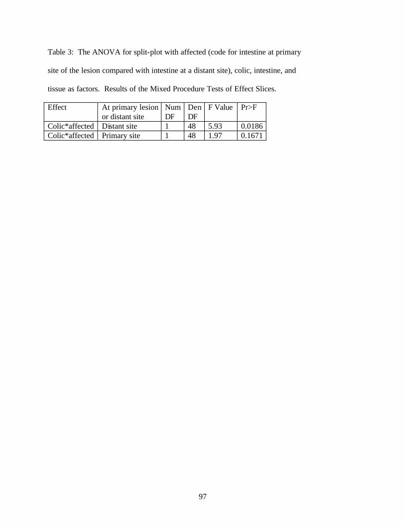
97
Table 3: The ANOVA for split-plot with affected (code for intestine at primary
site of the lesion compared with intestine at a distant site), colic, intestine, and
tissue as factors. Results of the Mixed Procedure Tests of Effect Slices. Effect At primary lesion
or distant site Num DF
Den DF
F Value Pr>F
Colic*affected Distant site 1 48 5.93 0.0186 Colic*affected Primary site 1 48 1.97 0.1671

98
CONCLUSION
Apoptotic cells were detected in the equine intestine using the TUNEL method. This
method was successful in detecting the apoptotic nuclei which stained a distinctive brown color.
Horses with gastrointestinal disease had a greater number of apoptotic cells compared to horses
with no gastrointestinal disease. Horses with simple obstructive disease had a higher number of
apoptotic cells in the circula r muscle compared to the other tissue layers in horses with
strangulation or no gastrointestinal disease. There was also a higher number of apoptotic cells in
the large colon compared to the small intestine for all the samples. There was a trend for a
higher number of apoptotic cells in the circular muscle of intestine over all. This finding of
increased apoptosis in the presence of disease may be of clinical importance as apoptosis has
been shown to be associated with inflammation and tissue injury in other species both in the
gastrointestinal system and other body systems.
There were a significantly higher number of apoptotic cells found in intestine distant to
the primary lesion in horses with strangulating obstruction compared to those with simple
obstruction and compared to the intestine taken at the primary strangulating obstruction lesion or
simple obstruction lesion. This indicated that intestine which is not directly affected by a lesion
has increased apoptosis which may be the result of systemic inflammatory mediators or free
radical production. This is may be important clinically as the intestine which remains following
a resection anastomosis for a strangulating lesion has increased apoptosis based on this finding.
Further investigation is required to determine if increased apoptosis causes inflammation and
tissue injury and affects intestinal function. As apoptosis can be controlled at the mitochondrial
level there may be potential therapeutic implications for the future. Apoptosis and the associated
inflammation has been inhibited in human smooth muscle and bovine and murine kidney.

99
VITA
Name : Emma Lucy Rowe
Date of Birth: 02/20/75
Tertiary education: Murdoch University School of Veterinary Studies 1992-1997
Bachelor of Science 1995
Bachelor of Veterinary Medicine and Surgery (Hons.) 1997