Detecting Pelvic Disease With Duplex Ultrasound Ron Bush, MD, FACS Midwest Vein & Laser Center...
20
Detecting Pelvic Disease With Duplex Ultrasound Ron Bush, MD, FACS Midwest Vein & Laser Center Dayton, Ohio
-
Upload
jonah-hart -
Category
Documents
-
view
218 -
download
0
Transcript of Detecting Pelvic Disease With Duplex Ultrasound Ron Bush, MD, FACS Midwest Vein & Laser Center...
Detecting Pelvic Disease WithDuplex Ultrasound
Ron Bush, MD, FACSMidwest Vein & Laser Center
Dayton, Ohio
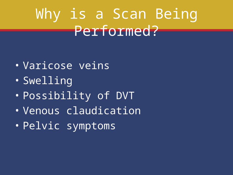
Why is a Scan Being Performed?
• Varicose veins• Swelling• Possibility of DVT• Venous claudication• Pelvic symptoms
Consider
• An ultrasound (US) of the pelvis includes an US of the femoral vein
• Abnormal femoral US occurs frequently in pelvic compression or obstruction
• Phasic flow is the normal pattern on doppler US
Ultrasound Findings: Cardiac
• Back and forth pulsatile flow (roller coaster) on US may indicate:
• ASD• Tricuspid regurgitation• AV fistula
Ultrasound Findings: Obesity
• Alteration in normal flow pattern• May be evidence of reflux at femoral &
popliteal level• Usually symmetric• Often overlooked as cause of venous
hypertension of the extremities
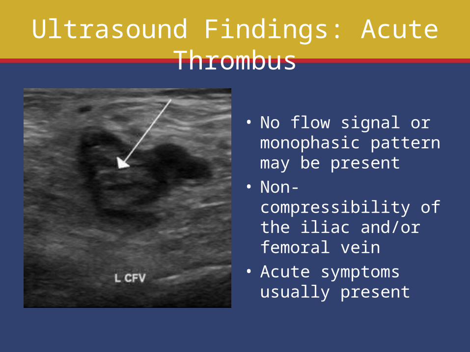
Ultrasound Findings: Acute Thrombus
• No flow signal or monophasic pattern may be present
• Non-compressibility of the iliac and/or femoral vein
• Acute symptoms usually present
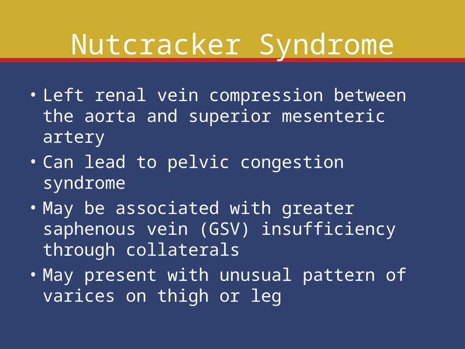
Nutcracker Syndrome
• Left renal vein compression between the aorta and superior mesenteric artery
• Can lead to pelvic congestion syndrome• May be associated with greater saphenous
vein (GSV) insufficiency through collaterals• May present with unusual pattern of varices
on thigh or leg
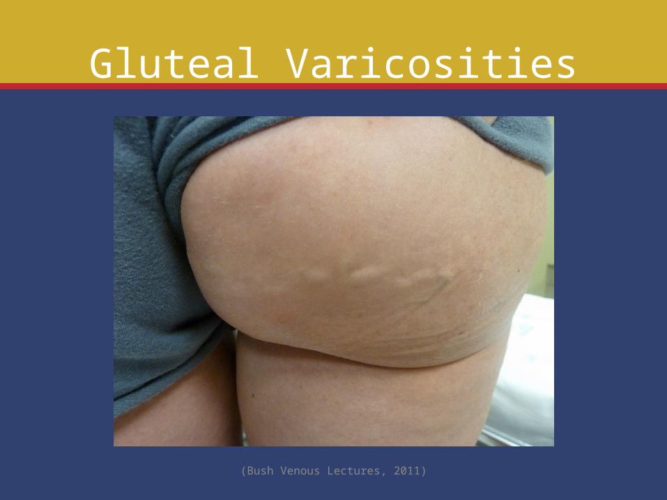
Gluteal Varicosities
(Bush Venous Lectures, 2011)

Ultrasound of LRV Compression
(Bush Venous Lectures, 2011)
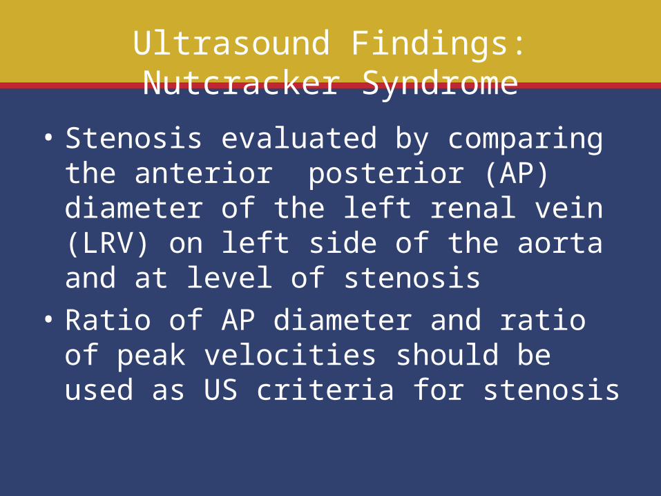
Ultrasound Findings: Nutcracker Syndrome
• Stenosis evaluated by comparing the anterior posterior (AP) diameter of the left renal vein (LRV) on left side of the aorta and at level of stenosis
• Ratio of AP diameter and ratio of peak velocities should be used as US criteria for stenosis
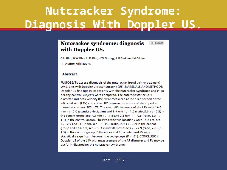
Nutcracker Syndrome: Diagnosis With Doppler US.
(Kim, 1996)
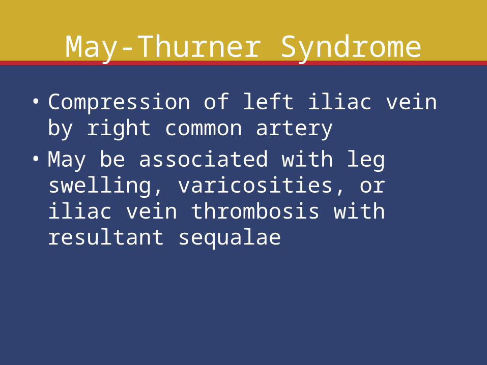
May-Thurner Syndrome
• Compression of left iliac vein by right common artery
• May be associated with leg swelling, varicosities, or iliac vein thrombosis with resultant sequalae
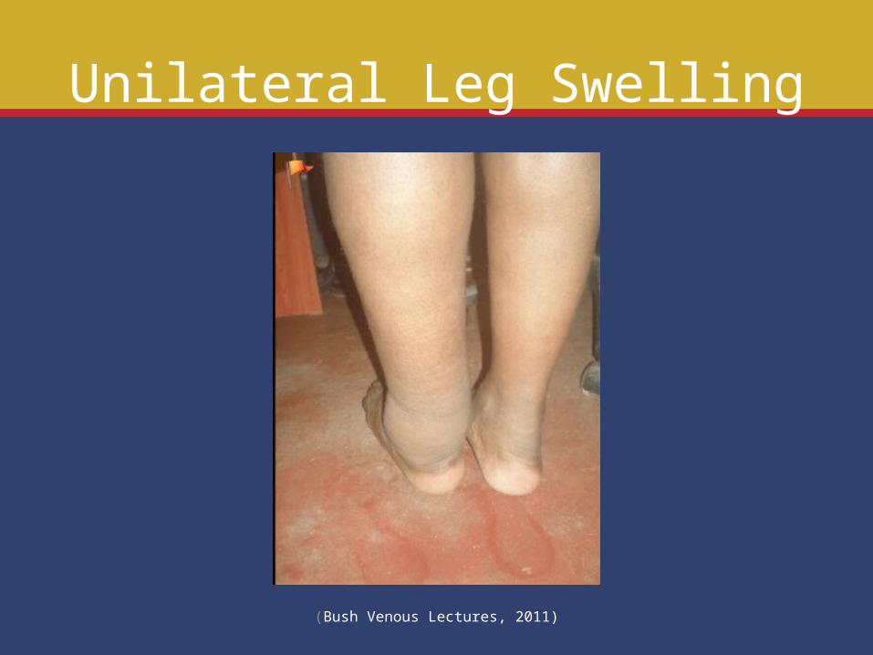
Unilateral Leg Swelling
(Bush Venous Lectures, 2011)
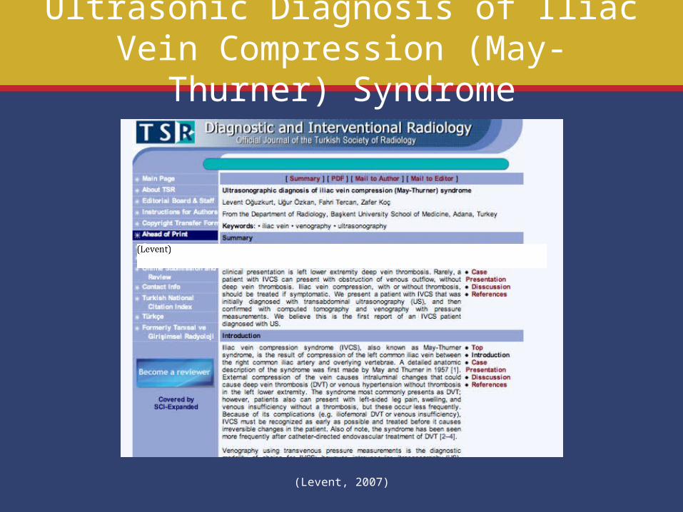
Ultrasonic Diagnosis of Iliac Vein Compression (May-Thurner) Syndrome
(Levent, 2007)
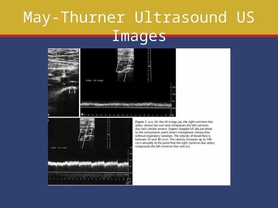
May-Thurner Ultrasound US Images
Ultrasound Findings: May-Thurner Syndrome
• Evidence of compression• Monophasic flow may be present distal to
obstruction• High velocity flow at area of stenosis
Ovarian Vein Reflux
• Manifestations may Include
• Pelvic congestive syndrome• Vulvar varices• Inguinal or thigh varices • Ovarian vein > 4mm
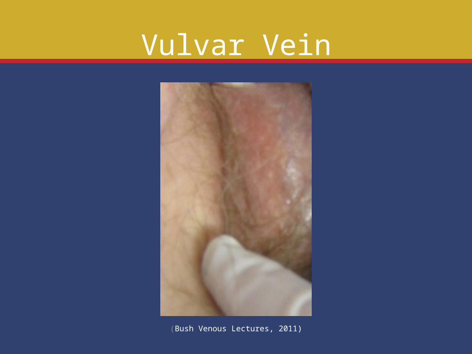
Vulvar Vein
(Bush Venous Lectures, 2011)
Conclusion
Any of the following should alert you to altered flow patterns in the pelvic veins:
• Varicosities in unusual locations• Unilateral leg swelling• Acute thrombosis of the iliac vein• Reflux or monophasic flow on femoral US• Vulvar varices
References Thombosis femoral vein image. Retrieved May 7, 2011 online from
http://www.answers.com/topic/deep-vein-thrombosis.
Nutcracker image. Retrieved May 7, 2011 online from www.phlebolymphology.org/2009/07/nutcracker-syndrome/
Kim SH, Cho SW, Kim HD, Chung JW, Park JH, Han MC. Nutcracker syndrome: diagnosis with
Doppler US. Radiology. 1996;198:93-97. Leg image. Retrieved May 7, 2011 online from
http://www.bushvenouslectures.com/blog/content.asp?id=348Oguzkurt L, Ozkan U, Tercan F, Koc Z, Sadick N, Trelies, M. A clinical histological and computer-based assessment of the Polaris LV, combination diode, and radio frequency system for leg treatment. Diagn Interv Radiol 2007;13:152-155.