Desvelando el ciclo del HCV
13
Leading Edge Review Cell 125, June 16, 2006 ©2006 Elsevier Inc. 1041 Introduction The transport of molecules between the nucleus and the cytoplasm is necessary for the exchange of information within the eukaryotic cell. Historically, the nuclear pore complex (NPC) has been presented as a static channel through which dynamic transport receptor-cargo com- plexes traverse the nuclear envelope. However, recent evidence illustrates that the NPC is itself a dynamic, adaptive macromolecular complex whose components directly influence the status of cargo transport within the cell. This view is supported by new higher-resolution images of the NPC ultrastructure. In addition, genomic and proteomic advances have allowed large-scale direct manipulation of the NPC, providing new insights into the translocation mechanism. Nuclear transport can be controlled by alteration of NPC composition and by transient association of dynamic pore components. The NPC may undergo structural changes during different transport or cell cycle stages. There is also remarkable emerging evidence of dynamic coupling between NPCs and transcriptionally active gene loci. The focus of this review is to re-evaluate our current understanding of nucleocytoplasmic exchange and to envision the NPC as an ever-changing portal with the potential to regulate crucial cellular functions both locally and globally. Nucleocytoplasmic Transport At the very edge of the nucleus, a pathway for movement of molecules across the nuclear envelope is provided by the aqueous channels within the NPCs. In the past, the function of the NPC has been relegated to constitutive maintenance of the nuclear permeability barrier. Molecules smaller than 25–40 kDa can passively diffuse across the NPC; however, highly efficient localization and all mac- romolecular transport events require facilitated, energy- dependent transport (reviewed in Fried and Kutay, 2003). Such active transport is dependent on either an intrinsic capacity to interact directly with the NPC or association with different types of transport receptors. The largest class of transport receptors is designated karyopherins, or Kaps, and includes different importins, exportins, and transportins that may be specialized for import or export. A very well characterized Kap, importin β (Kap95 in yeast), binds to import cargo through an adaptor protein, importin α (Kap60 in yeast), and has been shown to inter- act with several NPC proteins (reviewed in Pemberton and Paschal, 2005). The directionality of Kap-mediated transport is exquisitely controlled by the small GTPase Ran, whose nucleotide-bound state dictates cargo bind- ing and release in a compartmentalized manner (reviewed in Fried and Kutay, 2003). Overall, active nuclear trans- port is a highly orchestrated, rapid, and efficient process. Estimates from both in vitro transport rate studies and single-molecule imaging analysis suggest an import rate capacity of 1000 molecules per second per NPC, requir- ing that at least 10 molecules must traverse a given NPC simultaneously (Ribbeck and Gorlich, 2001; Yang et al., 2004). Thus, the NPC is capable of orchestrating multiple concurrent transport events. The NPC is a huge macromolecular assembly with a calculated mass of 40 and 60 MDa in yeast and verte- brates, respectively (Cronshaw et al., 2002; Rout et al., 2000). Despite the difference in size, the basic architec- ture of the NPC is conserved between yeast and meta- zoa. The overall structure of the NPC can be superficially divided into three basic elements: the nuclear basket, the central core, and the cytoplasmic fibrils. Recently, state of the art cryoelectron tomography with three- dimensional reconstruction has been used to present the highest-resolution structure (approximately 9 nm) of the NPC to date (Beck et al., 2004). This structure shows that the cytoplasmic filaments adopt a highly “kinked” Dynamic Nuclear Pore Complexes: Life on the Edge Elizabeth J. Tran 1 and Susan R. Wente 1, * 1 Department of Cell and Developmental Biology, Vanderbilt University Medical Center, U-3209 MRBIII, 465 21 st Avenue South, Nashville, TN 37232 USA *Contact: [email protected] DOI 10.1016/j.cell.2006.05.027 The exchange of molecules between the nucleus and cytoplasm is mediated through nuclear pore complexes (NPCs) embedded in the nuclear envelope. Altering the interac- tions between transport receptors and their cargo has been shown to be a major regulatory mechanism to control traffic through NPCs. New evidence now suggests that NPC proteins play active roles in translocation, and that transport is also controlled by dynamic changes in NPC composition and architecture. This view of ever-changing NPCs necessitates the re-evaluation of current models of nuclear transport and how this process is regulated.
description
Muy buena review completa sobre el ciclo de vida de lhcv
Transcript of Desvelando el ciclo del HCV

Leading Edge
Review
Dynamic Nuclear Pore Complexes: Life on the EdgeElizabeth J. Tran1 and Susan R. Wente1,*1Department of Cell and Developmental Biology, Vanderbilt University Medical Center, U-3209 MRBIII, 465 21st Avenue South, Nashville, TN 37232 USA*Contact: [email protected] 10.1016/j.cell.2006.05.027
The exchange of molecules between the nucleus and cytoplasm is mediated through nuclear pore complexes (NPCs) embedded in the nuclear envelope. Altering the interac-tions between transport receptors and their cargo has been shown to be a major regulatory mechanism to control traffic through NPCs. New evidence now suggests that NPC proteins play active roles in translocation, and that transport is also controlled by dynamic changes in NPC composition and architecture. This view of ever-changing NPCs necessitates the re-evaluation of current models of nuclear transport and how this process is regulated.
IntroductionThe transport of molecules between the nucleus and the cytoplasm is necessary for the exchange of information within the eukaryotic cell. Historically, the nuclear pore complex (NPC) has been presented as a static channel through which dynamic transport receptor-cargo com-plexes traverse the nuclear envelope. However, recent evidence illustrates that the NPC is itself a dynamic, adaptive macromolecular complex whose components directly influence the status of cargo transport within the cell. This view is supported by new higher-resolution images of the NPC ultrastructure. In addition, genomic and proteomic advances have allowed large-scale direct manipulation of the NPC, providing new insights into the translocation mechanism. Nuclear transport can be controlled by alteration of NPC composition and by transient association of dynamic pore components. The NPC may undergo structural changes during different transport or cell cycle stages. There is also remarkable emerging evidence of dynamic coupling between NPCs and transcriptionally active gene loci. The focus of this review is to re-evaluate our current understanding of nucleocytoplasmic exchange and to envision the NPC as an ever-changing portal with the potential to regulate crucial cellular functions both locally and globally.
Nucleocytoplasmic TransportAt the very edge of the nucleus, a pathway for movement of molecules across the nuclear envelope is provided by the aqueous channels within the NPCs. In the past, the function of the NPC has been relegated to constitutive maintenance of the nuclear permeability barrier. Molecules smaller than 25–40 kDa can passively diffuse across the NPC; however, highly efficient localization and all mac-romolecular transport events require facilitated, energy-dependent transport (reviewed in Fried and Kutay, 2003).
Such active transport is dependent on either an intrinsic capacity to interact directly with the NPC or association with different types of transport receptors. The largest class of transport receptors is designated karyopherins, or Kaps, and includes different importins, exportins, and transportins that may be specialized for import or export. A very well characterized Kap, importin β (Kap95 in yeast), binds to import cargo through an adaptor protein, importin α (Kap60 in yeast), and has been shown to inter-act with several NPC proteins (reviewed in Pemberton and Paschal, 2005). The directionality of Kap-mediated transport is exquisitely controlled by the small GTPase Ran, whose nucleotide-bound state dictates cargo bind-ing and release in a compartmentalized manner (reviewed in Fried and Kutay, 2003). Overall, active nuclear trans-port is a highly orchestrated, rapid, and efficient process. Estimates from both in vitro transport rate studies and single-molecule imaging analysis suggest an import rate capacity of 1000 molecules per second per NPC, requir-ing that at least 10 molecules must traverse a given NPC simultaneously (Ribbeck and Gorlich, 2001; Yang et al., 2004). Thus, the NPC is capable of orchestrating multiple concurrent transport events.
The NPC is a huge macromolecular assembly with a calculated mass of 40 and 60 MDa in yeast and verte-brates, respectively (Cronshaw et al., 2002; Rout et al., 2000). Despite the difference in size, the basic architec-ture of the NPC is conserved between yeast and meta-zoa. The overall structure of the NPC can be superficially divided into three basic elements: the nuclear basket, the central core, and the cytoplasmic fibrils. Recently, state of the art cryoelectron tomography with three-dimensional reconstruction has been used to present the highest-resolution structure (approximately 9 nm) of the NPC to date (Beck et al., 2004). This structure shows that the cytoplasmic filaments adopt a highly “kinked”
Cell 125, June 16, 2006 ©2006 Elsevier Inc. 1041

Figure 1. NPC Assembly and Nup Dynamics Are Correlated with Proposed Function within the NPCA cross-section of an NPC is shown, with the central region magnified in the right three panels. General structural features are based on data re-viewed in Schwartz (2005) and Table 1. The order of Nup assembly (late to early) into NPCs following mitosis (Hetzer et al., 2005, and references therein) and the relative Nup NPC residence time or shuttling activity (stable to transient) (Griffis et al., 2002; Pritchard et al., 1999; Rabut et al., 2004) are illustrated. The predicted function (structural to transport) for each Nup or subcomplex is also shown. Common colors between the three figures in each structure indicate correlations in assembly, dynamics, and function. Those that are early in assembly and stable in residence time are likely structural in function. In contrast, those that are late in assembly and transient in residence time are likely directly involved in transport.
structure, yielding a length of approximately 35 nm. A similar bent conformation is observed for the nuclear basket filaments, indicating that a transport complex travels 120 nm instead of the 200 nm distance previously predicted (Stoffler et al., 2003).
NPCs are composed of approximately 30 proteins, collectively called nucleoporins (Nups) (summarized in Cronshaw et al., 2002; Rout et al., 2000). These Nups have long been classified into groups based on the pres-ence or absence of primary sequence motifs. A conver-gence of recent direct structural analysis and structural modeling studies have gone one step further and now reveal three striking protein structure fold types that might constitute more than 85% of the complex (Devos et al., 2004, 2006; Rout et al., 2000). At the pore mem-brane, at least three proteins with transmembrane heli-ces, the first structural fold, have been functionally and structurally linked to the NPC (Mansfeld et al., 2006; Schwartz, 2005). These membrane-anchored proteins are presumably directly connected to a second struc-tural module composed of Nups of two specific fold types, the β propeller and α solenoid. Finally, a third dis-tinct group of peripheral Nups comprises at least a third of the total NPC mass and contains the FG Nups.
The FG Nups are so named for discrete domains har-boring repetitive stretches of Phe-Gly residues separated by polar spacers of variable length (reviewed in Rout and Wente, 1994). The FG domains are directly implicated
1042 Cell 125, June 16, 2006 ©2006 Elsevier Inc.
in active nuclear transport (see below). Interestingly, the FG domains themselves are thought to be natively unfolded (Denning et al., 2003). This would allow them to potentially have multiple topological locations in the NPC based on their site of anchoring by their respective non-FG domains. The non-FG domains of the FG Nups have predicted coiled-coil, β propeller, or unique β sandwich structures (Nup98 fold) (Hodel et al., 2002; Weirich et al., 2004). These might assemble on to the structural scaf-fold provided by the β propeller-α solenoid core (Devos et al., 2004, 2006, and references therein). In effect, the unfolded FG domains may coat the scaffold.
The prevalence of the β propeller and α solenoid domains was surprising, and a modular view of the NPC at the edge of the nuclear envelope has emerged (reviewed in Schwartz, 2005 and Figure 1). Moreover, the unfolded nature of the FG domains is a key tenet of nuclear transport models. Finally, intriguing structural similarities between these β propeller and α solenoid Nups and proteins in the Golgi-mediated transport sys-tem suggest common ancestral origins in evolution.
The NPC Is DynamicAs the static images in Figure 1 suggest, NPC structure could easily be viewed as being of a defined composition with each Nup at permanent locales. A wealth of studies has changed this view at multiple levels. In fact, the NPCs may undergo several types of dynamic modifications. This
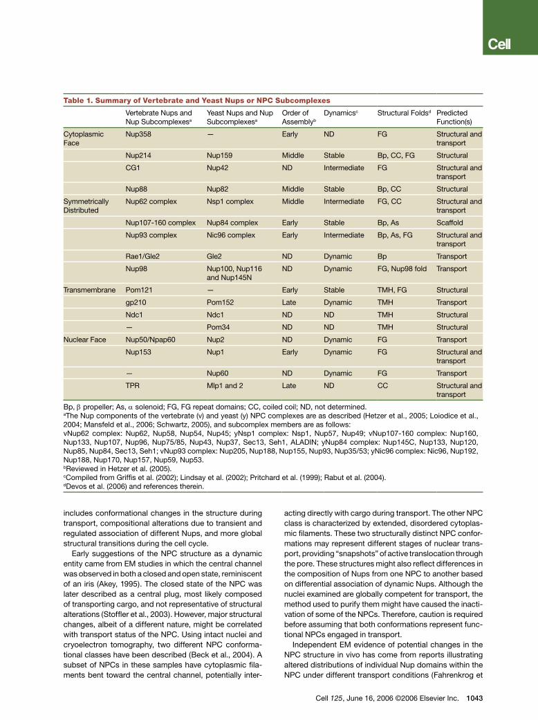
Table 1. Summary of Vertebrate and Yeast Nups or NPC Subcomplexes
Vertebrate Nups and Nup Subcomplexesa
Yeast Nups and Nup Subcomplexesa
Order of Assemblyb
Dynamicsc Structural Foldsd Predicted Function(s)
Cytoplasmic Face
Nup358 — Early ND FG Structural and transport
Nup214 Nup159 Middle Stable Bp, CC, FG Structural
CG1 Nup42 ND Intermediate FG Structural and transport
Nup88 Nup82 Middle Stable Bp, CC Structural
Symmetrically Distributed
Nup62 complex Nsp1 complex Middle Intermediate FG, CC Structural and transport
Nup107-160 complex Nup84 complex Early Stable Bp, As Scaffold
Nup93 complex Nic96 complex Early Intermediate Bp, As, FG Structural and transport
Rae1/Gle2 Gle2 ND Dynamic Bp Transport
Nup98 Nup100, Nup116 and Nup145N
ND Dynamic FG, Nup98 fold Transport
Transmembrane Pom121 — Early Stable TMH, FG Structural
gp210 Pom152 Late Dynamic TMH Transport
Ndc1 Ndc1 ND ND TMH Structural
— Pom34 ND ND TMH Structural
Nuclear Face Nup50/Npap60 Nup2 ND Dynamic FG Transport
Nup153 Nup1 Early Dynamic FG Structural and transport
— Nup60 ND Dynamic FG Transport
TPR Mlp1 and 2 Late ND CC Structural and transport
Bp, β propeller; As, α solenoid; FG, FG repeat domains; CC, coiled coil; ND, not determined.aThe Nup components of the vertebrate (v) and yeast (y) NPC complexes are as described (Hetzer et al., 2005; Loiodice et al., 2004; Mansfeld et al., 2006; Schwartz, 2005), and subcomplex members are as follows:vNup62 complex: Nup62, Nup58, Nup54, Nup45; yNsp1 complex: Nsp1, Nup57, Nup49; vNup107-160 complex: Nup160, Nup133, Nup107, Nup96, Nup75/85, Nup43, Nup37, Sec13, Seh1, ALADIN; yNup84 complex: Nup145C, Nup133, Nup120, Nup85, Nup84, Sec13, Seh1; vNup93 complex: Nup205, Nup188, Nup155, Nup93, Nup35/53; yNic96 complex: Nic96, Nup192, Nup188, Nup170, Nup157, Nup59, Nup53.bReviewed in Hetzer et al. (2005).cCompiled from Griffis et al. (2002); Lindsay et al. (2002); Pritchard et al. (1999); Rabut et al. (2004).dDevos et al. (2006) and references therein.
includes conformational changes in the structure during transport, compositional alterations due to transient and regulated association of different Nups, and more global structural transitions during the cell cycle.
Early suggestions of the NPC structure as a dynamic entity came from EM studies in which the central channel was observed in both a closed and open state, reminiscent of an iris (Akey, 1995). The closed state of the NPC was later described as a central plug, most likely composed of transporting cargo, and not representative of structural alterations (Stoffler et al., 2003). However, major structural changes, albeit of a different nature, might be correlated with transport status of the NPC. Using intact nuclei and cryoelectron tomography, two different NPC conforma-tional classes have been described (Beck et al., 2004). A subset of NPCs in these samples have cytoplasmic fila-ments bent toward the central channel, potentially inter-
acting directly with cargo during transport. The other NPC class is characterized by extended, disordered cytoplas-mic filaments. These two structurally distinct NPC confor-mations may represent different stages of nuclear trans-port, providing “snapshots” of active translocation through the pore. These structures might also reflect differences in the composition of Nups from one NPC to another based on differential association of dynamic Nups. Although the nuclei examined are globally competent for transport, the method used to purify them might have caused the inacti-vation of some of the NPCs. Therefore, caution is required before assuming that both conformations represent func-tional NPCs engaged in transport.
Independent EM evidence of potential changes in the NPC structure in vivo has come from reports illustrating altered distributions of individual Nup domains within the NPC under different transport conditions (Fahrenkrog et
Cell 125, June 16, 2006 ©2006 Elsevier Inc. 1043
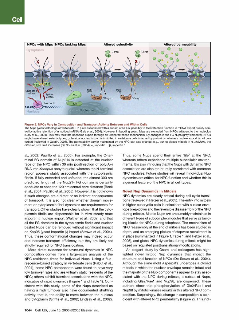
Figure 2. NPCs Vary in Composition and Transport Activity Between and Within CellsThe Mlps (yeast orthologs of vertebrate TPR) are associated with a subset of NPCs, possibly to facilitate their function in mRNA export quality con-trol by active retention of unspliced mRNA (Galy et al., 2004). However, in budding yeast, Mlps are excluded from NPCs adjacent to the nucleolus (Galy et al., 2004). This may facilitate ribosome export through an uncharacterized mechanism. By changes in the FG Nups (gray filaments), NPCs might have altered selectivity; e.g., classical nuclear import is inhibited in vertebrate cells infected by poliovirus, whereas nuclear export is not per-turbed (reviewed in Gustin, 2003). The permeability barrier maintained by the NPC can also change; e.g., during closed mitosis in A. nidulans, the diffusion size limit increases (De Souza et al., 2004). α, importin α; β, importin β.
al., 2002; Paulillo et al., 2005). For example, the C-ter-minal FG domain of Nup214 is detected at the nuclear face of the NPC within 30 min postinjection of poly(A+) RNA into Xenopus oocyte nuclei, whereas the N-terminal region appears stably associated with the cytoplasmic fibrils. If fully extended and unfolded, the almost 300 nm predicted length of the Nup214 FG domain is certainly adequate to span the 120 nm central core distance (Beck et al., 2004; Paulillo et al., 2005). However, it is not known if such changes are a direct or an indirect consequence of transport. It is also not clear whether domain move-ment or cytoplasmic fibril dynamics are requirements for transport. Other studies have clearly shown that the cyto-plasmic fibrils are dispensable for in vitro steady-state importin β nuclear import (Walther et al., 2002) and that all the FG domains in the cytoplasmic fibrils and nuclear basket Nups can be removed without significant impact on Kap95 (yeast importin β) import (Strawn et al., 2004). Thus, these conformational changes may indeed occur and increase transport efficiency, but they are likely not strictly required for NPC translocation.
More direct evidence for structural dynamics in NPC composition comes from a large-scale analysis of the NPC residence times for individual Nups. Using a fluo-rescence-based strategy in vertebrate cells (Rabut et al., 2004), some NPC components were found to have very low turnover rates and are virtually static residents of the NPC; others exhibit transient associations with the NPC, indicative of rapid dynamics (Figure 1 and Table 1). Con-sistent with this study, some of the Nups described as having a high turnover also have documented shuttling activity, that is, the ability to move between the nucleus and cytoplasm (Griffis et al., 2002; Lindsay et al., 2002).
1044 Cell 125, June 16, 2006 ©2006 Elsevier Inc.
Thus, some Nups spend their entire “life” at the NPC, whereas others experience multiple subcellular environ-ments. It is also intriguing that the Nups with dynamic NPC association are also structurally correlated with common NPC modules. Future studies will reveal if individual Nup dynamics are critical for NPC function and whether this is a general feature of the NPC in all cell types.
Novel Nup Dynamics in MitosisNPC dynamics are clearly critical during cell cycle transi-tions (reviewed in Hetzer et al., 2005). The entry into mitosis in higher eukaryotic cells is coincident with nuclear enve-lope breakdown and the reversible disassembly of the NPC during mitosis. Mitotic Nups are presumably maintained in different types of subcomplex modules that serve as build-ing blocks for NPCs during telophase. The mechanism of NPC reassembly at the end of mitosis has been studied in depth, and an emerging picture of stepwise recruitment is in place (summarized in Figure 1, Table 1, and Hetzer et al., 2005), and global NPC dynamics during mitosis might be based on regulated posttranslational modifications.
An elegant study by Osami and colleagues has high-lighted novel mitotic Nup dynamics that impact the structure and function of NPCs (De Souza et al., 2004). Although the slime mold Aspergillis undergoes a closed mitosis in which the nuclear envelope remains intact and the majority of the Nup components appear to stay asso-ciated with the NPC during mitosis, a subset of Nups, including Gle2/Rae1 and Nup98, are dispersed. These authors show that phosphorylation of Gle2/Rae1 and Nup98 by mitotic kinases results in this altered NPC com-position. Surprisingly, this change in composition is coin-cident with altered NPC permeability (Figure 2). This indi-

cates that at least some fungal species have ways to alter the nuclear permeability barrier without an open mitosis and complete NPC disassembly. Alterations of the NPC during the cell cycle may not be exclusive to this fungal species, as partial disassembly via release of peripheral Nups has also been noted for starfish oocytes prior to nuclear envelope breakdown (Lenart et al., 2003).
In contrast, the budding yeast Saccharomyces cerevi-siae NPC has not been reported to undergo any level of mitotic NPC disassembly. However, one study has dem-onstrated that specific transport pathways are altered during the budding yeast cell cycle (Makhnevych et al., 2003). This occurs by molecular rearrangements of Nup associations within the NPC. During interphase, Nup53 is bound to Nup170; however, during mitosis, Nup53 is no longer bound to Nup170 and is instead associated with Nic96. This rearrangement, likely a result of Nup53 phosphorylation, results in exposure of a high-affinity Kap121 binding site on Nup53 and inhibition of Kap121-dependent transport. How this alteration in Kap121 transport impacts mitotic progress remains unresolved. Taken together, these recent studies have demonstrated that structural and compositional changes in the NPC are mechanisms by which transport flux and individual pathways can be regulated.
Several NPC components whose function in transport is documented have also been tied to cellular activities distinct from their roles at the nuclear envelope. A wealth of recent data has documented the localization of Nups at kinetochores during mitosis. Multiple members of the ver-tebrate Nup107–160 complex show this kinetochore asso-ciation, as does Nup358 (reviewed in Chan et al., 2005). The Gle2/Rae1-Nup98 complex serves as an excellent example to illustrate the complex roles for Nups in mitotic mechanisms. Using a Xenopus egg extract system for mitotic spindle formation, Gle2/Rae1 was identified as a necessary component for promotion of microtubule assembly (Blower et al., 2005). GLE2/RAE1 depletion by RNAi in HeLa cells resulted in clear defects in spindle assembly, illustrating that Gle2/Rae1 has a role at this stage of mitosis. In a contrasting report, however, the van Deursen lab identified the Gle2/Rae1-Nup98 complex as an inhibitor of the anaphase promoting complex (APC) in a mouse model system (Jeganathan et al., 2005). These results suggested that Gle2/Rae1-Nup98 temporally con-trols the onset of anaphase by preventing untimely securin degradation mediated by the APC. Interestingly, the latter study saw no defect in spindle assembly. Therefore, the precise role of the Gle2/Rae1-Nup98 complex in mitosis is far from resolved.
Are All NPCs the Same?Several nuclear transport receptors exhibit differential requirements in multicellular organisms. For example, there are Kap family members with well documented changes in expression during development and, in some cases, tissue-specific expression of variants within an organism (reviewed in Poon and Jans, 2005). Such com-
positional variance was not generally thought to be the case for the Nups themselves. Several reports now sug-gest that the composition of the NPC can indeed be var-ied in a manner that might regulate transport. A notable recent example is that described above for the Aspergil-lis NPC changes during the cell cycle. Other mechanisms are highlighted here by which each NPC could be con-sidered structurally and functionally unique. With NPCs having the capacity to change or be altered to modulate transport pathways, it directly follows that the composi-tion of all NPCs may vary from tissue to tissue in a mul-ticellular organism, between developmental stages, and within the nuclear envelope of a single cell (Figure 2).
One example of a NPC protein that exhibits tissue-specific expression levels is gp210, a transmembrane spanning protein with dynamic NPC association (Rabut et al., 2004). In mouse embryos, gp210 levels vary widely across different tissues compared to Nup62 (Olsson et al., 2004). In another case, mutants of members only in Drosophila, a homolog of vertebrate Nup88, exhibit cell-type specific phenotypes that are linked to specific import defects (Uv et al., 2000). Given that NPC com-position varies among differentiated cell types, it is not surprising that NPCs are not the same during different stages of cell development. Early evidence for such compositional changes in the NPC arose from studies of Nup50 (Npap60) in rat spermatogenesis (Fan et al., 1997). Nup50 is 10–20 times more abundant in testis than in other rat tissues and exhibits strikingly different cellular localization patterns depending on the sperm maturation stage. For example, Nup50 is associated with the NPC in spermatocytes but is nucleoplasmic in spermatids, whereas other NPC components remain associated with the nuclear envelope. However, a role for the dynamic Nup50 localization patterns in chang-ing transport pathways has not been determined. In contrast, a more recent study has proposed a role for a splice variant of Nup358, BS-63 (Cai et al., 2002). BS-63 is found only in testis and associates with the NPC in spermatids. Interestingly, a yeast two-hybrid study revealed direct interaction between BS-63 and a germ cell-specific transcription factor, AF10 (Cai et al., 2002). AF10 may be imported through direct interaction with the NPC through BS-63, similar to β catenin. As previ-ously suggested, BS-63 may act as a developmental stage-specific docking site for specific transport recep-tor complexes (Cai et al., 2002).
Evidence of compositional variation of the NPC cer-tainly challenges the dogma that all NPCs within the same cell are identical. Mlp1 and Mlp2 (homologous to the ver-tebrate TPR) are highly similar coiled-coil proteins asso-ciated with the nuclear basket of the NPC in yeast. Strik-ingly, these proteins are not equally distributed around the nuclear envelope rim. Instead, they are excluded from regions adjacent to the nucleolus and are found only near NPCs associated with presumably active chromatin (Galy et al., 2004). Thus, Mlp proteins may be components of only a subset of NPCs, indicating that NPCs can be dis-
Cell 125, June 16, 2006 ©2006 Elsevier Inc. 1045

tinct in composition within a given cell. More importantly, this compositional difference has functional conse-quences. The Mlps have been linked to multiple aspects of nuclear physiology, including maintenance of mRNA export fidelity (Galy et al., 2004). These results are indic-ative of a coupling of upstream events, such as mRNA biogenesis, with nuclear transport through the activity of specific Nups and NPC-associated proteins (see below). As compared to NPCs without Mlps, the Mlp-associated NPCs might be committed to specific transport func-tions such as quality control in mRNA export through active mRNA retention (Figure 2). Although not yet docu-mented experimentally, the non-Mlp NPCs localized near the nucleolus might be specialized for transport events required in ribosome biogenesis.
Why Have a Dynamic Pore?The precise role of an actively adaptive pore in trans-port processes is not well characterized. However, the simplest scenario positions the NPC as a regulator of nucleocytoplasmic exchange. This hypothesis stands in clear contrast to the current field of transport, which places the vast majority of regulation on modulation of the Kap-cargo interaction (reviewed in Poon and Jans, 2005). Mechanisms for regulation of Kap-cargo interac-tions include both masking of a localization signal within the cargo and posttranslational modification of sites near or within the localization signal. These alterations affect the ability of a Kap to recognize its cargo or modulate the affinity of the Kap-cargo interaction. These changes also provide regulation of a specific nuclear transport pathway, a critical feature for any regulatory mechanism. How then would dynamic changes in the NPC modulate a precise transport event rather than globally affecting transport? One possibility is that multiple, nonequivalent transport pathways exist within a given pore complex.
The existence of multiple pathways is supported by bio-physical studies illustrating rates of import incompatible with single pathway models (Yang et al., 2004; Kubitscheck et al., 2005). There are an estimated 128 FG domains har-boring thousands of total FG repeats within a given NPC in yeast (Rout et al., 2000). The FG repeats provide mul-tiple, low-affinity NPC binding sites for transport receptor complexes during active transport (Ribbeck and Gorlich, 2001). Early evidence for distinct FG pathways came from the finding that a budding yeast mutant defective in the function of one FG Nup (nsp1-S5) only impaired distinct import pathways (Nehrbass et al., 1993). To fully investi-gate the contributions of specific FG domains within the yeast NPC, large-scale deletion analysis was performed across the entire complement of yeast Nups (Strawn et al., 2004). This work generated NPCs with minimal repertoires of FG domains and pinpointed the functionally important FG domains. The asymmetrically positioned FG domains in the peripheral NPC fibril structures are not essential. Perhaps more interesting, analysis of the Kap transport pathways in the minimal FG mutants revealed that differ-ent Kaps require different subsets of FG domains. This
1046 Cell 125, June 16, 2006 ©2006 Elsevier Inc.
in vivo data correlates with independent studies showing preferential in vitro binding of different Kaps to various FG Nups (Aitchison et al., 1996; Allen et al., 2001). Therefore, functionally independent routes through the NPC indeed exist, each potentially using the same biochemical mech-anism of translocation (see below) but through different binding sites in the pore. The presence of such multiple pathways would allow regulation of specific transport pathways at the level of the NPC.
Additional support for such a multiple transport path-way model comes from studies of viral inhibition of nuclear transport (reviewed in Gustin, 2003). Members of the picornavirus family (which includes poliovirus and rhinovirus) specifically target two FG Nups, Nup153 and Nup62, for degradation and effectively inhibit classical nuclear import without affecting export (Figure 2). It is speculated that viruses inhibit protein import to evade host immune responses while maintaining other cellular activities necessary for viral proliferation (Gustin, 2003). Selective degradation of specific Nups is also a char-acteristic of cellular defense mechanisms. Interestingly, apoptosis globally inhibits nuclear transport by degrad-ing both peripherally located Nups (the FG Nups Nup358, Nup214, and Nup153) as well two Nups within the NPC core without altering the overall NPC structure (Patre et al., 2006). These reports support a model whereby mul-tiple pathways through the NPC are controlled by dis-tinct Nup components and overall NPC composition.
A Unified Model for Translocation through the NPC?Several models have been proposed in recent years speculating on the mechanism for facilitated movement through the NPC. These include the virtual gating model, the affinity gradient model, the selective phase partition model, and the oily spaghetti model (summarized in Fried and Kutay, 2003). A unifying feature of these models is that each invokes some type of facilitated diffusion con-trolled by association and disassociation of transport receptors with FG Nups. FG Nups have also been thought critical for maintenance of the NPC permeability barrier (Ribbeck and Gorlich, 2001; Rout et al., 2000). Results again stemming from genetic analysis of NPC function in yeast have impacted the models for NPC translocation. First, the FG domains present at the asymmetric NPC peripheral structures are not strictly required, and there is no preference for FG domain type at the fibril structures (Strawn et al., 2004; Zeitler and Weis, 2004). This means that translocation is not driven by sequential transport receptor binding to different FG Nups with progressively increasing FG affinity from one NPC face to the other. In addition, the peripheral FG domains are not essential for trapping at or recruiting transport receptors to the NPC (although they may contribute to transport efficiency for some pathways). Second, nearly half of the total FG mass of the NPC can be deleted without affecting cell viability, and there is no correlation between the amount of FG mass deleted and the impact on active transport. Moreover, diffusive permeability measurements in sev-

Figure 3. Individual Nups and NPC-Associated Factors Actively Facilitate Nuclear Transport(A) The Gle2/Rae1-Nup98 complex is involved in targeting the mRNA export receptor, TAP/NXF1, to the NPC during mRNA export (Blevins et al., 2003) as well as promoting recycling of hnRNPs by affecting nuclear import (Fontoura et al., 2000).(B) Both Ran and Dbp5, drivers of protein or mRNA export respectively, are targeted to the NPC cytoplasmic face and spatially activated by NTPase activating factors (AP). On the left, the role of Ran in nuclear export is illustrated with the respective binding protein (BP) Nup358 and GTPase ac-tivating protein (RanGAP) indicated (Fried and Kutay, 2003). SUMOylation is required for RanGAP association (Matunis et al., 1998). In the middle, Dbp5 and its putative function in mRNA export is shown. Docking of Dbp5 at yeast Nup159 (vertebrate Nup214) results in juxtaposition with the NPC-associated protein, Gle1 (Hodge et al., 1999; Schmitt et al., 1999). Association of IP6 with Dbp5/Gle1 results in activation of Dbp5 ATPase activity (A.R. Alcázar-Román, E.J.T., S. Guo, and S.R.W., unpublished data). In the right merged panel, the common NTPase binding and activation mechanism in both export pathways is illustrated.
eral of the minimal FG NPCs did not reveal any defects (Strawn et al., 2004). Other studies have suggested that non-FG Nups in the NPC core structure are required for maintenance of the permeability barrier (Galy et al., 2003; Shulga et al., 2000). These findings indicate that the FG domains do not merely form a static permeability barrier and are, instead, involved in specific pathways. Indeed, the NPC structures required for active facilitated trans-location might be physically distinct and not coupled to those required for forming the permeability barrier.
Whereas some FG domains are critical for given trans-port receptor pathways, it is possible that the sheer bulk of FG mass in the NPC has been maintained in the NPC to allow for high transport efficiency. As a result of these in vivo studies and biophysical measurements of NPC residence times for model transport substrates, a new model has emerged, termed reduction of dimensionality (Peters, 2005). This model proposes that FG domains line the inner surface of the NPC, whereas the polar spac-ers separating the FG domains form outstretched loops within the NPC interior. This topological arrangement of FG domains would restrict Kap-cargo movement in only two dimensions, facilitating rapid transport by restricting random movement within the pore. Certainly, FG bulk mass would not be strictly required as long as enough were present to provide an appropriate binding surface. Presentation of a model that includes recent findings of
NPC dynamics is a daunting challenge. Definitive resolu-tion of the translocation mechanism will require further careful manipulation of the number and location of FG Nup binding sites in discrete NPC substructures.
Nups as Active Participants in TransportEvidence presented thus far demonstrates that the NPC is a dynamic macromolecular complex, which can spe-cifically regulate transport pathways. Although the NPC is not a static portal, the role of the NPC in transport could still be passive. However, several studies have now characterized specific Nups that function as direct transport facilitators and play an active role in transport. One example involves the role of Nup98, a dynamic FG Nup, in facilitating transport. First, Nup98 associates with RCC1 (Fontoura et al., 2000), the Ran guanine exchange factor (RanGEF) that promotes disassembly of Kap-cargo import complexes in the nucleus by catalyzing the exchange of GDP for GTP by Ran. Second, Nup98 also interacts with transportin (Fontoura et al., 2000), the Kap which recognizes an M9 import sequence in hnRNP A1 (Pemberton and Paschal, 2005). Interaction between transportin and Nup98 is inhibited by the presence of the M9 domain, suggesting that Nup98 may actively displace cargo from transportin receptor complexes at the NPC (Figure 3A). Indeed, Nup98 harbors a compet-ing M9-like sequence. A possible model for the role of
Cell 125, June 16, 2006 ©2006 Elsevier Inc. 1047

these interactions in nucleocytoplasmic transport would include initial binding of transportin to Nup98 followed by release of hnRNP A1. Localized generation of RanGTP at Nup98 by RCC1 would subsequently promote release of transportin from Nup98, facilitating recycling of this receptor back into the cytoplasm for future transport events. This putative mechanism may facilitate efficient disassembly of receptor complexes but may also aid rapid movement of cargo across the nuclear envelope by removing the need for receptor translocation across the entire length of the nuclear pore. However, as the precise cellular location of the Nup98 interaction with RCC1 has not been identified, it is also possible that it occurs within the nucleus itself rather than at the NPC.
Recent studies have identified other specific motifs in distinct Nups that broaden this paradigm for Nup-Kap binding in a mechanism for releasing cargo. Nup50 (Npap60) has a unique Kap binding site that interferes with Kap-cargo interactions (Gilchrist et al., 2002; Mat-suura and Stewart, 2005). In fact, Nup50 (yeast Nup2) uses a unique domain to actively displace importin α-bound nuclear localization sequences. This activity may promote efficient receptor-cargo complex disassembly upon import into the nucleus. Interestingly, importin α has two Nup50 binding sites, one of which overlaps with the Cse1 binding site (Matsuura and Stewart, 2004), the cognate export receptor for importin α. These over-lapping sites have led to the proposal that Nup50 may use these unique binding properties to couple nuclear import complex disassembly with rapid recycling of import receptors back to the cytoplasm (Matsuura and Stewart, 2005). Such Nup-mediated receptor complex dissociation and release may be a critical component of transport efficiency.
Another example of direct involvement of Nups in nucleocytoplasmic exchange is again provided by the Gle2/Rae1-Nup98 complex. Recently, the mRNA export receptor TAP/NXF1 was shown to bind to both Gle2/Rae1 and Nup98 individually (Blevins et al., 2003). In fact, Gle2/Rae1 and TAP/NXF1 associate with Nup98 at different binding sites to form a stable, ternary complex (see Figure 3A). Interaction between Nup98 and Gle2/Rae1 was, however, mutually exclusive with association between TAP/NXF1 and Gle2/Rae1. Therefore, a mecha-nism can be envisioned whereby Gle2/Rae1 may actively deliver TAP/NXF1 to the NPC and promote “hand-off” to other Nups, facilitating rapid mRNA export (Figure 3A).
The fact that the Gle2/Rae1-Nup98 complex functions in both the export of mRNA and import (recycling) of mRNA export factors into the nucleus illustrates that this complex is an active player in the gene expression path-way. Why have one complex involved in two fundamen-tal transport activities that are both required for mRNA export? This coupling of transport processes to a spe-cific protein complex would enable rapid and efficient control of gene expression. In fact, the vesicular sto-matitis virus (VSV) exploits this regulatory switch. VSV is a minus strand virus that upon infection specifically
1048 Cell 125, June 16, 2006 ©2006 Elsevier Inc.
inhibits host cell mRNA export through a virally encoded protein called M (Her et al., 1997). Gle2/Rae1 bridges the interaction between M protein and Nup98, leading to the conclusion that inhibition of the Gle2/Rae1-Nup98 com-plex is the means by which the M protein blocks mRNA export (Faria et al., 2005; von Kobbe et al., 2000). In fur-ther support of this model, increased Gle2 levels fully restore host cell mRNA export. Interestingly, both Gle2/Rae1 and Nup98 are upregulated during the cellular anti-viral response by interferon γ (Faria et al., 2005). There-fore, cells have a mechanism in place to “titrate” the viral M protein and restore cellular functions by exploiting the same molecular switch within the NPC. Overall, it is clear that viruses use specific components within the NPC to alter nuclear transport.
Nup Coupling Sites for the Direction of Transport and Cargo ModificationWhereas Nups can actively function in transport through both dynamic association with the NPC and direct interactions with transport receptors (e.g., Kaps or TAP/NXF1), reports of Nups acting as docking sites for other cellular transport factors provide further evi-dence for active roles of Nups in transport. This docking scenario is best illustrated by Nup358. The vertebrate Nup358, a component of the cytoplasmic fibrils, func-tions as a molecular “Grand Central Station” for disas-sembly of transport complexes and recycling of trans-port factors. Nup358/RanBP2 harbors four Ran binding domains and a stable interaction site for a SUMOylated Ran GTPase-activating factor, RanGAP1 (Matunis et al., 1998). SUMO modification has been shown to influence protein-protein interactions (reviewed in Hay, 2005). Strikingly, Nup358 functions directly in SUMO modi-fication of RanGAP1 along with the SUMO E2 ligase, Ubc9, by acting as an E3 ligase to stabilize a SUMO-modified RanGAP1 at the cytoplasmic fibrils (Pichler et al., 2002). As localization of RanGAP to the cytoplasm is a key determinant of directionality (reviewed in Fried and Kutay, 2003), it is clear that Nup358 is a critical component of nuclear transport.
Heterogeneous nuclear ribonucleoproteins (hnRNPs) may also be substrates for this SUMOylation complex, and SUMO modification of at least two hnRNPs (hnRNP C and hnRNP M) reduces their affinity for RNA (Vassi-leva and Matunis, 2004). HnRNP C proteins have been characterized as non-shuttling mRNA binding proteins (Pinol-Roma and Dreyfuss, 1992), and SUMOylation may aid in rapid recycling of SUMO-modified hnRNPs back into the nucleus and prevent their free diffusion into the cytoplasm. The fact that SENP2 (a SUMO protease in vertebrates) and Ulp1 (a SUMO deconjugating enzyme in yeast) have been structurally and functionally connected to the NPC is intriguing (Panse et al., 2003; Zhang et al., 2002). One group has shown connections between Ulp1 and the nuclear-faced Mlps and Nup60 in yeast, and others have Ulp1 anchored by Nup-associated Kaps (Panse et al., 2003; Zhao et al., 2004). This may allow for

an elegant cycle of SUMOylation of hnRNPs, or other transport cargo, in the cytoplasm, followed by rapid removal of the SUMO modification in the nucleus. How-ever, Ubc9 has also been localized to the nuclear basket, most likely via Nup153 (Zhang et al., 2002). Therefore, the precise mechanism of SUMO-mediated recycling of hnRNPs remains undetermined.
In addition to a role for SUMO modification in trigger-ing hnRNP release from mRNA, critical protein remodel-ing events in mRNA export are believed to be catalyzed at the NPC by the essential DEAD-box helicase protein, Dbp5. Like Ubc9, Dbp5 is also specifically localized at the cytoplasmic face of the NPC by association with a peripheral Nup, namely Nup214 (Nup159 in yeast) (Hodge et al., 1999; Schmitt et al., 1999). Using budding yeast as a model system, two independent laboratories have pro-vided compelling evidence for highly localized activation of Dbp5 in mRNA export. Whereas recombinant Dbp5 has a very low intrinsic ATPase activity, association with Gle1, an essential NPC-associated mRNA export factor, activates Dbp5 ATPase activity in a manner that is stim-ulated by soluble inositol hexakisphosphate (IP6) (A.R. Alcázar-Román, E.J.T., S. Guo, and S.R.W., unpublished data; K. Weis, personal communication). IP6 might serve as a stable cofactor for Gle1 activation of Dbp5; however, there is also the potential for regulation of inositide pools in response to extracellular stimuli. In yeast, Dbp5 is also functionally connected to Mex67, the yeast ortholog of TAP/NXF1. As dbp5 mutants have increased levels of Mex67-associated RNA (Lund and Guthrie, 2005), local-ized activation of Dbp5 at the NPC cytoplasmic face is likely required to facilitate recycling of Mex67 or other mRNA bound proteins into the nucleus for subsequent rounds of mRNA export.
In Figure 3B, the striking parallel control mechanisms that spatially dictate transport factor activities for the Kap and mRNA export pathways are shown. At the NPC cytoplasmic face, the Kap pathway requires Ran GAP for stimulating Ran GTPase activity (reviewed in Fried and Kutay, 2003), and the mRNA export pathway requires Gle1 and IP6 cofactors for stimulating Dbp5 ATPase activity. Thus, an NTPase activating protein is critical for both. In addition, juxtaposition of the respec-tive proteins is mediated by specific Nup binding sites and enhanced by potentially regulatory factors (SUMO modification and IP6, respectively).
Gene Gating: Functional Connectivity through the NPCIn addition to providing docking sites for enzymatic activi-ties associated with transport, recent evidence now illus-trates that Nups may regulate gene expression through functional connectivity in a process called “gene gating.” Gene gating was originally proposed in 1985 by Blobel (Blobel, 1985) and is based on the observation that NPCs do not distribute randomly around the nuclear envelope. This arrangement was predicted to mirror the distribution of transcriptionally active chromatin within the nucleus,
aiding in reformation of the nuclear envelope following mitosis as well as providing proper NPC distribution. Additionally, it would create a targeted pathway for export of mRNA transcripts to specific NPCs during interphase. It is now clear from multiple reports that certain Nups play an active role in both mitosis and NPC assembly (reviewed in Hetzer et al., 2005). However, only in the last few years have Nups been proposed to function in tether-ing transcriptionally active genes to the NPC.
The strongest support for the gene gating proposal comes from two reports by Silver and coworkers. Using ChIP assays and DNA microarray analysis, this report demonstrated that certain Nups associated prefer-entially with transcriptionally active genes in budding yeast (Casolari et al., 2004). Strikingly, genes under the control of the GAL promoter relocated to the nuclear periphery upon induction of transcription at these gene loci. This relocation was also observed for genes induced by addition of the α mating factor (Casolari et al., 2005). Both of these results correlated directly with alterations in gene expression.
Prior to these studies, however, several reports had linked components of the NPC to types of chromatin silencing such as boundary activity (the ability to pre-vent the spread of heterochromatin) (Ishii et al., 2002). How then can components of the NPC be linked to such opposing roles in gene expression? By examining the role of Nup2 in yeast, a protein previously shown to exhibit boundary activity when artificially tethered to the chromosome (Ishii et al., 2002), this precise question was recently addressed. Utilizing yeast DNA microarrays containing intergenic regions, in contrast to the ORF-specific microarrays used previously (Caso-lari et al., 2004, 2005), Aitchison and coworkers have found that Nup2 associates with chromatin at regions between heterochromatin and euchromatin (Dilworth et al., 2005). These investigators conclude that the chromatin association is mediated by formation of a tetrameric complex including Nup2, Nup60 (a nuclear basket component), Prp20 (yeast RanGEF), and a his-tone variant, Htz1 (Dilworth et al., 2005 and references therein). Htz1 has been proposed to function in bound-ary activity (Meneghini et al., 2003).
Using an innovative method of chromatin mapping, Laemmli and coworkers have pinpointed the interac-tion of Nup2 with the promoters of activated genes. This association is dependent on upstream transcriptional activating sequences and the TATA box (Schmid et al., 2006). The Laemmli and Aitchison studies now provide the first connection between Nups and factors that con-trol transcription. Moreover, Nup interactions with pro-moters may be an important mechanism for control of gene expression through a modified model of the gene gating theory. In this model, the NPC would function as a switchboard for gene expression regulation by providing a sorting site for activities such as transcriptional activa-tion and mRNA quality control (Figure 2). This functional connectivity would enable entire gene expression net-
Cell 125, June 16, 2006 ©2006 Elsevier Inc. 1049

works as well as regulatory elements to be linked from gene to cytoplasm through the NPC.
Given that all of the current supporting evidence for gene gating has been found in budding yeast, it leads one to question the evolutionary conservation of this phenomenon. In fact, cellular imaging of mammalian cells has revealed that mRNPs move within the nucleus primarily by diffusion (Shav-Tal et al., 2004). This find-ing would suggest a lack of physical gating of gene transcripts to the NPC. However, a recent report now illustrates that the NPC components are essential fac-tors required for dosage compensation in Drosophila, a phenomenon characterized by hypertranscription of the hemizygous male X chromosome (Mendjan et al., 2006). This work is consistent with gene gating in higher eukaryotes; however, more experiments are necessary to determine if gene gating is indeed a ubiquitous, evolu-tionarily conserved mechanism for gene expression.
Evolution of Today’s NPCThe fact that a single complex (and its components) can function in multiple different activities both directly and indirectly linked to its primary cellular role makes unraveling the origin of the modern day NPC challeng-ing. As has been proposed for the ribosome (Moore, 1993), the NPC is likely the result of fusion of multiple distinct activities followed by adaptation and refine-ment. The first of these activities would include both diffusive permeability and selective transport. The abil-ity to tether transport-specific effectors and directly control transport events would have been later addi-tions that were preserved during evolution as activities that influence transport efficiency.
As a continuous nuclear envelope dividing the chro-matin from the cytosol clearly cannot function without a transport portal, it is likely that the first rudimentary pore and any associated Nups would have originated in the first eukaryote upon creation of the first nucleus. How-ever, recent extensive phylogenetic analyses have illus-trated that several components of the soluble transport machinery, including Ran and Kaps, may be of bacte-rial origin (Mans et al., 2004). Notably absent in bacteria are ancestral homologs of Nups, although characteris-tic WD40 and TPR repeats are present. These findings led Koonin and coworkers to propose that the NPC was pieced together by contributions of domains from bacte-rial proteins (Mans et al., 2004). As this report notes, the absence of NPC components in bacteria is not support-ive of an endosymbiotic origin for the nucleus. There-fore, the critical unanswered question is: what was the evolutionary trigger that sparked formation of double-membrane compartments characteristic of eukaryotes? In contrast, identification of a shared domain struc-ture, namely the β propeller-α solenoid arrangement, between both components of both the NPC and COPII Golgi-mediated transport vesicles suggests a common evolutionary origin (Devos et al., 2004, 2006). In fact, this finding implicates a “protocoatomer” ancestor of
1050 Cell 125, June 16, 2006 ©2006 Elsevier Inc.
clathrin-coated vesicles whose evolutionary divergence resulted in components of both vesicular transport and the NPC. However, the profound lack of sequence con-servation is not supportive of a divergent evolutionary model for two relatively recent adaptations. Instead, we support a convergent evolutionary model whereby bacterial motifs such as the WD40 β propeller fold were used in two independent membrane bound structures.
Interestingly, the FG motifs characteristic of all eukaryotic NPCs, are not observed in bacteria and are likely a eukaryotic invention present in the last common eukaryotic ancestor (Mans et al., 2004). This suggests that early nuclear diffusive permeability barriers did not contain FG motifs. In fact, for the modern-day NPC, the non-FG Nups play key roles in forming the foundations of the permeability barrier. Whereas deletion of FG domains themselves has not been shown to impact dif-fusive permeability (Strawn et al., 2004), the absence of different non-FG Nups (yeast Nup170 or Nup188, and C. elegans Nup205 or Nup93) results in “leaky pores” (Shulga et al., 2000; Galy et al., 2003). Instead, FG motifs may have coevolved along with gene duplication and diversification of the Kap-based transport system, yielding the 20 different importin β paralogs and 10 or more FG Nups found in human cells today. This pro-posed scenario is consistent with the fact that bulk FG mass is not required for transport but specific FG Nups are necessary for distinct transport pathways (Strawn et al., 2004). However, given the lack of a true ancestral archetype, the evolutionary origin of the NPC is likely to remain speculative.
Future ChallengesGiven that the NPC is a dynamic structure that can adapt to both transport status and cellular cues, a future chal-lenge to the field will be the establishment of a testable model for nuclear transport. This model must incorpo-rate all of the following: not all NPCs are identical, the NPC actively rearranges both during transport and dur-ing the cell cycle, there are multiple different transport pathways through the NPC, and transport-associated activities can be coupled to the NPC. Clearly, this is a daunting challenge and requires studies in multiple experimental systems including both in vitro and whole animal models. A more immediate challenge, however, is understanding the coordination between transport and other cellular functions of Nups. As components of the NPC are clearly involved in different stages of mitosis, their precise roles in cell cycle, as well as their poten-tial functional connections to transport, remain to be determined. In fact, a complete functional catalog of Nups involved in mitosis may reveal intricate connec-tivities between reformation of the NPC as well as rees-tablishment of transport following mitosis, as originally suggested in the gene gating hypothesis (Blobel, 1985). Finally, with the “static NPC” model vanquished, investi-gations of Nups as regulatory entities may reveal elusive disease mechanisms.
ACkNoWLEDgmENTs
We thank our colleagues in the Wente laboratory for critical input. E.J.T. acknowledges support from a Ruth Kirschstein NIH NSRA 1F32GM075459-01 and NSRA NIH Virus, Nucleic Acids, and Cancer position on grant 5T32CA009385. Work in the laboratory is supported by NIH grants GM51219 and GM57438 to S.R.W. S.R.W. dedicates this paper to her parents, Harold R. Wente and Elizabeth A. Wente.
REFERENCEs
Aitchison, J.D., Blobel, G., and Rout, M.P. (1996). Kap104p: A karyo-pherin involved in the nuclear transport of messenger RNA binding proteins. Science 274, 624–627.
Akey, C.W. (1995). Structural plasticity of the nuclear pore complex. J. Mol. Biol. 248, 273–293.
Allen, N.P., Huang, L., Burlingame, A., and Rexach, M. (2001). Pro-teomic analysis of nucleoporin interacting proteins. J. Biol. Chem. 276, 29268–29274.
Beck, M., Forster, F., Ecke, M., Plitzko, J.M., Melchior, F., Gerisch, G., Baumeister, W., and Medalia, O. (2004). Nuclear pore complex structure and dynamics revealed by cryoelectron tomography. Sci-ence 306, 1387–1390.
Blevins, M.B., Smith, A.M., Phillips, E.M., and Powers, M.A. (2003). Complex formation among the RNA export proteins Nup98, Rae1/Gle2 and TAP. J. Biol. Chem. 278, 20979–20988.
Blobel, G. (1985). Gene gating: A hypothesis. Proc. Natl. Acad. Sci. USA 82, 8527–8529.
Blower, M.D., Nachury, M., Heald, R., and Weis, K. (2005). A Rae1-containing ribonucleoprotein complex is required for mitotic spindle assembly. Cell 121, 223–234.
Cai, Y., Gao, Y., Sheng, Q., Miao, S., Cui, X., Wang, L., Zong, S., and Koide, S.S. (2002). Characterization and potential function of a novel testis-specific nucleoporin BS-63. Mol. Reprod. Dev. 61, 126–134.
Casolari, J.M., Brown, C.R., Komili, S., West, J., Hieronymus, H., and Silver, P.A. (2004). Genome-wide localization of the nuclear transport machinery couples transcriptional status and nuclear organization. Cell 117, 427–439.
Casolari, J.M., Brown, C.R., Drubin, D.A., Rando, O.J., and Silver, P.A. (2005). Developmentally induced changes in transcriptional pro-gram alter spatial organization across chromosomes. Genes Dev. 19, 1188–1198.
Chan, G.K., Liu, S.T., and Yen, T.J. (2005). Kinetochore structure and function. Trends Cell Biol. 15, 589–598.
Cronshaw, J.M., Krutchinsky, A.N., Zhang, W., Chait, B.T., and Matu-nis, M.J. (2002). Proteomic analysis of the mammalian nuclear pore complex. J. Cell Biol. 158, 915–927.
De Souza, C.P., Osmani, A.H., Hashmi, S.B., and Osmani, S.A. (2004). Partial nuclear pore complex disassembly during closed mitosis in As-pergillus nidulans. Curr. Biol. 14, 1973–1984.
Denning, D.P., Patel, S.S., Uversky, V., Fink, A.L., and Rexach, M. (2003). Disorder in the nuclear pore complex: The FG repeat regions of nucleoporins are natively unfolded. Proc. Natl. Acad. Sci. USA 100, 2450–2455.
Devos, D., Dokudovskaya, S., Alber, F., Williams, R., Chait, B.T., Sali, A., and Rout, M.P. (2004). Components of coated vesicles and nuclear pore complexes share a common molecular architecture. PLoS Biol. 2, e380.
Devos, D., Dokudovskaya, S., Williams, R., Alber, F., Eswar, N., Chait, B.T., Rout, M.P., and Sali, A. (2006). Simple fold composition and modular architecture of the nuclear pore complex. Proc. Natl. Acad. Sci. USA 103, 2172–2177.
Dilworth, D.J., Tackett, A.J., Rogers, R.S., Yi, E.C., Christmas, R.H., Smith, J.J., Siegel, A.F., Chait, B.T., Wozniak, R.W., and Aitchison, J.D. (2005). The mobile nucleoporin Nup2p and chromatin-bound Prp20p function in endogenous NPC-mediated transcriptional control. J. Cell Biol. 171, 955–965.
Fahrenkrog, B., Maco, B., Fager, A.M., Koser, J., Sauder, U., Ullman, K.S., and Aebi, U. (2002). Domain-specific antibodies reveal multiple-site topology of Nup153 within the nuclear pore complex. J. Struct. Biol. 140, 254–267.
Fan, F., Liu, C.P., Korobova, O., Heyting, C., Offenberg, H.H., Trump, G., and Arnheim, N. (1997). cDNA cloning and characterization of Npap60: A novel rat nuclear pore-associated protein with an unusual subcellular localization during male germ cell differentiation. Genom-ics 40, 444–453.
Faria, P.A., Chakraborty, P., Levay, A., Barber, G.N., Ezelle, H.J., En-ninga, J., Arana, C., van Deursen, J., and Fontoura, B.M. (2005). VSV disrupts the Rae1/mrnp41 mRNA nuclear export pathway. Mol. Cell 17, 93–102.
Fontoura, B.M., Blobel, G., and Yaseen, N.R. (2000). The nucleopo-rin Nup98 is a site for GDP/GTP exchange on ran and termination of karyopherin beta 2-mediated nuclear import. J. Biol. Chem. 275, 31289–31296.
Fried, H., and Kutay, U. (2003). Nucleocytoplasmic transport: Taking an inventory. Cell. Mol. Life Sci. 60, 1659–1688.
Galy, V., Mattaj, I.W., and Askjaer, P. (2003). Caenorhabditis elegans nucleoporins Nup93 and Nup205 determine the limit of nuclear pore complex size exclusion in vivo. Mol. Biol. Cell 14, 5104–5115.
Galy, V., Gadal, O., Fromont-Racine, M., Romano, A., Jacquier, A., and Nehrbass, U. (2004). Nuclear retention of unspliced mRNAs in yeast is mediated by perinuclear Mlp1. Cell 116, 63–73.
Gilchrist, D., Mykytka, B., and Rexach, M. (2002). Accelerating the rate of disassembly of karyopherin.cargo complexes. J. Biol. Chem. 277, 18161–18172.
Griffis, E.R., Altan, N., Lippincott-Schwartz, J., and Powers, M.A. (2002). Nup98 is a mobile nucleoporin with transcription-dependent dynamics. Mol. Biol. Cell 13, 1282–1297.
Gustin, K.E. (2003). Inhibition of nucleo-cytoplasmic trafficking by RNA viruses: Targeting the nuclear pore complex. Virus Res. 95, 35–44.
Hay, R.T. (2005). SUMO: A history of modification. Mol. Cell 18, 1–12.
Her, L.S., Lund, E., and Dahlberg, J.E. (1997). Inhibition of Ran guano-sine triphosphatase-dependent nuclear transport by the matrix pro-tein of vesicular stomatitis virus. Science 276, 1845–1848.
Hetzer, M.W., Walther, T.C., and Mattaj, I.W. (2005). Pushing the en-velope: Structure, function, and dynamics of the nuclear periphery. Annu. Rev. Cell Dev. Biol. 21, 347–380.
Hodel, A.E., Hodel, M.R., Griffis, E.R., Hennig, K.A., Ratner, G.A., Xu, S., and Powers, M.A. (2002). The three-dimensional structure of the autoproteolytic, nuclear pore-targeting domain of the human nucleo-porin Nup98. Mol. Cell 10, 347–358.
Hodge, C.A., Colot, H.V., Stafford, P., and Cole, C.N. (1999). Rat8p/Db-p5p is a shuttling transport factor that interacts with Rat7p/Nup159p and Gle1p and suppresses the mRNA export defect of xpo1–1 cells. EMBO J. 18, 5778–5788.
Ishii, K., Arib, G., Lin, C., Van Houwe, G., and Laemmli, U.K. (2002). Chromatin boundaries in budding yeast: The nuclear pore connection. Cell 109, 551–562.
Jeganathan, K.B., Malureanu, L., and van Deursen, J.M. (2005). The Rae1-Nup98 complex prevents aneuploidy by inhibiting securin deg-radation. Nature 438, 1036–1039.
Kubitscheck, U., Grunwald, D., Hoekstra, A., Rohleder, D., Kues, T., Siebrasse, J.P., and Peters, R. (2005). Nuclear transport of single mol-
Cell 125, June 16, 2006 ©2006 Elsevier Inc. 1051

ecules: Dwell times at the nuclear pore complex. J. Cell Biol. 168, 233–243.
Lenart, P., Rabut, G., Daigle, N., Hand, A.R., Terasaki, M., and Ellen-berg, J. (2003). Nuclear envelope breakdown in starfish oocytes pro-ceeds by partial NPC disassembly followed by a rapidly spreading fenestration of nuclear membranes. J. Cell Biol. 160, 1055–1068.
Lindsay, M.E., Plafker, K., Smith, A.E., Clurman, B.E., and Macara, I.G. (2002). Npap60/Nup50 is a tri-stable switch that stimulates im-portin-alpha:beta-mediated nuclear protein import. Cell 110, 349–360.
Loiodice, I., Alves, A., Rabut, G., Van Overbeek, M., Ellenberg, J., Sibarita, J.B., and Doye, V. (2004). The entire Nup107–160 complex, including three new members, is targeted as one entity to kineto-chores in mitosis. Mol. Biol. Cell 15, 3333–3344.
Lund, M.K., and Guthrie, C. (2005). The DEAD-box protein Dbp5p is required to dissociate Mex67p from exported mRNPs at the nuclear rim. Mol. Cell 20, 645–651.
Makhnevych, T., Lusk, C.P., Anderson, A.M., Aitchison, J.D., and Wozniak, R.W. (2003). Cell cycle regulated transport controlled by alterations in the nuclear pore complex. Cell 115, 813–823.
Mans, B.J., Anantharaman, V., Aravind, L., and Koonin, E.V. (2004). Comparative genomics, evolution and origins of the nuclear enve-lope and nuclear pore complex. Cell Cycle 3, 1612–1637.
Mansfeld, J., Guttinger, S., Hawryluk-Gara, L.A., Pante, N., Mall, M., Galy, V., Haselmann, U., Muhlhausser, P., Wozniak, R.W., Mattah, I.W., et al. (2006). The conserved transmembrane nucleoporin NDC1 is required for nuclear pore complex assembly in vertebrate cells. Mol. Cell 22, 93–103.
Matsuura, Y., and Stewart, M. (2004). Structural basis for the assem-bly of a nuclear export complex. Nature 432, 872–877.
Matsuura, Y., and Stewart, M. (2005). Nup50/Npap60 function in nuclear protein import complex disassembly and importin recycling. EMBO J. 24, 3681–3689.
Matunis, M.J., Wu, J., and Blobel, G. (1998). SUMO-1 modification and its role in targeting the Ran GTPase-activating protein, Ran-GAP1, to the nuclear pore complex. J. Cell Biol. 140, 499–509.
Mendjan, S., Taipale, M., Kind, J., Holz, H., Gebhardt, P., Schelder, M., Vermeulen, M., Buscaino, A., Duncan, K., Mueller, J., et al. (2006). Nuclear pore components are involved in the transcriptional regula-tion of dosage compensation in Drosophila. Mol. Cell 21, 811–823.
Meneghini, M.D., Wu, M., and Madhani, H.D. (2003). Conserved his-tone variant H2A.Z protects euchromatin from the ectopic spread of silent heterochromatin. Cell 112, 725–736.
Moore, P.B. (1993). Ribosomes and the RNA World. (Cold Spring Harbor: Cold Spring Harbor Laboratory Press).
Nehrbass, U., Fabre, E., Dihlmann, S., Herth, W., and Hurt, E.C. (1993). Analysis of nucleo-cytoplasmic transport in a thermosensi-tive mutant of nuclear pore protein NSP1. Eur. J. Cell Biol. 62, 1–12.
Olsson, M., Scheele, S., and Ekblom, P. (2004). Limited expression of nuclear pore membrane glycoprotein 210 in cell lines and tissues suggests cell-type specific nuclear pores in metazoans. Exp. Cell Res. 292, 359–370.
Panse, V.G., Kuster, B., Gerstberger, T., and Hurt, E. (2003). Uncon-ventional tethereing of Ulp1 to the transport channel of the nuclear pore complex by karyopherins. Nat. Cell Biol. 5, 21–27.
Patre, M., Tabbert, A., Hermann, D., Walczak, H., Rackwitz, H.R., Cordes, V.C., and Ferrando-May, E. (2006). Caspases target only two architectural components within the core structure of the nuclear pore complex. J. Biol. Chem. 281, 1296–1304.
Paulillo, S.M., Phillips, E.M., Koser, J., Sauder, U., Ullman, K.S., Powers, M.A., and Fahrenkrog, B. (2005). Nucleoporin domain to-
1052 Cell 125, June 16, 2006 ©2006 Elsevier Inc.
pology is linked to the transport status of the nuclear pore complex. J. Mol. Biol. 351, 784–798.
Pemberton, L.F., and Paschal, B.M. (2005). Mechanisms of receptor-mediated nuclear import and nuclear export. Traffic 6, 187–198.
Peters, R. (2005). Translocation through the nuclear pore complex: Selectivity and speed by reduction-of-dimensionality. Traffic 6, 421–427.
Pichler, A., Gast, A., Seeler, J.S., Dejean, A., and Melchior, F. (2002). The nucleoporin RanBP2 has SUMO1 E3 ligase activity. Cell 108, 109–120.
Pinol-Roma, S., and Dreyfuss, G. (1992). Shuttling of pre-mRNA binding proteins between nucleus and cytoplasm. Nature 355, 730–732.
Poon, I.K., and Jans, D.A. (2005). Regulation of nuclear transport: Central role in development and transformation? Traffic 6, 173–186.
Pritchard, C.E., Fornerod, M., Kasper, L.H., and van Deursen, J.M. (1999). RAE1 is a shuttling mRNA export factor that binds to a GLEBS-like NUP98 motif at the nuclear pore complex through mul-tiple domains. J. Cell Biol. 145, 237–254.
Rabut, G., Doye, V., and Ellenberg, J. (2004). Mapping the dynamic organization of the nuclear pore complex inside single living cells. Nat. Cell Biol. 6, 1114–1121.
Ribbeck, K., and Gorlich, D. (2001). Kinetic analysis of translocation through nuclear pore complexes. EMBO J. 20, 1320–1330.
Rout, M.P., and Wente, S.R. (1994). Pores for thought: Nuclear pore complex proteins. Trends Cell Biol. 4, 357–365.
Rout, M.P., Aitchison, J.D., Suprapto, A., Hjertaas, K., Zhao, Y., and Chait, B.T. (2000). The yeast nuclear pore complex: Composition, architecture, and transport mechanism. J. Cell Biol. 148, 635–651.
Schmid, M., Arib, G., Laemmli, C., Nishikawa, J., Durussel, T., and Laemmli, U.K. (2006). Nup-PI: The nucleopore-promoter interaction genes in yeast. Mol. Cell 21, 379–391.
Schmitt, C., von Kobbe, C., Bachi, A., Pante, N., Rodrigues, J.P., Boscheron, C., Rigaut, G., Wilm, M., Seraphin, B., Carmo-Fonseca, M., and Izaurralde, E. (1999). Dbp5, a DEAD-box protein required for mRNA export, is recruited to the cytoplasmic fibrils of nuclear pore complex via a conserved interaction with CAN/Nup159p. EMBO J. 18, 4332–4347.
Schwartz, T.U. (2005). Modularity within the architecture of the nu-clear pore complex. Curr. Opin. Struct. Biol. 15, 221–226.
Shav-Tal, Y., Darzacq, X., Shenoy, S.M., Fusco, D., Janicki, S.M., Spector, D.L., and Singer, R.H. (2004). Dynamics of single mRNPs in nuclei of living cells. Science 304, 1797–1800.
Shulga, N., Mosammaparast, N., Wozniak, R., and Goldfarb, D.S. (2000). Yeast nucleoporins involved in passive nuclear envelope per-meability. J. Cell Biol. 149, 1027–1038.
Stoffler, D., Feja, B., Fahrenkrog, B., Walz, J., Typke, D., and Aebi, U. (2003). Cryo-electron tomography provides novel insights into nuclear pore architecture: Implications for nucleocytoplasmic trans-port. J. Mol. Biol. 328, 119–130.
Strawn, L.A., Shen, T., Shulga, N., Goldfarb, D., and Wente, S.R. (2004). Minimal nuclear pore complexes define FG repeat domains essential for transport. Nat. Cell Biol. 6, 197–206.
Uv, A.E., Roth, P., Xylourgidis, N., Wickberg, A., Cantera, R., and Samakovlis, C. (2000). members only encodes a Drosophila nucleo-porin required for rel protein import and immune response activation. Genes Dev. 14, 1945–1957.
Vassileva, M.T., and Matunis, M.J. (2004). SUMO modification of het-erogeneous nuclear ribonucleoproteins. Mol. Cell. Biol. 24, 3623–3632.

von Kobbe, C., van Deursen, J.M., Rodrigues, J.P., Sitterlin, D., Bachi, A., Wu, X., Wilm, M., Carmo-Fonseca, M., and Izurralde, E. (2000). Vesicular Stomatitis Virus Matrix Protein Inhibits Host Cell Gene Expression by Targeting the Nucleoporin Nup98. Mol. Cell 6, 1243–1252.
Walther, T.C., Pickersgill, H.S., Cordes, V.C., Goldberg, M.W., Allen, T.D., Mattaj, I.W., and Fornerod, M. (2002). The cytoplasmic filaments of the nuclear pore complex are dispensable for selective nuclear pro-tein import. J. Cell Biol. 158, 63–77.
Weirich, C.S., Erzberger, J.P., Berger, J.M., and Weis, K. (2004). The N-terminal domain of Nup159 forms a beta-propeller that functions in mRNA export by tethereing the helicase Dbp5 to the nuclear pore. Mol. Cell 16, 749–760.
Yang, W., Gelles, J., and Musser, S.M. (2004). Imaging of single-mol-ecule translocation through nuclear pore complexes. Proc. Natl. Acad. Sci. USA 101, 12887–12892.
Zeitler, B., and Weis, K. (2004). The FG-repeat asymmetry of the nu-clear pore complex is dispensible for bulk nucleocytoplasmic trans-port in vivo. J. Cell Biol. 167, 583–590.
Zhang, H., Saitoh, H., and Matunis, M.J. (2002). Enzymes of the SUMO modification pathway localize to filaments of the nuclear pore complex. Mol. Cell. Biol. 22, 6498–6508.
Zhao, X., Wu, C.Y., and Blobel, G. (2004). Mlp-dependent anchorage and stabilization of a desumoylating enzyme is required to prevent clonal lethality. J. Cell Biol. 167, 605–611.
Cell 125, June 16, 2006 ©2006 Elsevier Inc. 1053