Synthesis, characterization and biological activity of antimony(III)
Title Design, synthesis and biological characterization of novelinhibitors of CD38
Author(s)Dong, M; Si, YQ; Sun, SY; Pu, XP; Yang, ZJ; Zhang, LR; Zhang,LH; Leung, FP; Lam, CMC; Kwong, AKY; Yue, J; Zhou, Y;Kriksunov, IA; Hao, Q; Cheung Lee, H
Citation Organic And Biomolecular Chemistry, 2011, v. 9 n. 9, p. 3246-3257
Issued Date 2011
URL http://hdl.handle.net/10722/137500
Rights This work is licensed under a Creative Commons Attribution-NonCommercial-NoDerivatives 4.0 International License.
Organic &BiomolecularChemistry
Dynamic Article Links
Cite this: DOI: 10.1039/c0ob00768d
www.rsc.org/obc PAPER
Design, synthesis and biological characterization of novel inhibitors of CD38†Q2
Min Dong,a Yuan-Qi Si,a Shuang-Yong Sun,a Xiao-Ping Pu,a Zhen-Jun Yang,a Liang-Ren Zhang,a Li-HeZhang,*a Fung Ping Leung,b Connie Mo Ching. Lam,b Anna Ka Yee Kwong,b Jianbo Yue,b Yeyun Zhou,c IrinaA. Kriksunov,c Quan Haob and Hon Cheung Lee*b
Received 23rd September 2010, Accepted 17th February 2011DOI: 10.1039/c0ob00768d
Human CD38 is a novel multi-functional protein that acts not only as an antigen for B-lymphocyteactivation, but also an enzyme catalyzing the synthesis of a Ca2+ messenger molecule, cyclicADP-ribose, from NAD+. It is well established that this novel Ca2+ signaling enzyme is responsible forregulating a wide range of physiological functions. Based on the crystal structure of the CD38/NAD+
complex, we synthesized a series of simplified N-substituted nicotinamide derivatives (Compound 1–14).A number of these compounds exhibited moderate inhibition of the NAD+ utilizing activity of CD38,with Compound 4 showing the higher potency. The crystal structure of CD38/Compound 4 complex andcomputer simulation of Compound 7 docking to CD38 show a significant role of the nicotinamidemoiety and the distal aromatic group of the compounds for substrate recognition by the active site ofCD38. Biologically, we showed that both Compounds 4 and 7 effectively relaxed the agonist-inducedcontraction of muscle preparations form rats and guinea pigs. This study is a rational design ofinhibitors for CD38 that exhibit important physiological effects, and can serve as a model for futuredrug development.
Introduction
CD38 is a trans-membrane enzyme, originally identified as alymphocyte differentiation antigen.1 It is now known to be ubiq-uitously expressed in virtually all mammalian tissues examined.2
As a multi-functional protein and a member of the ADP-ribosyl5cyclase family, CD38 catalyzes the synthesis of cyclic ADP-ribose(cADPR) from NAD+, a cyclic nucleotide messenger mediatingCa2+ release from intracellular stores in a wide range of biologicalsystems from plant to human.3 Remarkably, CD38 can alsohydrolyze the product, cADPR, and the substrate, NAD+, to10produce ADP-ribose.4 That CD38 is the naturally occurringenzyme responsible for the synthesis of cADPR has been shown byablation of the CD38 gene in mice, which results in large reductionin endogenous cADPR in many tissues.5,6 The CD38 knockoutmice exhibit a variety of defects, establishing the importance of15CD38 as a regulator of diverse physiological functions,5,6 whichinclude immune cell differentiation,7 a-adrenoceptor signaling in
aState Key Laboratory of Natural and Biomimetic Drugs, School ofPharmaceutical Sciences, Peking University, Beijing 100191, China.E-mail: [email protected]; Fax: +86-10-82802724; Tel: +86-10-82801700bDepartment of Physiology, University of Hong Kong, Hong Kong, China.E-mail: [email protected]; Fax: +852-2817-1334; Tel: +852-2819-9163cMacCHESS, Cornell High Energy Synchrotron Source, Cornell University,Ithaca, NY 14853, USA† Electronic supplementary information (ESI) available. See DOI:10.1039/c0ob00768d
aorta,8 hormonal signaling in pancreatic acinar cells,9 migration ofdendritic cell precursors,10 bone resorption,11 insulin secretion,5,12
and social behavior changes.13 Clinically, CD38 expression is a 20negative prognostic marker for chronic lymphocytic leukemia.14,15
Moreover, CD38 is responsible for synthesizing yet anotherubiquitous Ca2+ messenger, nicotinic acid adenine dinucleotidephosphate (NAADP), from NADP and nicotinic acid via a base-exchange reaction.16,17 It should now be a generally accepted fact 25that CD38 is expressed both in intracellular organelles, such as thenucleus, ER, etc., as well as on the surface of some cells, particularthe blood cells. It is our belief that internal CD38 may be morerelevant for cell signaling.
That CD38 plays key roles in physiology provides an important 30impetus for this study to design and synthesize inhibitors ofCD38. Inhibitors of the enzymatic activities of CD38 have beendescribed, but none of them have been shown to have physiolog-ical effects. Slama et al. synthesized a non-hydrolyzable analogof NAD+, dinucleotide carbanicotinamide adenine dinucleotide 35(carba-NAD+), as a competitive inhibitor of the NAD+ glycohy-drolase activity of CD38 with the IC50 about 100 mM.18,19 Inhibitorsthat form covalent intermediates with the catalytic residues ofCD38 have also been described. These inhibitors are mainly NAD+
derivatives with modifications at the nicotinamide ribonucleoside. 40For example, a series of fluoro-substituted NAD+ derivativeshas been produced.20,21 The fluoro substitution at the 2¢-positionof the nicotinamide sugar moiety promotes the formation of astable covalent bond between the ribose and Glu226, the catalytic
This journal is © The Royal Society of Chemistry 2011 Org. Biomol. Chem., 2011, xxx, 1–12 | 1

residue of CD38, during catalysis. The covalent intermediate hasbeen captured by X-ray crystallography.22 Based on the structureof ara-F NAD, Schramm and coworkers reported that ara-F NMN and several b-nicotinamide 2¢-deoxyribosides are alsopotent inhibitors of CD38 with the K i values in the nanomolar5range.21,23
These inhibitors, being derivatives of NAD+, are chargedmolecules with limited permeability to cells and tissues andnone has been reported to exhibit biological effects. It is thepurpose of this study to develop membrane-permeant CD3810inhibitors as tools for physiological investigations and as po-tential drug candidates as well. Based on the crystal structureof the CD38/NAD+ complex that we reported previously,24 wesynthesized a series of simplified N-substituted nicotinamidederivatives (Fig. 2, 1–14) and showed that some of them, including15Compounds 4 and 7, are good inhibitors of the enzymatic activitiesof CD38. X-Ray crystallography shows that Compound 4 bindsinside the catalytic cavity of CD38 and reveals important detailsof the interacting residues at the active site. Moreover, whenapplied to guinea pigs tracheal muscle strips, both Compounds204 and 7 exhibit a potent relaxing effect on the agonist-inducedtension.
Fig. 1 Inhibitors of CD38.Q4
Results and discussion
Chemistry
The crystal structure of the CD38/NAD+ complex shows that25the nicotinamide group of the bound NAD+ enters the catalyticcavity first and interacts with residues Glu146 and Asp155 atthe active site through two hydrogen bonds to its amide. Theinteraction is further enhanced by the parallel p interactionsbetween its pyridine ring and the Trp189 indole ring.24b It is30reasoned that derivatives consisting of either one nicotinamide
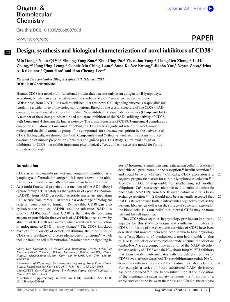
(Compounds 1–7) or bis-nicotinamide moiety (Compounds 8–11) may have sufficient affinity for the catalytic cavity of CD38to serve as inhibitors. In addition, a series of derivatives wasdesigned with aromatic ether strands replacing the adenosine- 35pyrophosphate moiety of the natural substrate, NAD+, so as toimprove the membrane permeability of the compounds. Finally,in Compounds 13 and 14, 4-amino-nicotinamide and 6-quinolinecarboxylic amide replaced and mimicked the nicotinamide ring,respectively. 40
Compounds 1 and 2 were synthesized as depicted in Scheme1.25 Starting from 1,3-dioxolane and acetyl chloride, thechloromethoxyl ethyl acetate was obtained in 62% yield, which wasstirred with nicotinamide for 10 h in DMF at room temperature26
to form Compound 1 with high yield (80%). Compound 2 was 45produced by the condensation of [2-(benzyloxy)ethoxy]methylchloride27 and nicotinamide in DMF in 60% yield.
Compounds 3–7 were synthesized by a similar strategy. Sub-stituted phenols or 8-hydroxyl-quinoline were condensed withchloroethanol in a 10% NaOH solution.28 The corresponding 50alcohols were chloromethylated by paraformaldehyde and dryHCl in DCE. The chloromethylation products were used directlyto react with nicotinamide to produce Compounds 3–7 in goodyields (Scheme 2).
8-OH-quinoline was coupled with 1,4-butenediol by Mitsunobu 55reaction in 70% yield. The chloromethylation product was ob-tained as described before and then condensed with nicotinamideto give Compound 12 in 55.8% for two steps. (Scheme 3)
The syntheses of the bis-nicotinamide Compounds 8–11 werecompleted by the condensation of the bis-chloromethylation 60products with two equivalents of nicotinamide, in high yields. 1,2-Bis- chloromethoxyethane, 1,4-bis-chloromethoxybutane, 1,4-bis-chloromethoxycrotonylene and 1,6-bis-chloromethoxyhexane29
were prepared from the corresponding dihydroxyl com-pounds by chloromethylation reaction, as described before. 65(Scheme 4)
4-Phenoxyphenol was coupled with chloroethanol and thenchloromethylated. The product was condensed with quinoline-6-carboxylic amide or 4-amino nicotinamide,30 respectively, in DMFfor 12 h at room temperature to produce Compounds 13 (63%) and 7014 (43%) in two steps (Scheme 5).
Biological activities
Inhibition of the activity of NADase. The newly synthesizedCompounds 1–14 were tested for their inhibitory properties againstthe NAD-glycohydrolase activity of the recombinant CD38, 75which was measured using a fluorimetric and highly sensitivecoupled enzyme assay as previously described.31 As shown inTable 1, compounds 3,4,5,7,12,13 and 14 exhibit weak inhibitoryactivities. Structurally, compounds with an aromatic group at thedistal end from the nicotinamide are more effective in inhibiting 80
Table 1 NADase inhibitory activity of 1–14 Q5
Inhibitor 1 2 3 4 5 6 7IC50/mMa NM NM 11.41 3.42 5.65 NM 10.00Inhibitor 8 9 10 11 12 13 14IC50/mMa NM NM NM NM 3.45 2.12 4.80
a NM = not measurable.
2 | Org. Biomol. Chem., 2011, xxx, 1–12 This journal is © The Royal Society of Chemistry 2011
Fig. 2 NAD+ and N-aromatic ether substituted nicotinamides.Q3
CD38. This is likely to be due to the enhanced hydrophobicinteractions with the active site. Compound 6 contains a strongelectron withdrawing m-CF3-phenol group that should interferewith hydrophobic interaction, and it indeed showed no affinityfor CD38. The positively charged bis-nicotinamide moiety in5Compounds 8–11 likewise should prohibit hydrophobic interactionand the compounds were, likewise, ineffective in inhibiting CD38.Interestingly, the longer aliphatic chain in Compound 12, ascompared to Compound 7, improves this compound’s interactionwith CD38. Comparing Compounds 13 and 4, replacing the nicoti-10namide with a quinoline ring, improved the affinity slightly. Onthe other hand, the additional amino group on the nicotinamidering in Compound 14 offered no improvement as compared toCompound 4.
Physiological effects on muscle preparations from rat and guinea 15pigs
It has previously been shown that CD38 gene ablation atten-uates the contraction induced by a-adrenoceptor stimulationin mouse aorta, indicating that the contraction is mediated bythe CD38/cADPR-pathway.8 We therefore tested the effect of 20compound 4 on the phenylephrine-induced contraction in rataortic ring preparations with intact endothelium. As shown in Fig.3A, compound 4 produced concentration-dependent relaxationwith a half-maximal effect (pD2) at about 36 mM. The effect isspecific because the structurally similar but inactive compound 259 produced no such vascular relaxing effect. Nicotinamide, acommonly used inhibitor of CD38, can effectively attenuate
This journal is © The Royal Society of Chemistry 2011 Org. Biomol. Chem., 2011, xxx, 1–12 | 3

Scheme 1 Synthesis of analogues 1 and 2.
Scheme 2 Synthesis of analogues 3–7.
Scheme 3 Synthesis of analogues 12.
4 | Org. Biomol. Chem., 2011, xxx, 1–12 This journal is © The Royal Society of Chemistry 2011
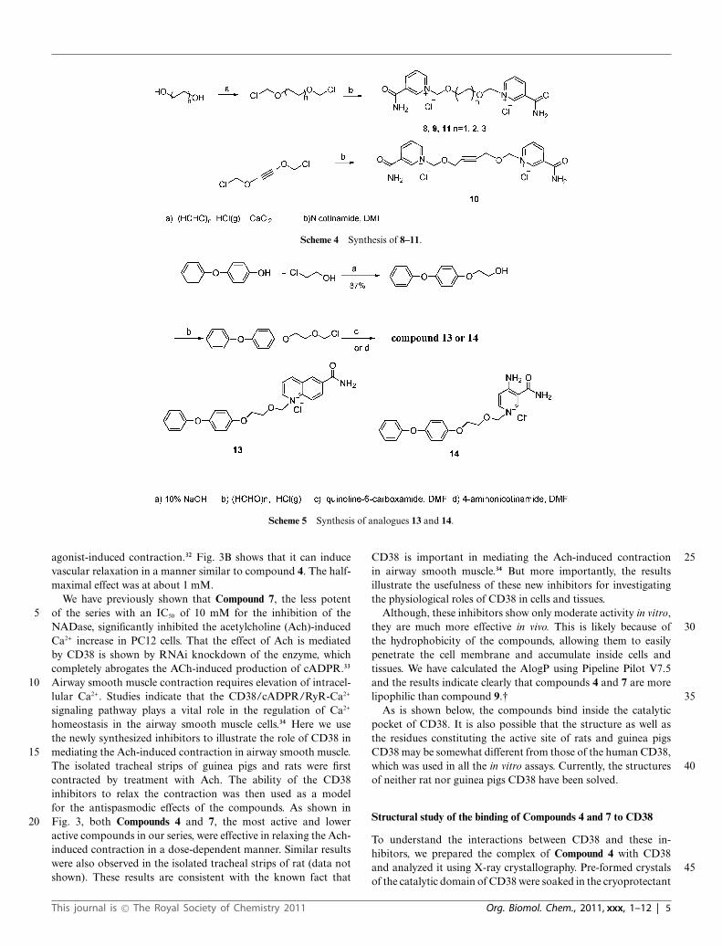
Scheme 4 Synthesis of 8–11.
Scheme 5 Synthesis of analogues 13 and 14.
agonist-induced contraction.32 Fig. 3B shows that it can inducevascular relaxation in a manner similar to compound 4. The half-maximal effect was at about 1 mM.
We have previously shown that Compound 7, the less potentof the series with an IC50 of 10 mM for the inhibition of the5NADase, significantly inhibited the acetylcholine (Ach)-inducedCa2+ increase in PC12 cells. That the effect of Ach is mediatedby CD38 is shown by RNAi knockdown of the enzyme, whichcompletely abrogates the ACh-induced production of cADPR.33
Airway smooth muscle contraction requires elevation of intracel-10lular Ca2+. Studies indicate that the CD38/cADPR/RyR-Ca2+
signaling pathway plays a vital role in the regulation of Ca2+
homeostasis in the airway smooth muscle cells.34 Here we usethe newly synthesized inhibitors to illustrate the role of CD38 inmediating the Ach-induced contraction in airway smooth muscle.15The isolated tracheal strips of guinea pigs and rats were firstcontracted by treatment with Ach. The ability of the CD38inhibitors to relax the contraction was then used as a modelfor the antispasmodic effects of the compounds. As shown inFig. 3, both Compounds 4 and 7, the most active and lower20active compounds in our series, were effective in relaxing the Ach-induced contraction in a dose-dependent manner. Similar resultswere also observed in the isolated tracheal strips of rat (data notshown). These results are consistent with the known fact that
CD38 is important in mediating the Ach-induced contraction 25in airway smooth muscle.34 But more importantly, the resultsillustrate the usefulness of these new inhibitors for investigatingthe physiological roles of CD38 in cells and tissues.
Although, these inhibitors show only moderate activity in vitro,they are much more effective in vivo. This is likely because of 30the hydrophobicity of the compounds, allowing them to easilypenetrate the cell membrane and accumulate inside cells andtissues. We have calculated the AlogP using Pipeline Pilot V7.5and the results indicate clearly that compounds 4 and 7 are morelipophilic than compound 9.† 35
As is shown below, the compounds bind inside the catalyticpocket of CD38. It is also possible that the structure as well asthe residues constituting the active site of rats and guinea pigsCD38 may be somewhat different from those of the human CD38,which was used in all the in vitro assays. Currently, the structures 40of neither rat nor guinea pigs CD38 have been solved.
Structural study of the binding of Compounds 4 and 7 to CD38
To understand the interactions between CD38 and these in-hibitors, we prepared the complex of Compound 4 with CD38and analyzed it using X-ray crystallography. Pre-formed crystals 45of the catalytic domain of CD38 were soaked in the cryoprotectant
This journal is © The Royal Society of Chemistry 2011 Org. Biomol. Chem., 2011, xxx, 1–12 | 5

Fig. 3 Biological effects of compounds 4, 7 and 9 on agonist-inducedmuscle contraction. A. Concentration-response curves for solvent (MQwater) control, compound 4 (active) and compound 9 (inactive), on thephenylephrine-induced vascular contraction in isolated rat aortic ringpreparations. B. Nicotinamide, a commonly used inhibitor of CD38,induced similar vascular relaxation. Results are means ± SEM. usingtissues from three to five rats in each group. C. The relaxing effects ofCompounds 4 and 7 on the acetylcholine-induced contraction in isolatedtracheal strips of guinea pigs. Data represent the mean ± SEM (n = 7). *P< 0.05, statistically significant as compared with Krebs–Henseleit solutiongroup.
buffer containing the compound to obtain the complex. We wereable to obtain only the complex with Compound 4 (the ESI shows
the statistics of data collection and structure refinement of thecomplex†). Fig. 4A shows that Compound 4 binds inside thecatalytic pocket of human CD38. Superimposed in the figure 5is the bound NAD previously determined by us.24 As can beseen, the nicotinamide groups of both Compound 4 and NADbind at the same position. They also interact identically with thesame residues, forming hydrogen bonds with Glu146 and Asp155,as well as hydrophobically stack with Trp189 (Fig. 4A). The 10structural results indicate that the inhibitory effect of Compound4 is likely to be due to its specific binding to the active site. TheN-substituted biphenyl ether group in Compound 4 distal to thenicotinamide ring, on the other hand, binds quite differently thanthe ribose and phosphate groups of NAD, interacting instead 15mainly with Trp176 through hydrophobic stacking (Fig. 4A).
Molecular dynamics simulation allows us to model the in-teraction of Compound 7 with CD38, even though we wereunable to obtain the crystal complex. The stimulation was carriedout starting with the crystallographic data obtained from the 20CD38/Compound 4 complex.
The result is shown in Fig. 5a and the superimposition of Q6Compounds 4 and 7 is shown in Fig. 5b. The docking studyindicated that the positioning of Compound 7 at the active site isapproximately the same as Compound 4 as observed in the crystal 25structure. Besides the same hydrogen bonding of the nicotinamidemoieties of Compound 7 and 4 with the active site,interactionsbetween the nitrogen atom of the quinoline of Compound 7 withresidue Ser186 and p-stacking interaction between the quinolinering and Trp176 of CD38 were also observed. The similarity of the 30interactions is consistent with both compounds having inhibitoryeffects on CD38.
Conclusion
There is a growing tendency to develop non-nucleotide com-pounds to mimic such signaling molecules.35 This study represents 35a rational design of a series of inhibitors of CD38, one of thekey enzymes in cellular Ca2+ signaling. The design was based onthe crystal structure of NAD binding to the active site of CD38and takes into account the need for membrane permeability. Wetargeted the nicotinamide portion of the NAD because it interacts 40strongly with the active site via not only hydrogen bonding butalso hydrophobic stacking. The reasoning was substantiated bythe crystal structure, showing that the nicotinamide portion of
Fig. 4 Structural alignment between CD38–Compound 4 and CD38–NAD complexes. (A) Surface presentation of the active pocket of CD38 (pale green).NAD (sticks presentation in magenta) penetrated to the bottom of the active pocket of CD38, while compound 4 (sticks presentation in grey) floated overthe active site. (B) The nicotinamide group of both compound 4 (grey) and NAD (magentas) is similarly positioned and stabilized by the interactionswith residues Glu146, Asp155 and Trp189 of CD38.
6 | Org. Biomol. Chem., 2011, xxx, 1–12 This journal is © The Royal Society of Chemistry 2011

Fig. 5 (a) The molecular dynamics simulation binding mode of compound 7 in the active pocket of human CD38. The carbon atoms of 7 are indicatedin cyan. All the nitrogen atoms are blue; oxygen atoms are red. Hydrogen bonds are represented by blue dotted lines. Corresponding residues are labeledand shown in blue lines. (b) Superimposition the binding poses of compound 4 in the enzyme–inhibitor cyrstal with the molecular dynamics simulationresult of 7. 4 is shown as the magenta stick; 7 is shown as the cyan stick.
Compound 4 binds identically as NAD. The replacement of the restof the highly charged moieties of the NAD with aromatic groupsensures membrane permeability and contributes to the greatlyincreased in vivo potency of the inhibitors. The potency increaseis observed both in cultured PC12 cells as we have previously5reported33 and muscle preparations from rats and guinea pigsdescribed here, verifying the general applicability of the newinhibitors. Structure–activity comparison of all the compoundsin this series provides clues for the next iteration to produce evenmore effective inhibitors and set the stage toward developing drug10candidates for CD38-related diseases.
Experiment section
Chemistry
All solvents and reagents were obtained from commercial sourcesand used without further purification unless otherwise stated.15HR-ESI-MS (electrospray ionization) was performed with BrukerBIFLEX III. 1H NMR and 13C NMR were recorded with aBruker AVANCE III 400 or a JEOL AL300 spectrometer. CD3OD,DMSO-d6 or D2O were used as a solvent. Chemical shifts arereported in parts per million downfield from TMS (1H and2013C). 19F NMR spectra were recorded on a Varian VXR-500spectrometer. Chemical shifts of 19F NMR are reported in ppmwith reference to CF3COOH as an external standard. Anhydroussolvents were obtained as follows: DMF was dried with CaH2 atroom temperature before being distilled in vacuo; ethyl acetate was25dried with P2O5 before distillation. 1,2-Dichlorethane, ethanol andmethanol were distilled over CaH2.
1-[(2-Acetoxyethoxy)methyl]-3-(aminocarbonyl)-pyridinium ch-loride (Compound 1). Dry nicotinamide (122 mg, 1 mmol) wasdissolved in dry DMF (3 mL), chloromethoxyl ethyl acetate (0.1430mL, 1 mmol) was added under N2. The mixture was stirred at roomtemperature for 1 h and a large amount of solid appeared. Dryethyl acetate (3 ml) was added and the mixture was stirred for 30min. The white precipitates were collected by filtration and washedwith dry ethyl acetate, then recrystallized from EtOH/EA to give35
the desired Compound 1 (221 mg, 80%) as a white solid. 1H NMR(300 MHz, CD3OD) d 2.03 (s, 3H), 3.98 (m, 2H),4.25 (m, 2H), 6.06(s, 2H), 8.30 (dd, 1H, J = 7.2, 5.1 Hz), 9.07 (d, 1H, J = 7.2 Hz), 9.21(d, 1H, J = 5.1 Hz), 9.53 (s, 1H).13C NMR (75 MHz, CD3OD) d20.7, 63.9, 70.8, 90.7, 129.3, 135.8, 144.2, 145.8, 146.5, 165.0, 172.4. 40ESI-MS: [M - Cl]+: 239.1. Anal. (C11H15ClN2O4·0.3H2O) C, H, N.
1-[(2-Benzyloxyethoxy)methyl]-3-(aminocarbonyl)-pyridiniumchloride (Compound 2). To a solution of 2-(benzyloxy)ethanol(0.85 ml, 6 mmol) in dry 1,2-dichloroethane (8 ml),paraformaldehyde (0.18 g, 6 mmol) was added. Dry HCl 45gas, obtained in situ from H2SO4 and NaCl, was bubbled throughthe reaction mixture over 10 h at 0 ◦C. The solution was thendried with anhydrous Na2SO4, filtered and concentrated underreduced pressure to give a yellow oily residue. The residue wasadded to a solution of nicotinamide (0.73 g, 6 mmol) in DMF 50(15 ml). The mixture was stirred at room temperature for 4 h.The white precipitates were collected by filtration and washedwith dry ethyl acetate and ethanol, then recrystallized from EtOHby freezing to give the desired Compound 2 (1.25 g, 65% for twosteps) as a white solid. 1H NMR (300 MHz, CD3OD) d 3.64 (m, 552H), 3.99 (m, 2H), 4.36 (s, 2H), 6.01 (s, 2H), 7.18–7.32 (m, 5H),8.05 (dd, 1H, J = 8.1, 6.3 Hz), 8.82 (d, 1H, J = 8.1 Hz), 9.12 (d,1H, J = 6.3 Hz), 9.45 (s, 1H).13C NMR (75 MHz, CD3OD) d165.0, 146.0, 145.7, 144.1, 139.0, 135.3, 129.5, 129.1, 129.0, 128.9,128.8, 91.4, 74.2, 73.3, 70.3. ESI-MS: [M - Cl]+ 287.1. Anal. 60(C16H19ClN2O3) C, H, N.
1-{[2-(4-Methoxy-phenoxy)ethoxy]methyl}-3-(aminocarbonyl)-pyridinium chloride (Compound 3). 4-Methoxyphenol (0.56 g,4.5 mmol) was dissolved in 10% sodium hydroxide solution (16 ml,40 mmol). 2-Chloroethanol (2.68 ml, 40 mmol) was then added 65and the mixture stirred for 24 h at room temperature. The solutionwas extracted with CH2Cl2. The extract was washed with waterthree times then dried by evaporation. The product obtained,2-(4-methoxyphenoxy)ethanol (0.68 g, 4.1 mmol, 90%), was usedin the next step directly and dissolved in 1,2-dichloroethane (10 70ml). Paraformaldehyde (0.13 g, 4.1 mmol) was added and dry HClgas was bubbled through the reaction mixture for 10 h at 0 ◦C.
This journal is © The Royal Society of Chemistry 2011 Org. Biomol. Chem., 2011, xxx, 1–12 | 7

The solution was then treated as in the synthesis of Compound 2and reacted with nicotinamide (0.5 g, 4.1 mmol) in DMF (8 ml).The mixture was stirred at room temperature for 8 h. The whiteprecipitates were collected by filtration and washed with dry ethylacetate and ethanol. The crude product was recrystallized from5EtOH/EA to obtain the desired Compound 3 (1.14 g, 82% fortwo steps) as a white solid. 1H NMR (300 MHz, CD3OD) d 3.72(s, 3H), 4.11 (m, 4H), 6.11 (s, 2H), 6.72–6.83 (m, 4H), 8.23 (dd,1H, J = 8.4, 6.0 Hz), 8.99 (dt, 1H, J = 8.4, 1.5 Hz), 9.22 (d, 1H,J = 6.0 Hz), 9.55 (s, 1H).13C NMR (75 MHz, CD3OD) d 165.0,10155.8, 153.5, 146.3, 145.7, 144.2, 135.5, 129.1, 91.2, 72.3, 68.5,56.0. ESI-MS: [M - Cl]+ 303.1. Anal. (C16H19ClN2O4) C, H, N.
1-{[2-(4-Phenoxy-phenoxy)ethoxy]methyl}-3-(aminocarbonyl)-pyridinium chloride (Compound 4). The procedure used wassimilar to that for the synthesis of Compound 3. 4-Phenoxyphenol15was condensed with 2-chloroethanol in 83% yield. The alcoholobtained was chloromethylated and reacted with nicotinamidesuccessively to yield Compound 4 (71.5% for two steps). 1H NMR(300 MHz, CD3OD) d 4.07–4.15 (m, 4H), 6.13 (s, 2H), 6.82–6.93(m, 6H), 7.04 (t, 1H, J = 7.5 Hz) 7.29 (dd, 2H, J = 8.5, 7.5 Hz)208.26 (dd, 1H, J = 8.1, 6.0 Hz), 9.02 (d, 1H, J = 8.1 Hz), 9.24 (d,1H, J = 6.0 Hz), 9.58 (s, 1H).13C NMR (75 MHz, CD3OD) d165.0, 159.7, 155.7, 152.2, 146.3, 145.7, 144.2, 135.6, 130.8, 129.1,123.8, 121.6, 118.8, 116.7, 91.1, 72.2, 68.5. ESI-MS: [M - Cl]+
365.1. Anal. (C21H21ClN2O4) C, H, N.25
1-{[2-(4-Nitro-phenoxy)ethoxy]methyl}-3-(aminocarbonyl)-py-ridinium chloride (Compound 5). The procedure used was similarto that for the synthesis of Compound 3. 4-Nitrophenol was con-densed with 2-chloroethanol in 87% yield. The alcohol obtainedwas chloromethylated and reacted with nicotinamide successively30to yield the Compound 5 (82% for two steps). 1H NMR (300 MHz,CD3OD) d 4.19 (m, 2H), 4.32 (m,2H), 6.14 (s, 2H), 7.05 (d, JAB
= 9.3 Hz,2H,A of aryl A2B2), 8.18 (d, JAB = 9.3 Hz,2H,B of arylA2B2), 8.30 (dd, 1H, J = 8.1, 6.0 Hz), 9.05 (d, 1H, J = 8.1 Hz), 9.25(d, 1H, J = 6.0 Hz), 9.58 (s, 1H).13C NMR (75 MHz, CD3OD)35d 165.0, 164.8, 146.5, 145.8, 144.2, 143.1, 135.7, 129.3, 129.2,126.8, 115.8, 90.9, 71.4, 68.8. ESI-MS: [M - Cl]+ 318.1. Anal.(C15H16ClN3O5) C, H, N.
1-{[2-(3-Trifluoromethyl-phenoxy)ethoxy]methyl}-3-(aminocar-bonyl)-pyridiniumchloride (Compound 6). The procedure follows40the synthesis of Compound 3. 3-Trifluoromethylphenol was con-densed with 2-chloroethanol in 95% yield. The alcohol obtainedwas chloromethylated and reacted with nicotinamide successivelyto yield the Compound 6 (40% for two steps). 1H NMR (300 MHz,CD3OD) d 4.16–4.25 (m, 4H), 6.13 (s, 2H), 7.12–7.25 (m, 3H),457.45 (m, 1H) 8.27 (dd, 1H, J = 8.1, 6.0 Hz), 9.03 (dt, 1H, J = 8.1,1.5 Hz), 9.24 (d, 1H, J = 6.0 Hz), 9.59 (s, 1H). 13C NMR (100MHz, CD3OD) d 164.9, 160.0, 146.4, 145.8, 144.2, 135.7, 132.9(q, JCF = 32 Hz), 131.6, 129.2, 125.4 (q, 1JCF = 270 Hz), 119.3,118.8 (q, JCF = 4 Hz), 112.4 (q, JCF = 4 Hz), 91.0, 71.8, 68.4. 19F50NMR (470 MHz, CD3OD) d 21.3. ESI-MS: [M - Cl]+ 341.1. Anal.(C16H16ClF3N2O3) C, H, N.
1-{[2-(8¢-Quinolyloxy)ethoxy]methyl}-3-(aminocarbonyl)-pyri-dinium chloride (Compound 7). The procedure follows thesynthesis of Compound 3. 8-Hydroxyqunoline was condensed55with 2-chloroethanol in 65% yield. A suspension of 2-(8¢-quinolinoxy)ethanol (1.28 g, 6.77 mmol), paraformaldehyde (0.2
g, 6.77 mmol) and 1.5 g 3 A MS in 10 ml 1,2-dichloroethane wasbubbled with HCl gas for 10 h at 0 ◦C. The molecular sieve wasfiltered and washed with DCE, the filtrate was concentrated under 60reduced pressure and treated with nicotinamide as before. Afterrecrystallized from MeOH/EA, Compound 7 was obtained as ayellow solid. (78% for two steps). 1H NMR (300 MHz, CD3OD)d 4.36 (t, 2H, J = 4.2 Hz), 4.67 (m, 2H, J = 4.2 Hz), 6.19 (s, 2H),7.70 (dd, 1H, J = 6.0, 2.7 Hz), 7.91 (m, 2H), 8.16 (dd, 1H, J = 8.4, 655.7 Hz), 8.25 (dd, 1H, J = 8.1, 6.0 Hz), 9.00 (d, 1H, J = 8.1 Hz),9.18–9.27 (m, 3H), 9.58 (s, 1H).13C NMR (75 MHz, CD3OD) d164.9, 150.0, 148.5, 146.2, 145.9, 145.5, 144.1, 135.7, 131.8, 131.7,131.0, 129.3, 123.8, 122.0, 115.3, 90.7, 71.1, 70.0. ESI-MS: [M -Cl]+ 324.2. Anal. (C18H18ClN3O3·3H2O) C, H, N. 70
1,2-Dimethoxy-ethylene-bis-N ,N ¢-3-(aminocarbonyl)-pyridini-um dichloride (Compound 8). 1,2-Bis-chloromethoxy-ethanewas prepared as described in the literature.29 The bis-chloromethylation product (0.78 ml, 5 mmol) was added to asolution of nicotinamide (1.34 g, 11 mmol) in 20 ml DMF. The 75mixture was stirred for 12 h at room temperature. Dry ethyl acetate(10 ml) was added and the mixture was stirred a further 30 min.The white precipitate was collected by filtration and washed withdry ethyl acetate and ethanol to give the desired Compound 8 (1.71g, 85%) as a white solid. 1H NMR (300 MHz, D2O) d 3.80 (s, 4H), 805.87 (s, 4H), 8.11 (dd, 2H, J = 8.1, 6.3 Hz), 8.84 (d, 2H, J = 8.1 Hz),8.96 (d, 2H, J = 6.3 Hz), 9.26 (s, 2H). 13C NMR (75 MHz, CD3OD)d 165.5, 146.5, 146.0, 144.4, 135.9, 129.4, 90.7, 71.4. ESI-MS: [M- Cl]+ 367.1. Anal. (C16H20Cl2N4O4·2H2O) C, H, N.
1,4-Dimethoxy-butylene-bis-N ,N ¢-3-(aminocarbonyl)-pyridini- 85um dichloride (Compound 9). The same procedure as for Com-pound 8 was followed to afford the desired Compound 9 (52.7% fortwo steps). 1H NMR (300 MHz, D2O) d 1.57 (s, 4H), 3.58 (s, 4H),5.86 (s, 4H), 8.14 (dd, 2H, J = 8.4, 6.0 Hz), 8.86 (d, 2H, J = 8.4Hz), 8.99 (d, 2H, J = 6.0 Hz), 9.26 (s, 2H). 13C NMR (125 MHz, 90CD3OD) d 165.1, 146.5, 145.8, 144.2, 135.8, 129.4, 91.0, 72.4, 26.8.ESI-MS: [M - Cl]+ 395.1. Anal. (C18H24Cl2N4O4·1.4H2O) C, H, N
1,4-Dimethoxy-butyne-bis-N ,N ¢-3-(aminocarbonyl)-pyridiniumdichloride (Compound 10). The same procedure as for Compound8 was followed to afford the desired Compound 10 (46% for two 95steps). 1H NMR (300 MHz, CD3OD) d 4.48 (s, 4H), 6.10 (s, 4H),8.31 (dd, 2H, J = 8.1, 6.3 Hz), 9.09 (dt, 2H, J = 8.1, 1.5 Hz), 9.25(d, 2H, J = 6.3 Hz), 9.61 (s, 2H). 13C NMR (75 MHz, CD3OD) d165.1, 146.8, 146.6, 145.0, 135.7, 129.3, 90.0, 83.7, 59.8. ESI-MS:[M - Cl]+ 391.1. Anal. (C18H20Cl2N4O4·0.8H2O) C, H, N. 100
1,4 -Dimethoxy-hexamethylene -bis - N ,N ¢ -3 - (aminocarbonyl)-pyridinium dichloride (Compound 11). The same procedure as forCompound 8 was followed to afford the desired Compound 11 (52%for two steps). 1H NMR (300 MHz, CD3OD) d 1.40 (m, 4H), 1.67(m, 4H), 3.69 (t, 4H, J = 6.3 Hz), 6.01 (s, 4H), 8.29 (dd, 2H, J = 1058.4, 6.3 Hz), 9.07 (dt, 2H, J = 8.4, 1.5 Hz), 9.20 (d, 2H, J = 6.3Hz), 9.50 (s, 2H). 13C NMR (75 MHz, CD3OD) d 165.1, 146.5,145.8, 144.2, 135.8, 129.3, 91.0, 72.6, 30.2, 26.6. ESI-MS: [M -Cl]+ 423.2. Anal. (C20H28Cl2N4O4) C, H, N.
(E)-1-{[4-(8¢-Quinolyloxy)but-2-enyloxy]methyl}-3-(aminocar- 110bonyl)-pyridinium chloride (Compound 12). To a solution of1,4-butenediol (0.88 g, 10 mmol), 8-hydroxyquinoline (0.725 g,5 mmol) and triphenylphosphine (PPh3) (1.57 g, 6 mmol) in
8 | Org. Biomol. Chem., 2011, xxx, 1–12 This journal is © The Royal Society of Chemistry 2011

THF (25 ml), diisopropyl azodicarboxylate (1.21 g, 6 mmol)was added dropwise. The reaction was stirred for 4 h at roomtemperature and refluxed for 12 h. The solution was concentratedunder reduced pressure. The crude mixture was purified byflash column chromatography (2 : 1–3 : 1 hexanes : ethyl acetate5eluent) to furnish pure alcohol (0.72 g, 70%). The alcohol waschloromethylated and treated with nicotinamide as in the synthesisof Compound 7. After recrystallized from MeOH/EA, Compound12 was obtained as a yellow-green solid (78% for two steps). 1HNMR (500 MHz, D2O) d 4.50 (d, 2H, J = 7.0 Hz), 4.94 (d, 2H,10J = 6.5 Hz), 5.99 (m, 1H), 6.09 (s, 2H), 7.50 (d, 1H, J = 6.5 Hz),7.86 (m, 2H), 8.11 (dd, 1H, J = 8.0, 5.5 Hz), 8.25 (t, 1H, J = 7.0Hz), 8.91 (d, 1H, J = 8.5 Hz), 9.03 (d, 1H, J = 5.0 Hz), 9.12 (d, 1H,J = 8.5 Hz), 9.19 (d, H, J = 6.5 Hz), 9.40 (s, 1H). 13C NMR (125MHz, D2O) d 165.8, 148.6, 148.0, 146.3, 145.6, 143.9, 143.4, 134.4,15131.2, 130.6, 130.1, 130.0, 129.3, 128.8, 123.1, 121.3, 114.9, 89.6,67.2, 66.0. ESI-MS: [M - Cl]+ 350.2. Anal. (C20H20ClN3O3·3H2O)C, H, N.
1-{[2-(4-Phenoxy-phenoxy)ethoxy]methyl}-6-(aminocarbonyl)-quinolinium chloride (Compound 13). The corresponding alcohol20was chloromethylated as in Compound 4 and reacted withquinoline-6-carboxamide successively to yield Compound 13 (63%for two steps).1H NMR (300 MHz, DMSO) d 4.07 (s, 4H), 5.77(s, 2H), 6.54 (s, 2H), 6.73–7.38 (m, 9H) 7.93 (s, 1H), 8.31 (dd, 1H,J = 8.4, 5.7 Hz), 8.53 (s, 1H), 8.65 (s, 2H), 9.00 (s, 1H), 9.44 (d,251H, J = 8.4 Hz), 9.78 (s, 1H). 13C NMR (125 MHz, D2O) d 169.8,160.1, 155.6, 152.3, 151.5, 150.5, 141.3, 136.8, 135.3, 131.4, 131.3,130.8, 123.8, 123.5, 121.5, 121.0, 118.8, 116.6, 89.2, 71.7, 68.6.ESI-MS: [M - Cl]+ 415.2. Anal. (C25H23ClN2O4·1.4H2O) C, H, N.
1-{[2-(4-Phenoxy-phenoxy)ethoxy]methyl}-3-(aminocarbonyl)-304-amino-pyridinium chloride (Compound 14). The correspondingalcohol was chloromethylated as in Compound 4 and reactedwith 4-aminonicotinamide successively to yield the Compound14 (43% for two steps). 1H NMR (300 MHz, DMSO) d 3.92 (s,2H), 4.10 (s, 2H), 5.55 (s, 2H), 6.90–7.10 (m, 8H), 7.35 (t, 2H,35J = 7.6 Hz), 7.91 (s, 1H), 8.30 (d, 2H, J = 6.9 Hz), 8.43 (bs,1H), 9.11 (m, 3H). 13C NMR (100 MHz, DMSO) d 166.5, 158.6,157.9, 154.4, 149.6, 143.9, 141.6, 129.9, 122.7, 120.6, 117.3, 115.7,112.0, 110.3, 85.7, 68.0, 66.9. ESI-MS: [M - Cl]+ 380.2. Anal.(C21H22ClN3O4·0.5H2O) C, H, N.40
Experimental details for the antagonist assay
Recombinant CD38 was prepared by a yeast expression systemas described in ref. 36. Inhibitors were dissolved in 50 mMHepes buffer (pH 7), except Compound 5, which was dissolvedin 50% DMSO (v/v). Recombinant CD38 (0.04 mg ml-1) and45BSA (50 mg ml-1) were mixed with different concentrations ofthe inhibitors and incubated for 1 h at room temperature. NAD(2 mM) was added to initiate the reactions and aliquots of werecollected at t = 0, 4, 8 and 12 min, respectively. The aliquots wereimmediately stopped with an equal volume of 0.6 M HCl followed50by neutralization with two volumes of 0.5 M sodium phosphatebuffer (pH 8). The amounts of NAD in the samples were measuredby the coupled enzyme assay.31 The percentages of inhibition of theNADase activity as compared to the control without the inhibitorwere plotted against various concentrations of the inhibitor to55obtain the IC50 value.
Biological effects on muscle preparations from rat and guinea pigs
1. Materials and methods. Animals: Male Sprague–Dawleyrats weighing 250–300 g were supplied from the Laboratory Ani-mal Unit of the University of Hong Kong. Some male Sprague– 60Dawley rats were also purchased from the Laboratory AnimalCenter of Peking University Health Science Center (Beijing,China). Rats were anesthetized by pentobarbitone sodium (50mg kg-1, by intraperitoneal injection) and then sacrificed bycervical dislocation. All experiments performed in this study were 65approved by the Committee on the Use of Live Animals inTeaching and Research of the University of Hong Kong and by theBeijing animal committee with the confirmation number: SCXK(Jing) 2006-0008.
Male Hartley guinea pigs were purchased from Beijing 70Fangyuanyuan Laboratory Animal Company and approved bythe local animal committee with the confirmation number: SCXK(Jing) 2009-0014. Animals were housed under standard conditions(temperature 22 ± 2 ◦C, relative humidity 55 ± 5%, 12 h light/darkcycle) with food and water available ad libitum. In the present 75study, all experiments were performed under the guidelinesof the Experimental Laboratory Animal Committee of PekingUniversity Health Science Center and were in strict accordancewith the principles and guidelines of the National Institutes ofHealth Guide for the Care and Use of Laboratory Animals. 80Reagents: phenylephrine (Phe) and acetylcholine chloride (ACh)were purchased from Sigma Chemicals (St. Louis, MO, USA).All chemicals (including compound 4, 7 and compound 9) weredissolved in miliQ water.
2. Tension measurement on isolated rat aortic ring preparations. 85The thoracic aorta was excised. After the surrounding connectivetissue had been carefully cleaned off, four 3 mm wide ring segmentswere prepared from each aorta. Each was dispensed between twostainless wire hooks in a 5 mL organ bath. The upper wire wasconnected to a force-displacement transducer (RM6240 system, 90Chengou Instrument Factory ) and the lower one was fixed at thebottom of the organ bath. The organ bath was filled with Krebssolution of the following composition (in mM): 119 NaCl, 4.7KCl, 25 NaHCO3, 2.5 CaCl2, 1 MgCl2, 1.2 KH2PO4, and 11 D-glucose. The bathing solution was gassed with 95% O2–5% CO2 95at 37 ◦C (pH ª 7.4). The rings were placed under an optimalbasal tone of 15 mN, determined from previous length–tensionexperiments. Changes in isometric tension were measured with aGrass force transducer and stored on RM6240 software for laterdata analysis. Twenty minutes after mounting in organ baths, the 100rings were first contracted with 0.3 mM phenylephrine (Phe) totest the contractility and then relaxed by 1 mM ACh. They wererinsed several times until baseline tone was restored. The ringswere thereafter allowed to equilibrate for 60 min. Baseline tonewas readjusted to 15 mN when necessary. Each set of experiments 105was performed on rings prepared from different rats. The use oflaboratory animals was approved by the Animal Research EthicalCommittee of the University of Hong Kong.
In the set of experiments, relaxation of Phe (1 mM)-contractedendothelium-intact rings was induced by compound 4 (30–300 110mM) or compound 9 (30–300 mM) or nicotinamide (0.01–6 mM).
The relaxant effects of the vasodilators were expressed as 100minus percentage reduction from the Phe-induced contractile
This journal is © The Royal Society of Chemistry 2011 Org. Biomol. Chem., 2011, xxx, 1–12 | 9

response. Non-linear regression curve fitting was performed onindividual cumulative concentration–response curves (GraphPadsoftware, Version 5.0). pD2 values (IC50) were calculated asnegative log molar of dilator that induced 50% of the maximalrelaxation. All data were shown as means ± SEM. Statistical5significance was determined by two-tailed Student’s t-test or one-way ANOVA followed by the Newman–Keuls test when more thantwo treatments were compared. A P value of less than 0.05 wasregarded as significant.
3. Tension measurement on isolated tracheal strips of guinea10pigs. Guinea pigs weighing 250–350 g were sacrificed by anoverdose of sodium pentobarbital (75 mg kg-1 intraperitoneally).The tracheas were removed and placed in ice-cold Krebs–Henseleitsolution bubbled through with 95% O2/5% CO2. The trachea wasthen isolated from surrounding connective tissue and cut spirally15into two strips 3 mm wide and 15 mm long. The composition ofKrebs–Henseleit solution was (in mM): NaCl 118.00, KCl 4.70,CaCl2 2.50, MgSO4·7H2O 1.20, KH2PO4, 1.20, NaHCO3 25.00,and glucose 11.00. The ends of each tracheal strip were then fixed,via two small clips, to the bottom of the chamber and to a force20displacement transducer for recording tension with a polygraph.The chamber (50 mL capacity) was filled with Krebs–Henseleitsolution at 37 ◦C and bubbled through with 95% O2/5% CO2.Each strip was subjected to a load of 2 g for at least 1 h, withfrequent changes of the bath fluid until a stable baseline tension25was obtained. 10 mM acetylcholine chloride was added into theKrebs–Henseleit solution to induce contraction of tracheal strips.After the tension become stable, compound 4 and compound 7of 10-8, 10-7, 10-6, 10-5 and 10-4 M were added every 10 min insequence, respectively. The tension of tracheal strips was recorded30by MedLab-U4C501H bio-signals collecting–processing system inthe whole experiment.37
4. Statistical evaluation. Antispasmodic percentage = (ten-sion before addition of compounds - tension after addition ofcompounds)/(tension before given compounds - basal tension)35¥ 100%. Tension changes of tracheal strips obtained from ad-ministration groups and the blank group were compared usingStudent’s t test. All values are represented as mean ± SEM. Twomeans were considered significantly different when P value was<0.05 or <0.01.40
Protein crystallography
The catalytic domain of human CD38 was expressed in a yeastexpression system and purified as reported previously.36 Using thehanging vapor diffusion method, CD38 crystals were obtainedby mixing 1 ml 10 mg ml-1 protein with 1 ml crystallization45solution containing 100 mM MES, pH 6.0, 10% PEG4000at room temperature. To obtain CD38–Compound 4 complex,native CD38 crystals were soaked for several minutes at roomtemperature in the crystallization mother liquid containing 40 mMCompound 4 and 30% glycerol.50
Data collection, reduction and structure refinement. All X-raydiffraction data were collected at the Cornell High-Energy Syn-chrotron Source (CHESS) A1 station under cryo-protection at 100K with a fixed wavelength of 0.976 A. A total of 360 images with anoscillation angle of 1◦ each were collected for each crystal using55a Quantum Q-210 CCD detector. The complete data sets were
processed using the program package DENZO/SCALEPACK.38
The crystallographic statistics are listed in Table 1. The shCD38apo structure served as the initial model for structure solutionwith the method of molecular replacement. Subsequent crystal- 60lographic refinements were done with the program REFMAC5.39
All substrates and products were built using the Program COOT.38
Computer simulation of compound 7 for the docking to CD38
The structure of Compound 7 first was constructed basedupon the crystal coordinates of Compound 4 followed by en- 65ergy minimization using Discovery Studio 2.1. It was thendocked into the binding pocket of CD38 using the AutoDock3.0.540 program. Considering the interactions with the keyresidues in CD38, one conformation with a relatively low en-ergy was selected as the starting conformation for the sub- 70sequent molecular dynamics simulation. The molecular topol-ogy file for Compound 7 was generated by the PRODRG241
server (http://davapc1.bioch.dundee.ac.uk/prodrg/). The partialatomic charges of the compound were calculated by Gaussian03program42 at the level of HF/6-31G*. The simulations were 75performed with the GROMACS43 (version: 3.3.1) software andthe force field GROMOS9644 43a1 was applied for the protein.The complex was put into a cubic periodic box with edgeapproximately 10 A from the system’s periphery in each dimension.Then 19 974 SPC water molecules were added into the box and 802 Cl- were also added in order to ensure the charge neutralityof the system. The final system contains 62 601 atoms. Duringthe entire simulation, all bond lengths were constrained by theLINCS algorithm.45 Long-range electrostatic interactions werecalculated using the PME method.46 The Berendsen thermostat47 85was applied using a coupling time of 0.1 ps to maintain thesystems at a constant temperature of 300 K and the pressurewas also maintained by coupling to a reference pressure of 1 barby Berendsen thermostat. The simulations began with 2000 stepsof steepest-descent algorithm to reach the tolerance. Then, the 90solvent equilibration was performed in 50 ps with the protein andthe ligand fixed. Following that, a second 50 ps simulation wascarried out with the main chain and the ligand fixed. Another20 ps simulation was used to relax the whole system except forthe Ca atoms and the ligand. The equilibration was completed 95after the 10 ps relaxation for the ligand. Finally, the productionsimulation of 5 ns was performed on the whole system. Thesystem was equilibrated after about 2 ns and the average structurewas obtained and minimized, which was considered as the stablebinding mode of Compound 7. 100
Acknowledgements
This study was supported by grants from the National Natu-ral Sciences Foundation of China to LH Zhang (NSFC-RGC20831160506), and the NSFC/RGC grant N_HKU 722/08 andGeneral Research Fund of Hong Kong: 769107, 768408, 769309, 105770610 (to H.C. Lee and Q Hao).
References
1 E. L. Reinherz, P. C. Kung, G. Goldstein, R. H. Leveyand S. F. Schlossman, Discrete stages of human intrathymicdifferentiation: analysis of normal thymocytes and leukemic lym- 110
10 | Org. Biomol. Chem., 2011, xxx, 1–12 This journal is © The Royal Society of Chemistry 2011

phoblasts of T-cell lineage, Proc. Natl. Acad. Sci. U. S. A., 1980, 77,1588–1592.
2 H. C. Lee, Enzymatic functions and structures of CD38 and homologs.,Chem. Immunol., 2000, 75, 39–59.
3 (a) H. C. Lee, Physiological functions of cyclic ADP-ribose and5NAADP as calcium messengers, Annu. Rev. Pharmacol., 2001, 41, 317–345; (b) H. C. Lee, R. Aarhus and D. Levitt, The crystal structure ofcyclic ADP-ribose, Nat. Struct. Biol., 1994, 1, 143–144; (c) H. C. Lee,T. F. Walseth and G. T. Bratt et al., Structural determination of a cyclicmetabolite of NAD+ with intracellular Ca2+-mobilizing activity, J. Biol.10Chem., 1989, 264, 1608–15.
4 M. Howard, J. C. Grimaldi, J. F. Bazan, F. E. Lund, L. Santos-Argumedo, R. M. Parkhouse, T. F. Walseth and H. C. Lee, Formationand hydrolysis of cyclic ADP-ribose catalyzed by lymphocyte antigenCD38, Science, 1993, 262, 1056–1059.15
5 I. Kato, Y. Yamamoto, M. Fujimura, N. Noguchi, S. Takasawa andH. Okamoto, CD38 disruption impairs glucose-induced increases inCyclic ADP-ribose, [Ca2+]i, and insulin secretion, J. Biol. Chem., 1999,274, 1869–1872.
6 S. Partida-Sanchez, D. A. Cockayne, S. Monard, E. L. Jacobson,20N. Oppenheimer, B. Garvy, K. Kusser, S. Goodrich, M. Howard,A. Harmsen, T. D. Randall and F. E. Lund, Cyclic ADP-riboseproduction by CD38 regulates intracellular calcium release, extra-cellular calcium influx and chemotaxis in neutrophils and is re-quired for bacterial clearance in vivo, Nat. Med., 2001, 7, 1209–251216.
7 G. Shubinsky and M. Schlesinger, The CD38 lymphocyte differentia-tion marker: new insight into its ectoenzymatic activity and its role asa signal transducer, Immunity, 1997, 7, 315–324.
8 M. Mitsui-Saito, I. Kato, S. Takasawa, H. Okamoto and T. Yanagisawa,30CD38 gene disruption inhibits the contraction induced by alphaa-drenoceptor stimulation in mouse aorta, J. Vet. Med. Sci., 2003, 65,1325–1330.
9 Y. Fukushi, I. Kato, S. Takasawa, T. Sasaki, B. H. Ong, M. Sato, A.Ohsaga, K. Sato, K. Shirato, H. Okamoto and Y. Maruyama, Iden-35tification of cyclic ADP-ribose-dependent mechanisms in pancreaticmuscarinic Ca2+ signaling using CD38 knockout mice, J. Biol. Chem.,2001, 276, 649–655.
10 S. Partida-Sanchez, S. Goodrich, K. Kusser, N. Oppenheimer, T. D.Randall and F. E. Lund, Regulation of dendritic cell trafficking by40the ADPribosyl cyclase CD38: impact on the development of humoralimmunity, Immunity, 2004, 20, 279–291.
11 L. Sun, J. Iqbal, S. Dolgilevich, T. Yuen, X. B. Wu, B. S. Moonga, O. A.Adebanjo, P. J. Bevis, F. Lund, C. L. Huang, H. C. Blair and M. Zaidi,Disordered osteoclast formation and function in a CD38 (ADP-ribosyl45cyclase)-deficient mouse establishes an essential role for CD38 in boneresorption, FASEB J., 2003, 17, 369–375.
12 J. D. Johnson, E. L. Ford, E. Bernal-Mizrachi, K. L. Kusser, D. S.Luciani, Z. Han, H. Tran, T. D. Randall, F. E. Lund and K. S. Polonsky,Suppressed insulin signaling and increased apoptosis in CD38-null50islets, Diabetes, 2006, 55, 2737–2746.
13 D. Jin, H. X. Liu and H. Hirai et al., CD38 is critical for social behaviourby regulating oxytocin secretion, Nature, 2007, 446, 41–45.
14 S. Deaglio, T. Vaisitti, S. Aydin, E. Ferrero and F. Malavasi, In-tandem insight from basic science combined with clinical research:55CD38 as both marker and key component of the pathogenetic networkunderlying chronic lymphocytic leukemia, Blood, 2006, 108, 1135–1144.
15 F. Morabito, R. N. Damle, S. Deaglio, M. Keating, M. Ferrarini andN. Chiorazzi, The CD38 ectoenzyme family: advances in basic science60and clinical practice, Mol. Med., 2006, 12, 342–344.
16 H. C. Lee, Mechanisms of calcium signaling by cyclic ADPribose andNAADP, Physiol. Rev., 1997, 77, 1133–1164.
17 R. Graeff, Q. Liu, I. A. Kriksunov, Q. Hao and H. C. Lee, Acidicresidues at the active sites of CD38 and ADP-ribosyl cyclase determine65nicotinic acid adenine dinucleotide phosphate (NAADP) synthesis andhydrolysis activities, J. Biol. Chem., 2006, 281, 28951–28957.
18 J. T. Slama and A. M. Simmons, Carbanicotinamide adenine din-ucleotide: synthesis and enzymological properties of a carbocyclicanalogue of oxidized nicotinamide adenine dinucleotide, Biochemistry,701988, 271, 183–193.
19 K. A. Wall, M. Klis, J. Kornet, D. Coyle, J. C. Ame, M. K. Jacobson andJ. T. Slama, Inhibition of the intrinsic NAD+ glycohydrolase activityof CD38 by carbocyclic NAD analogues, Biochem. J., 1998, 335, 631–636.75
20 H. M. Muller-Steffner, O. Malver, L. Hosie, N. J. Oppenheimer and F.Schuber, Slow-binding Inhibition of NAD+ Glycohydrolase by ArabinoAnalogues of a-NAD, J. Biol. Chem., 1992, 267, 9606–9611.
21 A. A. Sauve, H. T. Deng, R. H. Angeletti and V. L. A Schramm,Covalent Intermediate in CD38 Is Responsible for ADP-Ribosylation 80and Cyclization Reactions, J. Am. Chem. Soc., 2000, 122, 7855–7859.
22 (a) Q. Liu, R. Graeff and I. A. Kriksunov et al., Structural basis forenzymatic evolution from a dedicated ADP-ribosyl cyclase to a multi-functional NAD hydrolase, J. Biol. Chem., 2009, 284, 27637–27645;(b) Q. Liu, I. A. Kriksunov, H. Jiang, R. Graeff, H. Lin, H. C. Lee and 85Hao Q., Covalent, and noncovalent intermediates of an NAD utilizingenzyme, human CD38, Chem. Biol., 2008, 15, 1068–1078.
23 A. A. Sauve and V. L. Schramm, Mechanism-Based, Inhibitors ofCD38: A Mammalian Cyclic ADP-Ribose Synthetase, Biochemistry,2002, 41, 8455–8463. 90
24 (a) Q. Liu, I. A. Kriksunov and R. Graeff et al., Crystal structureof human CD38 extracellular domain, Structure, 2005, 13, 1331–1339;(b) Q. Liu, I. A Kriksunov, R. Graeff, C. Munshi, H. C. Lee and Q. Hao,Structural basis for the mechanistic understanding of human CD38-controlled multiple catalysis, J. Biol. Chem., 2006, 281, 32861–32869; 95(c) Q. Liu, I. A. Kriksunov, C. Moreau, R. Graeff, B. V. Potter, H C.Lee and Q. Hao, Catalysis-associated conformational changes revealedby human CD38 complexed with a non-hydrolyzable substrate analog,J. Biol. Chem., 2007, 282, 24825–24832.
25 S. Broussy, Y. Coppel, M. Nguyen, J. Bernadou and B. Meunier, 1H and 10013C NMR characterization of hemiamidal isoniazid-NAD(H) adductsas possible inhibitors of InhA reductase of mycobacterium tuberculosis,Chem.–Eur. J., 2003, 9, 2034–2038.
26 J. Pernak, J. Kalewska, H. Ksycinska and J. Cybulski, Synthesis andanti-microbial activities od some pyridinium salts with alkoxymethyl 105hydrophobic group, Eur. J. Med. Chem., 2001, 36, 899–907.
27 S. Saluja, R. Zou, J. C. Drach and L. B. Townsend, Structure–Activity Relationships among 2-Substituted 5,6-Dichloro-,4,6-Dichloro-,and 4,5-Dichloro-1-[(2- hydroxyethoxy)methyl]-and-1-[(1,3-dihydroxy-2-propoxy)methyl] benzimidazoles, J. Med. Chem., 1996, 39, 110881–891.
28 S. Inokuma, K. Kimura, T. Funaki and J. Nishimura, Synthesisof pyridinecrown ophanes exhibiting high Ag+-affinity, Heterocycles,2001, 54, 123–130.
29 G. Y. Yang, K. A. Oh, N. J. Park and Y. S. Jung, New oxime reactiva- 115tors connected with CH2O(CH2)nOCH2 linker and their reactivationpotency for organophosphorus agents-inhibited acetylcholinesterase,Bioorg. Med. Chem., 2007, 15, 7704–7710.
30 M. Y. Jang, S. De. Jonghe, L. J. Gao and P. Herdewijn, Regioselectivecross-coupling reactions and nucleophilic aromatic substitutions on a 1205,7-dichloropyrido[4,3-d]pyrimidine scaffold, Tetrahedron Lett., 2006,47, 8917–8920.
31 R. Graeff and H. C. Lee, A novel cycling assay for cellular cADP-ribosewith nanomolar sensitivity, Biochem J., 2002, 361, 379–384.
32 Z. D. Ge, D. X. Zhang, Y. F. Chen, F. X. Yi, A. P. Zou, W. B. Campbell 125and P. L. Li, Cyclic ADP-ribose contributes to contraction and Ca2+
Release by M1 muscarinic receptor activation in coronary arterialsmooth muscle, J. Vasc. Res., 2003, 40, 28–36.
33 J. Yue, W. Wei, C. M. C. Lam, Y. J. Zhao, M. Dong, L. R. Zhang, L. H.Zhang and H. C. Lee, The CD38/cADPR/Ca2+-pathway promotes cell 130proliferation and delays NGF-induced differentiation in PC12 cells, J.Biol. Chem., 2009, 284, 29335–29242.
34 (a) A. Deepak, T. A. Deshpande and S. D. White, et al., CD38/cyclicADP-ribose signaling: role in the regulation of calcium homeostasis inairway smooth muscle, Am. J. Physiol.: Lung Cell. Mol. Phys., 2005, 135288, 773–788; (b) G. T. Stevenson, CD38 as a therapeutic target, Mol.Med., 2006, 12, 345–346.
35 E. Naylor, A. Arredouani, S. R. Vasudevan, A. M. Lewis, R. Parkesh,A. Mizote, D. Rosen, J. M. Thomas, M. Izumi, A Ganesan, A Galioneand G. C. Churchill, Identification of a chemical probe for NAADP by 140virtual screening, Nat. Chem. Biol., 2009, 5, 220–226.
36 H. C. Lee and C. B. Munshi, Large scale production of human CD38in yeast by fermentation, Methods Enzymol., 1997, 280, 230–241.
37 K. Hirota, E. Hashiba, H. Yoshioka, S. Kabara and A. Matsuki, Effectsof three different L-type Ca2+ entry blockers on airway constriction 145induced by muscarinic receptor stimulation, Br. J. Anaesth., 2003, 90,671–675.
38 Z. Otwinowski and W. Minor, Processing of X-ray Diffraction DataCollected in Oscillation Mode, Methods Enzymol., 1997, 276, 307–326.
39 Collaborative Computational Project, Number 4. The CCP4 Suite: 150
This journal is © The Royal Society of Chemistry 2011 Org. Biomol. Chem., 2011, xxx, 1–12 | 11

Programs for Protein Crystallography , Acta Crystallogr., Sect. D: Biol.Crystallogr., 1994, D50, pp. 760–763.
40 G. M. Morris, D. S. Goodsell, R. S. Halliday, R. Huey, W. E. Hart,R. K. Belew and A. J. Olson, Automated docking using a Lamarckiangenetic algorithm and an empirical binding free energy function, J.5Comput. Chem., 1998, 19, 1639–1662.
41 A. W. Schuettelkopf and D. M. F. van. Aalten, PRODRG-a tool forhigh-throughput crystallography of protein–ligand complexes, ActaCrystallogr., Sect. D: Biol. Crystallogr., 2004, D60, 1355–1363.
42 M. J. Frisch, G. W. Trucks, H. B. Schlegel et al., Gaussian, Inc.,10Pittsburgh PA, 2003.
43 E. Lindahl, B. Hess and D. von der Spoel, Gromacs 3.0: A packagefor molecular simulation and trajectory analysis, J. Mol. Mod., 2001,7, 306–317.
44 W. F. van Gunsteren, S. R. Billeter, A. A. Eising, P. H. Hunen- 15berger, P. Kruger, A. E. Mark, W. R. P. Scott, I. G. Tironi,Biomolecular Simulation: The GROMOS96 manual and user guide.Zurich, Switzerland: Hochschulverlag AG an der ETH Zurich.1996.
45 B. Hess, H. Bekker, H. J. C. Berendsen and J. G. E. M. Fraaije, LINCS: 20A linear constraint solver for molecular simulations, J. Comput. Chem.,1997, 18, 1463–1472.
46 T. Darden, D. York and L. Pedersen, Particle mesh Ewald: An N-log(N) method for Ewald sums in large systems, J. Chem. Phys., 1993,98, 10089–10092. 25
47 H. J. C. Berendsen, J. P. M. Postma, W. F. van Gunsteren, A. Dinolaand J. R. Haak, Molecular dynamics with coupling to an external bath,J. Chem. Phys., 1984, 81, 3684–3690.
12 | Org. Biomol. Chem., 2011, xxx, 1–12 This journal is © The Royal Society of Chemistry 2011


![Synthesis, Characterization and Biological Evaluation of ... · pynthesis, Characterization and Biological evaluation of some novel myrazolo IR-a]Pyrimidine derivatives kilesh M.](https://static.fdocuments.in/doc/165x107/60f4066a1c78f1609b715fe2/synthesis-characterization-and-biological-evaluation-of-pynthesis-characterization.jpg)