- Description Flexitsystem® solution - Ancillaries ... · Cutting on wire(s) Technique Opératoire...
12
Surgical technique for HTO lateral external closing wedge Solution for closing high tibial osteotomy - Description Flexitsystem® solution - Ancillaries, instruments Flexitsystem®
Transcript of - Description Flexitsystem® solution - Ancillaries ... · Cutting on wire(s) Technique Opératoire...

Surgical technique for HTO lateral external closing wedge
Solution for closing high tibial osteotomy
- Description Flexitsystem® solution - Ancillaries, instruments
Flexitsystem®

Flexitsystem®
Genu varum: pathology
Genu varum is a deformation (orthostatic disorder) of the lower limb, which sits in the knee joint. Knee osteoarthritis is caused by destruction, progressive wear of the cartilage protecting the joint. Genu varum is characterized by osteoarthritis of the medial compartment.
Constraints genu varum "Most Frequent Deformation"
Constraint on
compartment internal

Flexitsystem
Genu varum: treatment by osteotomy outer closure
Benefits:
Possible correction in multiple planes
Nonunion rare: 1.5 to 3.5%
Transposition of the tibial tuberosity can
The limits are related to bone mass located above the
tibial tuberosity
Beyond 15 °, avoid closing osteotomies
Disadvantages:
Risk of Common fibular nerve lesions
Relaxation of the patellar tendon
Relaxation of ELL (External lateral ligament)
Fragility - support deferred
Effect of exteriorization of the shaft (TKA complicated installation)
Syndrome blood changing rooms, rare

Anatomical implants dedicated to genu varum pathology
Completely threaded locking
screws
Pin locking screws
Flexitsystem®
61,5
mm
24 mm 22,5 mm
Size 1
Shape
Material
Useful
- Stylish Curves protecting soft tissues - Small footprint under the skin - Mini-invasive (small skin cutting approach) - Head-buried - Mono-axial screw-locked system = monobloc system
- Elasticity of alloy titanium to damp loads (Ta6V-Eli) - Flexibility and fatigue resistance (confirmed by our mechanical tests) - Distribution of axial load, by the plate-screw system and bone hinge
-1 Size plate - Screwdriver 6-sided standard 3.5 mm - Locking mono-axial fixation - Screw-pin for guidance and fast synthesis/removal
Solution for Closing High Tibial Osteotomy
Screw orientation (transversal top cross section of tibia)

II-3 The range of implants Neosteo Flexitsystem®
• Screw heads buried. • Curves and rounded shapes, anatomical
shape • Three support points corresponding to a
natural pose, without contouring pre-operation.
• Monobloc system TA6V-Eli, compromise between stiffness and elasticity
• Remarkable orientation screws, no risk of tearing/pull-out
• 3 mm thick, compact under the skin
Simple lock: - Double threaded nets powerful under the
screw head. - Low friction, no cold welding. - Screwing / unscrewing to infinity and
beyond ! - Good resistance thread, with a maximum
torque of 8 Nm
Fully threaded screws: - Respect soft tissue cases
double corticotomy (the tip of the screw is rounded)
- Wide range of screw length (30-75 mm)
Screw locked key: - Smooth part easy to
guide into holes. - Double thread screwing
fast, only in cortical bone.
- Easy to remove
The advantages of our system

Short barrels, locking in the plate, light anti-heating, chips evacuation.
Drill guide Ø4,5mm Graduated drill bits
Direct measurement of the hole depth, related to the length of Neosteo screws.
Depth gauge
In addition to graduate drills, allows the measurement of the length of the screw implant.
Common OTV tray (closing and opening osteotomy)
II-3 The range of implants Neosteo Flexitsystem®

Hexagonal screwdriver 3.5mm
- Silicone handle, grips firmly with anti-slip gloves. - Important locking torque - Standard for locking screws.
HTO closing wedge cutting guide
• A cutting guide for the tibial and femoral closure. • The oscillating saw passes through the angled grooves (blade thickness
1.3 mm max , length 100mm recommended) • Ambidextrous instrument (works for left and right tibia) • Connects to the Neosteo handle
II-3 The range of implants Neosteo Flexitsystem®
Common OTV tray

In the frontal plane, the angular deformity of the knee is calculated on a single-leg stance radiograph. The surgeon measures the varus on the lower limb X-rays and decides of the angle correction (overall valgus:3 to 6°)
Incision on the outside of the tibial metaphysis, the underside of the bracket and the tibia fibula to the cervix. The common fibular nerve is identified. Release of the patellar tendon on its side and rear. Compliance with meniscal ligaments and patellar fins.
Pre-operative Approach
Flexitsystem® Technique Opératoire
Establishment of a pin obliquely upward The wire tip is 15 mm downwards from the joint, and 5mm from the medial cortex Control with an x-ray screening.
Cutting on wires plane

Osteotomy saw guided on wire(s), saving an internal hinge.
Cutting on wire(s)
Technique Opératoire
Remove the bone wedge without weakening the bone internal hinge, if necessary, with a bone chisel. Close osteotomy.
Put the guide in the first osteotomy cut. Choose the correction and cut with the oscillating saw through the grooves of the closing cutting guide. (Ep.1 blade, 3mm max)
Osteotomy closing Choosing the correction angle
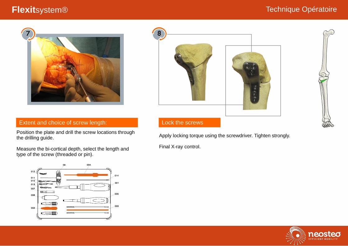
Position the plate and drill the screw locations through the drilling guide. Measure the bi-cortical depth, select the length and type of the screw (threaded or pin).
Apply locking torque using the screwdriver. Tighten strongly. Final X-ray control.
Extent and choice of screw length: Lock the screws
Flexitsystem® Technique Opératoire

Anatomical implants dedicated to a pathology genu varum

9 Guide de coupe OTV soustraction - HTO closing wedge cutting guide INS-009 1
Anatomical implants dedicated to a pathology genu varum