DermaSpan Acellular Dermal Matrix - Zimmer Biomet
16
DermaSpan ™ Acellular Dermal Matrix Wound Coverage Technique Surgical Protocol by Bradford Fine, DPM
Transcript of DermaSpan Acellular Dermal Matrix - Zimmer Biomet

DermaSpan™ Acellular Dermal MatrixWound Coverage Technique
Surgical Protocol byBradford Fine, DPM

Table of Contents
Debridement and Allograft Size ............................................................................... 2
Allograft Application................................................................................................. 3
Pie Crusting ............................................................................................................... 4
Author’s Post Op Protocol ........................................................................................ 4
Negative Pressure options ....................................................................................... 5
Appendix .................................................................................................................. 6
Ordering Information ............................................................................................... 8
Optional Meshing ..................................................................................................... 9
Indications and Contradictions .............................................................................. 10
2 | DermaSpan Meshed Acellular Dermal Matrix Wound Care Technique
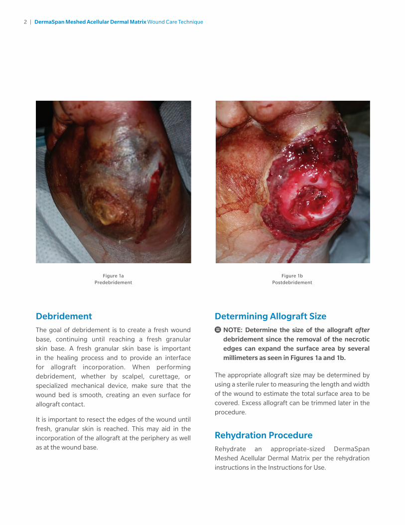
Figure 1aPredebridement
Figure 1bPostdebridement
Determining Allograft Size
NOTE: Determine the size of the allograft after debridement since the removal of the necrotic edges can expand the surface area by several millimeters as seen in Figures 1a and 1b.
The appropriate allograft size may be determined by using a sterile ruler to measuring the length and width of the wound to estimate the total surface area to be covered. Excess allograft can be trimmed later in the procedure.
Rehydration ProcedureRehydrate an appropriate-sized DermaSpan Meshed Acellular Dermal Matrix per the rehydration instructions in the Instructions for Use.
DebridementThe goal of debridement is to create a fresh wound base, continuing until reaching a fresh granular skin base. A fresh granular skin base is important in the healing process and to provide an interface for allograft incorporation. When performing debridement, whether by scalpel, curettage, or specialized mechanical device, make sure that the wound bed is smooth, creating an even surface for allograft contact.
It is important to resect the edges of the wound until fresh, granular skin is reached. This may aid in the incorporation of the allograft at the periphery as well as at the wound base.
3 | DermaSpan Meshed Acellular Dermal Matrix Wound Care Technique
Allograft Application1) Apply the DermaSpan Meshed ACD allograft to the
debrided wound bed ensuring the dermal (shiny) side is in contact with the wound. When applying to the wound bed, bleeding should be present.
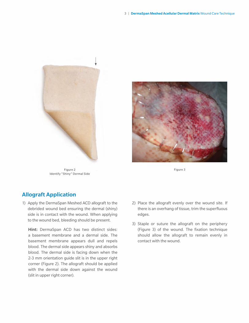
Hint: DermaSpan ACD has two distinct sides: a basement membrane and a dermal side. The basement membrane appears dull and repels blood. The dermal side appears shiny and absorbs blood. The dermal side is facing down when the 2-3 mm orientation guide slit is in the upper right corner (Figure 2). The allograft should be applied with the dermal side down against the wound (slit in upper right corner).
Figure 2 Identify “Shiny” Dermal Side
Figure 3
2) Place the allograft evenly over the wound site. If there is an overhang of tissue, trim the superfluous edges.
3) Staple or suture the allograft on the periphery (Figure 3) of the wound. The fixation technique should allow the allograft to remain evenly in contact with the wound.

4 | DermaSpan Meshed Acellular Dermal Matrix Wound Care Technique
Figure 5
Author’s Post Op ProtocolDressing techniques vary according to surgeon preference. Generally, a warm, moist environment protected from external contamination is conducive to wound healing. Some common dressings are listed below:
• Mineral oil: can be used as an adjunct to lend moisture to the wound bed and allograft.
• A sterile non-adhesive dressing: can be used as a contact layer between the allograft and dressings to help prevent pull-off damage when removing dry sterile dressings.
• Saline soaked gauze: is an option to cover the allograft and provide moisture.
• Negative pressure dressing: may assist with incorporation of the allograft.
Zimmer Biomet has not evaluated post operative wound care options.
Pie Crusting (Optional)If the surgeon determines additional draining may be required, prepare a slit in the graft with the scalpel much like you would to vent a pie crust (Figure 4). Another option is to extend one of the meshed holes of the graft.
Figure 4In some circumstances, a technique known as
“Pie Crusting” is sometimes utilized to help with drainage
5 | DermaSpan Meshed Acellular Dermal Matrix Wound Care Technique
Figure 6
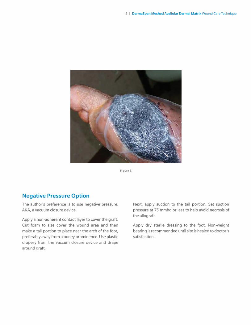
Negative Pressure OptionThe author’s preference is to use negative pressure, AKA, a vacuum closure device.
Apply a non-adherent contact layer to cover the graft. Cut foam to size cover the wound area and then make a tail portion to place near the arch of the foot, preferably away from a boney prominence. Use plastic drapery from the vaccum closure device and drape around graft.
Next, apply suction to the tail portion. Set suction pressure at 75 mmhg or less to help avoid necrosis of the allograft.
Apply dry sterile dressing to the foot. Non-weight bearing is recommended until site is healed to doctor’s satisfaction.
6 | DermaSpan Meshed Acellular Dermal Matrix Wound Care Technique
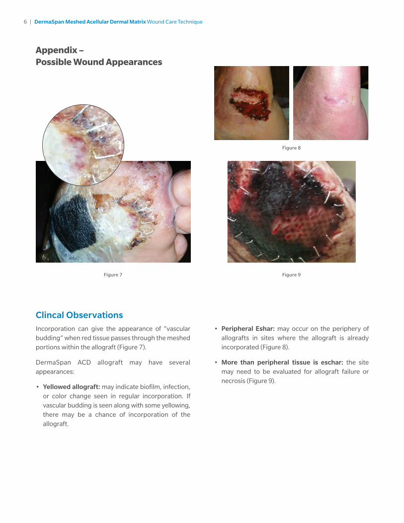
Appendix – Possible Wound Appearances
Figure 7
Figure 8
Figure 9
Clincal ObservationsIncorporation can give the appearance of “vascular budding” when red tissue passes through the meshed portions within the allograft (Figure 7).
DermaSpan ACD allograft may have several appearances:
• Yellowed allograft: may indicate biofilm, infection, or color change seen in regular incorporation. If vascular budding is seen along with some yellowing, there may be a chance of incorporation of the allograft.
• Peripheral Eshar: may occur on the periphery of allografts in sites where the allograft is already incorporated (Figure 8).
• More than peripheral tissue is eschar: the site may need to be evaluated for allograft failure or necrosis (Figure 9).

7 | DermaSpan Meshed Acellular Dermal Matrix Wound Care Technique
Figure 10
Clincal Observations (cont.)
• Black appearance: wound may look black and necrotic with hard, almost crusty eschar. If eschar is superficial, then the allograft may act like a normal scab and heal (Figure 10). With a thicker eschar, you may have to slightly lift the edges to look for healing, granulated tissue.
Figure 11
• Wet looking allograft: The allograft can have a wet and fibrotic appearance (Figure 11). Dressings may be changed every other day and the wound monitored for changes in exudate.
8 | DermaSpan Meshed Acellular Dermal Matrix Wound Care Technique
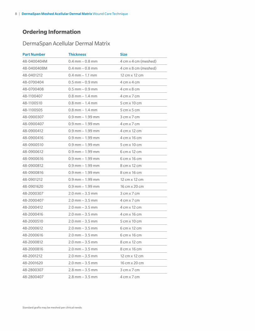
Ordering Information
DermaSpan Acellular Dermal Matrix
Part Number Thickness Size
48-0400404M 0.4 mm – 0.8 mm 4 cm x 4 cm (meshed)
48-0400408M 0.4 mm – 0.8 mm 4 cm x 8 cm (meshed)
48-0401212 0.4 mm – 1.1 mm 12 cm x 12 cm
48-0700404 0.5 mm – 0.9 mm 4 cm x 4 cm
48-0700408 0.5 mm – 0.9 mm 4 cm x 8 cm
48-1100407 0.8 mm – 1.4 mm 4 cm x 7 cm
48-1100510 0.8 mm – 1.4 mm 5 cm x 10 cm
48-1100505 0.8 mm – 1.4 mm 5 cm x 5 cm
48-0900307 0.9 mm – 1.99 mm 3 cm x 7 cm
48-0900407 0.9 mm – 1.99 mm 4 cm x 7 cm
48-0900412 0.9 mm – 1.99 mm 4 cm x 12 cm
48-0900416 0.9 mm – 1.99 mm 4 cm x 16 cm
48-0900510 0.9 mm – 1.99 mm 5 cm x 10 cm
48-0900612 0.9 mm – 1.99 mm 6 cm x 12 cm
48-0900616 0.9 mm – 1.99 mm 6 cm x 16 cm
48-0900812 0.9 mm – 1.99 mm 8 cm x 12 cm
48-0900816 0.9 mm – 1.99 mm 8 cm x 16 cm
48-0901212 0.9 mm – 1.99 mm 12 cm x 12 cm
48-0901620 0.9 mm – 1.99 mm 16 cm x 20 cm
48-2000307 2.0 mm – 3.5 mm 3 cm x 7 cm
48-2000407 2.0 mm – 3.5 mm 4 cm x 7 cm
48-2000412 2.0 mm – 3.5 mm 4 cm x 12 cm
48-2000416 2.0 mm – 3.5 mm 4 cm x 16 cm
48-2000510 2.0 mm – 3.5 mm 5 cm x 10 cm
48-2000612 2.0 mm – 3.5 mm 6 cm x 12 cm
48-2000616 2.0 mm – 3.5 mm 6 cm x 16 cm
48-2000812 2.0 mm – 3.5 mm 8 cm x 12 cm
48-2000816 2.0 mm – 3.5 mm 8 cm x 16 cm
48-2001212 2.0 mm – 3.5 mm 12 cm x 12 cm
48-2001620 2.0 mm – 3.5 mm 16 cm x 20 cm
48-2800307 2.8 mm – 3.5 mm 3 cm x 7 cm
48-2800407 2.8 mm – 3.5 mm 4 cm x 7 cm
Standard grafts may be meshed per clinical needs

9 | DermaSpan Meshed Acellular Dermal Matrix Wound Care Technique
Meshing of Standard (non-meshed) Allograft (Optional)In some applications, the surgeon may choose to mesh a standard (non-meshed) allograft prior to application. This should always be done after the rehydrating process.
The authors preference is to place the graft on a 1:1.5 carrier for use with a mesher. Use sterile saline to pre-wash the mesher, <25 ml.
10 | DermaSpan Meshed Acellular Dermal Matrix Wound Care Technique
Indications and Contraindications
INDICATIONS FOR USEDermaSpan ACD is to be used for the repair or replacement of damaged or inadequate integumental tissue or for other homologous uses of human integument. DermaSpan Meshed ACD is to be used as a covering for skin wounds (e.g. burns, ulcers) and should not be used in load-bearing applications. The standard allograft (non-meshed) may also be used for supplemental support, protection, reinforcement or covering of tendon or ligament, but is not intended to bear the load. Each package of DermaSpan ACD is intended for use in one patient on a single occasion by a licensed physician, surgeon, dentist or podiatrist.
CONTRAINDICATIONSUse of DermaSpan ACD in patients exhibiting autoimmune connective tissue disease is not recommended. DermaSpan should not be used in patients with sensitivities to processing agents (see WARNINGS).

12 | DermaSpan Meshed Acellular Dermal Matrix Wound Care Technique
Notes

All content herein is protected by copyright, trademarks and other intellectual property rights, as applicable, owned by or licensed to Zimmer Biomet or its affiliates unless otherwise indicated, and must not be redistributed, duplicated or disclosed, in whole or in part, without the express written consent of Zimmer Biomet.
For product information, including indications, contraindications, warnings, precautions, potential adverse effects and patient counseling information, see the package insert and www.zimmerbiomet.com.
This material is intended for health care professionals. Distribution to any other recipient is prohibited.
Zimmer Biomet does not practice medicine. This technique was developed in conjunction with [a] health care professional[s]. This document is intended for surgeons and is not intended for laypersons. Each surgeon should exercise his or her own independent judgment in the diagnosis and treatment of an individual patient, and this information does not purport to replace the comprehensive training surgeons have received. As with all surgical procedures, the technique used in each case will depend on the surgeon’s medical judgment as the best treatment for each patient. Results will vary based on health, weight, activity and other variables. Not all patients are candidates for this product and/or procedure. Caution: Federal (USA) law restricts this device to sale by or on the order of a surgeon. Rx only.
©2016 Zimmer Biomet
0699.1-US-en-REV1216
Legal ManufacturerBiomet OrthopedicsP.O. Box 58756 E. Bell DriveWarsaw, Indiana 46581-0587 USA
www.zimmerbiomet.com